Publisher’s version / Version de l'éditeur:
Glycobiology, 20, 12, pp. 1547-1573, 2010-08-30
READ THESE TERMS AND CONDITIONS CAREFULLY BEFORE USING THIS WEBSITE. https://nrc-publications.canada.ca/eng/copyright
Vous avez des questions? Nous pouvons vous aider. Pour communiquer directement avec un auteur, consultez la
première page de la revue dans laquelle son article a été publié afin de trouver ses coordonnées. Si vous n’arrivez pas à les repérer, communiquez avec nous à [email protected].
Questions? Contact the NRC Publications Archive team at
[email protected]. If you wish to email the authors directly, please see the first page of the publication for their contact information.
NRC Publications Archive
Archives des publications du CNRC
This publication could be one of several versions: author’s original, accepted manuscript or the publisher’s version. / La version de cette publication peut être l’une des suivantes : la version prépublication de l’auteur, la version acceptée du manuscrit ou la version de l’éditeur.
For the publisher’s version, please access the DOI link below./ Pour consulter la version de l’éditeur, utilisez le lien DOI ci-dessous.
https://doi.org/10.1093/glycob/cwq122
Access and use of this website and the material on it are subject to the Terms and Conditions set forth at
Structural and mechanistic classification of uronic acid-containing
polysaccharide lyases
Garron, Marie-Line; Cygler, Miroslaw
https://publications-cnrc.canada.ca/fra/droits
L’accès à ce site Web et l’utilisation de son contenu sont assujettis aux conditions présentées dans le site LISEZ CES CONDITIONS ATTENTIVEMENT AVANT D’UTILISER CE SITE WEB.
NRC Publications Record / Notice d'Archives des publications de CNRC:
https://nrc-publications.canada.ca/eng/view/object/?id=8899cf95-74fa-40ce-8f57-15085cad602e https://publications-cnrc.canada.ca/fra/voir/objet/?id=8899cf95-74fa-40ce-8f57-15085cad602eAdvance Access publication on August 30, 2010
REVIEW
Structural and mechanistic classification of uronic
acid-containing polysaccharide lyases
Marie-Line Garron2,and Miroslaw Cygler1,2,3
2
Department of Biochemistry, McGill University, 3655 Promenade Sir William Osler, Montreal, Quebec, Canada H3G 1Y6; and3Biotechnology Research
Institute, NRC, 6100 Royalmount Avenue, Montreal, Quebec, Canada H4P 2R2
Received on June 1, 2010; revised on August 6, 2010; accepted on August 11, 2010
Polysaccharide lyases (PLs) have been assigned to 21 fam-ilies based on their sequences, with ~50 singletons awaiting further classification. For 19 of these families, the structure of at least one protein is known. In this review, we have analyzed the available structural information and show that presently known PL families belong to six general folds. Only two general catalytic mechanisms have been observed among these PLs: (1) metal-assisted neutraliza-tion of the acidic group of the sugar next to the cleaved bond, with, rather unusually, arginine or lysine playing the role of Brønsted base and (2) neutralization of the acidic group on the sugar by a close approach of an amino or acidic group forcing its protonation and Tyr or Tyr-His acting as the Brønsted base and acid.
Keywords: polysaccharide lyases / glycosaminoglycans /
classification/ enzymatic mechanism / fold analysis
Introduction
Carbohydrates are a large class of essential molecules common to all organisms. They are found most abundantly as polymers, either as oligo- or polysaccharides, usually linked to proteins or lipids. Carbohydrates possess a multiplicity of chiral centers
and a large number of specific modifications, including
acety-lation, methyacety-lation, oxidation, and sulfonation, creating great chemical diversity from simple carbohydrate building blocks (Gabius 2000; Bertozzi and Kiessling 2001; Cummings and Stephen 2007). Polysaccharides are polymers formed of carbo-hydrate repeating units joined by glycosidic bonds. These are predominantly linear polymers but may contain various degrees of branching. Polysaccharides are essential cellular components of all living organisms and play multiple biological roles (Gagneux and Varki 1999;Schaefer and Schaefer 2010). They
are involved in various cellular processes, including signal transmission (Mythreye and Blobe 2009;Schaefer and Schaefer 2010), immune response (Chen et al. 2008;Marth and Grewal 2008), bacterial pathogenesis (Boneca 2005), and cancer progression (Vlodavsky et al. 2007;Iozzo et al. 2009). Polysac-charides are most frequently found on cell surfaces in uni- and multicellular organisms (Gabius 2006) and in the extracellular matrix of higher eukaryotes (Iozzo 1998) and are essential com-ponents of cell wall of plants (Knox 2008;Sarkar et al. 2009). Within the extracellular matrix, they fulfill architectural func-tions providing elasticity (Knudson and Knudson 2001;Scott 2003) and serve as storage for many growth factors and other proteins (Macri et al. 2007). They form the building blocks of cell walls, providing physical rigidity and protection against the environment. Oligosaccharides are utilized for signaling dur-ing protein folddur-ing (Parodi 2000) and decorate many secreted proteins (Ohtsubo and Marth 2006). Many of these biologically important polysaccharides contain uronic acid within their repeating units.
Not surprisingly, all organisms contain a large number of
en-zymes dedicated to polysaccharide synthesis, modification, and
degradation (Lairson et al. 2008). Some bacteria and plants re-lay on polysaccharides as a carbon source and contain an unusually large contingent of such enzymes (e.g. commensal
Bacteroides thetaiotaomicron). The up-to-date classification of enzymes that degrade, modify, or create glycosidic bonds is maintained in the Carbohydrate Active enZYmes (CAZy) database (http://www.cazy.org/) (Cantarel et al. 2009). Carbo-hydrate modifying enzymes are divided into four classes: glycoside hydrolases (GHs), glycosyltransferases (GTs), poly-saccharide lyases (PLs), and carbohydrate esterases (CEs). The structure and mechanisms of GHs and GTs have been exten-sively studied and frequently reviewed, most recently in (Henrissat et al. 2008). On the other hand, only limited reviews of specific families of PLs have been published (Jenkins et al. 1998;Hashimoto et al. 2005; Linhardt et al. 2006). The pur-pose of this review is to analyze and compare the structural and mechanistic features of PLs and to classify them based on their fold and catalytic mechanism.
Two chemical reactions are predominantly utilized for enzy-matic depolymerization of polysaccharide chains: hydrolysis
and lytic β-elimination (Yip and Withers 2004). Hydrolysis
proceeds through the addition of a water molecule to break the glycosidic bond, creating a new reducing end on one frag-ment, with either retention or inversion of the configuration at C-1, and a saturated hexose ring on the nonreducing end of the other fragment (McCarter and Withers 1994). β-Eliminative
1To whom correspondence should be addressed: Tel: + 1-514-496-6321;
Fax: +1-514-496-5143; e-mail: [email protected]
at Canada Institute for STI on March 30, 2011
glycob.oxfordjournals.org
cleavage of a glycosidic bond can occur when the sugar is sub-stituted with an acidic group next to the carbon forming the glycosidic bond and results in the formation of a reducing end on one fragment and an unsaturated ring on the nonre-ducing end of the second fragment. The chemical steps of this mechanism were proposed byGacesa (1987). Glucan poly-saccharides can also be degraded by phosphorolysis by such enzymes as α-glucan phosphorylases (Withers et al. 1981). Eukaryotic enzymes that depolymerize polysaccharides predominantly utilize hydrolytic mechanisms while the lytic mechanism is found in many enzymes of bacterial or fungal or-igin as well as in plants and algae. In this review, we will discuss only PLs.
PLs act on polysaccharides containing a hexose oxidized at C-5 position to a carboxylic group and use β-elimination mechanism to cleave the glycosidic bond at the C-4 position. Three chemical steps have been proposed to occur during the β-elimination reaction (Figure 1A) (Gacesa 1987). First, the carboxyl group of the substrate is neutralized, often by a nearby positive charge, to reduce the pKa of the C-5 proton. Second, an enolate anion intermediate is created by proton abstraction at C-5. Then, electron transfer occurs from the carboxyl group to form a double bond between C-4 and C-5 with concomitant cleavage of the C-4–O-1 glycosidic bond. This bond cleavage creates two chemically distinct ends; the sugar on the new non-reducing end is unsaturated and contains a double bond between C4 and C5, while the sugar on the new reducing end remains saturated. Depending on the configurations at C-4 and C-5, the abstracted proton and the C-4 bridging oxygen can be either on the same side (syn configuration) or opposite sides (anti configuration) of the uronic acid ring. According to commonly used nomenclature (Davies et al. 1997), the sugars are numbered +1, + 2, etc. from the cleavage site toward the reducing end and −1, −2, etc. toward the nonreducing end.
All identified PLs are classified into families within the
CAZy database (http://www.cazy.org/) based on recognizable sequence homologies. They are presently divided into 21 fami-lies (1 to 22, family 19 has been moved to glycosyl hydrolases, Table I), with ~50 sequences as yet unclassified. The number of families is expected to grow with the rapid progress in sequenc-ing of new genomes through many large-scale sequencsequenc-ing efforts (http://www.ncbi.nlm.nih.gov/sites/entrez?db=genome). Within the last several years, the three-dimensional structures of many PLs have been determined, allowing extension of the evo-lutionary relationships between these proteins beyond what was possible to deduce from sequence analysis alone. Presently, 19 of 21 PL families have a known three-dimensional structure of at least one representative (Table I). Only two families, PL12 and PL17 have as yet unassigned folds. Of these, heparan sulfate (HS) lyase family PL12 is the largest without a representative structure.
Polysaccharide substrates bind to their respective lyases along an extended surface on the enzyme, with several consec-utive sugars making contacts with the protein. The sites on the enzyme surface interacting with individual sugars are termed “subsites” and are given numbers according to the position of the sugar they interact with, as described above. Therefore, the enzyme’s substrate-binding site is divided into +n and −n subsites (n = 1,2,3,…), with cleavage occurring between sugars located in the + 1 and −1 subsites. In the majority of PL
enzymes, the subsites −2 to +2 are the most important determi-nants of substrate specificity.
In this review, we analyze the available data on the structure and catalytic mechanisms of PLs and group the PL families into classes based on their folds.
Substrates of PLs
PL substrates have different origins: bacterial, plant, or animal. Nevertheless, they present relatively similar chemical struc-tures composed of repetitive blocks of pyranoses. Each block
is composed of two tofive saccharides linked by a (1-4)
gly-cosidic bond. PLs described here require an acidic group at +1 sugar. Based on the nature of the +1 sugar, the substrates of PLs can be divided into three groups: (1) galacturonic acid, (2) glucuronic/iduronic acid, and (3) mannuronate/guluronate.
Polysaccharides in thefirst group include pectate and pectin,
the second group includes hyaluronan, chondroitin, chondroitin sulfate (CS), dermatan sulfate (DS), heparin, HS, heparosan, glucuronan, and xanthan, and the third group contains alginates.
Pectin/pectate (galacturonate)
Pectate/pectin is the common term to indicate the structural acidic polysaccharide from the cell wall of higher plants (Roberts 1990;Cosgrove 1997;Ridley et al. 2001). This poly-mer is widely used in the food industry as a gelling and thickening agent or stabilizer. Based on the carbohydrate com-ponents and their degree of modification, two types of polysaccharide regions are distinguished: the “smooth” region containing predominantly polygalacturonan (PGA) and the “hairy” region that contains rhamnogalacturonan I and II (RG I, RG II). The PGA contains α-1,4-linked GalA sugars that can be partially 6-O-methylated and acetylated at C-2 and/or C-3 positions (Ridley et al. 2001) (Figure 1B). The RG I and RG II in the hairy regions are highly modified on
the L-rhamnose (Rha) and also partially acetylated at the C-2
and C-3 positions of GalA (Ridley et al. 2001). The enzymes degrading PGA are designated pectate or pectin lyases. Pectin lyases are mainly synthesized by fungi and act on less nega-tively charged, highly methylated pectins, whereas bacterial pectate lyases (Pels) degrade highly negatively charged non-methylated or low-esterified substrates and cleave α-1,4 linkages between galacturonosyl residues. Generally, Pels require Ca2 +ions for activity and have an optimal pH in the basic range. The exceptions are Pels from family PL2, which preferentially utilize Mn2 +ions. Contrary to Pels, pectin lyases have optimal pH around 5.5 and do not require metal ions for activity. Pectin and pectate lyases have been classified within six PL families, PL1, PL2, PL3, PL9, PL10, and PL22. These enzymes are predominantly endolytic and extracellular (Abbott and Boraston 2008); no structure of an exolytic pectate lyase has so far been determined.
Contrary to pectate lyases, enzymes cleaving the RG I re-gions have been less well studied. Rhamnogalacturonan
lyases (RLs) act specifically on [2)-α-L-Rha-(1-4)-α-D-GalA
(1] bonds of the RG I main chain. The C-4-Rha is usually a branch point where arabinan, galactan, and arabinogalactan are attached, creating the “hairy” region. This branching influ-ences the extent of degradation, for example deacetylation or
M-L Garron and M Cygler
at Canada Institute for STI on March 30, 2011
glycob.oxfordjournals.org
1549
Polysacchar
ide
reduction of arabinan sidechains increases enzymatic activity (Mutter et al. 1998). RLs are classified into two families, PL4 and PL11, which are mainly comprised of fungal and bac-terial enzymes, respectively. A bacbac-terial RG degradation pathway has been recently described in Bacillus subtilis strain 168, containing two PL11 RLs, an exolytic and an endolytic, participating together in the degradation of the RG I region (Ochiai, Itoh, Kawamata et al. 2007).
Polysaccharides containing glucuronic/iduronic acid
A large group of PL substrates in this class are glycosamino-glycans (GAGs). They are linear anionic polysaccharides, mainly of eukaryotic origin. Their disaccharide repeating units contain a hexosamine linked 1-4 to an uronic acid. GAGs, with the exception of hyaluronic acid (HA), are O-linked to core proteins via a serine residue, forming proteoglycans, which are involved in the regulation of various cellular processes such as cell adhesion, signaling, cell proliferation, or in
flam-mation. GAGs such as heparin, HS, heparosan, chondroitin sulfate, chondroitin, DS, and HA are substrates of PLs. GAG lyases are produced by various bacteria that can use the result-ing mono- and disaccharides as a carbon source. They are classified as members of families PL6, PL8, PL12, PL13, PL16, and PL21. In addition to GAGs, two other polysacchar-ides are found that contain glucuronic acid: xanthan and glucuronan. Xanthan is degraded by xanthan lyases (XLs) be-longing to the PL8 family, while glucuronan is degraded by glucuronan lyases belonging to the PL14 and PL20 families.
HS and Heparin. HS and heparin are negatively charged, lin-ear polysaccharides, composed of repeating disaccharide units made up of uronic acid and glucosamine residues (GlcN,
2-amino-2-deoxy-α-D-glucopyranose) (Esko and Selleck 2002)
(Figure 1B). HS has a diverse primary structure and is characterized by a high percentage of the GlcA (β-D-glucopyranosyluronic acid). Both GlcA and its minor C-5 epimer, iduronic acid (IdoA, α-L-idopyranosyluronic acid) can be infrequently sulfated at the Fig. 1. (A) General mechanism of β-elimination reaction; (B) substrates of polysaccharide lyases and products of the cleavage reaction.
M-L Garron and M Cygler
at Canada Institute for STI on March 30, 2011
glycob.oxfordjournals.org
C-2 position. The GlcN is frequently N-acetylated (GlcNAc) and, to a limited extent, sulfated at the C-2, C-3, and N-positions. The modifications in HS are concentrated within specific regions of the polysaccharide, known as the high S domains, giving rise to short sequence motifs responsible for the interac-tions between HS and a diverse repertoire of proteins leading to its multiple biological roles. HS-GAGs are abundant at the cell surface as part of proteoglycan cell surface receptors (Jackson et al. 1991; Kjellen and Lindahl 1991).
Heparin has a high content (~90%) of IdoA that is frequently
sulfated at the C-2 position (Linhardt 2003). The glucosamine
residue in heparin is predominantly substituted with N-sulfo groups (GlcNS), with only a small number of N-acetyl groups and 6-O-sulfo groups, as well as rare 3-O-sulfo groups. Cleav-age of the heparin GAG chain is an important part of its biological activity, which leads to the release of heparin-binding proteins, e.g. various growth factors, as well as the creation of short-specific oligosaccharides that regulate hemostasis through
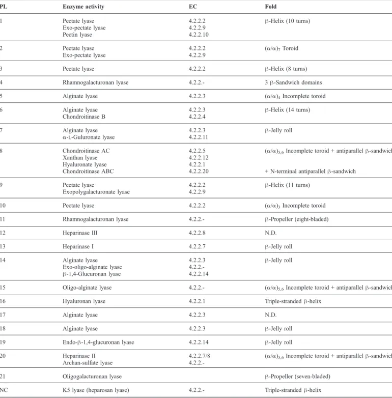
Table I.Polysaccharide lyase families with associated activities and assigned fold
PL Enzyme activity EC Fold
1 Pectate lyase 4.2.2.2 β-Helix (10 turns)
Exo-pectate lyase 4.2.2.9
Pectin lyase 4.2.2.10
2 Pectate lyase 4.2.2.2 (α/α)7Toroid
Exo-pectate lyase 4.2.2.9
3 Pectate lyase 4.2.2.2 β-Helix (8 turns)
4 Rhamnogalacturonan lyase 4.2.2.- 3 β-Sandwich domains
5 Alginate lyase 4.2.2.3 (α/α)4Incomplete toroid
6 Alginate lyase 4.2.2.3 β-Helix (14 turns)
Chondroitinase B 4.2.2.4
7 Alginate lyase 4.2.2.3 β-Jelly roll
α-L-Guluronate lyase 4.2.2.11
8 Chondroitinase AC 4.2.2.5 (α/α)5,6Incomplete toroid + antiparallel β-sandwich
Xanthan lyase 4.2.2.12
Hyaluronate lyase 4.2.2.1
Chondroitinase ABC 4.2.2.20 + N-terminal antiparallel β-sandwich
9 Pectate lyase 4.2.2.2 β-Helix (11 turns)
Exopolygalacturonate lyase 4.2.2.9
10 Pectate lyase 4.2.2.2 (α/α)3Incomplete toroid
11 Rhamnogalacturonan lyase 4.2.2.- β-Propeller (eight-bladed)
12 Heparinase III 4.2.2.8 N.D.
13 Heparinase I 4.2.2.7 β-Jelly roll
14 Alginate lyase 4.2.2.3 β-Jelly roll
Exo-oligo-alginate lyase
4.2.2.-β-1,4-Glucuronan lyase 4.2.2.14
15 Oligo-alginate lyase 4.2.2.- (α/α)5,6Incomplete toroid + antiparallel β-sandwich
16 Hyaluronan lyase 4.2.2.1 Triple-stranded β-helix
17 Alginate lyase 4.2.2.3 N.D.
18 Alginate lyase 4.2.2.3 β-Jelly roll
19 Endo-β-1,4-glucuronan lyase 4.2.2.14 β-Jelly roll
20 Heparinase II 4.2.2.7/8 (α/α)5,6Incomplete toroid + antiparallel β-sandwich
Archan-sulfate lyase
4.2.2.-21 Oligogalacturonan lyase β-Propeller (seven-bladed)
NC K5 lyase (heparosan lyase) 4.2.2.- Triple-stranded β-helix
at Canada Institute for STI on March 30, 2011
glycob.oxfordjournals.org
interactions with proteins of the blood coagulation cascade (Capila and Linhardt 2002; Munoz and Linhardt 2004).
Four types of bacterial heparin/HS lyases have been identi-fied. Heparinase I (HepI) is specific for heparin and cleaves the bond on the nonreducing end of iduronic acid, heparinase III (HepIII) cleaves HS at the nonreducing end of glucuronic acid, while heparinase II (HepII) can depolymerize both heparin and
HS. These enzymes have been classified to three different PL
families. HepI belongs to the PL13 family, HepIII to PL12, and HepII was recently assigned to a new family, PL21. Heparosan K5 lyase is an E. coli phage enzyme that cleaves the unsulfated bacterial polysaccharide heparosan (the biosynthetic precursor to heparin and HS) and HS next to GlcA but is intolerant of sulfated polysaccharides (Rek et al. 2007).
Chondroitin and DSs. CS and DS are major components of
cartilage, blood vessels, and tendons (Yoon and Halper 2005;
Roughley 2006). CS is widely used as a dietary supplement and is believed to be beneficial in the treatment of osteoarthritis due to its anti-inflammatory action (Lauder 2009) and, together with DS, has potential for applications in regenerative medicine and in the treatment of viral infections (Yamada and Sugahara 2008). CS and DS are composed of disaccharide repeating units of α/β-D-uronate-(1,3)-β-D-N-acetylgalactosamine (GalNAc) (Figure 1B). CS contains the C-5 epimer GlcA while DS con-tains the IdoA (Ernst et al. 1995). Five types of CS are distinguishable based on their sulfation pattern: CS-A con-tains sulfate at the C-4 position, CS-B (now known as DS) is sulfated at C-4, CS-C is sulfated at C-6, CS-D is sulfated at C-2 and C-6, and CS-E is sulfated at C-4 and C-6. Addi-tional sulfation can occur at the C-2 position of IdoA and at the C-6 position of GalNAc in DS. Chondroitin, lacking sul-fate groups, is the biosynthetic precursor of both CS and DS and is synthesized by commensal and pathogenic bacteria (DeAngelis et al. 2002).
As is the case with heparinases, several types of bacterial
chondroitin lyases have been identified differing in substrate
specificity (Linhardt et al. 2006). Chondroitin lyase AC
(Cho-nAC) degrades CS-A and C, chondroitin lyase B (ChonB) is specific for DS, and chondroitin lyase ABC (ChonABC) shows broad specificity and can cleave CS-A/C and DS. The ChonAC and ChonABC enzymes belong to the PL8 family while ChonB belongs to the PL6 family.
Hyaluronan. Hyaluronan (HA) is a long, unsulfated polysac-charide synthesized directly in the plasma membrane, rather than in the Golgi like other GAGs. Abundant in higher organ-isms, HA is present in tissues such as skin, cartilage, or neural
tissue (McDonald and Camenisch 2002; Kogan et al. 2007).
Due to its wide distribution, HA is used in various medical ap-plications including eye surgery, osteoarthritis, and tissue engineering. Some bacteria and viruses synthesize capsular HA that allows them to evade the host immune system (DeAngelis 2002). HA is composed of repeating units of [3)-β-D-N-acetylglucosamine-(1,4)-β-D-GlcA-(1] (Figure 1B). This disaccharide block is similar to that found in CS, and HA lyases (HLs) also show some activity toward CS. Bacterial HLs are similar in sequence to ChonAC and ChonABC and belong to the same PL8 family. Bacteriophage invading Streptococcus strains also produces enzymes with HL activity but unrelated se-quence. These HLs are also found in some Streptococcus strains and have been classified within the PL16 family.
Xanthan. Xanthan is an extracellular hetero-polysaccharide synthesized by the pathogenic bacterium, Xanthomonas
campestris. Xanthan is a common food additive used for its
abil-ity to increase viscosabil-ity of liquids. It is composed of a branched
pentasaccharide repeat unit, in which the disaccharide, β-D
-glucose-(1,4)-β-D-glucose, forms the main chain polymer. The
second disaccharide’s glucose is substituted at the C-3 position
by a trisaccharide [β-D-mannose-(1,4)-β-D-glucuronic
acid-(1,2)-β-D-mannose-(1,3] (Figure 1B) (Garcia-Ochoa et al.
2000). Both mannose units can be acetylated at the C-6 position and a pyruvate can be added on C-4 or C-6 position via a ketal linkage (Hashimoto et al. 1998). XL specifically degrades the link [β-D-mannose-(1,4)-β-D-glucuronic acid] on the branched section. So far, only two xanthan lyases from Bacillus sp. GL1 and Paenibacillus alginolyticus XL-1 have been characterized (Hashimoto et al. 1998; Ruijssenaars et al. 2000). Like the majority of lyases cleaving the glycosidic bond next to GlcA, xanthan lyase belongs to the PL8 family.
Glucuronan. Glucuronan is a less abundant polysaccharide, synthesized by some bacteria, fungi, and algae. This polysac-charide is of interest to the food and pharmacological industries for its rheological and biological properties. It is composed of (1,4) linked β-D-glucuronic acids and is partially acetylated at the C-3 and/or C-2 positions (Figure 1B) (Miyazaki et al. 1975;Heyraud et al. 1993;Redouan et al. 2009). Glucu-ronan is thefirst example of a homoglucuronic acid polymer synthesized by bacteria. Several glucuronan lyases have been identified and are classified within families PL14 and PL20.
Alginate (mannuronate)
Alginate is a linear polysaccharide produced by some bacteria belonging to the Pseudomonas and Azotobacter genera and by
brown seaweed (Fischer and Dorfel 1955; Rehm and Valla
1997). It contains α-L-guluronate (G) or its C-5 epimer, β-D-mannuronate (M), connected by (1-4) linkages (Figure 1B). In
Pseudomonas species, this polysaccharide plays a role in
viru-lence related to alginate-mediated biofilm formation that helps to avoid phagocytosis (Russell and Gacesa 1989;Pritt et al. 2007). Alginate from brown seaweed is widely used in the food and pharmaceutical industries for its capacity to chelate ions and for its gelling properties. Three distinct regions can be found along the alginate chain, the homopolymeric poly(M), poly(G) blocks and an alternate poly(MG) block. Poly(M) bacterial al-ginate is more frequently acetylated at the C-2 and/or C-3 position of mannuronate than alginate from seaweed ( Remmin-ghorst and Rehm 2006). Accordingly, various bacterial strains contain lyases that can degrade these polysaccharides. Alginate lyases have been found within several PL families, namely in PL5, PL6, PL7, PL14, PL15, PL17, and PL18.
PL fold classes
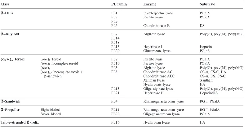
At the time of writing, 19 out of 21 PL families have at least one structural representative. The structures show that the number of folds observed among PL enzymes is smaller than the number of families and indicate distant evolutionary relationships between several families, which could not be detected from their sequences alone. We have organized the PL families into six general folds and classified PLs accordingly into classes (Table II).
M-L Garron and M Cygler
at Canada Institute for STI on March 30, 2011
glycob.oxfordjournals.org
Right-handed β-helix class
The right-handed β-helix is the most abundant fold among PL families. Four families, PL1, PL3, PL6 and PL9, share this fold. PL1, PL3, and PL9 are pectate lyase families, PL6 con-tains alginate and DS lyases. PL1, with more than 400 assigned sequences and over 35 known three-dimensional structures, is the largest family of PLs and contains proteins from bacteria, plants, and fungi.
The right-handed β-helix wasfirst observed in the structure
of the pectate lyase C (PelC) from Erwinia chrysanthemi (Yoder
et al. 1993) (Figure 2A). Each turn of this helical architecture is composed of three β-strands PB1, PB2, and PB3 connected by turns T1 (between PB1 and 2), T2 (PB2 and 3), and T3 (PB3 and 1) (Figure 2B). The β-strands are rather short, containing three tofive residues with strands PB1 and PB2 being nearly antipar-allel to each other. Proteins in this class contain from 8 to 14 helical turns, creating three parallel β-sheets extending along the helix axis (Jenkins et al. 1998). The T2 turns usually contain only two residues, with the second residue adopting the αL
con-formation that introduces a 90° bend to the polypeptide backbone. The second T2 residue in consecutive turns is fre-quently an asparagine that forms the so called “asparagine ladder” (Yoder et al. 1993). The T3 turns are longer than T1 or T2 and can participate in the active site. With the exception of proteins with the fewest number of turns (eight turns), the N-terminal end of the β-helix is capped by an amphipathic α-helix preceding thefirst PB1 strand. The N-terminal and C-terminal extensions usually form irregular structures that pack against small hydrophobic patches on the PB2 and T2 sides of the β-helix, respectively. The inner part of the β-helix is lined with
stacks of hydrophobic and aromatic residues. Sequence align-ment of pectate lyases from the PL1 family shows that PB2, PB3, and T2 are the most conserved parts of the structure. The PB1 strands and T3 turns, involved in the substrate-binding site and contributing to the formation of the active site, show greater variability, adapting to the nature of their specific substrates.
The β-helix formed by members of the PL3 family differs
somewhat from that found in other PL families. This is the shortest β-helix, with only eight turns and is devoid of the
N-terminal capping α-helix (Figure 2C). An asparagine stack
occurs at thefirst position of T2 turns, rather than at the second position observed in other families, and these sidechains are facing toward the exterior of the β-helix. No aromatic stacks are present inside the β-helix, and the rigidity of the structure is maintained by interactions of hydrophobic sidechains and disulfide bonds, five of which are present in PelI (P3B8Y), (Creze et al. 2008). Some of the enzymes from the PL3 family contain an additional fibronectin III-like domain that presum-ably mediates interactions with other protein(s) (Creze et al. 2008).
β-Jelly roll class
Five families of PLs, PL7, PL13, PL14, PL18, and PL20, share a β-jelly roll fold (Figure 3). The enzymes belonging to the β-jelly roll PL class degrade diverse polysaccharide substrates. Members of families PL7, PL14, and PL18 degrade alginate, PL13 depolymerize heparin, and PL20 break down glucuro-nate. In this structural family, PL7, PL13, and PL18 contain mainly bacterial enzymes, whereas PL14 contains viral
en-Table II.Fold classes and PL families associate with each class
Class PL family Enzyme Substrate
β-Helix PL1 Pectate/pectin lyase PGalA
PL3 Pectate lyase PGalA
PL9
PL6 Chondroitinase B DS
β-Jelly roll PL7 Alginate lyase Poly(G), poly(M), poly(MG)
PL14 PL18
PL13 Heparinase I Heparin
PL20 Glucuronate lyase PGlcA
(α/α)nToroid (α/α)7Toroid PL2 Pectate lyase PGalA
(α/α)3Incomplete toroid PL10 Pectate lyase PGalA
(α/α)4 PL5 Alginate lyase Poly(G), poly(M), poly(MG)
(α/α)5,6Incomplete toroid +
β-sandwich
PL8 Chondroitinase AC CS-A, CS-C, HA
Chondroitinase ABC CS-A, DS, CS-C
Xanthan lyase Xanthan
Hyaluronate lyase HA
PL15 Oligo-alginate lyase Poly(G), poly(M), poly(MG)
PL21 Heparinase II Heparin/HS
β-Sandwich PL4 Rhamnogalacturonan lyase RG I, PGalA
β-Propeller Eight-bladed PL11 Rhamnogalacturonan lyase RG I, PGalA
Seven-bladed PL22 Oligogalacturonan lyase PGalA
Triple-stranded β-helix PL16 Hyaluronan lyase HA
Polygalacturonic acid (PGalA); polyglucuronic acid (PGlcA); dermatan sulfate (DS); polyguluronate (Poly(G)); polymannuronate (Poly(M)); chondroitin sulfate A and C (CS-A, CS-C); hyaluronic acid (HA); heparan sulfate (HS); rhamnogalacturonan I (RG I).
at Canada Institute for STI on March 30, 2011
glycob.oxfordjournals.org
Garron
and
M
Cygler
at Canada Institute for STI on March 30, 2011 glycob.oxfordjournals.org
zymes and PL20 eukaryotic enzymes. The canonical fold is made up of two antiparallel seven-stranded β-sheets bent in the middle by nearly 90° (Yamasaki et al. 2004). The resulting curvature creates a groove in which the substrate binds and de-fines the location of the active site (Figure 3). Some enzymes with this fold have one or two additional strands in one or both sheets. The loops linking the inner and outer β-sheets are var-iable in length and sequence. In some structures, these loops are very short, e.g. in the family of PL7 alginate lyases, while in other structures they can be quite long and fold into indepen-dent domains. The largest proteins in this class are PL13 enzymes that have two additional extensions in the middle of the sequence (Figure 3B), e.g. the thumb domain in HepI (Han et al. 2009). The long loops are often rigidified by the presence of structural ions (Ca2 +), although these metal-binding sites are
not conserved within the proteins of this class. In HepI, the Ca2 + ion binds on the side of the β-sandwich and joins two long
loops that stabilize the base of the thumb domain (Han et al. 2009). In PL18, the Ca2 +ion is situated on the opposite side of the β-sheet compared to that in HepI (PDB code 1J1T, un-published). This Ca2 +ion adds rigidity to a long loop. Finally,
in PL20 enzymes, the Ca2 +ion is located at the top of the outer sheet, and it is involved in stabilization of the N-terminal end (Konno et al. 2009).
(α/α)nToroid class
Several PL families contain a domain that is formed by a repe-tition of a pair of antiparallel α-helices (helical hairpin), with each helix composed of 10–20 residues. The hairpins are ar-ranged counterclockwise (looking from the top of the hairpin). The number of hairpins, n, varies from three to seven. We clas-sify these folds together within an (α/α)ntoroid class. This class
can be divided into several subclasses: single-domain (α/α)n
Fig. 3. The β-jelly roll fold. (A) The typical fold for enzymes from families PL7, PL13, and PL18 (PDB code 2ZAA); (B) the structure of PL13 HepI (PDB code 3INA) with small domains inserted into the canonical fold (green and cyan on the right). Bound oligosaccharides are shown in stick mode.
Fig. 2. The right-handed β-helix fold drawn in a cartoon form. The polypeptide is rainbow colored, from blue at the N-terminus to red at the C-terminus. (A) The structure of the canonical pectate lyase PelC (PDB code 2EWE) with nine full turns of the β-helix. The three calcium ions with full occupancy are shown as yellow spheres, and the ion present only in the R218K mutant is shown in gray. Bound tetrasaccharide is shown in stick mode; (B) the view down the β-helix axis with strands PB1, PB2, and PB3 and turns T1, T2, and T3 marked explicitly. Residues forming the asparagine ladder in T2 are shown in a stick representation; (C) the structure of pectate lyase from Bacillus sp. strain ksm-p15 having the shortest, eight-turn β-helix and no additional extension (PDB code 1EE6). This and otherfigures were prepared with PyMOL (www.pymol.org).
at Canada Institute for STI on March 30, 2011
glycob.oxfordjournals.org
Garron
and
M
Cygler
at Canada Institute for STI on March 30, 2011 glycob.oxfordjournals.org
toroids with three, four, or seven α-helical hairpins and multiple
domain proteins with (α/α)5,6toroid domain, a C-terminal
do-main composed of a β-sandwich of four antiparallel β-sheets and, frequently, an additional β-sheet N-terminal domain.
The (α/α)7forms a full toroid while the structures with
smal-ler number of hairpins form incomplete toroids. A characteristic feature of the α-helical hairpins is a short connection between
the helices and significantly longer connections between the
consecutive hairpins. In all cases, the substrate-binding sites
are located on the side of these long connections, near the top of the (semi)-toroid. Despite different number of α-helical hair-pins in various PL families, these structures can be superposed with reasonable root mean squares deviations, starting from the N-terminal hairpins.
The single-domain (α/α)ntoroid subclasses encompass the
PL2, PL5, and PL10 families. The PL10 family has only three hairpin repeats, (α/α)3 (Charnock et al. 2002) (Figure 4A), alginate lyases from PL5 contain four hairpin repetitions, Fig. 4. The (α/α)ndomain fold. (A) (α/α)3Incomplete toroid (PDB code 1GXO); (B) (α/α)4incomplete toroid (PDB code 1HV6); (C) (α/α)7toroid (PDB code
2V8K); (D) the multidomain (α/α)5,6incomplete toroid (PDB code 1RWH); (E) the superposition of these four folds showing good superposition of thefirst several
α-helical hairpins. Colors: (α/α)31GXO (PL10)—blue, (α/α)41HV6 (PL5)—green, (α/α)5,61RWH (PL8)—red, (α/α)5,62FUT (PL21)—orange, and (α/α)7
2V8K (PL2)—yellow.
at Canada Institute for STI on March 30, 2011
glycob.oxfordjournals.org
(α/α)4(Yoon et al. 2001) (Figure 4B), and PL2 has seven
re-peats, (α/α)7(Abbott and Boraston 2007) (Figure 4C). They
differ somewhat in the curvature of the (semi)-toroid formed by the helical hairpins but thefirst several hairpins can be super-imposed relatively well (Figure 4E).
The multidomain (α/α)5,6toroid subclass contains additional
domains. The (α/α)5,6 toroid domain contains five α-helical
hairpins and in some proteins a sixth hairpin is assembled from
two additional antiparallel helices, one at the N-terminus and the other at the C-terminus of this domain. This domain is followed by a C-terminal antiparallel β-sandwich domain
containing four β-sheets (Féthière et al. 1999; Hashimoto
et al. 2003) (Figure 4D). Some lyases, such as ChonABC and HL, contain also a small N-terminal β-sandwich domain (Li and Jedrzejas 2001; Huang et al. 2003; Shaya, Hahn, Bjerkan et al. 2008). In this subclass, the C-terminal domain Fig. 5. The β-propeller fold. The eight-bladed propeller is preceded by a β-sheet domain (PDB code 2Z8S). The substrate (gray sticks) binds on the opposite end of the propeller domain to the N-terminal domain. Seven β-sheet blades are stabilized by Ca2+ions (yellow spheres), and two additional Ca2 +ions bind near the substrate.
Fig. 6. β-Sandwich fold. The structure of PL4 rhamnogalacturonan lyase from Aspergillus aculeatus (PDB code 1NKG) contains three domains colored blue, green, and red, respectively. The active site is within the domain. The bound Ca2+(yellow) and sulfate ions are shown explicitly as spheres.
M-L Garron and M Cygler
at Canada Institute for STI on March 30, 2011
glycob.oxfordjournals.org
participates in the formation of the substrate-binding site and contributes one amino acid to the catalytic tetrad. The PL8 family belongs to this subclass and contains lyases that can depolymerize several different substrates: CS-A, -B, or -C, HA, and xanthan. All these enzymes, with exception of HLs, contain structural ions, calcium or sodium, coordinated mainly by aspartate sidechains. Despite similarities in their structures, none of these metal ion sites are conserved across this enzyme family. Apparently, these sites are not essential for maintaining this particular fold since their location is var-iable. For example, xanthan lyase from Bacillus sp. has a Ca+ 2 ion in the native structure (PDB code 1J0M) and no ion in the complex with mannose (PDB code 2E22) (Hashimoto et al. 2003;Maruyama et al. 2007).
Two other small families share with PL8, the multidomain
(α/α)5,6 toroid subclass, namely PL15 and PL21. The PL15
family contains bacterial oligo-alginate or exotype alginate
lyases. The structure of the first enzyme from this family
was determined recently (PBD code 3AFL) (Ochiai et al.
2010). Structural comparison shows that PL15 lyase is more similar to family PL21 than to PL8 lyases. Like ChonABC and hyaluronan lyases, this alginate lyase possesses a small β-sandwich domain at the N-terminus, but its orientation dif-fers from those in the ChonABC and hyaluronan lyases. A structural ion is also found in this family. A potential chloride ion links the N-terminal helical domain to the C-terminal β-sandwich but like for PL8 this ion is not essential (Ochiai et al. 2010). The PL21 family contains HepII enzymes. Like other PLs from this subclass, HepII contains a structural
ion (zinc) that links several long loops in the C-terminal β-sandwich domain (Shaya et al. 2006).
β-Propeller class
The lyases from two PL families display β-propeller fold, PL11 and PL22. B. subtilis RG PL11 lyases YesW and YesX
differ in their mode of action: YesW is an endolytic enzyme
while YesX is an exolytic enzyme (Ochiai, Itoh, Maruyama
et al. 2007). Both enzymes share the same fold and their exo/endo specificity is linked to the presence of an extended loop in the exolytic YesX that blocks polysaccharide binding on the “−” side (Ochiai et al. 2009). Both enzymes are com-posed of two domains. The N-terminal domain contains two three-stranded antiparallel β-sheets and is followed by an eight-bladed β-propeller domain (Figure 5). Each blade con-tains four antiparallel β-strands and each blade, except for blade D, contains a structural Ca2 +ion. One end of the propel-ler is occluded by the N-terminal domain and is plugged by the C-terminal α-helix. The substrate binds to the opposite open end of the β-propeller (Figure 5).
The oligogalacturonate lyase from Vibrio parahaemolyticus (PDB code 3C5M, F. Forouhar et al., unpublished) is a seven-bladed β-propeller with no additional domains. In contrast to PL11 lyases, no structural ions are present; however, one
Mn2 + ion is located at the entrance to the deep depression
at one end of the propeller, presumably the location of the substrate-binding site.
Among the carbohydrate processing enzymes, the β-propeller fold is not limited to PLs but is also found among GHs. For
ex-Fig. 7. Triple-stranded β-helix (PDB code 2YVV). The three intertwined subunits are shown in different colors: yellow, blue, and magenta. Bound disaccharides
are shown in stick mode. at Canada Institute for STI on March 30, 2011
glycob.oxfordjournals.org
ample, the 6-bladed β-propeller fold occurs in family GH33
whilefive blades are observed in family GH62 (CAZy database
(Cantarel et al. 2009)).
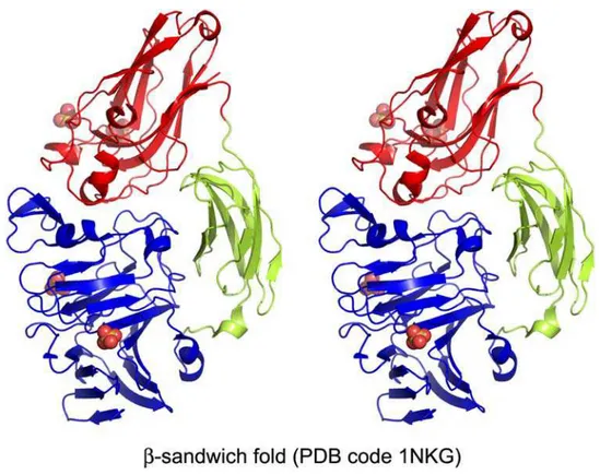
β-Sandwich class
The structure of the PL4 family of RLs has no structural similar-ity with RLs from the PL11 family or with any other lyases. While PL11 contains essential bacterial sequences, the PL4 fam-ily mainly comprises eukaryotic enzymes of fungal and plant origin. The only structural representative with this fold in PL4 is
the eukaryotic RL from Aspergillus aculeatus (McDonough
et al. 2004). The A. aculeatus lyase contains three domains (Figure 6). The N-terminal domain is the largest and is predicted to contain the catalytic site. The central and C-terminal domains are presumed to be involved in binding the polysaccharide substrate. These domains were identified as a fibronectin type III-like domain and a carbohydrate binding module-like domain, respectively. The N-terminal β-sandwich domain is formed from two antiparallel eight-stranded β-sheets (McDonough et al. 2004).
Triple-stranded β-helix
The triple-stranded β-helix is found in bacteriophage tail spike proteins (TSPs) that are essential for phage virulence. The three strands are intertwined to form a single β-helix or fold independently into three β-helices when the chains are held together through the interactions of N- and/or C-terminal domains. Some TSPs possess catalytic activity to degrade poly-saccharides of the bacterial envelope. Example of glycosidases
include endo-α-L-1,3-rhamnosidase from Salmonella phage
P22 and from family GH90 (PDB code 1TSP (Steinbacher et
al. 1994)) and bacteriophage K1F endo-4,8-sialidase from family GH58 (PDB code 1VOF (Stummeyer et al. 2005)).
This fold is also found in PLs (hyaluronidase) from family PL16. The PL16 family of bacteriophage-encoded HL is found in the genomes of virulent Streptococcus species and in phages invading Streptococcus. The phage HL helps the phage to pen-etrate the capsule of its bacterial host by degrading the HA layer and reducing the capsule viscosity. These HLs are distinct from PL8 HLs not only in their fold but also in that they are
specific for HA and cannot digest CS substrates. They are
com-posed of three intertwined polypeptides forming a
triple-Table IV.Active site of metal-dependent lyases
Active site → Isostructural Similar Different
Fold β-Helix
(α/α)3
Toroid
(α/α)7
Toroid β-Helix β-Propeller
PL PL1 PL3 PL9 PL10 PL2 PL6 PL11 PL22
Enzyme Pel Pectin lyase Pel Pel Pel Pel ChB RL OligoGal.
PDB codea 2EWE 1QCX 3B8Y 1RU4 1GXO 2V8K 1OFL 2Z8S 3C5M
Neutralizer 1-2 Ca2 + Arg176
(replaces Glu166)
Ca2+ Ca2+ Ca2+ Mn2+ Ca2+ Ca2 + Mn2 +
Metal stabilizers Asp131, Glu166, Asp170 Asp173b, Asp195b, Glu194b, Asp209, Asp233, Asp234, Asp237 Asp451 Glu130, His109, His172 Glu243, Glu245, Asp213 Site 1 Asp153, Asn596 Site 2 His363, His399, Asp401, Glu422 His287, His353, His355, Gln350
Base Arg218 Arg236 Lys224 Lys273 Arg524 Arg171 Lys250 Lys535bor
Arg452b, Arg349b Acid H2O H2O H2O H2O H2O H2O Arg271 H2Ob H2Ob a Representative structure. b To be confirmed.
Table III. Catalytic residues of PLs
Fold (α/α)5,6Toroid + β-sandwich β-Jelly roll
(α/α)4
PL family PL8 PL15 PL21 PL7 PL13 PL18 PL20 PL5
Enzyme ChonAC XL HL ChABC Oligo-alg L HepII Alg L HepI Alg L Gluc L. Alg L
PDB codea 1RWH 1JON 1I8Q 2Q1F 3AFL 2FUT 1UAI 3IKW 1J1T 2ZZJ 1HV6
+1 Substrate GlcA GlcA GlcA GlcA/IdoA G/M GlcA/IdoA G/M IdoA G/M GlcA M
anti- base His345 His311b His202 His119 His151 His92 Ile93 His192 anti- acid Tyr461 Tyr365b Tyr257 Tyr195 Tyr357 Tyr186 Tyr246
Catalytic tetrad syn- base/acid Tyr242 Tyr255 Tyr488 Tyr461 Tyr365b Tyr257 Tyr200
His233 His246 His479 His454 His531b His406 Arg72 Arg83 Arg52 His53 Arg239 Arg296 Arg309 Arg542 Arg514
Glu407 Glu421 Glu657 Glu628
Neutralizer Asn183 Asn196 Asn429 Asp398 Arg314b Glu205 Gln117 Gln149 Gln90 Gln91 Asn191
Glu74 Glu85 Glu54 Glu55 Glu236
a
Representative structure.
bTo be con
firmed.
M-L Garron and M Cygler
at Canada Institute for STI on March 30, 2011
glycob.oxfordjournals.org
Table V.Association between the substrate and mechanism. Families with no structural information are shown in italics
Substrate Class Mechanism PL family
Heparin β-Jelly roll Tyr/His, Gln PL13
(α/α)5Toroid Tyr/His, Glu PL21
HS (α/α)5Toroid Tyr/His, Glu PL21
N.D. (Hep3) N.D. PL12
HA (α/α)5Toroid Tyr/His, Asn PL8
Triple-stranded β-helix Tyr/Arg, Gln PL16
CS (α/α)5Toroid Tyr/His, Asn PL8
DS β-Helix Ca2 + PL6
(α/α)5Toroid Tyr/His, Asp PL8
Xanthan (α/α)5Toroid Tyr/His, Asn PL8
Pectate β-Helix Ca2 + PL1, PL3, PL9
(α/α)3,7Toroid Mn
2+
, Ca2 + PL2, PL10
β-Propeller (seven-bladed) Mn2+ PL22
Pectin β-Helix Arg/Arg PL1
Alginate β-Jelly roll Tyr/His, Gln PL7, PL18
N.D. PL14
β-Helix Ca2 + PL6
(α/α)4Toroid Tyr/His, Asn PL5
(α/α)5,6Toroid Tyr/His, Arg PL15
N.D. (AlyII) N.D. PL17
Glucuronan β-Jelly roll Tyr, Gln PL20
N.D. PL14
Rhamnogalacturonan β-Sandwich N.D. PL4
β-Propeller (eight-bladed) Ca2 + PL11
Fig. 8. The substrate-binding site of PelC with active site residues and calcium ions (PDB code 2EWE). The three most important Ca2+ions are colored in yellow, the fourth (observed only in the R218K mutant) is gray. The position of Arg218 is modeled based on the native structure. The numbering of the Ca2 +ions corresponds to the references in the text. The key contacts are marked with dashed lines. The sugars on either side of the cleaved bond are marked “+1” and “−1”.
at Canada Institute for STI on March 30, 2011
glycob.oxfordjournals.org
stranded β-helix (Figure 7) (Smith et al. 2005; Mishra et al. 2009). The three polypeptide chains form an N-terminal α/β capping domain followed by two helical coiled-coil, a central right-handed β-helix in the shape of an irregular triangular tube and another coiled-coil. The β-strands of the central β-helix are bent, with their concave surface pointing outside the helix, which creates a depression along each side of the β-helix. This depression is the binding site for HA polysaccharide chains (Figure 7).
A TSP KflA from coliphage K5A is another example of a
PL, which cleaves K5 capsular polysaccharide. This enzyme has not yet been assigned to a PL family due to the lack of
sufficient sequence similarity to already classified enzymes.
The structure shows that the three polypeptide chains are inter-twined at the N- and C-terminal ends but in the central part the
chains fold into individual β-helices (Thompson et al. 2010).
This fold resembles more the GH90 than the PL16 family.
Active site topologies and catalytic mechanisms
The β-elimination reaction results in breaking the glycosidic bond adjacent to a carboxylate group to form a new reducing end and an unsaturated sugar at the nonreducing end. This pro-cess occurs in three steps: (1) neutralization of the C-5 carboxyl group that lowers the pKa of the H-5 proton and in-creases its susceptibility for abstraction, (2) proton abstraction leading to an enolate intermediate, and (3) elimination of the glycosidic bond with concomitant electron transfer from the carboxylate group to form a double bond between C-4 and C-5. The β-elimination reaction requires participation of a Brønsted base to accept the H-5 proton and a Brønsted acid to
donate the proton to the leaving group recreating a new reducing end.
The crystal structures of PLs from various families, fre-quently with bound substrates or products, together with mutagenesis data, have in most cases allowed insight into the reaction mechanisms at the molecular level. These catalytic mechanisms fall into two general categories based on the na-ture of the group neutralizing the C-5 carboxyl and the residues
playing the role of Brønsted base and acid (Figure 1A): (1)
metal (Ca2+, rarely Mn2 +)-assisted neutralization of the acidic group, with Lys or Arg as a Brønsted base and a water mole-cule as a Brønsted acid or (2) neutralization of the acidic group by Asn/Gln or a protonated Asp/Glu, with Tyr or His as a Brønsted base and Tyr as a Brønsted acid. The available data (multiple crystal structures, sequence conservation of active site residues) strongly suggest that the type of enzymatic mech-anism is conserved within each PL family (with some variability in the details, acquired during evolution). Our anal-ysis shows that conservation of the enzymatic mechanism extends over the entire fold class (or a subclass in the case of (α/α)ntoroid enzymes). Such conservation of mechanistic
features provides additional support that there exists a common ancestry among the enzymes from the same class, extending beyond the recognized PL families. Moreover, several different fold classes share the same mechanism with sometimes surpris-ing similarities of their active sites, indicatsurpris-ing a convergent evolution of several different folds to adopt the same molecular solution to the problem of polysaccharide bond cleavage.
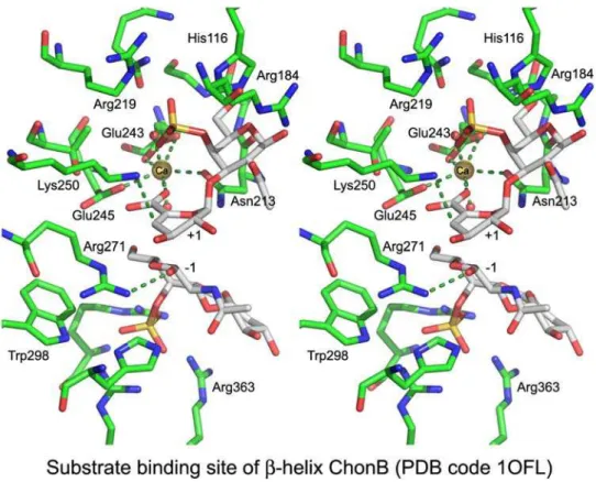
The metal ion-assisted mechanism is found in the β-helix class, (α/α)3/7classes, and β-propeller classes (Table III), while the Asx/Glx-assisted mechanism is found in the remaining clas-Fig. 9. The substrate-binding site of ChonB (PDB code 1OFL). The Ca2 +ion (yellow ball) neutralizes the acidic group of +1 sugar; Lys250 is proposed to act as the Brønsted base and Arg271 as the Brønsted acid. Relevant close contacts are marked with dashed lines.
M-L Garron and M Cygler
at Canada Institute for STI on March 30, 2011
glycob.oxfordjournals.org
ses: (α/α)4barrel, the multidomain (α/α)5,6toroid, the β-jelly
roll, the triple-stranded β-helix, and (likely) the β-sandwich (Table IV). Interestingly, there is a correlation between the type of substrate and the mechanism of a corresponding lyase that depolymerizes this substrate. Pectin/pectate degradation is associated exclusively with metal-assisted mechanism while all other polysaccharides for which the enzymatic mechanisms have been worked out are processed by lyases utilizing the Tyr/His mechanism (Table V). DS is an exception, in that en-zymes utilizing either mechanism are known: ChonB utilizes a Ca2 +-dependent mechanism while ChonABC employs a His/ Tyr-dependent mechanism. The mechanism utilized by several families is still not fully characterized. These include RLs from PL4 and PL11, alginate and glucuronan lyases from PL14, and oligogalacturonan lyases from PL22. Nevertheless, all these lyases are likely associated with metal-assisted mechanism be-cause a metal ion was found in their putative active sites.
Calcium-assisted β-elimination
Enzymatic studies of lyases from several PL families showed that the presence of calcium increases enzymatic activity, therefore raising the question of its role in catalysis. As men-tioned above, only some enzymes utilize a Ca2 +ion(s) in their active sites and it is plausible that a more general role for cal-cium is to neutralize the highly negatively charged lyase
substrates. Structural studies identified families PL1, PL2,
PL3, PL6, PL9, PL10, PL11, and PL22 as requiring metal ions to neutralize the acidic group of the uronic acid (Table I).
The β-Helix Class. We will discuss the active site in this class using the E. chrysanthemi PelC structure (PDB code
1AIR) as a prototype of this fold (Scavetta et al. 1999). PelC utilizes up to four Ca2 +ions to help bind the pectate substrate. Other Pels employ fewer Ca2 +ions for this purpose. The cal-cium ions bound to PelC neutralize the negative charge of the polyGalA substrate resulting from the presence of the carboxylic groups. Ca2 + ions Ca1, Ca2, and Ca3 (Figure 8) are on the same side of the substrate oligosaccharide and are coordinated by sidechains from an acidic patch constituted by six amino acids (Asp129, Asp131, Asp160, Asp162, Asp170, and Glu166). Calcium Ca4 has fewer contacts and appears to have partial occupancy suggesting its lesser importance. The side of the polyGalA substrate opposite to the Ca2 +ions interacts with
three basic amino acids, Lys172, Arg223, and Arg245. The carboxylic group of the sugar in the +1 position is neutralized by interaction with Ca2 +ions Ca2 and Ca3 and with Lys190. In the PelC structure, only Arg218 is well positioned to be the Brønsted base (Figure 8) and, indeed, mutation of this residue to a lysine inactivates the enzyme (Herron et al. 2003). More-over, this arginine is strictly conserved through the PL1 family, and although unusual in the role of a proton acceptor, its func-tion is consistent with the optimal pH for pectate lyases of between eight and nine. The role of an arginine as a general base is not unique to pectate lyases but has also been observed in several other enzymes including IMP dehydrogenase, fuma-rate reductase, and L-aspartate oxidase (Guillen Schlippe and Hedstrom 2005). As no other residues are in sufficiently close proximity to the bridging O4 oxygen between −1 and +1 sugars, a water molecule is the most likely the proton donor. In other PL1 pectate lyases, the three main calcium sites are well conserved, although some variations occur in the arrange-ment of amino acids around the Ca2 site. Of the residues Fig. 10. The convergent evolution of active sites in β-helix (PDB code 2EWE, cyan backbone), (α/α)3(PDB code 1GXO, green backbone), and (α/α)7(PDB code
2V8K, yellow backbone) folds. In each case, an arginine (marked Arg) plays the role of the Brønsted acid maintaining similar position of its guanidinium group relative to the +1 sugar despite approaching from different directions. The Ca2 +(or Mn2 +in case of 2V8K) neutralizes the acidic moiety of +1 sugar approaching from different sides.
at Canada Institute for STI on March 30, 2011
glycob.oxfordjournals.org
forming the basic patch in PelC, only Arg223, which hydrogen bonds with the C-3 and C-4 hydroxyl groups of the sugar in the +1 subsite, is conserved in PL1 family, suggesting that these interactions are important for proper orientation of the sub-strate in the active site.
Interestingly, the Ca2-assisted mechanism is not fully con-served in all enzymes of the PL1 family. Several sequences
of eukaryotic pectin lyases eliminate the need for Ca2 + ions
by introducing the guanidinium group of an arginine in an ap-propriate position to neutralize the charge of the acidic group of the substrate. Pectin is a highly methylated substrate, more
susceptible to degradation (Abbott and Boraston 2008). In the
active site of pectin lyases, the residues of the acidic patch in-volved in Ca2 +binding are not conserved, and the binding site
is enriched in apolar sidechains, predominantly the aromatic residues, tryptophan and histidine (Mayans et al. 1997). This is consistent with the requirements introduced by the less polar, methylated pectin substrate. A Ca2 +ion does not bind in the active site of pectin lyases and instead an arginine sidechain, which replaces one of the Ca2 +-coordinating glutamates, neu-tralizes the acidic group of the uronic acid in the +1 subsite. This arginine corresponds to Arg176 in pectin lyase A (PelA), replacing the Ca2 +ligand Glu166 in PelC. However, the cata-lytic arginine that plays the role of a Brønsted base in PelC is strictly conserved in pectin lyases and, similarly, a water mole-cule most likely plays the role of a Brønsted acid.
A somewhat different active site has evolved in families PL3 and PL9. Pectate lyases from these two families have generally
lower specific activity than PL1 pectate lyases (Roy et al.
1999; Creze et al. 2008). For example, Pel9A (PL9 family) is a constitutive pectate lyase that alerts bacteria to the presence of substrates and leads to the activation of the expression of other, more active degrading enzymes (Jenkins et al. 2004). These lyases utilize a lysine as a Brønsted base instead of an arginine playing this role in other families. PL9 members retain the Ca2 +ion for neutralization of the acidic group of the sub-strate, similar to other pectate lyases. Only one Ca+ 2 ion is present in the lyases from PL3 and PL9 families and it corre-sponds structurally to Ca2 in the PelC structure. This Ca2+is located between the carboxylic groups of sugars in the −1 and + 1 positions. In the PL9 family, four aspartate residues (Asp209, Asp233, Asp234, and Asp237) stabilize the calcium involved in neutralization of the +1 carboxylic group. Unfor-tunately, the available structures of lyases from family PL3 are without Ca2 +ion in the active site and, therefore, its exact po-sition is uncertain (Creze et al. 2008). Nevertheless, Asp173, Asp195, and Glu194 present in PelI (PL3 family) form an acidic patch near the +1 sugar and are the likely ligands of Ca2 +ion. Superposition of representatives from PL3, PL9, and PL1 fam-ilies based only on their substrates shows that the location of the crucial Ca2 +ions and the Brønsted base (Arg or Lys) is con-served among them. Therefore, the key residues in the metal-assisted mechanism are present in all these enzymes—the basic residue (Arg or Lys) playing the role of a Brønsted base, a water molecule acting as a Brønsted acid, and Ca2 +to neutralize the acidic group of the +1 sugar.
Whereas a link between the substrate and the enzymatic mechanism seems to be clear for the majority of PLs, the PL6 family presents an interesting exception. This small family (~20 sequences) contains alginate lyases and the only
chon-droitinase outside of the PL8 family, ChonB, specific for
IdoA-containing DS. The PL6 family is the only lyase family
associated with the Ca2 +-assisted mechanism that contains
enzymes degrading substrates other than pectate/pectin. More-over, all other enzymes cleaving the glycosidic bond next to
IdoA use a different enzymatic mechanism (see below). In
the ChonB structure, calcium is located in an acidic patch formed by Asn213, Glu243, and Glu245 and two water mole-cules. In addition to using calcium for charge neutralization, ChonB also utilizes a basic residue, Lys250, as the Brønsted base. However, unlike the other β-helix metal-assisted en-zymes, it utilizes the sidechain of Arg271 as the Brønsted
acid rather than a water molecule (Figure 9). The role of
Arg271 was confirmed by mutagenesis (Michel et al. 2004). Although the overall fold of PL6 family lyases is the same as that of other β-helix lyases, the orientations of their sub-strates differ considerably and the location of the active site and Ca2 +-coordinating residues are not superposable with the other β-helix PLs.
The (α/α)3/7Toroid Class. Pectate lyases having an (α/α)3/7
toroid fold belong to the PL2 and PL10 families and also utilize a metal-assisted mechanism. Whereas majority of the pectate lyases are extracellular enzymes, the PL2 family contains
peri-plasmic and cytoperi-plasmic enzymes (Rodionov et al. 2004). This
difference in subcellular localization affects the type of metal involved in their catalytic activity. Experiments with PL2A from Yersinia enterocolitica with an (α/α)7toroid fold showed
that Mn2 +rather than Ca2 +is a more effective cation for recov-ering activity following metal depletion by EDTA addition (Abbott and Boraston 2007). The structure of this enzyme has been determined bound to either Mn2 + ion or to trigalac-turonic acid, and superposition of both these structures shows that Mn2 +binds opposite to the carboxylic group of the sugar in the +1 subsite. The Mn2 +is coordinated by His109, His172, Glu130, and three water molecules. Despite a different fold and different metal ion, the active site topology is very similar to that observed in β-helix fold PL1 enzymes (Abbott and Boraston 2007). Superposition of the active site residues of PelC(PL1) and Pel2A(PL2) based on the sugars in the +1 po-sition shows spatial conservation of several residues: Glu130 of PL2A and Glu166 of PelC are involved in metal coordina-tion while Arg272 of PL2A and Arg233 of PelC stabilize the C2 and C3 hydroxyl groups of the +1 sugar (Figure 10). The position of the Brønsted base is not strictly conserved but the guanidinium group of Arg171 of PL2A is in a similar position to that of Arg218 in PelC. In both cases, it is expected that a water molecule acts as the Brønsted acid.
Analysis of the active site of the polygalacturonic acid lyase Pel10Acm from Cellvibrio japonicus (PL10) belonging to the (α/α)3incomplete toroid fold shows that it also has spatial
sim-ilarities to the enzymes from the PL1 family (Charnock et al.
2002). Superposition of Pel10Acm and PelC based on the po-sitions of their +1 sugars of their respective substrates shows several structurally conserved amino acids (Figure 10). The Ca2 +site in Pel10Acm is very close to Ca2 in PelC and inter-acts in a similar way with the carboxylic groups of the −1 and +1 sugars. In both families, the Ca2 +ion is stabilized by a bidentate interaction with Asp451 in Pel10Acm and the corresponding Glu166 in PelC. This Ca2 + is roughly equidistant from both carboxylic groups and its interaction is completed by three M-L Garron and M Cygler
at Canada Institute for STI on March 30, 2011
glycob.oxfordjournals.org
water molecules. Pel10Acm also possesses an arginine, Arg524, in the same position as the Brønsted base Arg218in PelC. Based on the active site configuration of Pel10Acm, a water molecule is expected to be the Brønsted acid. In addi-tion to catalytic amino acids, the basic patch formed by Arg610 and Arg625 in Pel10Acm corresponds to Arg223 and Arg245 in PelC.
These similarities in the active sites between pectate lyases
belonging to three different folds, (α/α)3incomplete toroid,
(α/α)7toroid, and β-helix, represent a clear example of a
con-vergent evolution to a common active site employing a similar catalytic mechanism.
The β-Propeller Class. The enzymes with this fold belong to the PL11 and PL22 families. The structures of endo- and
exo-RLs from PL11 family show the presence of two Ca2 + ions
within the substrate binding cleft (Ochiai et al. 2009). The
Ca2 +ions are coordinated by the Asp153 and Asn596 side-chains, the Ala594 and Asn592 main chain oxygens and two water molecules (site 1) and by His363, His399, Asp401, and Glu422 (site 2). The structures of several complexes with dif-ferent sugars bound in the active site combined with molecular modeling led to the conclusion that the Ca2 +ions neutralize the carboxylic group of the +1 sugar (Ochiai et al. 2009). The nature of the other catalytic residues requires further investigation. Based on the comparison with other metal-dependent PLs, we propose that Lys535 or Arg452 could act as the Brønsted base.
In PL22 oligogalacturonan lyase, there is a Mn2+situated at the open part of the β-propeller (PDB code 3C5M). This ion is
stabilized by three histidines (His287, His353, and His355) and Gln350. This structural observation is in agreement with the observation that a related E. chrysanthemi lyase is more active
in the presence of Mn2 + than Ca2 + (Shevchik et al. 1999).
Based on metal-assisted mechanism used by all the pectate lyases, we hypothesize that, as in PL2, the Mn2 +ion would act as the neutralizer. Moreover, Arg349, situated in the vicin-ity of the Mn2 +-binding site, could play the role of a Brønsted base and a water molecule to be the Brønsted acid.
Histidine/tyrosine active site
Whereas the calcium-assisted mechanism is mainly associated with the β-helix fold, pectate lyases, and anti elimination, the His/Tyr mechanism is used by various enzymes acting on GlcA/IdoA-containing polysaccharides and by alginate lyases. The presence of C-5 epimers in the substrates is associated with the His/Tyr-containing active sites and indicates the ability of these lyases to perform both anti as well as syn elimination. Principally, two classes share this active site architecture, the multidomain (α/α)4/5,6toroid and the β-jelly roll. This active
site topology appears to be more consistent between different
folds than is the case with the calcium-assisted mechanism. The (α/α)nToroid Class. The multidomain (α/α)ntoroid
sub-class—This subclass contains three families, PL8, PL15, and
PL21. As described above, PL8 and PL21 families contain lyases that act on very similar substrates with GlcA or its C5 epimer IdoA in the +1 position, while PL15 lyases act on algi-nate. In the PL8 family, except for ChonABC, all enzymes
Fig. 11. The substrate-binding site and the catalytic tetrad in (α/α)5,6incomplete toroid lyases shown for Arthrobacter aureus ChonAC (PDB code 1RWH).
The hydrogen bonds are marked by dashed lines. The active site tetrad is comprised of Tyr242, His233, Arg296, and Glu407 and is shown with carbon atoms colored yellow. The other residues surrounding the substrate are colored green, and the oligosaccharide is shown with gray carbon atoms. The neutralization of the acidic group of +1 sugar is accomplished by short hydrogen bonds between Asn183 and the acidic group of +1 sugar that promote its protonation.
at Canada Institute for STI on March 30, 2011
glycob.oxfordjournals.org
cleave (1-4) bond linking glucuronic acid to hexosamine (galactosamine for ChonAC, glucosamine for HL, and man-nose for XL) performing syn elimination. ChonABC is a unique enzyme that can digest CS (e.g. GlcA) as well as DS (e.g. IdoA). Four amino acids (Tyr-His-Arg-Glu) involved in
the catalytic mechanism have been identified (Figure 11)
(Féthière et al. 1999;Li et al. 2000;Huang et al. 2001). This tetrad is strictly conserved within the PL8 family. A role for the Brønsted base has been suggested either for a histidine (Li et al. 2000) or for a tyrosine (Huang et al. 2001). The high reso-lution structure of Arthrobacter aurescens ChonAC clearly identified tyrosine in the appropriate distance and orientation to serve as the proton abstracting base (Figure 11) (Lunin et al. 2004). The same tyrosine also plays the role of a Brønsted acid. The dual role of the tyrosine in the catalytic mechanism of chondroitinase AC was further confirmed by kinetic studies (Rye et al. 2006). Moreover, deuterium kinetic isotope experi-ments showed that the deprotonation is the rate-limiting step of the reaction and suggested a step-wise rather than concerted mechanism (Rye and Withers 2002). The histidine is protonated and, together with a conserved asparagine, neutralizes the charge on the acidic group of the +1 glucuronic acid enabling the elimination reaction. The presence of two hydrogen bonds between the asparagine sidechain and the acidic group, found in the high resolution crystal structure, suggests strongly that the acidic group of GlcA is protonated in the enzyme-bound state. The neutralization of the acidic group by these enzymes is therefore accomplished not by a proximal positive charge but by forcing protonation of the acid. Another feature of these enzymes, extrapolated from the structure of ChonAC, is that the binding of the substrate forces the + 1 GlcA into a twist-boat conformation (Figure 11), likely decreasing the activation energy barrier for the reaction by forcing the substrate to adopt a conformation closer to that of the transition state (Lunin et al. 2004).
The same catalytic tetrad as in ChonAC and HLs is also
present in xanthan lyases (Maruyama et al. 2005) and in
Cho-nABC (Huang et al. 2003; Shaya, Hahn, Park et al. 2008). Analysis of the structure of the Bacillus sp. GL1 xanthan lyase with oligosaccharides and the effect of mutagenesis on enzyme activity led to the proposal of a similar mechanism to that de-scribed above (Maruyama et al. 2005). Although no structure of ChonABC with a bound oligosaccharide is available, the importance of the tetrad for catalysis was confirmed by muta-genesis (Prabhakar et al. 2005;Shaya, Hahn, Park et al. 2008). It was proposed that neutralization of the acidic group of GlcA in the + 1 subsite is accomplished by an aspartate sidechain, replacing the asparagine present in ChonAC, and requires its protonation (Shaya, Hahn, Bjerkan et al. 2008). The digestion of CS by ChonABC is likely performed in the same manner as in ChonAC, i.e. tyrosine serves both as the Brønsted base and acid (Shaya, Hahn, Park et al. 2008). The degradation of DS containing IdoA (anti elimination) has been proposed to in-volve His345 as a Brønsted base and its role in catalysis was confirmed by mutagenesis (Shaya, Hahn, Park et al. 2008). This histidine is located on the opposite side of the wide sub-strate binding cleft relative to the tetrad. The role of a Brønsted acid would be performed by the tyrosine from the tetrad. The role of His345 in catalysis was confirmed by mutagenesis (Shaya, Hahn, Park et al. 2008). Both DS and CS substrates use the same binding site but utilize different amino acids in the role of Brønsted base, with the active sites for these two substrates partially overlapping.
HepII, constituting PL21 family, possesses a similar ability as ChonABC to perform syn as well as anti elimination reactions: GlcA-containing HS and IdoA-containing heparin. The catalyt-ically essential residues are Tyr257 and two histidines, His202 and His406. For the IdoA-containing substrate, it was proposed that His406 acts as a Brønsted base and Tyr257 performs as a Brønsted acid, while for GlcA-containing substrate Tyr257
per-Fig. 12. Active site of (α/α)4fold PL (PDB code 1HV6). The trisaccharide present in the structure occupies “−1, −2, and −3” subsites. We have positioned
approximately the sugar in +1 subsite (semi-transparent with cyan backbone). Tyr246 was proposed to act as the base and the acid, Asn191 was proposed to neutralize the acidic group, and His192 stabilizes the system. These three residues are colored yellow.
M-L Garron and M Cygler
at Canada Institute for STI on March 30, 2011
glycob.oxfordjournals.org