Biological responses in stented arteries
Chiraz Chaabane
1, Fumiyuki Otsuka
2, Renu Virmani
2, and Marie-Luce Bochaton-Piallat
1*
1
Department of Pathology and Immunology, Faculty of Medicine, University of Geneva, Rue Michel Servet -1, 1211 Geneva 4, Switzerland; and2
CVPath Institute, Inc., Gaithersburg, MD, USA Received 22 February 2013; revised 1 May 2013; accepted 5 May 2013; online publish-ahead-of-print 10 May 2013
Abstract Vascular walls change their dimension and mechanical properties in response to injury such as balloon angioplasty and endovascular stent implantation. Placement of bare metal stents induces neointimal proliferation/restenosis which pro-gresses through different phases of repair with time involving a cascade of cellular reactions. These phases just like wound healing comprise distinct steps consisting of thrombosis, inflammation, proliferation, and migration followed by remod-elling. It is noteworthy that animals show a rapid progression of healing after stent deployment compared with man. During stenting, endothelial cells are partially to completely destroyed or crushed along with medial wall injury and stretching promoting activation of platelets, and thrombus formation accompanied by inflammatory reaction. Macro-phages and platelets play a central role through the release of cytokines and growth factors that induce vascular smooth muscle cell accumulation within the intima. Smooth muscle cells undergo complex phenotypic changes including migration and proliferation from the media towards the intima, and transition from a contractile to a synthetic phenotype; the molecular mechanisms responsible for this change are highlighted in this review. Since studies in animals and man show that smooth muscle cells play a dominant role in restenosis, drugs like rapamycin and paclitaxel have been coated on stent with polymers to allow local slow release of drugs, which have resulted in dramatic reduction of restenosis that was once the Achilles’ heel of interventional cardiologists.
-Keywords Restenosis † Smooth muscle cells † Endothelial cells † Extracellular matrix † S100A4
-This article is part of the Spotlight Issue on: Biomechanical Factors in Cardiovascular Disease.
1. Introduction
Restenosis or the re-narrowing of the arteries occurs following mechan-ical injury, such as balloon angioplasty and endovascular stent implant-ation. Placement of conventional, bare metal stents (BMS), which requires the use of balloon, initiates immediately a variety of reactions including de-endothelialization of the surface, crushing, and lifting of the plaque from the underlying arterial wall, and tearing and stretching of the medial wall, with eventual neointimal growth as a general healing response. The initial reaction to the mechanical damage induced by stent implantation or balloon angioplasty is platelet activation and thrombus formation accompanied by inflammatory reaction even in the presence of dual antiplatelet therapy. However, the extent of plate-let deposition is significantly higher in the absence of dual antiplateplate-let therapy. This is followed by the activation of chemokines and cytokines which lead to the proliferation and migration of smooth muscle cells (SMCs) within the media and the intima. These cellular responses lead to intimal thickening by SMCs and the surrounding extracellular matrix (ECM) with or without the progression to restenosis.1,2
However, with the advent of drug-eluting stents (DES) since the early to mid-2000, neointimal proliferation and restenosis have been dramatically reduced through the suppression of SMC proliferation and migration accompanied by delayed healing. Differences between animal models and human responses exist such as the underlying athero-sclerotic plaque and its influence on the success and failure of these devices in man; nonetheless, important mechanisms have been eluci-dated by animal studies and will be discussed where appropriate.
2. Pathophysiology of restenosis in
stented arteries
Restenosis is an exaggerated response, and is defined as .75% cross-sectional area luminal narrowing of the stented area by neointimal tissue consisting of SMCs and proteoglycan-collagenous matrix. The time course of re-endothelialization and neointimal growth after stent deployment are different between animal species (e.g. in pig and rabbit) and human iliac and coronary arteries (Figure1).3,4In animal
*Corresponding author. Tel:+1 41 22 379 5764; fax: +1 41 22 379 5746, Email: [email protected]
models, peak neointimal growth is observed at 28 days following BMS placement, whereas in humans, it is identified at 6 – 12 months (Figure 2).4 In the animal models with normal underlying media, restenosis is an uncommon response and only occurs in the presence of severe injury to the underlying medial wall with tearing and a profuse inflammatory response. In pathological human studies, its inci-dence is high: 30 – 40%, and is usually a consequence of severe injury and exaggerated inflammatory response. Recently, we have also shown that atherosclerotic change within the neointima can occur but usually only after 2 years of stent implantation.5
The best animal model that more closely simulate human disease is the porcine coronary artery model; however, the rate of healing is ex-tremely rapid with greater proliferation when compared with man. For endothelialization studies, we have used the rabbit iliac artery model which has a slightly slower rate of re-endothelialization when compared with the porcine model.3Other smaller models like the rat and mice have been used but the arteries are too small and the flow dis-turbances too great due to thick struts that it is not possible to extrapo-late the results to man.
Cellular cascade response to mechanical vascular injury initiated im-mediately after stent deployment can be divided into three different time phases: an early phase consisting of platelet activation and inflammation, followed by an intermediate phase of granulation tissue Figure 1 BMS healing in animals vs. humans. Line plot showing the
temporal relation of peak neointimal growth in animals and humans fol-lowing the placement of bare stainless steel stent. The plots are pre-dominantly derived from morphometric analysis of pig and human coronary stents. In animals, peak neointimal growth in stainless steel stents is observed at 28 days, compared with 6 – 12 months in humans. (Reproduced with permission from Virmani et al.4)
Figure 2 Arterial healing following balloon expandable BMS implantation in human coronary arteries. (A) Platelet-rich thrombus (arrows) is identified around a stent strut (*) 1 day after the placement of Driver stent. (B) A fibrin-rich thrombus (arrowhead) is focally present around a stent strut 1 day after the placement of Express stent. (C – F ) Platelet- (arrow) and fibrin- (arrowheads) rich thrombus (C ) with numerous inflammatory cell infiltration around stent struts (double arrows, C – E) and focal endothelialization (F ). Five days after the placement of Multi-Link Vison stent. Giant cells are occasionally observed (arrows, D). The presence of inflammatory cells consisting of neutrophils and lymphocytes (boxed area in D) are highlighted in (E and G – I ). Early neointima present 23 days after Multi-Link Vision stent placement. (G) Intimal cells within extracellular matrix seen above the stent strut. (H) KP-1 immunostaining identifying macrophages (arrows) adjacent to the strut at base of neointima. (I) Actin staining identifying smooth muscle cells close to luminal surface of neointima (arrows). (J and K ). Smooth muscle cell-rich neointima in stented coronary artery (duration .6 months). Actin staining shows smooth muscle cells close to luminal surface of the neointima in (I ). (A – G) are stained with haematoxylin and eosin, while (J ) is stained with Movat pentachrome.
corresponding to SMC migration and proliferation, and a late phase of tissue remodelling.1,6,7
2.1 The early phase
In the early phase after stent deployment, endothelial cells (ECs) are par-tially to completely destroyed or crushed promoting activation and ag-gregation of platelets, infiltration of circulating leucocytes, and release of growth factors and cytokines.1,2,8
2.1.1 Endothelium injury
It is noteworthy that the early event after BMS implantation involves endothelium injury.9The response of the endothelium to stenting can be divided into three phases: endothelial denudation, re-endothelializa-tion, and/or neoendothelium.6Indeed, in human, in response to coron-ary stent implantation, complete to partial destruction of the EC layer is observed, this initiates the formation of a thin layer of thrombus covering the vascular and stent surface (in the presence of dual antiplatelet therapy and heparin). Complete coverage of the neointima by vascular ECs occurs several weeks after stenting.6 In animal models, many studies have demonstrated that dysfunction of the regenerating ECs after stent implantation promotes mobilization of bone marrow-derived endothelial progenitor cells to accelerate vascular repair through rapid re-endothelialization and attenuate neointimal formation.4,10The con-tribution of the circulating endothelial progenitor cell has not been well demonstrated in patients undergoing stenting although their mobil-ization has been demonstrated to stent site in arteriovenous shunts and an increase in circulating endothelial progenitor cells has been reported with the use of statins.10
2.1.2 Inflammatory response
Endothelium injury and foreign body stent placement lead to the response of the activation of platelets and their deposition at the site of the lesion, with recruitment of circulating leucocytes.2,4Leucocyte– platelet interac-tions are critical in the initiation and progression of neointimal formation. Many studies have shown the importance of inflammation as an early event after stenting in different animal models using specific antibodies to leucocytes (CD45), macrophages (CD14), and monocytes (CD115). The number of adherent monocytes is correlated with neointimal area. Few investigators have correlated the number of macrophages in the neointima to the extent of neointimal formation and have shown that as the inflammation increases so does neointima increase. In human studies, a strong link has been demonstrated between an early chronic in-flammation and intimal thickening in stented coronaries.11Others believe that neutrophils and not macrophages play an important role in the induc-tion of SMC proliferainduc-tion and intimal thickening. In the rabbit model, Welt et al.12have shown that with the use of monoclonal antibody to Mac-1 there was a reduction in neutrophil infiltration and decrease in SMC pro-liferation.
2.2 Granulation tissue formation
The ECs proliferate and migrate over the injured areas while SMCs and macrophages replace the fibrin clot with granulation tissue. The newly formed vascularized tissue includes macrophages which are responsible for phagocytosis of cell debris as well as secretion of growth factors chemokines and cytokines. The macrophages enhance the inflammatory response, but along with platelets and ECs serve to secrete growth factors. These growth factors including fibroblast growth factor (FGF), insulin-like growth factor (IGF), transforming growth factor beta (TGF-b), platelet-derived growth factor (PDGF), and vascular
endothelial growth factor (VEGF) all facilitate the SMC and EC prolifer-ation which are required for the wounded surface to heal.
2.3 Tissue remodelling phase
The hallmark of this phase is the modification of SMCs (see in what follows) activated by growth factors/cytokines produced by the injured endothelium and media, platelets, and infiltrated inflammatory cells as well as by the compressive forces generated in the vessel wall by the stent placement along with low shear stress-induced by stent struts. SMCs undergo complex changes including migration and prolifer-ation from the media towards the intima. Proliferprolifer-ation of SMCs in the media and the subsequent migration into the intima13require the tran-sition of SMCs from a contractile to a synthetic phenotype (see in what follows) with the eventual ECM deposition in the intima.14The ECM plays an important role as it interacts with cells to influence adhesion, mi-gration, proliferation, and remodelling of the wound. In the first phase of matrix formation is the formation of ‘provisional matrix’ which is formed by the plasma proteins such as fibrin, fibrinogen, and fibronectin.15The ECM allows for inflammatory cells like the macrophage and SMCs to adhere and begin the repair process. The SMCs secret hyaluronan and proteoglycans, especially versican that interact and stabilize the fibrin-enriched ECM. This matrix allows more SMCs to bind and prolif-erate while also trapping inflammatory cells that release matrix metallo-proteinases (MMPs) that digest the hyaluronan so that a more permanent matrix collagen type I and III production can occur allowing the wound to completely heal with eventual fibrosis. Collagen also ad-versely affects the SMCs by arresting its proliferation and eventually resulting in altered contractile forces with eventual SMC apoptosis and cell loss. These processes are closely interrelated and difficult to separate in sequential phases.
3. Prospective view on how to
prevent in-stent restenosis
Prior to the introduction of drug-eluting stents for the reduction of re-stenosis many oral pharmacological agents had shown promise in animal studies but all failed when tried in man. The concept of drug therapy to reduce restenosis began in the era of balloon angioplasty. One of the first concepts was that platelets and thrombi were responsible for SMC pro-liferation through the release of PDGF, as a result numerous antiplatelet and antithrombotic drugs were tried with uniform failure in man al-though each had shown promise in animal models. This leads to search for a better understanding of restenosis after vascular injury that occurs during percutaneous coronary intervention (PCI).
Three basic mechanisms have been identified: elastic recoil, negative remodelling, and neointimal hyperplasia. Coronary stenting prevents elastic recoil but is associated with greater neointimal hyperplasia, i.e. al-though many cytokines and mediators are involved, the final common pathway is the entry of SMCs into the cell cycle. It has now been well shown in the porcine model that neointimal hyperplasia is proportional to the extent of vascular injury and it has also been validated in man. SMCs in the media of normal arteries are quiescent with a very low rate of proliferation (SMCs are mostly in quiescent and non-proliferative phase G0); however, once injury occurs, there is an imbalance between stimulatory growth factors and cytokines (e.g. PDGF, interleukin-1, -6, and tumour necrosis factor-a) and inhibitory factors (e.g. endothelial-derived nitric oxide, heparin sulfate proteoglycans) inducing SMC activa-tion, i.e. the SMC change from a quiescent contractile phase into a
synthetic phenotype. Once activated the SMCs then enter into the cycle of division which is divided into four phases: G0, G1, S, G2, and M. A number of growth factors like FGF-2, epidermal growth factor, PDGF, and IGF are responsible for the initiation of VSMC proliferation via the tyrosine kinase receptors (TKRs) (Figure3). The tyrosine kinase recep-tor can also activate the mitogen-activated protein kinase (MAPK) pathway. The cell cycle is the final common pathway that leads to intimal hyperplasia following vascular injury and therefore targeting the regulators of the cell cycle with either cytostatic drugs which alter the expression of cell-cycle promoting or inhibitory proteins or through cytotoxic drugs which result in cell death and necrosis, inducing inflammation and ultimately may even promote restenosis.
The better understanding of the importance of SMC proliferation and migration resulted in the use of immunosuppressant drug like sirolimus
and its analogues (everolmus, zotarolimus, biolimus A9, myolimusand, and others) delivered on stents through the use of polymers. Sirolimus inhibits the mammalian target of rapamycin and prevents the degrad-ation of p27kip1, a cyclin-dependent kinase inhibitor that plays an im-portant role in regulating vascular SMC migration and proliferation.5,6 More pertinent to the process of arterial healing, sirolimus is also a potent inhibitor of EC proliferation by deactivating the p70 S6 kinase pathway, an essential step for cell-cycle progression in response to growth factors. The best results have been reported with the use of the cytostatic drug than cytotoxic drugs16(Figures3and4); however, both have the short-coming of being non-specific in that they inhibit the proliferation of both SMCs and ECs and also permanent polymers induce inflammation. As stated earlier and shown in Table1, cytotoxic drugs like actinomycin D delivered via permanent polymer and
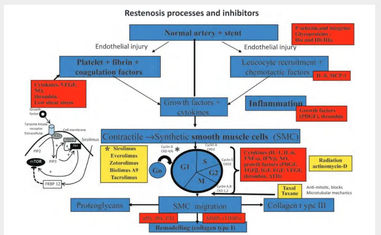
Figure 3 Overview of the molecular mechanisms of restenosis and their inhibitors. The normal coronary artery is injured secondarily from ballooning and stenting resulting in endothelial cells (EC) loss followed by mural thrombus consisting of platelet aggregation with fibrin deposition. The platelets within the thrombus release a number of chemokines essential for the initiation of inflammation. This includes activation of P-selectin and integrins such as b2-integrin Mac-1 and thrombin. Local production of chemokines like interleukin (IL)-1, IL-6, interferon (IFN)g, and tumour necrosis factor-alpha (TNF-a) by macrophages result in the induction of inflammation at the injury site. The injury is not only to the endothelium but also to the underlying medial wall. There is an imbalance between stimulatory growth factors (PDGF, FGF, TGF-b, and IGF-1) and chemokines and the inhibitory factors like endothelial-derived nitric oxide and heparin sulfate proteoglycans that result in the activation of smooth muscle cells (SMCs). The SMCs within the media transform from a quiescent contractile to a synthetic cell which not only proliferate but also migrate into the intima. Within the intima they further proliferate but also secrete extracellular matrix. SMC proliferation involves cell division with different cyclins that are required at different steps of the cell cycle. CDK (cyclin-dependent kinase) activity is also regulated by cell-cycle inhibitory proteins called cyclin-dependent kinase inhibitors (CKIs), which counteract CDK activity. SMC membranes are also stimulated by several classes of tyrosine kinase receptors (TKR). One of the intracellular signalling pathways involves the phosphatidylinositol (PI)3-kinase (PI3K) pathway. PI3K is a lipid kinase that phosphorylates PI. The dominant lipid product generated by PI3K—PI 3,4-biphosphate (PIP2) and PI 3,4,5-triphosphate (PIP3), the latter recruits phosphoinositide-dependent kinase-1 and Akt. Akt is then capable of phosphorylating the mammalian target of rapamycin (mTOR). The yellow squares show the various inhibitors and at which site of the cell cycle they are involved, especially sirolimus and its analogues which inhibit mTOR, whereas paclitaxel blocks microtubular mechanisms. The insert on the left is reproduced with permission from Charron et al.16
high-dose paclitaxel coated directly on a stent without the use of polymer failed in clinical trials because of toxic effects and restenosis at 6 months. The anti-proliferative drug tacrolimus failed because it is 100 times less potent for inhibitory activity on human SMCs. The future may lie in the use of totally erodible polymers without the use of metal, and therefore drug effects will not persist and there is absence of persistent inflammation associated with greater neointimal formation or induces atherosclerotic changes observed in metal-stented segments. Also, one can envision the use of more specific drugs that only target SMCs or use of molecules that enhance endothelialization and gene therapy.
4. Role of EC shear stress
Local haemodynamic factors, in particular low endothelial shear stress has been well recognized as the region where atherosclerotic plaques form and these sites typically include bifurcations at the outer wall and inner curvatures of the arteries. It has been shown that in regions of dis-turbed flow, the balance of gene expression is tipped towards athero-susceptibility. Total endothelial protein kinase C activity in the disturbed flow regions is 145 – 240% that of undisturbed flow locations (P , 0.05), whereas the undisturbed flow regions are not significantly different from each other.17 ECs express proinflammatory mediators like vascular cell adhesion molecule-1 (VCAM-1) which activates p38 and c-Jun N-terminal kinase MAPK by phosphorylation. MAPK phosphatase 1 expression is induced by shear stress, while VCAM-1 is down-regulated.18Similarly, in stented arteries, a stent that protrudes into the lumen will alter the coronary flow and geometry and results in changes in local shear stress distribution.19This results in the develop-ment of vortices where the flow has to turn sharply promoting fluid sep-aration. The blood travelling tangentially to the stent strut surfaces induces large shearing force20 and at these sites, platelet activation occurs with the release of thromboxane and adenosine diphosphate and other procoagulant factors which at critical concentrations activate the coagulation cascade. The destruction of the endothelium during balloon angioplasty and stenting results in the loss of the endothelium removing the benefit of the anti-coagulants produced by the endothe-lium such as nitric oxide, prostacyclin, tissue factor pathway inhibitor, tissue plasminogen activator, thrombomodulin, and heparan. It has been shown in animal experiments that increasing wall shear stress Figure 4 Principal sites of action of anti-proliferative drugs.
Repro-duced with permission from Charron et al.16
. . . . Table 1 Summary of clinical trials of DES (modified from Charron et al.16)
Study (reference) Design Number of points In-stent late loss (mm), DES vs. BMS
In-stent binary restenosis (%) DES vs. BMS Sirolimus RAVEL Multicentre, PRDBC 238 20.01 vs. 0.80, P , 0.001 0.0 vs. 26.6, P , 0.001 SIRUS Multicentre, PRDBC 1101 0.17 vs. 1.00, P , 0.01 3.2 vs. 35.4, P , 0.001 E-SIRUS Multicentre, PRDBC 350 0.20 vs. 1.05, P , 0.0001 3.9 vs. 41.7, P , 0.0001 C-SIRUS Multicentre, PRDBC 102 0.12 vs.1.05, P , 0.001 0.0 vs. 45.5, P , 0.001 Paclitaxel DELIEVER Multicentre, PRSBC 1043 0.81 vs. 0.98, P ¼ 0.0025 14.9 vs.20.6, P ¼ 0.076 TAXUS II* Multicentre, PRDBC 267 (slow) 0.31 vs.0.79, P , 0.001 2.3 vs. 17.9, P , 0.001 TAXUS II** Multicentre, PRDBC 269 (mod) 0.30 vs.0.77, P , 0.001 4.7 vs. 20.2, P , 0.001 TAXUS IV* Multicentre, PRDBC 1314 (slow) 0.39 vs.0.92, P , 0.0001 5.5 vs. 24.4, P , 0.0001 Everolimus
FUTURE I Single centre, PRSBC 42 0.11 vs.0.85, P , 0.0001 0.0 vs.9.1, P ¼ NS FUTURE II Multicentre, PRSBC 64 0.12 vs.0.85, P , 0.0001 0.0 vs. 19.4, P ¼ 0.039 SPIRIT I Multicentre, PRDBC 60 0.10 vs. 0.84, P , 0.0001 0.0 vs. 26.9, P ¼ 0.01 SPIRIT III Multicentre, PRDBC 1002 0.14 vs. 0.28, P ¼ 0.004 4.7 vs. 8.9 P ¼ 0.07 SPIRIT IV Multicentre, PRDBC 3687 – TLF ¼ 4.2 vs. 6.8, P , 0.001 Tacrolimus (PRESENT I) Single centre, registry 22 0.81 + 0.39 19.0
17-beta-estradiol (EASTER) Single centre, registry 30 0.54 + 0.44 6.7
Actinomycin D (ACTION) Multcentre, PRSBC; high-dose group 360 0.93 vs.0.76, P ¼ 0.03 17.9 vs. 11.0, P ¼ 0.38
The cell cycle: a critical therapeutic target to prevent vascular proliferation.
ACTION, ACTinomycin eluting stent ImprovesOutcomes by reducing Neointimal hyperplasia; C, Canada; E, Europe, EASTER, ESTrogen and Stents to Eliminate Restenosis; FUTURE, First Use To Underscore restenosis Reduction with Everolimus; NS, not significant; PRDBC, Prospective, randomized, double-blind, controlled; PRESENT, PREliminary Safety Evaluation of Nanoporous Tacrolimus-eluting stents; PRSBC, Prospective, randomized, single-blind, controlled; RAVEL, Randomized study with the sirolimus-eluting VElocity balloon-expandable stent in the treatment of patients with de novo native coronary artery Lesions; SIRIUS, SIRolimUS-eluting stent in de novo native coronary lesions; TLF, target lesion failure.
from 0.38 to 0.82 N/m2reduces neointimal hyperplasia21and did not change inflammation or injury score. Also, low flow velocity (shear stress) inhibits re-endothelialization of the vessel surfaces and struts. Similarly, it has been shown that when cultured SMCs are in synthetic phenotype, they respond to shear stress in a similar manner to endothe-lial cells, altering the production of growth factors. High shear stress inhi-bits SMC proliferation in vitro via TGF-b1 and tissue-type plasminogen activator.22
Low endothelial shear stress and/or disturbed flow modulate EC mechanosensors (endothelial mechanotransduction). Deformation of the luminal surface of ECs affects structural components such as caveo-lae or glycocalyx, and is transmitted throughout the EC via the cytoskel-eton to intercellular junction complexes and transmembrane proteins (such as cadherins, catenins, platelet EC adhesion molecule-1, integrins). This results in the autophosphorylation of focal adhesion kinases and other MAP kinases that induce the expression of atherogenic genes such as PDGFs responsible for the activation of SMCs23,24
5. Role of mechanical stretch
on SMCs
To maintain a patent lumen and prevent elastic recoil by the artery, im-plantation of the stent stretches the vessel; this yields extremely high, non-physiological, circumferential, and radial wall stress (Laplace law).25These compressive forces produced by stent placement activate intracellular pathways in vascular SMCs (SMC mechanotransduction). Mechanical stretch is transmitted to the SMCs through focal adhesion that links the ECM and the intracellular cytoskeleton.26,27Focal adhe-sions are macromolecular protein complexes consisting of transmem-brane integrins that are associated with intracellular adaptors (paxillin, vinculin, talin) and signalling proteins (focal adhesion kinases). In turn, SMCs undergo phenotypic adaptation. Mechanical stretch induces enhanced SMC proliferation and migration by activating RhoA/Rho kinase, MAPK, TKR (e.g. PDGF receptor), and PI3K/Akt pathways. Stretch-induced SMC proliferation could be mediated by IGF-1, PDGF-BB, and FGF-2. It has been reported that MMPs have differential responses to stretch. The effect of mechanical stretch on SMC survival/ apoptosis is controversial. Interestingly, mechanical stretch induces apoptosis on differentiated SMCs but not on proliferating SMCs. Never-theless, the mechanisms controlling the response of SMCs to mechanical stress remain to be clarified.
SMCs which are embedded in the vascular wall may be exposed to blood flow and shear stress. However, early after endothelial denuda-tion, the superficial layer of SMCs directly in contact with the blood flow is rapidly activated and dedifferentiated, while when intimal thicken-ing appears, the majority of medial SMCs are no longer activated. This result indicates that blood flow acts on arterial SMCs.28,29By mechan-isms similar to those described for ECs (discussed earlier) shear stress promotes PDGF-BB and FGF-2 release from SMCs and in turn their pro-liferation and migration. Shear stress can also modulate SMCs from con-tractile to synthetic phenotype.30
6. Role of SMC heterogeneity
During their accumulation in the intima, SMCs undergo complex changes and switch from a contractile (rich in contractile elements, low proliferative, and migratory activities) to a synthetic phenotype
(rich in endoplasmic reticulum, high proliferative, and migratory activities).
6.1 Features of SMC phenotypes
The contractile phenotype is typical of SMCs in healthy artery wall and corresponds to differentiated SMCs; these SMCs contain many micro-filament bundles and their main function is the contraction – relaxation of the vessel. They express well-defined differentiation markers belong-ing to the cytoskeletal protein family, which are responsible for cell con-traction. Among them, a-smooth muscle actin (a-SMA), the actin isoform typical of vascular SMCs, desmin, an intermediate filament protein expressed in all muscle cells, and SM myosin heavy chains (SMMHCs) isoforms SM1 and SM2, specific of vascular SMCs, are the most studied.14,31,32Their expression is dependent on different cis – ele-ments in their promoter region that are activated by specific binding factors; in particular the CArG (i.e. CC[A/T-rich]6GG) sequence
motif binds serum response factor and myocardin.33 a-SMA is expressed in vascular SMCs even at early stage SMC lineages of develop-ment, and is only exceptionally absent in SMCs, therefore representing the most widespread marker of vascular SMCs, whereas desmin and SMMHCs are markers of well-differentiated SMC markers. Other less extensively studied cytoskeletal proteins, such as smoothelin, SM22a, calponin, heavy-caldesmon, and meta-vinculin, serve as late-differentiation markers (Figure 5). Smoothelin, an a-SMA binding protein, exists in two isoforms: smoothelin-A (59 KDa) and smoothelin-B (110 KDa); the B isoform is predominantly expressed in vascular SMCs. Smoothelin-B plays a crucial role in vascular SMC
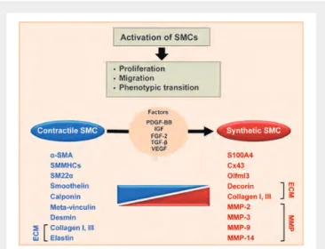
Figure 5 Schema showing smooth muscle cell phenotypic changes after stent implantation. Mechanical vascular injury initiated after stent implantation induces cellular cascade response. Endothelium injury activates platelet deposition at the site of lesion, which induces recruitment of inflammatory cells as precursors to intimal thickening formation. This leads to the activation of smooth muscle cell migration, proliferation, and phenotypic transition from contractile to synthetic phenotype. Transition from a contractile to a synthetic phenotype is associated with changes in smooth muscle cell gene/protein expres-sion: decrease of contractile smooth muscle cell gene/proteins (in blue) and increase of smooth muscle cell synthetic gene/proteins (in red). Well-known growth factors involved in contractile to synthetic phenotypic switch are mentioned.
contraction by still undiscovered mechanisms besides being a reliable marker of the contractile phenotype.34
The synthetic phenotype is typical of SMCs in the media of developing arteries and intima of pathological arteries (atherosclerotic and resteno-tic lesions) and corresponds to poorly differentiated or dedifferentiated SMCs. It is characterized by a cytoplasm with a predominance of rough endoplasmic reticulum and contains a well-developed Golgi apparatus. These changes are correlated to the decrease or loss of the above-mentioned SMC differentiation markers and the ability of SMCs to produce ECM proteins.
The question remains open whether any SMCs of the media could be activated towards a synthetic phenotype or only a pre-existing SMC sub-population is prone to accumulate into the intimal thickening. This pos-sibility is based on the observation that the atheromatous plaque has a monoclonal or oligoclonal origin.35,36This notion has been reinforced by the description of morphologically distinct SMC populations.37 Initial-ly, two populations were identified in vitro in the rat carotid artery and aorta: (i) a spindle-shaped (S) phenotype obtained from the normal media and (ii) an epithelioid (E) phenotype isolated from the intimal thickening 15 days after endothelial injury. We isolated SMC clones exhi-biting S- and E-phenotypes in the rat aorta. E-SMC clones are prevalent in balloon-induced intimal thickening and represent an atheroma-prone phenotype. Distinct SMC phenotypes have been described in vessels of other species such as cow pulmonary artery.38From the normal media of the porcine coronary artery, we isolated two distinct populations, S-and R-SMCs (in this case the rhomboid [R]-SMCs are similar to the rat E-SMCs).39 In the same model, we also demonstrated that a stent-induced intimal thickening yields a large proportion of R-SMCs. R-SMCs share similar features with rat E-SMCs, i.e. high proliferative, mi-gratory, and proteolytic activities and a dedifferentiated phenotype indi-cating that they represent a likely candidate for the formation of intimal thickening and hence an atheroma-prone phenotype.
Exporting the concept of SMC heterogeneity from animal models to human has been hampered by the limited availability of human arterial SMCs and difficult experimental standardization. As a result, the isola-tion of differing SMC subpopulaisola-tions in humans has been sporadically reported over the last decade.37The most conclusive work to date dem-onstrating the presence of SMC populations in human is based on the finding that E-SMCs can be cloned from the media of undiseased arter-ies.40The existence of distinct SMC subpopulations has been reported in saphenous vein of non-diabetic and diabetic patients.41
Saphenous vein SMCs from diabetic patients exhibited an R-phenotype and were more migratory, but less proliferative, when compared with those from non-diabetic patients.
The role for adventitial myofibroblasts in neointima formation in re-stenosis after balloon angioplasty or stent placement, as well as in the de-velopment of atherosclerosis, has long been controversial. Some early studies had suggested that the migration of adventitial myfibroblasts to the media and intima was pivotal to neointima formation, including in rest-enotic lesions after stent placement;42,43recent evidence has corrobo-rated the notion of a negligible contribution of myfibroblasts to this process.44,45Nevertheless, the study of SMC differentiation markers in different well-characterized human coronary lesions, including restenotic lesions after stent implantation, showed that coronary intimal SMCs exhibit a phenotypic profile typical of myofibroblasts,46a stromal cell crucial for wound healing and fibrosis development. Myofibroblasts share with SMCs the expression of a-SMA. SMC-to-myofibroblast differ-entiation may contribute to plaque remodelling, in particular by producing ECM component similar to that of wound healing and fibrotic tissues.
It is well accepted that SMCs within the intima migrate from the media. However, studies have suggested the role of adult vascular progenitor cells in the development of atherosclerosis and vein graft atheroscler-osis.47,48SMC progenitor cells have been mainly identified in the bone marrow (multipotent vascular stem cell progenitors and mesenchymal stem cells), in the circulating blood, and in the adventitia (resident SMC progenitor cells). These progenitor cells have the ability to differ-entiate into SMCs.49,50Although SMC progenitors could play a role in the development of restenotic lesions, their contribution to human in-stent restenosis has not been clearly demonstrated up to date. 6.1.1 S100A4
The comparison of S- and R-SMCs by 2D-gel electrophoresis followed by tandem mass spectrometry allowed us to identify S100A4 as being a marker of the R-SMC population in vitro.51S100A4 is also a marker of intimal SMCs in vivo, both in pig and man: it is strongly expressed in intimal SMCs of porcine coronary artery stent-induced intimal thicken-ing and of human atherosclerotic and restenotic lesions, whereas it is hardly detectable in the underlying media of these lesions.51S100A4 is also up-regulated in arterial SMCs of children with pulmonary hyperten-sion.52More recently, we have observed that medial SMCs of human sac-cular intracranial aneurysms exhibit a dedifferentiated phenotype as demonstrated by decreased a-SMA and SMMHC expression, dis-appearance of smoothelin, and the unexpected strong up-regulation of S100A4 when they are compared with that of non-aneurysmal arter-ies. Therefore, S100A4 represents a reliable marker of the synthetic/ dedifferentiated SMC phenotype in vivo. Nevertheless, the possibility that a distinct SMC subpopulation is present in the healthy artery remains to be demonstrated.
S100A4 is a small calcium-binding protein,53,54known as a mediator of cancer metastasis.54S100A4 is involved in cell proliferation and migra-tion by inhibiting the phosphorylamigra-tion of target proteins in a calcium-dependent manner such as non-muscle myosin heavy chains and p53.54–56 As an extracellular protein, S100A4 through the receptor for advanced glycation end products (RAGE)55induces migration of various cells including tumour cells57,58and ECs and is correlated with increased MMP production.57,58 Some experiments suggest that S100A4 could be involved in SMC phenotypic changes. Extracellular S100A4 leads to human pulmonary artery SMC proliferation and migra-tion through NF-kB activamigra-tion in a RAGE-dependent manner.59,60 S100A4 has also been detected in the culture medium of human pulmon-ary artery SMCs under sustained hypoxia: its blockade with S100A4 neu-tralizing antibody attenuates SMC migration.51,61 We observed that S100A4 is up-regulated in migrating R-SMCs and is implicated in porcine R-SMC proliferation.51Taken together, a better understanding of S100A4 expression, release, and regulation in the SMCs will help to shed light on the mechanisms of SMC phenotypic changes and accumu-lation in the intima.
6.1.2 Other genes and/or proteins
Osteopontin, an ECM protein involved in bone mineralization is a marker of rat E-SMCs. In vivo, it is transiently up-regulated in rat experi-mentally induced intimal thickening and accumulates in calcified areas of the human atheromatous plaque.62Connexin 43, a component of gap junction channels, is strongly expressed in human and mouse athero-sclerotic plaque,63,64 and porcine coronary artery stent-induced intimal thickening.65This correlates in vitro with the high expression of connexin 43 in R-SMCs.65Interestingly, connexin 43 down-regulation reduces atherosclerotic plaque development in mice.63,64 Recently,
we have demonstrated that olfactomedin-like 3, a novel angiogenic factor involved in pericyte migration and coverage of tumour vessels, is elevated in R-SMCs, whereas it is undetectable in S-SMCs.66Studies of these proteins could give further insight into the mechanisms of SMC phenotypic modulation.
MicroRNAs (miRNAs) are short non-coding single-strand RNAs, which regulate post-transcriptionally many genes by binding to the 3′ un-translated regions of target mRNAs. Very recently, some of them have been directly implicated in SMC phenotypic transition and intimal thick-ening formation. In particular, miR-145 is a marker of differentiated SMCs. Overexpression of miR-143 and miR-145 promotes SMC differ-entiation in vitro.67miR-145 is reduced in experimentally induced intimal thickening in the rat carotid artery.68Besides, neointimal formation is reduced in miR-143 and miR-145-knockout mice following carotid artery ligation.69 Quite in contrast, miR-221 and miR-223 are up-regulated in proliferating SMCs in vitro and in intimal SMCs in vivo after balloon-injury of rat carotid arteries.70
6.2 SMC proliferation and migration
SMC proliferation and migration are essential processes in experimental intimal thickening, atherosclerotic and restenotic lesion formation. Pro-liferative and migratory activities have been studied in the distinct SMC phenotypes identified in vitro. Although they stop growing at confluence as a result of cell-contact inhibition, animal E- and R-SMCs show a higher proliferative activity than S-SMCs. It is noteworthy that growth in the absence of serum has been observed in rat E-SMCs and cow R-SMCs.71 Rat E-SMCs produce PDGF-BB, which is a potent SMC mitogen.72Pig R-SMCs do not display autonomous growth;39in this respect, they are similar to human. Apoptosis is an important mechanism in the regulation of intimal thickening evolution. An enhanced suscepti-bility of E-SMCs to apoptosis induced by reactive oxygen species,73 ret-inoic acid, and antimitotic drugs74has been described.
Cell migration, a major event of the intimal thickening, is a complex process that includes the degradation of ECM by proteolytic enzymes. In pig,39rat,75and man,40E- and R-SMCs exhibit a higher migratory ac-tivity compared with S-SMCs. This is correlated to high tissue-type plas-minogen activator activity in rat E-SMCs76and high urokinase activity in pig R-SMCs.39Likewise, rat E-SMCs may produce plasminogen activa-tors, and MMP-2 under particular growth conditions.77Taken together, the studies demonstrating the enhanced proliferative, migratory, and apoptotic activities of E- and R-SMCs in various species fit well with the expected features of participants in intimal thickening (Figure5).
6.3 SMCs and ECM production
ECM of the intimal thickening is synthesized by neointimal SMCs. It mod-ulates important events during the evolution of the restenosis process, including cell proliferation, migration, growth factor expression, and re-modelling. ECM of the intimal thickening to a great extent differs from the ECM of the underlying media that mainly contains fibrillar collagen I and III, proteoglycans and thick fibres of elastin.78ECM of the intimal thickening consists of varying concentrations of proteoglycans (versican, biglycan, and decorin), hyaluronan, and fragmented collagen (types I and III). In stented human coronary arteries for over 18 months, the ECM composition is similar to wound healing or fibrotic tissues. The neoin-tima remodels over time with changes in proteoglycans, such as increase of decorin, and replacement of type III collagen with type I collagen.78 This phenomenon is accompanied by reduced SMC density.79
Intimal SMCs have the ability to remodel their surrounding ECM through the action of MMPs. MMPs are divided into five classes based
on their structure and substrate specificities: collagenases, gelatinases, stromelysins, membrane type MMPs (MT-MMPs), and others. Active MMPs are inhibited by tissue inhibitors (TIMPs).80,81During arterial wound healing (with or without stent implantation), the gelatinases, MMPs-2 and -9, and later MT1-MMP, are implicated in the migration and proliferation of VSMCs, and degradation of ECM components in various animals and man. Nevertheless, knockout of MMP-2, MMP-9, and MMP-14 delay intimal thickening formation in mouse models, which shows that each is necessary.80,81 More recently, it has been shown that MMP-3 is also involved in the activation of MMP-9-mediated VSMC migration and neointimal formation in mice.82These studies in animals and man indicate that several MMPs are involved in ECM remod-elling and restenosis progression (Figure5).
6.4 Factors influencing SMC phenotypes
The injured endothelium, platelets, and inflammatory cells release a variety of factors influencing SMC behaviour. Many of them are well characterized such as PDGF-BB, FGF-2, IGF, TGF-b, angiotensin II, endothelin-1, and interleukins. The chance to orchestrate SMC switch-ing, thus influencing in-stent restenosis may be instrumental for the de-velopment of preventing and therapeutic strategies. Inhibitors of SMC proliferation and/or differentiation factors, e.g. heparin and TGF-b, and stimulators of SMC proliferation and/or dedifferentiation factors, e.g. PDGF-BB and FGF-2 are among the most studied agents employed to modulate SMC phenotype. Some of them show species-specific and phenotype-dependent effects.
Heparin and TGF-b inhibit proliferation and increase differentiation in pig SMCs39 regardless of their phenotype, while only R-SMCs are affected in the bovine model.38The effects on cell morphology are absent or partial depending on the initial phenotype; moreover, a com-plete morphological switch is never achieved. We studied the effects of different cytostatic drugs on porcine S- and R-SMC. Although imatinib may represent valid therapeutic options by promoting SMC quiescence and differentiation,83SMCs regain a contractile phenotype only partially. In human SMCs, only E-SMCs show a strong response to PDGF-BB and FGF-2 in terms of proliferation and to PDGF-BB alone in terms of migration.40 In pig, SMCs both increase migration and proliferation and induce a switch from the S- to the R-phenotype.39The PDGF-driven phenotypic switch is accompanied by an up-regulation of S100A451as well as connexin 43. PDGF – BB induced S-to-R phenotypic change is prevented by a reduction of connexin 43 expression suggesting that con-nexin 43 may play a role in the development of restenosis.65We have also observed that co-culture of ECs with porcine S-SMCs induces a transition towards the R-phenotype; in this experiment, ECs never reached quiescence thus mimicking a dysfunctional endothelium.39 Taken together, these studies offer insights on how microenvironmental factors can influence essential cellular processes (i.e. proliferation, mi-gration, and dedifferentiation) and selectively modulate the behaviour of distinct SMC subpopulations.
7. Shortcomings of existing animal
models
One of the major criticisms of the preclinical animal studies of DES that were carried out in the porcine model was their inability to predict DES associated late stent thrombosis. This was due to the lack of appreciation of differences in the healing responses of the arterial wall when com-pared with BMS at 28 days. Animal studies are performed in healthy
juvenile animals which heal within 28 days following BMS implantation and in DES although there is persistence of peristrut fibrin, nevertheless there is complete endothelialization observed even at 28 days. Endothe-lialization has been shown to be significantly more rapid in the porcine than in the rabbit model, however, both heal rapidly when compared with man, where endothelialization is only complete by 3 – 4 months fol-lowing BMS implantation. Another aspect that deserves mention is the presence of underlying atherosclerosis in man and other plaque factors such as thrombus, as well as the presence of lipid core likely influ-ences the drug retention and therefore healing which is also further delayed with incomplete endothelialization seen even up to 1 year, espe-cially in patients presenting with acute coronary syndrome when com-pared with stable angina.
8. Untoward effects of DES
Restenosis in BMS is usually diffuse, whereas following DES, the process is very focal and is believed to be the result of excessive injury in both, but in DES there may be non-uniform drug distribution due to the increase in inter-strut distance. Arterial healing is generally delayed in DES when compared with BMS, which is characterized by the decrease in intimal thickening due to a suppression of SMCs with persistence of fibrin de-position in the peristrut regions and poor endothelialization since the anti-proliferative effects of drugs are not specific to SMCs but effect EC re-growth (Figure 6). Although restenosis was dramatically reduced with the use of DES, these beneficial effects were at the cost of a higher incidence of late and very late stent thrombosis. Late stent thrombosis was attributed to delayed healing and the presence of uncov-ered struts due to the decrease in endothelialization of stent struts. The use of permanent polymers was also attributed to excessive
inflammation and hypersensitivity reaction. The second generation of stents has reduced late stent thrombosis likely as a result of both thin struts and better polymers. Bioabsorbable polymer usage has also been associated with the decrease in late stent thrombosis and reduc-tion in target lesion revascularizareduc-tion (Leaders trial). Restenosis al-though dramatically reduced by DES in the first year, but has gradually increased with time in both the first and second generation of DES.
9. Conclusion
Besides differences in neointimal formation after stent implantation in humans and animals, experiments in pig and rabbit have been instrumen-tal to decipher the complex mechanisms underlying the restenotic process. Stent implantation leads to a series of early events including endothelium injury, platelet deposition, and inflammatory response. These events are associated with the mechanical stretch and low shear stress induced by stent struts which promote SMC accumulation in the intima. Therefore, SMCs play a central role in the granulation tissue development, wound healing, tissue remodelling, and finally progression to in-stent restenosis. During these processes, SMCs undergo profound modifications. They acquire high proliferative and migratory capabilities and switch to a synthetic phenotype. Many studies, including ours, on ar-terial SMC heterogeneity yield the isolation in vitro of distinct SMC sub-populations and the identification of genes/proteins typical of the atheroma-prone phenotype. The identification of different SMC pheno-types that can be characterized by specific genes and/or proteins better clarify the role of SMCs in intimal thickening formation. In particular, investigations aimed at specifically influencing the cellular processes (i.e. proliferation, migration, and differentiation) of the atheroma-prone SMC (e.g. porcine R-SMCs) accumulation in the intima, without altering
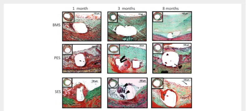
Figure 6 Delayed arterial healing following DES vs. BMS implantation. Time course of arterial healing in BMS, paclitaxel-eluting stents (PES: Taxus DES), and sirolimus-eluting stents (SES: Cypher DES) from 1 to 8 months after stent implantation. Although some peristrut inflammation is observed in BMS at 1 month, complete arterial healing, including a well-established neointimal layer, is seen at 3 and 8 months’ duration. PES shows early fibrin deposition sur-rounding stent struts (*), which persists up to 8 months, as a sign of delayed healing. In contrast, SES shows the predominance of inflammatory cells, including giant cell formation (black arrowheads), at early time points (1 and 3 months), whereas fibrin deposition is stronger at 8 months. Reproduced with permission from Luscher et al.84
the behaviour of quiescent contractile SMCs (e.g. porcine S-SMCs) could be instrumental to design human-tailored therapies that prevent in-stent restenosis. S100A4 identified as being typical of the atheroma-prone phenotype and relevant to the human restenotic lesions repre-sents a potential target for this purpose.
Through a greater understanding of the mechanisms responsible for restenosis following BMS, DES evolved and these have successfully reduced restenosis. However, even DES have their short comings in-cluding late stent thrombosis and increasing target vessel revasculariza-tion with time; further improvement in stents are needed and perhaps the totally bioerodable scaffold with improvements and newer agents that target only SMCs may become the next revolution in interventional technology.
Conflict of interest: none declared.
Funding
The authors acknowledge the support of the Swiss national Science Founda-tion (grant nos 310030_130700/1 and 310030_146790/1), the FondaFounda-tion Simone et Gustave Prevot, and the Foundation Arte`res.
References
1. Mitra AK, Agrawal DK. In stent restenosis: bane of the stent era. J Clin Pathol 2006;59: 232 – 239.
2. Otsuka F, Finn AV, Yazdani SK, Nakano M, Kolodgie FD, Virmani R. The importance of the endothelium in atherothrombosis and coronary stenting. Nat Rev Cardiol 2012;9: 439 – 453.
3. Finn AV, Nakazawa G, Joner M, Kolodgie FD, Mont EK, Gold HK et al. Vascular responses to drug eluting stents: importance of delayed healing. Arterioscler Thromb Vasc Biol 2007; 27:1500 – 1510.
4. Virmani R, Kolodgie FD, Farb A, Lafont A. Drug eluting stents: are human and animal studies comparable?. Heart 2003;89:133 – 138.
5. Nakazawa G, Otsuka F, Nakano M, Vorpahl M, Yazdani SK, Ladich E et al. The pathology of neoatherosclerosis in human coronary implants bare-metal and drug-eluting stents. J Am Coll Cardiol 2011;57:1314 – 1322.
6. Grewe PH, Deneke T, Machraoui A, Barmeyer J, Muller KM. Acute and chronic tissue response to coronary stent implantation: Pathologic findings in human specimen. J Am Coll Cardiol 2000;35:157 – 163.
7. Inoue T, Croce K, Morooka T, Sakuma M, Node K, Simon DI. Vascular inflammation and repair: Implications for re-endothelialization, restenosis, and stent thrombosis. JACC Car-diovasc Intervent 2011;4:1057 – 1066.
8. Welt FG, Tso C, Edelman ER, Kjelsberg MA, Paolini JF, Seifert P et al. Leukocyte recruit-ment and expression of chemokines following different forms of vascular injury. Vasc Med 2003;8:1 – 7.
9. Kipshidze N, Dangas G, Tsapenko M, Moses J, Leon MB, Kutryk M et al. Role of the endo-thelium in modulating neointimal formation: Vasculoprotective approaches to attenuate restenosis after percutaneous coronary interventions. J Am Coll Cardiol 2004;44: 733 – 739.
10. Padfield GJ, Newby DE, Mills NL. Understanding the role of endothelial progenitor cells in percutaneous coronary intervention. J Am Coll Cardiol 2010;55:1553 – 1565. 11. Burke AP, Kolodgie FD, Farb A, Weber D, Virmani R. Morphological predictors of
arter-ial remodeling in coronary atherosclerosis. Circulation 2002;105:297 – 303.
12. Welt FG, Edelman ER, Simon DI, Rogers C. Neutrophil, not macrophage, infiltration pre-cedes neointimal thickening in balloon-injured arteries. Arterioscler Thromb Vasc Biol 2000; 20:2553 – 2558.
13. Ross R. Atherosclerosis: an inflammatory disease. N Engl J Med 1999;340:115 – 126. 14. Campbell JH, Campbell GR. Smooth muscle phenotypic modulation – a personal
experi-ence. Arterioscler Thromb Vasc Biol 2012;32:1784 – 1789.
15. Wight TN, Potter-Perigo S. The extracellular matrix: An active or passive player in fibro-sis? Am J Physiol Gastrointest Liver Physiol 2011;301:G950 – G955.
16. Charron T, Nili N, Strauss BH. The cell cycle: a critical therapeutic target to prevent vas-cular proliferative disease. Can J Cardiol 2006;22(Suppl. B):41B – 55B.
17. Magid R, Davies PF. Endothelial protein kinase c isoform identity and differential activity of pkczeta in an athero-susceptible region of porcine aorta. Circ Res 2005;97:443 – 449. 18. Zakkar M, Chaudhury H, Sandvik G, Enesa K, Luong le A, Cuhlmann S et al. Increased endothelial mitogen-activated protein kinase phosphatase-1 expression suppresses proinflammatory activation at sites that are resistant to atherosclerosis. Circ Res 2008; 103:726 – 732.
19. Koskinas KC, Chatzizisis YS, Antoniadis AP, Giannoglou GD. Role of endothelial shear stress in stent restenosis and thrombosis: pathophysiologic mechanisms and implications for clinical translation. J Am Coll Cardiol 2012;59:1337 – 1349.
20. Jimenez JM, Davies PF. Hemodynamically driven stent strut design. Ann Biomed Eng 2009; 37:1483 – 1494.
21. Carlier SG, van Damme LC, Blommerde CP, Wentzel JJ, van Langehove G, Verheye S et al. Augmentation of wall shear stress inhibits neointimal hyperplasia after stent im-plantation: Inhibition through reduction of inflammation? Circulation 2003;107: 2741 – 2746.
22. Ueba H, Kawakami M, Yaginuma T. Shear stress as an inhibitor of vascular smooth muscle cell proliferation. Role of transforming growth factor-beta 1 and tissue-type plasminogen activator. Arterioscler Thromb Vasc Biol 1997;17:1512 – 1516.
23. Davies PF. Hemodynamic shear stress and the endothelium in cardiovascular patho-physiology. Nat Clin Pract Cardiovasc Med 2009;6:16 – 26.
24. Chatzizisis YS, Coskun AU, Jonas M, Edelman ER, Feldman CL, Stone PH. Role of endo-thelial shear stress in the natural history of coronary atherosclerosis and vascular remod-eling: molecular, cellular, and vascular behavior. J Am Coll Cardiol 2007;49:2379 – 2393. 25. Timmins LH, Miller MW, Clubb FJ Jr, Moore JE Jr. Increased artery wall stress
post-stenting leads to greater intimal thickening. Lab Invest 2011;91:955 – 967.
26. Haga JH, Li YS, Chien S. Molecular basis of the effects of mechanical stretch on vascular smooth muscle cells. J Biomech 2007;40:947 – 960.
27. Shyu KG. Cellular and molecular effects of mechanical stretch on vascular cells and cardiac myocytes. Clin Sci (Lond) 2009;116:377 – 389.
28. Louis H, Lacolley P, Kakou A, Cattan V, Daret D, Safar M et al. Early activation of internal medial smooth muscle cells in the rabbit aorta after mechanical injury: Relationship with intimal thickening and pharmacological applications. Clin Exp Pharmacol Physiol 2006;33: 131 – 138.
29. Tada S, Tarbell JM. Interstitial flow through the internal elastic lamina affects shear stress on arterial smooth muscle cells. Am J of Physiol Heart Circ Physiol 2000;278: H1589 – H1597.
30. Shi ZD, Tarbell JM. Fluid flow mechanotransduction in vascular smooth muscle cells and fibroblasts. Ann Biomed Eng 2011;39:1608 – 1619.
31. Yoshida T, Owens GK. Molecular determinants of vascular smooth muscle cell diversity. Circ Res 2005;96:280 – 291.
32. Sartore S, Franch R, Roelofs M, Chiavegato A. Molecular and cellular phenotypes and their regulation in smooth muscle. Rev Physiol Biochem Pharmacol 1999;134:235 – 320. 33. Kawai-Kowase K, Owens GK. Multiple repressor pathways contribute to phenotypic
switching of vascular smooth muscle cells. Am J Physiol Cell Physiol 2007;292:C59 – C69. 34. Rensen SS, Niessen PM, van Deursen JM, Janssen BJ, Heijman E, Hermeling E et al. Smoothelin-b deficiency results in reduced arterial contractility, hypertension, and cardiac hypertrophy in mice. Circulation 2008;118:828 – 836.
35. Benditt EP, Benditt JM. Evidence for a monoclonal origin of human atherosclerotic plaques. Proc Natl Acad Sci USA 1973;70:1753 – 1756.
36. Murry CE, Gipaya CT, Bartosek T, Benditt EP, Schwartz SM. Monoclonality of smooth muscle cells in human atherosclerosis. Am J Pathol 1997;151:697 – 705.
37. Coen M, Bochaton-Piallat ML. Phenotypic smooth muscle cell heterogeneity: implica-tions for atherosclerosis. In: George SJ, Johnson J, eds. Molecular and Cellular Mechanisms Underlying Atherosclerosis. Weinheim: Wiley-VCH; 2010. p. 327 – 342.
38. Frid MG, Dempsey EC, Durmowicz AG, Stenmark KR. Smooth muscle cell heterogen-eity in pulmonary and systemic vessels. Importance in vascular disease. Arterioscler Thromb Vasc Biol 1997;17:1203 – 1209.
39. Hao H, Ropraz P, Verin V, Camenzind E, Geinoz A, Pepper MS et al. Heterogeneity of smooth muscle cell populations cultured from pig coronary artery. Arterioscler Thromb Vasc Biol 2002;22:1093 – 1099.
40. Li S, Fan YS, Chow LH, Van Den Diepstraten C, van Der Veer E, Sims SM et al. Innate diversity of adult human arterial smooth muscle cells: cloning of distinct subtypes from the internal thoracic artery. Circ Res 2001;89:517 – 525.
41. Madi HA, Riches K, Warburton P, O’Regan DJ, Turner NA, Porter KE. Inherent differ-ences in morphology, proliferation, and migration in saphenous vein smooth muscle cells cultured from nondiabetic and type 2 diabetic patients. Am J Physiol Cell Physiol 2009;297:C1307 – C1317.
42. Scott NA, Cipolla GD, Ross CE, Dunn B, Martin FH, Simonet L et al. Identification of a potential role for the adventitia in vascular lesion formation after balloon overstretch injury of porcine coronary arteries. Circulation 1996;93:2178 – 2187.
43. Shi Y, Pieniek M, Fard A, O’Brien J, Mannion JD, Zalewski A. Adventitial remodeling after coronary arterial injury. Circulation 1996;93:340 – 348.
44. Christen T, Verin V, Bochaton-Piallat M, Popowski Y, Ramaekers F, Debruyne P et al. Mechanisms of neointima formation and remodeling in the porcine coronary artery. Circulation 2001;103:882 – 888.
45. De Leon H, Ollerenshaw JD, Griendling KK, Wilcox JN. Adventitial cells do not contrib-ute to neointimal mass after balloon angioplasty of the rat common carotid artery. Circu-lation 2001;104:1591 – 1593.
46. Hao H, Gabbiani G, Camenzind E, Bacchetta M, Virmani R, Bochaton-Piallat ML. Pheno-typic modulation of intima and media smooth muscle cells in fatal cases of coronary artery lesion. Arterioscler Thromb Vasc Biol 2006;26:326 – 332.
47. Tsai TN, Kirton JP, Campagnolo P, Zhang L, Xiao Q, Zhang Z et al. Contribution of stem cells to neointimal formation of decellularized vessel grafts in a novel mouse model. Am J Pathol 2012;181:362 – 373.
48. Torsney E, Hu Y, Xu Q. Adventitial progenitor cells contribute to arteriosclerosis. Trends Cardiovasc Med 2005;15:64 – 68.
49. Campagnolo P, Wong MM, Xu Q. Progenitor cells in arteriosclerosis: Good or bad guys? Antioxid Redox Signal 2011;15:1013 – 1027.
50. Orlandi A, Bennett M. Progenitor cell-derived smooth muscle cells in vascular disease. Biochem Pharmaco 2010;79:1706 – 1713.
51. Brisset AC, Hao H, Camenzind E, Bacchetta M, Geinoz A, Sanchez JC et al. Intimal smooth muscle cells of porcine and human coronary artery express s100a4, a marker of the rhomboid phenotype in vitro. Circ Res 2007;100:1055 – 1062.
52. Greenway S, van Suylen RJ, Du Marchie Sarvaas G, Kwan E, Ambartsumian N, Lukanidin E et al. S100a4/mts1 produces murine pulmonary artery changes resembling plexogenic arteriopathy and is increased in human plexogenic arteriopathy. Am J Pathol 2004;164: 253 – 262.
53. Marenholz I, Heizmann CW, Fritz G. S100 proteins in mouse and man: From evolution to function and pathology (including an update of the nomenclature). Biochem Biophys Res Commun 2004;322:1111 – 1122.
54. Sherbet GV. Metastasis promoter s100a4 is a potentially valuable molecular target for cancer therapy. Cancer Lett 2009;280:15 – 30.
55. Mishra SK, Siddique HR, Saleem M. S100a4 calcium-binding protein is key player in tumor progression and metastasis: Preclinical and clinical evidence. Cancer Metastasis Rev 2012; 31:163 – 172.
56. Helfman DM, Kim EJ, Lukanidin E, Grigorian M. The metastasis associated protein s100a4: Role in tumour progression and metastasis. Br J Cancer 2005;92:1955 – 1958. 57. Bjornland K, Winberg JO, Odegaard OT, Hovig E, Loennechen T, Aasen AO et al. S100a4
involvement in metastasis: deregulation of matrix metalloproteinases and tissue inhibi-tors of matrix metalloproteinases in osteosarcoma cells transfected with an anti-s100a4 ribozyme. Cancer Res 1999;59:4702 – 4708.
58. Mathisen B, Lindstad RI, Hansen J, El-Gewely SA, Maelandsmo GM, Hovig E et al. S100a4 regulates membrane induced activation of matrix metalloproteinase-2 in osteosarcoma cells. Clin Exp Metastasis 2003;20:701 – 711.
59. Lawrie A, Spiekerkoetter E, Martinez EC, Ambartsumian N, Sheward WJ, MacLean MR et al. Interdependent serotonin transporter and receptor pathways regulate s100a4/ mts1, a gene associated with pulmonary vascular disease. Circ Res 2005;97:227 – 235. 60. Spiekerkoetter E, Guignabert C, de Jesus Perez V, Alastalo TP, Powers JM, Wang L et al.
S100a4 and bone morphogenetic protein-2 codependently induce vascular smooth muscle cell migration via phospho-extracellular signal-regulated kinase and chloride intracellular channel 4. Circ Res 2009;105:639 – 647.
61. Frid MG, Li M, Gnanasekharan M, Burke DL, Fragoso M, Strassheim D et al. Sustained hypoxia leads to the emergence of cells with enhanced growth, migratory, and promito-genic potentials within the distal pulmonary artery wall. Am J Physiol Lung Cell Mol Physiol 2009;297:L1059 – L1072.
62. Giachelli CM, Speer MY, Li X, Rajachar RM, Yang H. Regulation of vascular calcification: Roles of phosphate and osteopontin. Circ Res 2005;96:717 – 722.
63. Haefliger JA, Nicod P, Meda P. Contribution of connexins to the function of the vascular wall. Cardiovasc Res 2004;62:345 – 356.
64. Chadjichristos CE, Kwak BR. Connexins: new genes in atherosclerosis. Ann Med 2007;39: 402 – 411.
65. Chadjichristos CE, Morel S, Derouette JP, Sutter E, Roth I, Brisset AC et al. Targeting con-nexin 43 prevents platelet-derived growth factor-bb-induced phenotypic change in porcine coronary artery smooth muscle cells. Circ Res 2008;102:653 – 660.
66. Miljkovic-Licina M, Hammel P, Garrido-Urbani S, Lee BP, Meguenani M, Chaabane C et al. Targeting olfactomedin-like 3 inhibits tumor growth by impairing angiogenesis and peri-cyte coverage. Mol Cancer Ther 2012;11:2588 – 2599.
67. Cordes KR, Sheehy NT, White MP, Berry EC, Morton SU, Muth AN et al. Mir-145 and mir-143 regulate smooth muscle cell fate and plasticity. Nature 2009;460: 705 – 710.
68. Cheng Y, Liu X, Yang J, Lin Y, Xu DZ, Lu Q et al. Microrna-145, a novel smooth muscle cell phenotypic marker and modulator, controls vascular neointimal lesion formation. Circ Res 2009;105:158 – 166.
69. Elia L, Quintavalle M, Zhang J, Contu R, Cossu L, Latronico MV et al. The knockout of mir-143 and -145 alters smooth muscle cell maintenance and vascular homeostasis in mice: Correlates with human disease. Cell Death Diff 2009;16:1590 – 1598.
70. Liu X, Cheng Y, Zhang S, Lin Y, Yang J, Zhang C. A necessary role of mir-221 and mir-222 in vascular smooth muscle cell proliferation and neointimal hyperplasia. Circ Res 2009; 104:476 – 487.
71. Hao H, Gabbiani G, Bochaton-Piallat ML. Arterial smooth muscle cell heterogeneity: implications for atherosclerosis and restenosis development. Arterioscler Thromb Vasc Biol 2003;23:1510 – 1520.
72. Berk BC. Vascular smooth muscle growth: autocrine growth mechanisms. Physiol Rev 2001;81:999 – 1030.
73. Li WG, Miller FJ Jr, Brown MR, Chatterjee P, Aylsworth GR, Shao J et al. Enhanced h(2)o(2)-induced cytotoxicity in ‘epithelioid’ smooth muscle cells: Implications for neointimal regression. Arterioscler Thromb Vasc Biol 2000;20:1473 – 1479.
74. Orlandi A, Francesconi A, Cocchia D, Corsini A, Spagnoli LG. Phenotypic heterogeneity influences apoptotic susceptibility to retinoic acid and cis-platinum of rat arterial smooth muscle cells in vitro: Implications for the evolution of experimental intimal thickening. Arterioscler Thromb Vasc Biol 2001;21:1118 – 1123.
75. Bochaton-Piallat ML, Ropraz P, Gabbiani F, Gabbiani G. Phenotypic heterogeneity of rat arterial smooth muscle cell clones. Implications for the development of experimental intimal thickening. Arterioscler Thromb Vasc Biol 1996;16:815 – 820.
76. Bochaton-Piallat M-L, Gabbiani G, Pepper MS. Plasminogen activator expression in rat arterial smooth muscle cells depends on their phenotype and is modulated by cytokines. Circ Res 1998;82:1086 – 1093.
77. Lau HK. Regulation of proteolytic enzymes and inhibitors in two smooth muscle cell phe-notypes. Cardiovasc Res 1999;43:1049 – 1059.
78. Adiguzel E, Ahmad PJ, Franco C, Bendeck MP. Collagens in the progression and complica-tions of atherosclerosis. Vasc Med 2009;14:73 – 89.
79. Farb A, Kolodgie FD, Hwang JY, Burke AP, Tefera K, Weber DK et al. Extracellular matrix changes in stented human coronary arteries. Circulation 2004;110:940 – 947. 80. Newby AC. Matrix metalloproteinase inhibition therapy for vascular diseases. Vasc
Phar-macol 2012;56:232 – 244.
81. Lijnen HR. Metalloproteinases in development and progression of vascular disease. Pathophysiol Haemostasis Thrombosis 2003;33:275 – 281.
82. Johnson JL, Dwivedi A, Somerville M, George SJ, Newby AC. Matrix metalloproteinase (mmp)-3 activates mmp-9 mediated vascular smooth muscle cell migration and neoin-tima formation in mice. Arterioscler Thromb Vasc Biol 2011;31:e35 – e44.
83. Prunotto M, Bacchetta M, Jayaraman S, Galloni M, Van Eys G, Gabbiani G et al. Cytostatic drugs differentially affect phenotypic features of porcine coronary artery smooth muscle cell populations. FEBS Lett 2007;581:5847 – 5851.
84. Luscher TF, Steffel J, Eberli FR, Joner M, Nakazawa G, Tanner FC et al. Drug-eluting stent and coronary thrombosis: biological mechanisms and clinical implications. Circulation 2007;115:1051 – 1058.