HAL Id: inserm-00175957
https://www.hal.inserm.fr/inserm-00175957
Submitted on 15 Jan 2010
HAL is a multi-disciplinary open access
archive for the deposit and dissemination of
sci-entific research documents, whether they are
pub-lished or not. The documents may come from
teaching and research institutions in France or
abroad, or from public or private research centers.
L’archive ouverte pluridisciplinaire HAL, est
destinée au dépôt et à la diffusion de documents
scientifiques de niveau recherche, publiés ou non,
émanant des établissements d’enseignement et de
recherche français ou étrangers, des laboratoires
publics ou privés.
Effects of blood pressure lowering on cerebral white
matter hyperintensities in patients with stroke: the
PROGRESS (Perindopril Protection Against Recurrent
Stroke Study) Magnetic Resonance Imaging Substudy.
Carole Dufouil, John Chalmers, Oghuzham Coskun, Véronique Besancon,
Marie-Germaine Bousser, Pierre Guillon, Stephen Macmahon, Bernard
Mazoyer, Bruce Neal, Mark Woodward, et al.
To cite this version:
Carole Dufouil, John Chalmers, Oghuzham Coskun, Véronique Besancon, Marie-Germaine Bousser,
et al.. Effects of blood pressure lowering on cerebral white matter hyperintensities in patients with
stroke: the PROGRESS (Perindopril Protection Against Recurrent Stroke Study) Magnetic Resonance
Imaging Substudy.: The PROGRESS MRI Substudy.. Circulation, American Heart Association, 2005,
112 (11), pp.1644-50. �10.1161/CIRCULATIONAHA.104.501163�. �inserm-00175957�
ISSN: 1524-4539
Copyright © 2005 American Heart Association. All rights reserved. Print ISSN: 0009-7322. Online
72514
Circulation is published by the American Heart Association. 7272 Greenville Avenue, Dallas, TX
DOI: 10.1161/CIRCULATIONAHA.104.501163
2005;112;1644-1650; originally published online Sep 6, 2005;
Circulation
the PROGRESS MRI Substudy Investigators
Bruce Neal, Mark Woodward, Nathalie Tzourio-Mazoyer, Christophe Tzourio and for
Marie-Germaine Bousser, Pierre Guillon, Stephen MacMahon, Bernard Mazoyer,
Carole Dufouil, John Chalmers, Oguzhan Coskun, Véronique Besançon,
Recurrent Stroke Study) Magnetic Resonance Imaging Substudy
in Patients With Stroke: The PROGRESS (Perindopril Protection Against
Effects of Blood Pressure Lowering on Cerebral White Matter Hyperintensities
http://circ.ahajournals.org/cgi/content/full/112/11/1644
located on the World Wide Web at:
The online version of this article, along with updated information and services, is
http://www.lww.com/reprints
Reprints: Information about reprints can be found online at
journalpermissions@lww.com
410-528-8550. E-mail:
Fax:
Kluwer Health, 351 West Camden Street, Baltimore, MD 21202-2436. Phone: 410-528-4050.
Permissions: Permissions & Rights Desk, Lippincott Williams & Wilkins, a division of Wolters
http://circ.ahajournals.org/subscriptions/
Subscriptions: Information about subscribing to Circulation is online at
at McGill University on May 18, 2007
circ.ahajournals.org
Effects of Blood Pressure Lowering on Cerebral White
Matter Hyperintensities in Patients With Stroke
The PROGRESS (Perindopril Protection Against Recurrent Stroke Study)
Magnetic Resonance Imaging Substudy
Carole Dufouil, PhD; John Chalmers, MD, PhD; Oguzhan Coskun, MD; Véronique Besançon, MD;
Marie-Germaine Bousser, MD; Pierre Guillon, PhD; Stephen MacMahon, PhD;
Bernard Mazoyer, MD, PhD; Bruce Neal, MD, PhD; Mark Woodward, PhD;
Nathalie Tzourio-Mazoyer, MD, PhD; Christophe Tzourio, MD, PhD;
for the PROGRESS MRI Substudy Investigators
Background—The prevalence of white matter hyperintensities (WMHs) detected on cerebral MRI is associated with
hypertension, but it is not known whether blood pressure lowering can arrest their progression. We report here the results
of an MRI substudy of PROGRESS (Perindopril Protection Against Recurrent Stroke Study), a randomized trial of
blood pressure lowering in subjects with cerebrovascular disease.
Methods and Results—The substudy comprised 192 participants who had a cerebral MRI both at baseline and after a mean
follow-up time of 36 months (SD
⫽6.0 months). At the first MRI, WMHs were graded with a visual rating scale from
A (no WMH) to D (severe WMH). Participants were assigned to a combination of perindopril plus indapamide (or their
placebos; 58%) or to single therapy with perindopril (or placebo). At the time of the second MRI, the blood pressure
reduction in the active arm compared with the placebo arm was 11.2 mm Hg for systolic blood pressure and 4.3 mm Hg
for diastolic blood pressure. Twenty-four subjects (12.5%) developed new WMHs at follow-up. The risk of new WMH
was reduced by 43% (95% CI
⫺7% to 89%) in the active treatment group compared with the placebo group (P⫽0.17).
The mean total volume of new WMHs was significantly reduced in the active treatment group (0.4 mm
3[SE
⫽0.8])
compared with the placebo group (2.0 mm
3[SE
⫽0.7]; P⫽0.012). This difference was greatest for patients with severe
WMH at entry, 0.0 mm
3(SE
⫽0) in the active treatment group versus 7.6 mm
3(SE
⫽1.0) in the placebo group
(P
⬍0.0001).
Conclusions—These results indicate that an active blood pressure–lowering regimen stopped or delayed the progression
of WMHs in patients with cerebrovascular disease. (Circulation. 2005;112:1644-1650.)
Key Words: stroke
䡲 cerebrovascular disorders 䡲 magnetic resonance imaging 䡲 hypertension 䡲 trials
W
hite matter hyperintensities (WMHs) are often
ob-served on brain MRIs in elderly persons
1–5and in
patients with stroke.
6 – 8WMHs, which include areas of
demyelination as well as silent infarcts, are associated with
cognitive impairment or dementia,
4,9 –13depression,
14 –16and
gait disturbances.
17,18Apart from age, the main risk factors
for WMHs are vascular, particularly high blood pressure.
1,2,4Cross-sectional population-based MRI studies have shown a
positive linear relationship between blood pressure and
se-verity of WMHs.
5,19From these studies, it also appears that
people with uncontrolled hypertension have a higher
preva-lence of severe WMH than people without hypertension or
with controlled hypertension.
See p 1525
MRI follow-up studies have shown that the total load of
WMHs may increase over time but will never decrease.
17,20,21To date, it is not known whether it is possible to stop or delay
this progression. Because of the strong relationship between
blood pressure and WMH, it seems plausible that lowering
blood pressure would reduce the incidence of WMHs,
al-though this has never been demonstrated.
8To test this
hypothesis, we conducted the first MRI-based assessment of
WMHs in a substudy of a randomized trial of blood pressure
lowering in patients with a history of cerebrovascular disease,
the Perindopril Protection Against Recurrent Stroke Study
(PROGRESS).
22Received August 18, 2004; revision received March 21, 2005; accepted May 25, 2005.
From the INSERM U708, Paris, France (C.D., V.B., C.T.); The George Institute for International Health, Sydney, Australia (J.C., S.M., B.N., M.W., C.T.); the UMR6194 CNRS-CEA, and Unite IRM, CHU de Caen, France (O.C., P.G., B.M., N.T.-M.); and the Service de Neurologie, Hôpital Lariboisière, Paris, France (M.-G.B., C.T.).
Correspondence to Dr Christophe Tzourio, INSERM Unit 708, Hôpital La Salpêtrière, 75651 Paris Cedex 13 France. E-mail tzourio@chups.jussieu.fr © 2005 American Heart Association, Inc.
Circulation is available at http://www.circulationaha.org DOI: 10.1161/CIRCULATIONAHA.104.501163
1644
Methods
Study Design and Participants
The design of the PROGRESS study has been described in detail elsewhere.22 Briefly, 6105 participants were recruited from 172
collaborating centers in 10 countries between May 1995 and No-vember 1997. Participants were eligible if they had a history of cerebrovascular disease (stroke or transient ischemic attack but not subarachnoid hemorrhage) within the previous 5 years. In addition, participants were required to have no clear indication for (such as heart failure) or contraindication to treatment with an ACE inhibitor. Active treatment comprised a flexible treatment regimen based on perindopril (4 mg daily), with the addition of indapamide (2.5 mg daily, or 2 mg daily in Japan) in those participants for whom the responsible study physician believed that there was no specific indication for nor contraindication to the use of a diuretic. Those participants assigned placebo received tablets identical in appearance to the active agents. The rationale for the use of combination therapy (perindopril and indapamide or double placebo) rather than single-drug therapy (perindopril or single placebo), wherever possible, was to optimize the fall in blood pressure.
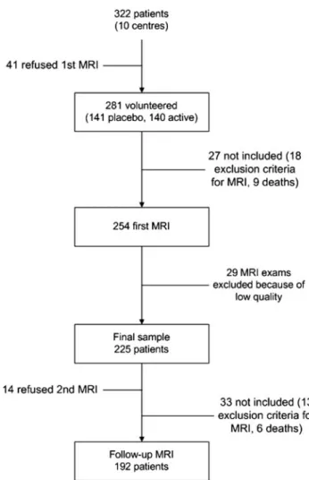
The cerebral MRI substudy was initiated in France in 1995. The St. Antoine ethical committee approved the study, and all patients signed an informed consent. The exclusion criteria included the usual contraindications for MRI, such as prosthetic valves, pacemakers, cerebral aneurysm clips, and a history of intraocular metal fragments, cochlear implants, or claustrophobia. To be eligible, a center needed to have a MRI scanner available and to be able to export the native data. Ten centers were eligible and agreed to participate. Of the 322 patients (159 in the placebo group, 163 in the active treatment group) randomized in these 10 centers, 281 (87%) volunteered to participate (141 in the placebo group, 140 in the active treatment group), but of these, 9 died before their MRI appointment and 18 met 1 of the exclusion criteria (Figure 1). The remaining 254 patients (131 in the placebo group and 123 in the active treatment group) had their first MRI within 6 months of randomization. Among these patients, 29 examinations could not enter the image-processing phase because of low quality, and hence, the final sample included 225 patients (116 in the placebo group and 109 in the active treatment group). The patients randomized who did not participate in the MRI substudy (n⫽97) were older (mean age 64.0 years [SD⫽11.0] versus 60.4 [SD⫽10.8], P⫽0.007) and were more often women (37.5% versus 22.1%, P⫽0.004) than those who participated. These 2 groups did not differ significantly for treatment allocation (44.2% of those who did not participate were assigned to placebo versus 51.6% of those who participated, P⫽0.23) or for hypertension at baseline (59.6% versus 50.9%, P⫽0.14). Similar differences were observed when the group of 192 patients who had both MRI examinations was com-pared with the 130 patients who did not.
A second MRI examination was scheduled to be performed before the end of the follow-up, on average 3 years after the initial scan (range of 24 to 49 months). Follow-up rate was 86% (n⫽192). Among the 33 patients who did not have a follow-up scan, 14 refused, 13 met 1 of the exclusion criteria, and 6 had died (Figure 1). Subjects who did not have a follow-up MRI (n⫽33) were on average older (mean age 68.9 years [SD⫽9.0] versus 59.0 years [SD⫽10.4],
P⬍0.001) and had more frequent severe cerebral WMH on first MRI
(48.5% versus 14.0% respectively, P⬍0.001) than subjects who had a follow-up MRI (n⫽192). Gender distribution, baseline blood pressure levels, and intake of antihypertensive medication were similar in both groups. The follow-up rate was slightly higher for those who were in the placebo group (89%) than for those who were in the active treatment group (82%), but the difference was not significant (P⫽0.14). At each step of the study, the rates of refusals or dropouts did not differ significantly between the active treatment and placebo groups.
Magnetic Resonance Imaging
Baseline cerebral MRI was performed with 1.0-Tesla scanners in 5 centers and 1.5-Tesla scanners in 5 other centers. The following procedures were implemented after visits of the MRI coordinating
team in each center. For each subject, the orbitomeatal line served as the reference line for brain positioning in the field of view, to ensure maximum field homogeneity over the volume of interest. A para-medial sagittal, T1-weighted thick slice was first acquired to delimit the length of the field of view in the z-axis. A high-resolution, T1-weighted brain volume was subsequently acquired with a 3D, fast spoiled gradient echo sequence (3D-FSPGR, 128 1.4-mm-thick slices, 256⫻256 matrix, 0.9⫻0.9 mm2pixel, 24-cm transversal field
of view). Proton-density/T2-weighted images were obtained with a fast multislice double-echo 2D axial acquisition (repetition time 3500 ms, time of echo 1⫽85 ms, time of echo 2⫽140 ms, 5-mm-thick contiguous slices, 256⫻256 matrix, 0.9⫻0.9 mm2pixel,
24-cm transversal field of view). Image-acquisition parameters were identical for both the initial and follow-up MRI examinations.
Because the major goal of the present study was to assess on the follow-up MRI examination the evolution of WMHs, a special procedure had to be implemented to ensure that image interpretation conditions were as similar as possible for both examinations in a given subject. The steps of this procedure were as follows: (1) The follow-up T2 volume was first aligned to the initial one with the automatic image registration algorithm.23(2) Histogram equalization
was then applied to the 2 T2 volumes, thereby ensuring comparable image characteristics in terms of signal intensity. (3) Finally, a PC-based, user-friendly graphic interface was developed to allow the radiologist to visualize side by side the slices of both the initial and follow-up MRI examinations (Figure 2).
Figure 1. Flowchart of participants in the PROGRESS MRI
substudy.
Dufouil et al
The PROGRESS MRI Study
1645
at McGill University on May 18, 2007
circ.ahajournals.org
WMH Assessment
A trained neuroradiologist (OC) blinded to clinical data and treat-ment allocation established all the ratings. At first MRI examination, the magnetic resonance images were rated visually with respect to the presence of WMH with a modified version of a validated scale.5,24This scale provided an overall WMH grade ranging from A
to D, as follows: A, no lesion; B (mild WMH), deep WMHⱕ3 mm or periventricular hyperintensitiesⱕ5 mm; C (moderate WMH), 1 to 10 deep WMHs (4 to 10 mm) or periventricular hyperintensities (6 to 10 mm); and D (severe WMH), more than 10 deep WMHs (4 to 10 mm) or confluent deep WMHs or periventricular hyperintensities ⱖ11 mm. Because of the limited number of subjects, in some analyses we combined categories B and C and used a 3-level grading scale. Comparing baseline and follow-up scans on the same screen, the radiologist also determined the presence of each new WMH that occurred during follow-up, the volume of which was assessed after individual delimitation of the WMH on the computer screen. Volume of incident WMHs was calculated by summing the surfaces of the WMHs on consecutive T2-weighted images, and the slice thickness was used as a third dimension. The presence of prevalent stroke scars was rated by size (small, medium, or large). Stroke scars limited to the white matter were distinguished from WMHs, because they were hypointense on T1-weighted images. Volume of stroke scars was not included in the calculation of volume of prevalent or incident WMH.
Statistical Analysis
All randomized participants who had 2 MRI examinations (n⫽192) were included in the analysis. For baseline comparisons, we used simple2tests for categorical variables and ANOVA for continuous
variables.
We performed logistic regression to assess the relationship be-tween treatment and presence of incident WMH at second MRI. We conducted ANCOVA to compare the total volume of incident WMHs between the active treatment and placebo groups. For both logistic regression and ANCOVA, the initial analyses included adjustment for age, sex, and center. Further adjustments were made for height, stroke type, baseline blood pressure level, antihyperten-sive treatment intake, interval (in months) between the 2 MRI scans, and baseline severity of WMH. To test the hypothesis that the baseline severity of WMH may modify the relationship between treatment allocation and total volume of incident WMH, we added
the WMH-by-treatment interaction term to the above model and tested for its significance. Because the interaction term was signifi-cant, we performed stratified analysis by baseline severity of WMH. We also tested interaction between treatment and age, sex, center, and stroke type. Because distribution of WMH volumes is skewed, we used log-transformed WMH volumes to test for significance. All analyses were performed with SAS version 8.02 (SAS Institute Inc).
Results
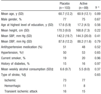
Table 1 describes baseline characteristics by treatment
allo-cation of all randomized study participants who had the 2
MRI examinations. The age of the study participants ranged
from 35 to 85 years, and approximately 75% of them were
men. At study entry, approximately half of the patients were
taking antihypertensive medications, and 52% of the subjects
had hypertension. There was no significant difference
be-tween the 2 treatment groups for age, gender, and vascular
factors (Table 1). With regard to study regimen, 112 patients
(58%) were given the combination of perindopril and
indap-amide (or their placebos), and 80 were given perindopril
alone (or its placebo).
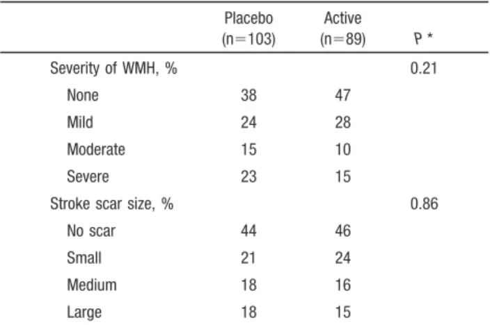
On baseline MRI, 42% of the patients had no WMH, 26%
had mild WMH, 13% had moderate WMH, and 19% had
severe WMH (Table 2). A stroke scar was visible at cerebral
MRI in 55% of the patients. Among patients with a visible
stroke scar, its size was small (
⬍15 mm) in 41%, medium in
30%, and large in 29%. There were no significant differences
for baseline cerebral MRI characteristics between placebo
and active treatment groups. Factors associated with baseline
severity of WMH for the entire sample are shown in Table 3.
There was a significant trend for a relationship between
Figure 2. Illustration of the technique used for MRI assessment.
On the left side of the Figure, there is an axial T2-weighted image of first MRI examination of a PROGRESS patient. On the right side, the image is taken from the second examination per-formed at the end of the follow-up. With registration techniques and histogram equalization, it was possible to make this second examination as similar as possible to the first examination, as seen in the Figure, thus facilitating the comparison between the 2 examinations by the neuroradiologist. Despite the small loss of resolution in the second examination due to the technique, it is possible to visualize new lesions close to the left occipital horn.
TABLE 1. Baseline Characteristics of the PROGRESS MRI Study Participants Placebo (n⫽103) Active (n⫽89) P * Mean age, y (SD) 60.7 (12.2) 60.9 (12.1) 0.89 Male gender, % 77 75 0.67
Age at highest level of education, y (SD) 17.6 (5.9) 17.2 (4.5) 0.56 Mean height, cm (SD) 170.3 (9.0) 168.8 (7.3) 0.22 Mean SBP, mm Hg (SD) 142.2 (19.7) 144.3 (20.0) 0.41 Mean DBP, mm Hg (SD) 87.8 (12.2) 88.2 (11.6) 0.79 Antihypertensive medication (%) 51 48 0.52 Hypertension, %† 50 53 0.65 Current smoker, % 19 20 0.96 History of diabetes, % 15 16 0.97 Mean weekly alcohol consumption (SD)‡ 6.6 (9.7) 5.5 (9.0) 0.37
Type of stroke, %§ 0.65
Ischemic 73 77
Hemorrhagic 11 8
Transient ischemic attack 16 15 SBP indicates systolic blood pressure; DBP, diastolic blood pressure. *Comparison of active treatment and placebo groups based on2test for
qualitative variables and Student’s t test for quantitative variables. †Systolic blood pressureⱖ160 or diastolic blood pressure ⱖ90 mm Hg. ‡In number of standard drinks of alcohol.
§Qualifying (most recent) event for study entry.
increasing age and severity of WMH. The prevalence of
severe WMH was also associated with antihypertensive drug
treatment at entry and with blood pressure level at
random-ization. Mean SBP was 13 mm Hg higher in the group of
patients who had severe WMH than in those who had no
WMH at entry (Table 3).
The second MRI was performed on average 36 months
after baseline MRI (37 months in the placebo group, 36
months in the active treatment group; P
⫽0.10). At the time of
the second MRI, decreases in systolic and diastolic blood
pressure levels compared with baseline measure were
signif-icantly larger in the treated group than in the placebo group.
The mean decrease of systolic blood pressure was
12.5 mm Hg (SD
⫽22.0) in the treated group compared with
1.3 mm Hg (SD
⫽20.0) in the placebo group (P⫽0.0004), and
the mean decrease of diastolic blood pressure was 8.2
(SD
⫽12.3) in the treated group compared with 3.9
(SD
⫽15.5) in the placebo group (P⫽0.04). The proportion of
patients who continued to take randomized therapy at the
time of the second MRI was 79% in the active treatment
group and 88% in the placebo group (P
⫽0.42).
During follow-up, 24 patients developed new WMH, 16
(16%) in the placebo group and 8 (9%) in the active treatment
group (P
⫽0.17; Table 4). Overall, the total volume of new
WMHs was 1.8 mm
3(SE
⫽0.5). The mean (SE) volume of
new WMHs increased with baseline grade of WMH, from
0.05 (0.8) for patients who had no WMH at baseline to
1.2 mm
3(1.2) for those who had a mild to moderate grade and
6.5 mm
3(2.0) for patients with a severe baseline grade of
WMH. Other baseline variables were not associated with the
total volume of new WMHs. The total volume of new WMHs
was significantly lower in the active treatment group
(mean
⫽0.4 mm
3, SE
⫽0.8 mm
3) compared with the placebo
group (mean
⫽2.0 mm
3, SE
⫽0.7 mm
3; P
⫽0.012; Table 4).
We found a significant interaction between baseline severity
of WMH and treatment allocation on the total volume of
incident WMH (P
⫽0.001). We therefore performed stratified
analysis by severity of WMH at baseline and observed that
the treatment effect on the total volume of new WMH was
particularly marked in patients with severe WMH at baseline
(Table 4). Further adjustments for height, stroke type,
base-line blood pressure level, antihypertensive treatment intake,
interval (in months) between the 2 MRI scans, and baseline
severity of WMH gave similar results (Table 4).
Exclusion of subjects who had a stroke (n
⫽20) during
follow-up did not modify the pattern of results. We also
performed separate analyses by study drug regimen
(perindopril-indapamide combination and perindopril
single-TABLE 2. Baseline MRI Characteristics by Treatment
Placebo (n⫽103) Active (n⫽89) P * Severity of WMH, % 0.21 None 38 47 Mild 24 28 Moderate 15 10 Severe 23 15
Stroke scar size, % 0.86
No scar 44 46
Small 21 24
Medium 18 16
Large 18 15
*Comparison of active treatment and placebo groups based on2test.
TABLE 3. Factors Associated With WMH Grade at Baseline
Baseline Grade of WMH None (n⫽86) Mild (n⫽53) Moderate (n⫽26) Severe (n⫽27) P * Mean age, y (SD) 55.9 (10.4) 59.7 (9.2) 61.1 (10.5) 65.5 (9.1) 0.0001 Male gender, % 80 76 69 85 0.49 Mean height, cm (SD) 170.6 (7.8) 168.4 (9.0) 168.2 (8.2) 170.1 (8.3) 0.37 Age at highest level of education, y (SD) 18 17 19 17 0.13 Mean SBP, mm Hg (SD) 136.0 (15.6) 144.6 (21.8) 146.6 (19.3) 149.3 (21.7) 0.003 Mean DBP, mm Hg (SD) 85.1 (10.8) 90.0 (13.4) 90.1 (10.8) 88.9 (12.9) 0.07
Antihypertensive medication, % 30 51 58 67 0.006
Current smoker, % 20 21 19 15 0.50
Diabetes, % 13 6 27 22 0.04
Mean weekly alcohol consumption, mean (SD)† 4.9 (7.3) 7.2 (9.6) 5.7 (7.4) 6.2 (9.4) 0.48
Type of stroke, %‡ 0.64
Ischemic 44 27 15 14
Hemorrhagic 50 20 5 25
Transient ischemic attack 45 32 13 10
SBP indicates systolic blood pressure; DBP, diastolic blood pressure.
*P value from2test for qualitative variables and ANOVA for quantitative variables.
†In number of standard drinks of alcohol. ‡Qualifying (most recent) event for study entry.
Dufouil et al
The PROGRESS MRI Study
1647
at McGill University on May 18, 2007
circ.ahajournals.org
drug therapy), and the results were similar in the 2 subgroups
(data not shown). Interactions between treatment and sex
(P
⫽0.83), age (P⫽0.47), center (P⫽0.96), and stroke type
(P
⫽0.55) on the volume of WMH were not significant.
Discussion
In this placebo-controlled, double-blind trial of a blood
pressure–lowering regimen that combined an ACE inhibitor
(perindopril) and a diuretic (indapamide) in patients with a
history of cerebrovascular disease, patients who received
active treatment had a significantly lower total volume of
incident WMH than patients who received placebo over 3
years of follow-up. The beneficial effect of this blood
pressure–lowering regimen remained significant after
adjust-ment for several variables, including age, gender, stroke type,
baseline blood pressure, and severity of WMH at baseline.
We also observed a 43% (95% CI
⫺7% to 89%) risk
reduction of the occurrence of new WMH in the active
treatment group compared with the placebo group, although
the difference did not reach statistical significance (8/89
[9.0%] versus 16/103 [15.5%], respectively; P
⫽0.17).
WMHs are strongly associated with hypertension,
5,19,25and
the results of some large observational cohort studies suggest
that patients whose hypertension is better controlled have a
more limited progression of WMH.
5,25The present study, the
first to have implemented an MRI-based assessment of WMH
in a randomized trial, confirms that it is possible to limit
WMH progression with a perindopril-based blood pressure–
lowering regimen in patients with cerebrovascular disease. A
post hoc analysis also indicates that the greatest beneficial
effect of antihypertensive therapy on WMH progression was
observed in the group of patients who had severe WMH at
entry. This result is consistent with previous studies showing
that over time, patients with greater lesion volume at baseline
have a greater increase in the number or total volume of
WMHs.
6,17,20,21,26The present findings must be considered in light of the
limited power of the study. During the mean 3-year follow-up
period, 24 of 192 patients developed new WMH, a ratio
consistent with previous studies,
21but these relatively small
numbers affect the power to detect the effect of treatment.
They also do not allow us to give precise estimates of the
treatment effect on the incidence of WMH or the volume of
incident WMH by regimen (perindopril alone or perindopril
plus indapamide) or by stroke type.
7,27With regard to volume
of WMH, we decided per protocol to measure only the
volume of new WMH. Another option would be to also study
the growth of baseline WMH, because it could participate in
the overall increase in volume of WMH. Although the
question addressed would be slightly different, this option
should be considered in future studies to increase their power
to detect a treatment effect. In our estimation of the volume of
new WMH, we carefully excluded scars caused by stroke that
occurred during follow-up, because the treatment effect for
stroke could mask its effect on WMH. Furthermore, of the 20
patients who had a stroke during follow-up, only 2 had new
WMHs, and exclusion of these patients from the analysis did
not modify the results with regard to meaning and statistical
significance. Selection of patients is a potential limitation of
the present study, because those who agreed to participate in
the MRI substudy were healthier than those who refused. This
selection bias, which is usual in MRI studies, had no effect on
the balanced distribution of the variables between the placebo
and the active treatment arm. Furthermore, because the
strongest treatment effect was seen in patients with the most
severe grades of WMH at baseline, that is, in patients more
likely to refuse than agree to participate, the selection bias
observed is probably conservative. We therefore believe that
this selection had no bearing on the validity of the present
results, although it could have affected the study power.
Despite its limited power, our study has several strengths.
Among the patients included in the selected centers, 87%
(281/322) agreed to participate in the study, and among those
who had their first MRI examination and who could
partici-pate, 93% (192/206) agreed to have a second MRI
examina-tion. Overall, despite deaths and patients who met exclusion
criteria for MRI, the participation rate was 85% (192/225)
between both examinations. The study is further strengthened
by the methods used to estimate the volume of WMH.
Limitations of visual scales to evaluate WMH severity, as
well as WMH changes over time, have been discussed
extensively.
28 –30We used a semiautomated method to
iden-TABLE 4. Presence and Volume of Incident WMH by Treatment
Total (n⫽192) Placebo (n⫽103) Active (n⫽89) P Value, Model 1* P Value, Model 2† Incident WMH, n (%) 24 (13) 16 (16) 8 (9) 0.17 0.10 Mean volume of incident WMH, mm3(SE) 1.8 (0.5) 2.0 (0.7) 0.4 (0.8) 0.012 0.009
Volume of incident WMH by initial grade of WMH, mm3(SE)
No WMH 0.05 (0.8) 0 0.09 (0.8) 0.76 0.81
Mild to moderate WMH 1.2 (1.2) 1.3 (1.0) 0.9 (1.0) 0.58 0.71 Severe WMH 6.5 (2.0) 7.6 (1.0) 0 ⬍0.0001 ⬍0.0001 *P value generated from logistic regression for qualitative variables or from ANCOVA with adjustment for age, sex, and center.
†P value generated from logistic regression for qualitative variables or from ANCOVA with adjustment for age, sex, center, height, stroke type, baseline blood pressure levels, and antihypertensive treatment intake, interval (in number of months) between the 2 MRI scans, and baseline severity of WMH (if applicable).
tify incident WMHs and to measure their volume. Special
care was taken in standardizing the quality and reading
conditions of the T2 scans acquired 3 years apart in the same
patient. Finally, this substudy had the same favorable
char-acteristics as the main PROGRESS study in terms of good
adherence to treatment and maintained blood pressure
differ-ence throughout follow-up.
The selection of patients mentioned above limits our ability
to extend the results of this substudy to all patients enrolled in
the PROGRESS trial. The present study showed, in a
partic-ular subset of patients, that it is possible to limit the
progression of WMH. These results now need to be
con-firmed and extended in further clinical trials, such as in
hypertensive patients free of cerebrovascular disease. Our
results could help design such future studies in terms of
number of patients, duration of follow-up, stratification on
baseline severity of WMH, and methods used to estimate the
volume of WMH.
Acknowledgments
The main PROGRESS study was funded by grants from Servier, the Health Research Council of New Zealand and the National Health and Medical Research Council of Australia. The PROGRESS MRI substudy was conducted under an agreement between INSERM (Institut National de la Santé et de la Recherche Médicale) and Servier. The main study and the substudy were both designed, conducted, analyzed, and interpreted by the investigators indepen-dently of all sponsors. The MRI database was managed at UMR 6194 CNRS-CEA (Caen, France), directed by Professor B. Mazoyer. The authors constitute the writing committee for the PROGRESS MRI Substudy Investigators, who are listed below.
PROGRESS MRI Substudy Centers
and Investigators
Hôpital d’Angers: Drs H. Brugeilles, P. Lejeune, C. Moreau (Neu-rology); Dr A. Pasco (Radiology). Hôpital de Caen: Dr S. Iglesias, Professor F. Viader (Neurology); Dr JM. Constans, Professor P. Courtheoux (Radiology). Hôpital de Grenoble: Dr A. Jaillard, Dr G. Besson, Professor M. Hommel (Neurology); Dr S. Grand, Professor J.F. Lebas (Radiology). Hôpital de Lille: Dr C. Lucas, Professor D. Leys (Neurology); Dr X. Leclerc, Professor J.P. Pruvost (Radiol-ogy). Hôpital de Maubeuge: Drs V. Neuville, T. Rosolacci (Neurol-ogy). Hôpital de Meaux: Drs A. Ameri, F. Chedru (Neurology); Dr P. Bérou (Radiology). Hôpital de Nantes: Drs B. Guillon, G. Hinzelin, Professor Feve (Neurology); Drs E. Auffray, H. Desal, Professor De Kersaint-Gilly (Radiology). Hôpital Saint Antoine and Lariboisière, Paris: Drs V. Biousse, K. Berthet, K. Vahedi, Professor M.G. Bousser (Neurology). Hôpital Saint Antoine: Drs V. Bousson, C. Levy, L Brunereau, Professor J. Tubiana (Radiology). Hôpital Tenon, Paris: Dr S. Alamovitch, Professor E. Roullet (Neurology). Hôpital de Saint-Denis: Dr T. De Broucker (Neurology); Dr N. Gauthier, Stroh-Marcy (Radiology). Hôpital de Valenciennes: Dr P. Dobbelaere (Radiology). Hôpital des 15-20, Paris: Dr M.T. Iba-Zizen, Professor S. Cabanis (radiology).
Disclosures
Dr Chalmers has received research grants from Servier for PROGRESS and ADVANCE and has received honoraria from Servier for speaking at meetings. Drs MacMahon, Neal, Woodward, and Tzourio have received honoraria for speaking.
References
1. Bots ML, van Swieten JC, Breteler M, de Jong PTVM, van Gijn J, Hofman A, Grobbee DE. Cerebral white matter lesions and atheroscle-rosis in the Rotterdam Study. Lancet. 1993;341:1232–1237.
2. Breteler M, van Swieten JC, Bots ML, Grobbee DE, Claus JJ, van den Hout JHW, van Harskamp F, Tanghe HLJ, de Jong PTVM, van Gijn J, Hofman A. Cerebral white matter lesions, vascular risk factors and cognitive function in a population-based study: the Rotterdam study. Neurology. 1994;44:1246 –1252.
3. Lindgren A, Roijer A, Rudling O, Norrving B, Larsson EM, Eskilsson J, Wallin L, Olsson B, Johansson BB. Cerebral lesions on magnetic res-onance imaging, heart disease, and vascular risk factors in subjects without stroke: a population-based study. Stroke. 1994;25:929 –934. 4. Longstreth WT, Manolio TA, Arnold A, Burke GL, Bryan N, Jungreis
CA, Enright PL, O’Leary D, Fried L. Clinical correlates of white matter findings on cranial magnetic resonance imaging of 3301 elderly people: the Cardiovascular Health Study. Stroke. 1996;27:1274 –1282. 5. Dufouil C, de Kersaint Gilly A, Besancon V, Levy C, Auffray E, Brunnereau
L, Alperovitch A, Tzourio C. Longitudinal study of blood pressure and white matter hyperintensities: the EVA MRI cohort. Neurology. 2001;56:921–926. 6. van Zagten M, Boiten J, Kessels F, Lodder J. Significant progression of white matter lesions and small deep (lacunar) infarcts in patients with stroke. Arch Neurol. 1996;53:650 – 655.
7. Miyao S, Takano A, Teramoto J, Takahashi A. Leukoaraiosis in relation to prognosis for patients with lacunar infarction. Stroke. 1992;23: 1434 –1438.
8. Inzitari D. Leukoaraiosis: an independent risk factor for stroke? Stroke. 2003;34:2067–2071.
9. Breteler M, van Amerongen NM, van Swieten JC, Claus JJ, Grobbee DE, van Gijn J, Hofman A, van Harskamp F. Cognitive correlates of ventric-ular enlargement and cerebral white matter lesions on MRI: the Rot-terdam Study. Stroke. 1994;25:1109 –1115.
10. Bronge L, Wahlund LO. White matter lesions in dementia: an MRI study on blood-brain barrier dysfunction. Dement Geriatr Cogn Disord. 2000; 11:263–267.
11. Garde E, Mortensen EL, Krabbe K, Rostrup E, Larsson HB. Relation between age-related decline in intelligence and cerebral white-matter hyperintensities in healthy octogenarians: a longitudinal study. Lancet. 2000;356:628 – 634.
12. Koga H, Yuzuriha T, Yao H, Endo K, Hiejima S, Takashima Y, Sadanaga F, Matsumoto T, Uchino A, Ogomori K, Ichimiya A, Uchimura H, Tashiro N. Quantitative MRI findings and cognitive impairment among community dwelling elderly subjects. J Neurol Neurosurg Psychiatry. 2002;72:737–741.
13. Vermeer SE, Prins ND, den Heijer T, Hofman A, Koudstaal PJ, Breteler MMB. Silent brain infarcts and the risk of dementia and cognitive decline. N Engl J Med. 2003;348:1215–1222.
14. Videbech P. MRI findings in patients with affective disorder: a meta-analysis. Acta Psychiatr Scand. 1997;96:157–168.
15. Hickie I, Scott E, Wilhelm K, Brodaty H. Subcortical hyperintensities on magnetic resonance imaging in patients with severe depression: a longi-tudinal evaluation. Biol Psychiatry. 1997;42:367–374.
16. O’Brien J, Ames D, Chiu E, Schweitzer I, Desmond P, Tress B. Severe deep white matter lesions and outcome in elderly patients with major depressive disorder: follow up study. Br Med J. 1998;317:982–984. 17. Whitman GT, Tang T, Lin A, Baloh RW. A prospective study of cerebral
white matter abnormalities in older people with gait dysfunction. Neu-rology. 2001;57:990 –994.
18. Starr JM, Leaper SA, Murray AD, Lemmon HA, Staff RT, Deary IJ, Whalley LJ. Brain white matter lesions detected by magnetic resonance imaging are associated with balance and gait speed. J Neurol Neurosurg Psychiatry. 2003;74:94 –98.
19. Liao DP, Cooper L, Cai JW, Toole JF, Bryan NR, Hutchinson RG, Tyroler HA. Presence and severity of cerebral white matter lesions and hypertension, its treatment, and its control: the ARIC study. Stroke. 1996;27:2262–2270.
20. Schmidt R, Enzinger C, Ropele S, Schmidt H, Fazekas F. Progression of cerebral white matter lesions: 6-year results of the Austrian Stroke Pre-vention Study. Lancet. 2003;361:2046 –2048.
21. Schmidt R, Fazekas F, Kapeller P, Schmidt H, Hartung HP. MRI white matter hyperintensities: three-year follow-up of the Austrian stroke pre-vention study. Neurology. 1999;53:132–139.
22. PROGRESS Collaborative Group. Randomised trial of a perindopril-based blood-pressure-lowering regimen among 6105 indi-viduals with previous stroke or transient ischaemic attack. Lancet. 2001; 358:1033–1041.
23. Woods RP, Grafton ST, Holmes CJ, Cherry SR, Mazziotta JC. Automated image registration, I: general methods and intrasubject, intramodality validation. J Comput Assist Tomogr. 1998;22:139 –152.
Dufouil et al
The PROGRESS MRI Study
1649
at McGill University on May 18, 2007
circ.ahajournals.org
24. Scheltens P, Barkhof F, Leys D, Pruvo JP, Nauta JJ, Vermersch P, Steinling M, Valk J. A semiquantitative rating scale for the assessment of signal hyperintensities on magnetic resonance imaging. J Neurol Sci. 1993;114:7–12.
25. de Leeuw FE, de Groot JC, Oudkerk M, Witteman JCM, Hofman A, van Gijn J, Breteler M. Hypertension and cerebral white matter lesions in a prospective cohort study. Brain. 2002;125:765–772.
26. Taylor WD, MacFall JR, Provenzale JM, Payne ME, McQuoid DR, Steffens DC, Krishnan KRR. Serial MR imaging of volumes of hyper-intense white matter lesions in elderly patients: correlation with vascular risk factors. Am J Roentgenol. 2003;181:571–576.
27. van Swieten JC, Kappelle LJ, Algra A, Van Latum JC, Koudstaal PJ, van Gijn J. Hypodensity of the cerebral white matter in patients with transient ischemic attack or minor stroke: influence on the rate of
subsequent stroke: Dutch TIA Trial Study Group. Ann Neurol. 1992; 32:177–183.
28. Scheltens P, Erkinjuntti T, Leys D, Wahlund LO, Inzitari D, del Ser T, Pasquier F, Barkhof F, Mantyla R, Bowler J, Wallin A, Ghika J, Fazekas F, Pantoni L. White matter changes on CT and MRI: an overview of visual rating scales. Eur Neurol. 1998;39:80 – 89.
29. Kapeller P, Barber R, Vermeulen RJ, Ader H, Scheltens P, Freidl W, Almkvist O, Moretti M, del Ser T, Vaghfeldt P, Enzinger C, Barkhof F, Inzitari D, Erkinjunti T, Schmidt R, Fazekas F. Visual rating of age-related white matter changes on magnetic resonance imaging: scale com-parison, interrater agreement, and correlations with quantitative mea-surements. Stroke. 2003;34:441– 445.
30. Pantoni L, Simoni M, Pracucci G, Schmidt R, Barkhof F, Inzitari D. Visual rating scales for age-related white matter changes (leukoaraiosis): can the heterogeneity be reduced? Stroke. 2002;33:2827–2833.