Characterization of two Bacillus subtilis proteins required for the initiation, restart, and control of DNA replication
by
Megan E. Rokop
B.S., Biology Brown University, 1999
Submitted to the Department of Biology in Partial Fulfillment of the Requirements for the Degree of
Doctor of Philosophy in Biology at the
Massachusetts Institute of Technology September, 2004
© Massachusetts Institute of Technology All rights reserved
Signature of author ... .... .. . A. . ... Department of Biology September 7, 2004
Certified
by ... ...
..
/...- ...
Alan D. Grossman Professor of Biology Thesis AdvisorAccepted
by ...
:
...
...
Stephen P. Bell Professor of Biology Co-chair, Biology Graduate CommitteeCharacterization of two Bacillus subtilis proteins required for the initiation, restart,
and control of DNA replication
by
Megan E. Rokop
Submitted to the Department of Biology on September 7, 2004, in partial fulfillment of the requirements for the degree of Doctor of Philosophy in Biology
ABSTRACT
DnaB and DnaD are essential proteins that function in the initiation and control of DNA replication in Bacillus subtilis. I found that DnaB and DnaD are required to load the replicative helicase onto chromosomal origins during replication initiation. DnaB and DnaD are also
involved in loading helicase during replication restart at sites of stalled replication forks.
Despite the fact that DnaB and DnaD are thought to work together to load helicase, DnaB and DnaD are found in separate subcellular compartments. I showed that DnaB is found in the membrane fraction of cells, and DnaD is found in the cytoplasmic fraction. This separation
could prevent helicase loading during the majority of the cell cycle. I isolated a missense
mutation in dnaB, dnaBS371P, that disrupts the spatial separation of DnaB and DnaD. I isolated dnaBS371P as a suppressor of the temperature sensitivity of dnaBts cells and dnaDts cells. dnaBS3 71P also suppresses the growth defects of ipriA cells, which cannot restart replication at stalled forks. I found that a significant fraction of DnaD is found in the membrane fraction of dnaBS371P cells. In addition, I observed a direct interaction between DnaBS371P and DnaD that is not observed between the wild-type proteins. I hypothesize that the DnaB-DnaD
interaction is regulated, thereby controlling when these two proteins converge at the membrane to coordinate helicase loading.
dnaBS3 71P cells lack proper control of replication, suggesting that the spatial separation of DnaB and DnaD is an important mechanism of replication control in B. subtilis. I showed that dnaBS371P cells over-initiate replication when grown slowly in minimal medium, but contain decreased DNA content when grown faster, in rich medium. This is consistent with an inability of dnaBS371P cells to adjust the frequency of initiation according to growth rate. I also found that cells over-producing DnaBS371P are filamentous, contain decreased DNA contents, and are hypersensitive to DNA-damaging agents. These abnormalities may result from a defect in
replication restart at damaged or stalled replication forks. Thus, whereas dnaBS3 71P suppresses the defects of mutant cells that cannot initiate or restart replication, expressing DnaBS371P in wild-type cells causes defects in initiation and restart.
Thesis Advisor: Alan D. Grossman Title: Professor of Biology
Acknowledgements
There are many people who I would like to thank for their help during my time in graduate school. I would like to thank Alexi Goranov and Jade Wang for performing experiments that are described in Chapter 3 of this thesis. I thank Jenny Auchtung for many things, including helping me to set up the ChIP assay in Chapter 2, giving me many helpful comments on several chapters of this thesis, and being a friend for years. I thank Bill Burkholder for being an incredible baymate for several years, and for teaching me everything I ever wanted to know about bacterial genetics. I thank David Rudner and Soni Lacefield Shimoda for
seemingly endless advice about experimental protocols and for plenty of reagents.
There are also many people who were generous enough to donate lots of time to reading and commenting on my thesis. These people include Lourdes Aleman, Milan deVries, C. Lee, April Risinger, and Lyle Simmons. I would especially like to thank Lourdes and Lyle for being
incredibly helpful and remaining energetic through multiple rounds of revision. I would also like to thank Milan for the advice, support, and mounds of constructive criticism that he has given me
ever since our first day of graduate school.
Many people provided me with reagents, experimental advice, and helpful tidbits of information along the way. I would like to thank Christy Collins Taylor, Judy Healy, Katherine Lemon, Petra Levin, and numerous members of the Kaiser, Bell, and Solomon labs for help in these areas. In particular, I thank Judy Healy for being a great lab room-mate and, thankfully, a
good listener. In addition, many past and present members of the Grossman lab were kind and helpful to me, especially Natalia Comella and Rita Monson, who are wonderful lab-mates and friends. I am lucky and grateful to have had them around throughout my experience in graduate school.
Finally, I would like to thank my advisor, Alan Grossman, and the members of my thesis committee, Steve Bell, Chris Kaiser, Frank Solomon, and Andrew Wright, for the advice and support they gave me over the past years. My special thanks are reserved for Frank, for whom I have a tremendous amount of respect. Thank you, Frank, for being there from the very
Table of Contents Abstract Acknowledgements Table of Contents List of Tables List of Figures Chapter 1: Chapter 2: Introduction
Control of DNA replication initiation by recruitment of an essential initiation protein to the membrane of Bacillus
subtilis Appendix A: Appendix B: Appendix C: Chapter 3: Chapter 4:
Isolation and characterization of suppressors of the temperature sensitivity of dnaBts and dnaDts Bacillus subtilis cells
The essential replication initiation factor DnaB from Bacillus subtilis is not an integral membrane protein The dnaBS371P allele causes a reduction in the ability of DnaB to multimerize in the yeast two-hybrid assay Effects of a mutation in dnaB on Bacillus subtilis DNA replication and cell division
Discussion 3 5 7 9 11 13 89 133 161 183 191 239
List of Tables
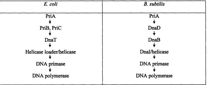
Chapter 1 Table 1 The pathways of replication restart in E. coli and B. 77
subtilis.
Chapter 2 Table I B. subtilis strains used. 125
Appendix A Table 1 B. subtilis strains used. 155
Table 2 A summary of suppressors of the temperature 156 sensitivity of dnaD23ts cells.
Table 3 A summary of suppressors of the temperature 157 sensitivity of dnaB134ts cells.
Table 4 A summary of suppressors of the temperature 158 sensitivity of dnaB19ts cells.
Appendix B Table 1 B. subtilis strains used. 179
Chapter 3 Table 1 B. subtilis strains used. 230
Table 2 DNA content of dnaBS371P cells and cells over- 230 producing DnaBS371P grown in defined minimal
medium at 37C, as measured by DNA/protein ratios.
Table 3 Percentage of cells containing each number of 231 origins per cell, as measured by number of
SpoOJ-GFP foci.
Table 4 DNA content of dnaBS371P cells and cells over- 231 producing DnaBS371P grown in rich medium at
Chapter 1
Chapter 2
Appendix A
List of Figures
Figure 1 A comparison of prokaryotic and eukaryotic replication initiation.
Figure 2 The steps of replication initiation in E. coli. Figure 3 The single, circular B. subtilis chromosome. Figure 4 The cell cycle in slow-growing prokaryotes. Figure 5 The cell cycle in fast-growing prokaryotes. Figure 6 The importance of replication timing.
Figure 7 Contrasting synchronously and asynchronously replicating cells.
Figure 8 SeqA-mediated origin sequestration in E. coli.
Figure 9 A model for the spatial control over replication initiation in B.
subtilis.
Figure 10 The positioning of the FtsZ cell division ring is influenced by nucleoid occlusion and the Min division inhibitors.
Figure 11 Asynchronous replication can lead to asymmetric cell division.
Figure 1 DnaD and DnaB function is necessary prior to the loading of helicase at oriC.
Figure 2 DnaD and DnaB both share sequence similarity with Gp49, a putative phage replication initiation protein.
Figure 3 DnaD is recruited to the membrane fraction of cells by DnaBS371P.
Figure 4 DnaD interacts with DnaBS371P.
Figure 5 Phenotypes associated with the dnaBS371P allele. Figure 1 DnaB interacts with DnaD23A154-155.
Figure 2 Fragments of wild-type DnaB that have been tested for interaction with full-length wild-type DnaD.
15 19 23 27 31 35 39 45 61 67 71 95 101 105 109 113 139 149
Appendix B
Appendix C Chapter 3
Chapter 4
Figure 1 The putative transmembrane domain of DnaB is well-conserved among low G+C content Gram-positive bacteria. Figure 2 DnaB can be solubilized from the membrane fraction of cells
using high salt.
Figure 3 The N-terminus of DnaB is located intracellularly.
Figure 1 DnaB interacts with itself more strongly than DnaBS371P. Figure 1 dnaBS3 71P cells exhibit a skewed distribution of cell lengths. Figure 2 dnaBS371P cells contain asymmetric cell division rings that
form between unequal amounts of DNA.
Figure 3 Cells over-producing DnaBS371P grown in rich medium contain low origin/terminus copy number ratios and stalled forks.
Figure 4 Cells over-producing DnaBS371P are hypersensitive to mitomycin-C.
Figure 5 DnaBS371P and over-produced wild-type DnaB suppress the defects of ApriA cells better than over-produced DnaBS371P. Figure 1 A single protein from Clostridium acetobutylicum shares
sequence similarity with both B. subtilis DnaD and DnaB.
165 169 173 185 201 205 213 217 221 243
Chapter 1:
All cells must replicate their genomes prior to cell division in order to pass on a complete genome to each newborn cell. DNA replication is highly regulated to ensure that newborn cells neither lack any necessary genetic information, nor suffer the consequences of containing excess DNA. The amount of DNA replication that occurs is primarily controlled at the level of
replication initiation. The steps of replication initiation appear to be similar in Gram-negative and Gram-positive bacteria, although some of the proteins involved in these steps differ between the two groups. Prokaryotes possess multiple mechanisms for control over initiation, but Gram-negative and Gram-positive bacteria seem to possess different proteins that regulate this process. This thesis focuses on the roles of two essential proteins found only in Gram-positive bacteria that function in both replication initiation and replication control in the model Gram-positive organism Bacillus subtilis.
The three phases of DNA replication. DNA replication consists of three phases: replication initiation, replication elongation, and replication termination. Replication is mainly controlled at the initiation phase. The events that occur during replication initiation are common to both prokaryotes and eukaryotes. In both groups, replication initiation begins with the binding of an initiator protein to a specific DNA sequence called an origin of replication. In bacteria, this initiator protein is DnaA; in eukaryotes, the initiator complex consists of multiple ORC (origin recognition complex) proteins (Figure 1). After DnaA or ORC has bound to origins,
double-stranded DNA in the origin region is unwound to create a bubble of single-double-stranded DNA (Figure 1). In both cases, the region of DNA at the origin that is unwound is A+T rich. This DNA unwinding is achieved by active DnaA protein in bacteria and by an unknown mechanism in eukaryotes (Baker and Bell, 1998; Bell and Dutta, 2002; Davey and O'Donnell, 2000;
binding of the
initiator
I (initiator isprotein to the origin
|
DnaA in prokaryotes,ORC in eukaryotes)
(loading by
loading
Cdc6 and Cdtl ladingin eukaryotes)
of helicase
DNA \ (unwinding by
unwindingh
DnaA in prokaryotes)(unwinding by
DNA
an unknown\unwinding
mechanism in eukaryotes)loading of
helicase
(loading by helicase loader in prokaryotes)entry of
DNA primase and
DNA polymerase
. },.999~~- - - - - r\-AQ
Figure 1.
Figure 1. A comparison of prokaryotic and eukaryotic replication initiation. The steps of replication initiation are conserved in prokaryotes and eukaryotes. In both groups, replication initiation begins with the binding of the initiator protein (DnaA in bacteria, ORC in eukaryotes) to the origin of replication. Following initiatior binding, double-stranded DNA is unwound to
single-stranded DNA by the action of DnaA in prokaryotes, and by an unknown mechanism in eukaryotes. Helicase (or the MCM complex in eukaryotes) is loaded onto DNA at the origin by a protein known as helicase loader in prokaryotes, and by the Cdc6 and Cdtl proteins in
eukaryotes. Helicase is loaded following DNA unwinding in prokaryotes, and most likely prior to DNA unwinding in eukaryotes. Finally, DNA primase and DNA polymerase load at the unwound DNA, and the process of replication elongation begins.
Kornberg and Baker, 1992; Leatherwood, 1998; Lemon et al., 2002; Messer and Weigel, 1996; Messer et al., 2001; Messer, 2002; Moriya et al., 1999; Yoshikawa and Wake, 1993).
During replication initiation, many replication proteins are recruited to the origin of
replication, including the replicative helicase, DNA primase, and DNA polymerase (Figure 1). The replicative helicase (which most likely consists of the MCM proteins in eukaryotes) is loaded onto DNA at the origin by a protein called helicase loader in bacteria, or by the Cdc6 and Cdtl proteins in eukaryotes. In prokaryotes, the replicative helicase is loaded onto the single-stranded DNA that has been created at the origin by the unwinding activity of DnaA (Baker and Bell, 1998; Bell and Dutta, 2002; Davey and O'Donnell, 2000; Kornberg and Baker, 1992; Leatherwood, 1998; Lemon et al., 2002; Messer and Weigel, 1996; Messer et al., 2001; Messer, 2002; Moriya et al., 1999; Yoshikawa and Wake, 1993). In contrast, in eukaryotes, the MCM proteins are most likely loaded onto double-stranded DNA at the origin, before any DNA unwinding occurs (Forsburg, 2004; Pape et al., 2003; Shin et al., 2003). In either case, upon activation, helicase begins to unwind extensive regions of DNA near the origin. Finally, DNA primase loads at the single-stranded region of the origin and synthesizes short RNA primers that will prime the leading and lagging strands. Replication initiation ends with the recruitment of DNA polymerases to the sites of these primers, at which point, replication elongation begins (Figure 1) (Baker and Bell, 1998; Bell and Dutta, 2002; Davey and O'Donnell, 2000; Kornberg and Baker, 1992; Leatherwood, 1998; Lemon et al., 2002; Messer and Weigel, 1996; Messer et al., 2001; Messer, 2002; Moriya et al., 1999; Yoshikawa and Wake, 1993). All of the events listed above have been most well characterized in the Gram-negative bacterium E. coli. The events of replication initiation in E. coli are diagrammed in greater detail in Figure 2.
DnaA-induced unwinding \~~----helicase loading and unwinding
replication
t
elongationprimase loading and I primer synthesis
olyme loadir
Figure 2. .
Figure 2. The steps of replication initiation. The steps of replication initiation have been most well characterized in the Gram-negative bacterium E. coli. In E. coli, the binding of DnaA to oriC (the chromosomal origin) leads to nucleoprotein complex formation. Active DnaA protein induces the unwinding of DNA within the origin region, creating single-stranded DNA. Helicase is loaded onto this single-stranded DNA by the helicase loader protein. Primase is then recruited to the long stretches of single-stranded DNA created by helicase, where primase generates RNA primers. The replicative DNA polymerase is then recruited to sites of primed single-stranded DNA to begin the process of replication elongation. All of the steps listed here most likely occur in a similar fashion in the Gram-positive bacterium B. subtilis.
Many organisms, such as eukaryotes and the bacterium B. subtilis, utilize multiple, distinct versions of DNA polymerase during the elongation phase of DNA replication (Dervyn et al., 2001; Hubscher et al., 2002; McHenry, 2003). In these organisms, one version appears to be dedicated to the leading strand, and another version may be dedicated to lagging strand
replication. It is thought that some organisms have multiple replicative polymerases to serve the different needs for the leading and lagging strands. For instance, the lagging strand polymerase is recycled after the completion of each Okazaki fragment, while the leading strand polymerase replicates its strand in a much more processive manner (McHenry, 2003). However some organisms, such as E. coli, possess a single type of DNA polymerase that appears to be
responsible for the elongation of both the leading and lagging strands. In E. coli, a single type of replicative polymerase may fulfill these different needs by possessing differential properties due to asymmetric association with other replisome subunits (Glover and McHenry, 2001; McHenry, 2003).
Replication elongation in both prokaryotes and eukaryotes proceeds bidirectionally outwards from the origin of replication to the site of termination. Bacteria such as Escherichia coli and Bacillus subtilis each possess a single circular chromosome with one origin of replication (Figure 3). In these organisms, replication termination occurs in a chromosomal region approximately half-way around the circular chromosome from the origin. Thus, in E. coli as in B. subtilis, a single circular chromosome exists that contains a single origin of replication (oriC) at the 0° position on the circle, and a predominant site of replication termination located near the 180° position on the circle (Figure 3). The Replication termination protein Rtp (or Tus in E. coli) binds specifically to the ter (termination) sites in the region of termination and inhibits helicase, thereby terminating replication at these sites (Kornberg and Baker, 1992; Lemon et al., 2002;
0°: the site of DNA
replication initiation (oriC)
172°: the main ter site in the
region of replication termination
Figure 3. The single, circular B. subtilis chromosome. B. subtilis contains a single circular chromosome that contains about 4,000 genes in 4 megabases of DNA. One chromosomal origin of replication, oriC, is found on this circular chromosome. Approximately half-way around the circular chromosome exists the major site of replication termination, located in a large region of termination that contains several ter sites. All of the structural features of the B. subtilis
Messer and Weigel, 1996; Messer et al., 2001; Messer, 2002; Moriya et al., 1999; Yoshikawa and Wake, 1993).
In organisms whose genomes possess multiple origins of replication, replication elongation proceeds until the two sides of the replication bubble meet up with its neighboring replication forks to the right and left. Thus, replication termination in these organisms typically occurs in a
sequence-non-specific manner where replication forks converge (Vengrova et al., 2002). The coordination of DNA replication and cell division. During the prokaryotic and eukaryotic mitotic cell cycles, DNA replication initiation and cell division alternate. These processes are coordinated with each other so that each newborn cell inherits one full copy of the genome. In eukaryotic cells, DNA replication and cell division are temporally separated. Thus, a cell going through the mitotic cell cycle duplicates its genome, waits, and then goes through the process of cell division (Figure 4). Under slow growth conditions, the bacterial cell cycle
mimics that of eukaryotes. Thus, slow-growing bacteria can similarly be thought as having separate G1, S, G2, and M phases, just as eukaryotes do (Figure 4) (Kornberg and Baker, 1992; Nordstrom and Austin, 1993; Zyskind and Smith, 1992).
During fast growth, however, bacteria do not temporally separate DNA replication and cell division. This is because replication of the -4 Mb genome possessed by bacteria such as E. coli and B. subtilis cannot occur in less than about 40 minutes, but bacterial generation times can be as fast as 20 minutes. When growing very quickly, bacterial cells re-initiate replication before the previous round of replication has completed. This replication re-initiation results in
dichotomous, or multi-fork, replication, during which multiple elongating replication forks exist on a single chromosome (Figure 5) (Kornberg and Baker, 1992; Nordstrom and Austin, 1993; Zyskind and Smith, 1992).
D
M phase
phase
GI phase
I
G2 phase
S phase
Figure 4.
~--- --Figure 4. The cell cycle in slow-growing prokaryotes. In eukaryotes and slow-growing prokaryotes, DNA replication and cell division are temporally separated, occurring in distinct phases of the cell cycle. Shown here is the cell cycle, containing the G1 (gap 1), S (synthesis), G2 (gap 2), and M (mitosis) phases. Next to each phase is a simplified diagram of a prokaryotic cell with a single circular chromosome in each phase of the cell cycle.
Figure 5. The cell cycle in fast-growing prokaryotes. Bacteria growing under fast growth conditions do not have separate phases of the cell cycle for DNA replication and cell division. Instead, cell division occurs while replication elongation is ongoing. The bacterial chromosomes of B. subtilis and E. coli cannot be replicated in less than approximately 40 minutes, although these bacteria can grow with doubling times of 20 minutes. Thus, these cells respond by initiating additional rounds of replication on actively replicating chromosomes before cell division occurs. This creates cells with 4 or even 8 chromosomal origins, leading to the creation of newborn cells with 2 or 4 origins. Thus, newborn bacterial cells grown under fast growth conditions are born with chromosomes that are already undergoing replication elongation.
cell division and replication
elongation occur simultaneously
I
the first round of
re
replication completes
newborn cells are already
undergoing elongation
1-initiation occurs on an
actively replicating
chromosome
Figure 5. f ~ _ _ iExcess initiation of DNA replication. E. coli and B. subtilis undergo multi-fork replication in order to grow with doubling times of 20 minutes, and thus these bacteria are equipped to tolerate a certain amount of replication re-initiation (Kornberg and Baker, 1992; Nordstrom and Austin, 1993; Zyskind and Smith, 1992). This is in contrast to eukaryotic cells, many of which cannot tolerate any amount of excess DNA replication. In budding yeast, for example, a single round of over-replication leads to cell cycle arrest and lethality (Nguyen et al., 2001; Wilmes et al., 2004), whereas E. coli cells can tolerate having 4 or 8 origins per cell under fast growth conditions (Kornberg and Baker, 1992; Nordstrom and Austin, 1993; Zyskind and Smith, 1992). However, too much replication initiation is intolerable to bacterial cells (Boye et al., 2000; Braun et al., 1987; Donachie and Blakely, 2003; Katayama, 2001; Simmons and Kaguni, 2003;
Simmons et al., 2004).
It is important to regulate the amount and timing of replication initiation in a cell, because either too much or too little initiation causes cellular defects (Figure 6) (Boye et al., 2000; Donachie and Blakely, 2003; Katayama, 2001). If the frequency and timing of DNA replication initiation are affected and too few initiation events occur per cell division event, cell division will result in the formation of anucleate cells (cells lacking genetic information). If too many
initiation events occur per bacterial cell division, a collection of consequences can result,
including replication fork stalling during the elongation phase and aberrant timing or positioning of cell division events (Figure 6) (Bach and Skarstad, 2004; Boye et al., 2000; Donachie and Blakely, 2003; Katayama, 2001; Simmons et al., 2004; Weitao et al., 1999). Over-initiation of replication is often also accompanied by increased replication asynchrony. Replication
Figure 6. The importance of replication timing. Either too few or too many replication initiation events per cell division event cause disastrous consequences to the cell. Too few replication events per cell division event can lead to the formation of anucleate cells (cells
lacking all genetic information). Too many replication initiation events per cell division event can lead to replication asynchrony, replication fork stalling, and aberrant cell division.
too much
L
initiatic
stalled forks
cell division defects
replication asynchrony
anucleate cells
Figure 6.
CO
I
too little
tiation
w Ic>
firing of its sister origin in the same cell (Figure 7). Synchronously replicating cells contain 1, 2, 4, or 8 origins, whereas asynchronously replicating cells can contain 3, 5, 6, or 7 origins. Such asynchrony leads to a single cell possessing multiple chromosomes that are at different stages in the replication cycle (Figure 7) (Katayama, 2001; Nordstrom and Austin, 1993; Skarstad et al., 1986).
Mechanisms of replication control. To avoid the problems associated with over-initiation, DNA replication is highly regulated. Regulation of replication occurs mainly at the stage of initiation (Boye et al., 2000; Donachie and Blakely, 2003; Katayama, 2001). In bacteria,
chromosomal replication and cell division each require a characteristic amount of time to occur, regardless of growth rate. Thus, the length of the cell cycle is mainly adjusted for different growth rates based on how long a newborn cell waits to initiate DNA replication. In minimal medium at low temperatures, bacterial cells grow very slowly, and many newborn cells are born with a single unreplicated circular chromosome. These cells wait for a period of time before undergoing replication initiation (Figure 4). In bacterial cells growing in rich medium at higher, more optimal temperatures, new cells are born with chromosomes that have initiated replication prior to the birth of the new cell (Figure 5). Thus, the cell is born with two or even four copies of the origin of replication (Kornberg and Baker, 1992; Nordstrom and Austin, 1993; Zyskind and Smith, 1992).
Although most known mechanisms of replication control operate at the stage of initiation, the bacterium B. subtilis possesses a mechanism of post-initiation control. When B. subtilis
undergoes a starvation adaptation process known as the stringent response, new rounds of replication initiate at oriC but then stall reversibly a couple of hundred kilobases away, at sites
Figure 7. Contrasting synchronously and asynchronously replicating cells. Synchronously replicating cells contain chromosomes with origins that always initiate replication if their sister origins have initiated replication. Synchronously replicating cells can contain 2 or 4 (or 8) origins. Asynchronously replicating cells contain sister origins that have not fired in unison, and can therefore contain 3 origins, for example. Replication elongation in asynchronously
replicating cells leads to cells possessing chromosomes that are at different stages in the replication cycle. Asynchronously replicating cells can also contain 5, 6, or 7 origins.
synchronously replicating cells
2 origins
flcm
4 origins
19~~~~~~~~~~~~~~~~1~~~~
asynchronously replicating cells
3 origins 5 origins 5 origins
r
cm
7 origins 6 origins 7 origins Figure 7. , , f I,-",:-0 -.1r~~tp
0(~
N r 10C:D
on the chromosome called the left and right STer (stringent termination or arrest) sites (Autret et al., 1999; Levine et al., 1991; Levine et al., 1995). These replication arrest sites may also operate in B. subtilis to manage excess replication initiation events under conditions when the stringent response is not activated. There is a temperature sensitive mutation that exists in the B. subtilis replication initiation machinery (dnaB134ts) that allows two rounds of replication to initiate shortly after cells containing this mutation are shifted from the non-permissive temperature to the permissive temperature. Evidence suggests that forks derived from one of these two rounds of replication are stalled at the stringent arrest sites, and are only allowed to proceed after the first round of replication terminates (Henckes et al., 1989).
The existence of stringent arrest sites on either side of oriC on the B. subtilis chromosome is an example of a mechanism through which bacteria may control the progression of excess replication initiation events. However, most bacterial replication control mechanisms prevent excess initiation from occurring in the first place (Boye et al., 2000; Donachie and Blakely, 2003; Katayama, 2001). In the following sections, the major mechanisms of control over initiation in E. coli and B. subtilis are discussed. Mechanisms of bacterial replication control have been most extensively studied in E. coli. Three major means of regulation of replication initiation exist in E. coli: DnaA activation and inactivation, origin sequestration, and DnaA sequestration.
The activation and inactivation of DnaA in E. coli. Replication initiation is regulated in part at the level of DnaA activity (Messer and Weigel, 1996; Messer et al., 2001; Messer, 2002; Mizushima et al., 1997; Nishida et al., 2002; Sekimizu et al., 1987). DnaA is a nucleotide binding protein and a member of the AAA+ ATPase family (Koonin, 1993; Neuwald et al.,
1999). DnaA-ATP is active for replication, whereas DnaA-ADP is not (Messer and Weigel, 1996; Messer et al., 2001; Messer, 2002; Mizushima et al., 1997; Nishida et al., 2002; Sekimizu et al., 1987). In E. coli, most DnaA is in the ATP bound form before replication initiation occurs. After initiation occurs, the pool of DnaA quickly changes so that DnaA-ADP becomes the predominant form (Kurokawa et al., 1999). This is thought to be a way in which further
initiation events are prevented for a portion of the cell cycle immediately following origin firing (Boye et al., 2000; Donachie and Blakely, 2003; Katayama, 2001).
The quick shift in predominance from DnaA-ATP to DnaA-ADP that occurs after replication initiation is due to the inactivation of DnaA by the E. coli replication elongation machinery (Katayama et al., 1998; Katayama and Sekimizu, 1999). Two proteins work together to inactivate DnaA during replication elongation by stimulating the intrinsic ATPase activity of DnaA. These two proteins are DnaN (Boye et al., 2000; Bruck and O'Donnell, 2001; Donachie and Blakely, 2003; Katayama, 2001; Kuriyan and O'Donnell, 1993) and Hda (Kato and
Katayama, 2001). DnaN is a component of the replication machinery called the sliding clamp, or [ clamp, which is analogous to Proliferating Cell Nuclear Antigen (PCNA) in eukaryotes. The E. coli sliding clamp is a protein ring that binds to DNA polymerase and also encircles
replicating DNA. The link that the sliding clamp forms between DNA polymerase and the DNA accounts for the high processivity of E. coli DNA polymerase, which replicates DNA at a rate of 500-1000 nucleotides per second (Bruck and O'Donnell, 2001; Kuriyan and O'Donnell, 1993). Hda is an E. coli protein that resembles DnaA and is also a AAA+ ATPase family member (Kato and Katayama, 2001). Hda acts as a physical bridge between DnaA and sliding clamp (Kurz et al., 2004; Su'etsugu et al., 2004). The function of Hda is necessary for the in vitro reconstitution of the inactivation of DnaA by the replication elongation machinery (Kato and Katayama, 2001)
and for controlled, synchronous replication initiation (Camara et al., 2003; Kato and Katayama, 2001).
The importance of the regulation of DnaA activity is demonstrated by studies of mutant forms of DnaA that have increased levels of activity. One such mutant, DnaAcos, is a form of DnaA that cannot hydrolyze ATP at its non-permissive temperature (Katayama, 1994; Katayama and Kornberg, 1994; Katayama and Crooke, 1995; Katayama et al., 1995). Expressing DnaAcos in E. coli cells results in replication asynchrony at its permissive temperature, and severe over-initiation at its non-permissive temperature (Braun et al., 1987; Kellenberger-Gujer et al., 1978;
Simmons and Kaguni, 2003; Simmons et al., 2004). Elevating levels of DnaA or DnaAcos in the cell appears to swamp E. coli cells with so many replication forks that elongation arrests due to replication fork stalling and collapse (Simmons et al., 2004).
Controlling DNA replication through regulating DnaA activity most likely occurs in other bacteria in addition to E. coli. Elevating levels of DnaA in B. subtilis by five-fold leads to cellular toxicity and the induction of the SOS response, indicating the presence of single-stranded DNA that may have resulted from stalled or collapsed replication forks (Ogura et al., 2001). DnaA activity is not regulated through Hda in most bacteria, however, because bacteria outside of the - and y- proteobacteria (such as E. coli and its close relatives) do not contain homologs of hda. It is hypothesized that B. subtilis may contain a functional analog of Hda, called YabA, which is described below (Noirot-Gros et al., 2002). However more studies need to be performed on YabA before any firm conclusions are drawn regarding its function.
Origin sequestration in E. coli. E. coli also regulates the initiation of DNA replication by sequestering chromosomal origins at the membrane immediately after replication initiation occurs (Boye et al., 2000; Donachie and Blakely, 2003; Katayama, 2001; Landoulsi et al., 1990;
Ogden et al., 1988). This appears to prevent re-initiation at the origins for about one-third of the cell cycle (Boye et al., 2000; Donachie and Blakely, 2003; Katayama, 2001; Landoulsi et al.,
1990).
Sequestration of origins in the membrane fraction of cells occurs due to a system involving the action of two proteins, the sequestration factor SeqA, and the Dam methylase (Figure 8). The Dam protein methylates the adenine nucleotide in the GATC sites on the E. coli
chromosome, and SeqA opposes the function of Dam (Boye et al., 2000; Donachie and Blakely, 2003; Katayama, 2001). After a round of semiconservative replication, a fully methylated chromosome yields two hemi-methylated chromosomes. Subsequently, the Dam methylase converts the hemi-methylated DNA into the fully methylated form (Boye et al., 2000; Donachie and Blakely, 2003; Katayama, 2001). Before Dam can act upon the hemi-methylated GATC sites near oriC, however, the membrane-associated SeqA protein binds to these sites due to a binding preference for hemi-methylated GATC sites found enriched in the origin region of the chromosome (Brendler et al., 1995; d'Alencon et al., 1999; Kang et al., 1999; Shakibai et al.,
1998; Skarstad et al., 2000; Slater et al., 1995; Taghbalout et al., 2000). This sequesters the origin region of the chromosome at the membrane (d'Alencon et al., 1999; Ogden et al., 1988; Shakibai et al., 1998; Slater et al., 1995), presumably rendering it inaccessible to methylation and to re-initiation (Figure 8) (Campbell and Kleckner, 1990; Kang et al., 1999; Russell and Zinder,
1987; Taghbalout et al., 2000; Torheim and Skarstad, 1999; Wold et al., 1998).
Cells lacking SeqA function display over-initiation (Bach and Skarstad, 2004; Boye et al., 1996; Lu et al., 1994; von Freiesleben et al., 1994; Weitao et al., 1999), as these cells cannot sequester origins in the membrane fraction of cells (Shakibai et al., 1998; Slater et al., 1995). E. coli cells that lack seqA also display phenotypes that often accompany over-initiation, such as
Dam methylase cannot act at replicated origins
in seqA+ cells
L
sequestration ends
Dam methylase acts at replicated origins
in AseqA cells
2
11
Figure 8. Legend
- -- * fully methylated ds DNA
' SeqA
* hemi-methylated ds DNA
I v K I
-Figure 8. SeqA-mediated origin sequestration in E. coli. E. coli DNA is methylated at the adenines in GATC motifs that are enriched in the origin-proximal region of the chromosome. Upon replication of the chromosome, these sites become hemi-methylated (methylated only on the parental strand). The Dam methylase functions to methylate the daughter strand of DNA.
The SeqA protein binds to the hemi-methylated GATC sites near to oriC, however, and
sequesters that region of the chromosome for about one third of the cell cycle, thereby preventing full methylation and replication re-initiation at oriC.
increased replication asynchrony and asymmetric cell division (Bach and Skarstad, 2004; Boye et al., 1996; Lu et al., 1994; von Freiesleben et al., 1994; Weitao et al., 1999). Similar
abnormalities are displayed by cells in which the GATC sites near to oriC have been mutated so that they are unrecognizable by SeqA (Bach and Skarstad, 2004). Because SeqA prevents the function of Dam methylase at origins, a deletion of seqA results results in a similar phenotype as producing Dam. Accordingly, cells producing the Dam methylase exhibit over-initiation (Boye and Lobner-Olesen, 1990).
The SeqA-mediated sequestration mechanism of replication control only appears to exist in E. coli and its close relatives (Hiraga, 2000). Genomic analysis reveals that no sequenced Gram-positive bacterial species contain homologs of SeqA, and many Gram-negative bacteria also lack homologs of SeqA. Only y-proteobacteria, the group containing E. coli and its closest relatives, contain SeqA homologs.
DnaA sequestration in E. coli. Another characterized mechanism of E. coli replication control involves the data locus. data is a locus on the E. coli chromosome that has a high affinity for the DnaA protein (Kitagawa et al., 1996; Roth and Messer, 1998). data contains five DnaA binding sites, several of which have been shown to be critical for data function (Kitagawa et al., 1996; Kitagawa et al., 1998; Ogawa et al., 2002). The data locus is thought to bind and
sequester excess DnaA protein in the cell, preventing it from functioning at the origin of
replication to stimulate re-initiation (Kitagawa et al., 1996). data has been shown in vivo to have the ability to sequester 40-60% of the DnaA in the cell, thereby affecting both replication
initiation and transcription from DnaA-regulated promoters (Kitagawa et al., 1996). The sequestration of DnaA by data causes the addition of a few extra copies of data to result in
delayed initiation, and the addition of many copies of datA to be lethal (Morigen et al., 2003). This lethality can be suppressed by the over-production of DnaA (Morigen et al., 2003).
Cells in which the data locus has been deleted exhibit increased replication asynchrony and over-initiation (Kitagawa et al., 1998), demonstrating that data does play an important role in E. coli replication control. No site analogous to the E. coli data locus has been found in Bacillus subtilis, although extensive searches for such a region have not been reported.
Known mechanisms of control over B. subtilis replication initiation. The roles of four B. subtilis proteins in control of initiation have been previously characterized. These proteins are
DnaA, YabA, SpoOJ, and Soj. The roles of these four proteins in B. subtilis replication control are discussed below.
The amount of DnaA in the cell influences replication control in B. subtilis, as it does in E. coli (Moriya et al., 1990; Ogura et al., 2001). DnaA-ATP most likely stimulates replication initiation at oriC in B. subtilis, and over-production of DnaA probably leads to increased cellular DnaA-ATP pools. Notably, the situation is not as simple as an overexpression of DnaA leading to increased replication. This is because DnaA negatively regulates the expression of DnaN, an essential component of the replication elongation machinery. Thus, overexpression of DnaA alone actually leads to cellular toxicity, but co-overexpression of DnaA and DnaN does lead to over-initiation (Ogura et al., 2001).
Three known B. subtilis genes exist that, when deleted, cause over-initiation. These three known genes are yabA, spoOJ, and soj. YabA, as aforementioned, has been proposed to regulate DnaA activity by performing a similar function to E. coli Hda, which is required for the
replication elongation-dependent inactivation of DnaA. However, neither Hda nor any other E. coli protein is homologous to YabA. A deletion of the yabA gene causes over-initation. In
addition, YabA acts as a bridge between B. subtilis DnaA and DnaN (the sliding clamp) in yeast three-hybrid experiments (Noirot-Gros et al., 2002). More experiments must be done, however, to establish a conclusive role for YabA in replication control.
SpoOJ and Soj both appear to negatively regulate replication initiation (Lee et al., 2003; Lee and Grossman, 2004). SpoOJ is a member of the conserved ParB family of chromosome partitioning proteins that binds to several binding sites located in the origin-proximal region of the B. subtilis chromosome (Bignell and Thomas, 2001; Hiraga, 2000; Lin and Grossman, 1998; Surtees and Funnell, 2003). Soj is the corresponding ParA homolog in B. subtilis, and is a transcription factor and a putative ATPase (Bignell and Thomas, 2001; Hiraga, 2000; Marston and Errington, 1999; Quisel et al., 1999; Surtees and Funnell, 2003).
spoOJ mutant cells exhibit over-initiation, increased DNA content, and increased asynchrony (Lee et al., 2003; Lee and Grossman, 2004; Ogura et al., 2003). It is not clear yet by what mechanism SpoOJ inhibits replication. It has been proposed that the binding of SpoOJ to the origin creates a nucleoprotein complex that may prevent access of essential replication proteins to the origin, thereby negatively regulating replication initiation at oriC (Lee et al., 2003; Lee and Grossman, 2004; Ogura et al., 2003).
The effect of Soj on replication initiation is less clear. In one report, a deletion of soj led to over-initiation (Lee and Grossman, 2004), whereas another report concluded that the
overexpression of Soj leads to over-initiation (Ogura et al., 2003). It has been proposed that opposite effects of Soj-ADP and Soj-ATP on replication control could explain this discrepancy (Lee and Grossman, 2004). Soj may regulate replication by influencing the assembly of proteins at oriC, or Soj may affect replication indirectly through its activity as a transcription factor (Lee
et al., 2003; Lee and Grossman, 2004; Ogura et al., 2003). Notably, there are no chromosomally encoded homologs of SpoOJ or Soj in E. coli.
Replication initiation proteins specific to low G+C content Gram-positive bacteria. The lack of conservation of genes and loci involved in control of replication initiation between the Gram-negative bacteria and Gram-positive bacteria may reflect differences in the protein players involved in the process of replication initiation in these bacteria. All characterized bacteria have homologs of the E. coli replication proteins DnaA, helicase, primase, and DNA polymerase. The genomes of low G+C Gram-positive bacteria also contain three genes that encode replication initiation proteins that have no E. coli homologs. These three genes, dnaB, dnaD, and dnaI, are essential for replication initiation in B. subtilis (Bruand and Ehrlich, 1995; Bruand et al., 1995b; Hoshino et al., 1987; Ogasawara et al., 1986; Sueoka, 1998). Genes encoding DnaB, DnaD, and DnaI can also be found in the genomes of other low G+C Gram-positive bacteria including Staphylococcus, Enterococcus, Lactobacillus, Listeria, Lactococcus, and Streptococcus species.
Temperature sensitive mutations in the dnaB, dnaD, and dnaI genes were used to demonstrate that these genes are essential for replication initiation in B. subtilis. In dnaBts, dnaDts, or dnalts cells incubated at the non-permissive temperature, DNA replication ceases gradually. This is because ongoing rounds of replication elongation are able to finish, but no new rounds of replication are initiated. Thus, these mutations are in genes that are essential for the process of replication initiation in B. subtilis (Burnett and Wake, 1977; Gross et al., 1968; Karamata and Gross, 1970; White and Sueoka, 1973). Subsequently, a dnaDts mutation has been isolated in Staphylococcus aureus. Studies of this mutation demonstrate that DnaD is also essential for replication initiation in S. aureus (Li et al., 2004).
DnaB, DnaD, and DnaI contribute to the loading of helicase onto origins. DnaI is most likely the functional analog of E. coli helicase loader. Both DnaI and E. coli helicase loader are ATPases belonging to the AAA+ family (Davey et al., 2002; Koonin, 1993; Lee and Bell, 2000; Neuwald et al., 1999), and both proteins bind to their respective replicative helicases (Imai et al., 2000; Kobori and Kornberg, 1982; Lanka and Schuster, 1983; Noirot-Gros et al., 2002;
Soultanas, 2002; Velten et al., 2003; Wickner and Hurwitz, 1975). In addition, both proteins expose the ATPase and unwinding activities of helicase, and yet at high concentrations inhibit these activities of helicase (Davey et al., 2002; Velten et al., 2003; Wahle et al., 1989a; Wahle et
al., 1989b). For E. coli, it has been proposed that helicase loader might use its two opposing effects on helicase activity to act as a switch protein that holds and inhibits helicase when it is not loaded on DNA or is in the process of being loaded. Then, once helicase is properly loaded, helicase loader hydrolyzes ATP, which releases active replicative helicase for DNA duplex unwinding (Davey et al., 2002; Lee and Bell, 2000).
DnaB and DnaD are also thought to act at the step in initiation of loading the replicative helicase onto origins. It was originally proposed that DnaB and DnaD might be involved in helicase loading at oriC because both proteins are necessary to load helicase onto some B. subtilis plasmids (Bruand et al., 1995a; Bruand et al., 2001b). Subsequently, it was shown that DnaB physically interacts with DnaI-helicase complexes in vitro, and that DnaB stimulates the ATPase and unwinding activities of helicase (when complexed with DnaI) three-fold (Velten et al., 2003). In addition, dnaB is found in an operon with dnaI (Bruand and Ehrlich, 1995), which is significant because proteins that function together are often co-transcribed in bacteria.
In Chapter 2, we show that DnaB and DnaD function is necessary prior to the loading of helicase during initiation at oriC in vivo. We monitored helicase loading at origins in live cells,
and found that dnaBts and dnaDts mutations block replication initiation at the step before helicase loading (Rokop et al., 2004). DnaD may assist in helicase loading by acting as a physical link between the DnaB-DnaI-helicase complex and DnaA bound to oriC, as DnaD has been shown to interact with both DnaA (Ishigo-oka et al., 2001) and DnaB (Rokop et al., 2004). The physical interaction between DnaD and DnaB is examined and discussed in Chapter 2 and Appendix A.
DnaB, DnaD, and DnaI are involved in the process of replication restart. DnaB, DnaD, and DnaI are not only involved in helicase loading during replication initiation at oriC. These proteins are also involved in loading helicase at replication forks that have stalled along the chromosome (Bruand et al., 1995a; Bruand et al., 2001b; Marsin et al., 2001). Fork stalling can result from many different factors, such as protein roadblocks, DNA damage, and DNA
structural changes such as supercoiling status. The process of reloading helicase at stalled replication forks is called replication fork restart, and this process is organized by PriA (Cox et al., 2000; Cox, 2001; Marians, 2000; Sandler and Marians, 2000). There are numerous
recombination pathways that can convert a stalled or damaged replication fork into a fork that is ready to undergo PriA-mediated replication restart. The pathway that is utilized depends on the cause of the stalled fork (McGlynn and Lloyd, 2002; Michel, 2000; Michel et al., 2001).
Replication restart is a very important cellular process, as priA mutant cells are barely viable. A deletion ofpriA results in poor growth, inviability in rich medium, SOS induction, and
sensitivity to DNA damaging agents in both E. coli and B. subtilis (Polard et al., 2002; Sandler et al., 1996). The inviability ofpriA mutant cells grown in rich medium has been used to support the hypothesis that most cells encounter blockage to a replication fork in each generation. Conservatively low estimates of the frequency of replication fork stalling are that 15-20% of
cells experience fork blockage and replication restart in every generation. The need for replication restart increases with growth rate, because the number of replication forks per chromosome and the number of replication forks per cell increase with growth rate (Cox et al., 2000; Cox, 2001; Marians, 2000; Sandler and Marians, 2000). PriA recognizes stalled forks by binding to a specific structure called the D loop. Binding of PriA to a D loop initiates the process of loading helicase at the stalled replication fork. By directing helicase loading at sites of replication fork stalling, PriA acts as the "replication initiator" for all sites on the chromosome except oriC (Cox et al., 2000; Cox, 2001; Marians, 2000; Sandler and Marians, 2000).
Both biochemical and genetic evidence supports the involvement of DnaB and DnaD in replication restart along with PriA. DnaD and DnaB are both multimeric DNA binding proteins that bind preferentially to forked DNA substrates that resemble replication fork intermediates (Marsin et al., 2001). The binding affinity of DnaD for forked DNAs is stimulated by PriA, and the binding affinity of DnaB for forked DNAs is stimulated by DnaD (Marsin et al., 2001). Higher order complexes can be detected forming on forked DNAs only if these proteins are added in the order: PriA, DnaD, DnaB (Marsin et al., 2001). In addition, a missense mutation in the dnaB gene suppresses all characterized defects ofpriA null mutant cells (Bruand et al., 2001b).
Contrasting E. coli and B. subtilis replication restart. Replication restart differs between E. coli and B. subtilis in several ways. One difference is that E. coli has a set of proteins that are dedicated to replication restart, whereas B. subtilis uses the same proteins during replication intiation and replication restart. In B. subtilis, the proteins known to be involved in replication restart are PriA, DnaD, DnaB, DnaI, helicase, and DNA primase. Thus, all proteins involved in B. subtilis replication initiation are involved in replication restart except for DnaA, for which
PriA is substituted (Bruand et al., 1995a; Bruand et al., 2001b; Marsin et al., 2001). In contrast, E. coli uses several proteins during replication restart that do not play any known role in
replication initiation. These proteins are the PriB, PriC, and DnaT proteins. Thus, the E. coli replication restart pathway converges with the E. coli replication initiation pathway after the action of PriA, PriB, PriC, and DnaT, and upon entry of helicase loader, helicase, and DNA primase (Table 1) (Cox et al., 2000; Cox, 2001; Marians, 2000; Sandler and Marians, 2000).
Another difference between replication restart in E. coli and in B. subtilis lies in the different mutations that eliminate the need for PriA in each of these bacterial species. A missense
mutation in the gene encoding helicase loader suppresses the growth defects ofpriA null mutant E. coli cells (Sandler et al., 1996). In contrast, a missense mutation in dnaB suppresses the growth defects ofpriL null mutant B. subtilis cells (Bruand et al., 200lb). This is despite the fact that B. subtilis DnaI is functionally analogous to E. coli helicase loader in many ways (such as being a AAA+ ATPase that both exposes and inhibits the ATPase and unwinding activities of helicase) (Koonin, 1993; Neuwald et al., 1999; Velten et al., 2003). In contrast, DnaB is not a AAA+ ATPase, and DnaB cannot expose or inhibit the activities of helicase (Velten et al., 2003). Yet a mutation in dnaB, and not in dnaI, suppresses the defects of B. subtilis priA null mutant cells. One explanation for this result is that E. coli helicase loader is responsible for both loading helicase and regulating the frequency and timing of helicase loading. These two functions may be separated in B. subtilis, so that DnaI is helicase loader, and DnaB regulates when helicase loader can perform its function. In this model, mutations that suppress the defects ofpriA mutant cells would be found in the protein that regulates the timing and frequency of helicase loading events, thereby allowing helicase loading to take place in a more unregulated fashion.
A mutant form of E. coli helicase loader that eliminates the need for PriA has been shown in vitro to be capable of loading helicase onto DNA in the absence of PriA. This mutant form of helicase loader is thus considered to be a promiscuous form of helicase loader. Typically, helicase loader requires PriA to displace single-stranded-DNA-binding-protein (SSB) from unwound DNA so that it can load helicase onto the naked single-stranded DNA. However the mutant helicase loader can either load helicase onto SSB-coated DNA, or is capable of stripping SSB off of DNA itself without help from PriA (Liu et al., 1999; Xu and Marians, 2000).
Interestingly, E. coli cells utilizing the 4priA-suppressing form of E. coli helicase loader have no obvious growth phenotype, even though one would think that promiscuous helicase loading could be detrimental due to an unregulated production of single-stranded DNA in the cell (Sandler et al., 1996). Similarly, B. subtilis cells containing dnaBS371P (the PriA-suppressing form of dnaB) do not grow with a rate that is obviously different from that of wild-type cells. Upon closer examination, however, we have found that dnaBS3 71P actually does cause cellular defects, as dnaBS3771P cells exhibit aberrant replication control and cell division defects. In addition, cells over-producing DnaBS371P form filamentous and anucleate cells when grown in rich medium, most likely due to replication fork stalling and collapse. These phenotypes of dnaBS371P cells and cells over-producing DnaBS371P are examined and discussed in Chapters
2 and 3.
DnaB and DnaD affect recombinational repair. The process of replication restart is intimately connected to the processes of recombinational repair, as many stalled replication forks must be repaired or processed by recombination proteins before they are ready for replication restart (McGlynn and Lloyd, 2002; Michel, 2000; Michel et al., 2001). There is some evidence that DnaB and DnaD may also affect recombinational repair in B. subtilis cells. First, dnaDts
and dnaBts mutations stimulate recombination in B. subtilis cells at the non-permissive
temperature. This stimulation of recombination could result because these mutations may render the cells unable to restart replication, which would lead to the prolonged presence of replication
fork intermediates (Bruand et al., 2001 a). Second, in Staphylococcus aureus, a temperature sensitive mutation in dnaD leads to stalled replication forks, DNA degradation, and sensitivity to both the DNA damaging agent mitomycin C and to UV irradiation at the non-permissive
temperature (Li et al., 2004). Third, B. subtilis cells over-producing a mutant form of DnaB (DnaBS371P) display severe effects on replication and cell division that appear to result from a reduced ability to repair stalled replication forks. As discussed in Chapter 3, DnaBS371P-over-producing cells are hypersensitive to DNA damaging agents, which is an indication of damaged DNA that may exist at stalled forks that have not undergone proper repair. Finally, it is possible that DnaD also has a role in more general aspects of DNA repair, as dnaD is found in an operon with nth, which encodes a homolog of endonuclease III (a known DNA repair enzyme in E. coli)
(Bruand et al., 1995b).
DnaB is a proposed anchor of chromosomal origins in the membrane. In addition to contributing to aspects of DNA metabolism that include replication initiation and replication restart, DnaB is necessary for the enrichment of plasmid and chromosomal origins in the cell membrane. Origin regions of the chromosome and of plasmids are enriched in the membrane fraction of cells (Funnell, 1996; Sueoka and Hammers, 1974; Sueoka, 1998; Yamaguchi and Yoshikawa, 1977). In dnaBts cells incubated at the non-permissive temperature, chromosomal origins are no longer enriched in the membrane fraction of cells (Funnell, 1996; Sato et al., 1991; Sueoka, 1998; Winston and Sueoka, 1980). Some temperature sensitive alleles of dnaB also affect the association of plasmids with the cell membrane, as well as affecting their ability to
replicate (Alonso et al., 1988; Funnell, 1996; Langer and Alonso, 1994; Sueoka, 1998; Watabe and Forough, 1987; Winston and Sueoka, 1980). Thus, it has been hypothesized that DnaB anchors origins in the membrane of B. subtilis cells. Accordingly, we show in Chapter 2 that DnaB is found in the membrane fraction of B. subtilis cells (Rokop et al., 2004). DnaB is not an integral membrane protein, however, as it can be solubilized from the membrane fraction of cells by high salt (Appendix B). Therefore, DnaB may require another factor to be
membrane-associated.
Notably, the subcellular localization of DnaB as visualized by fluorescence microscopy does not lend support to the notion that DnaB is an anchor for chromosomal origins. The subcellular location of DnaB has been examined both by immuno-fluorescence (Imai et al., 2000) and by live cell microscopy using a DnaB-GFP fusion protein (Healy, Lemon, and Grossman,
unpublished results). In both cases, DnaB did not appear to localize to mid-cell and cell-quarter positions in the cell, as origins of replication do. In addition, studies in which the localization of origin markers and DnaB were examined simultaneously in the same cells showed that DnaB does not colocalize with chromosomal origins unless replication initiation is inactivated using a
dnaAts allele (Imai et al., 2000) (Healy, Lemon, and Grossman, unpublished results). Notably,
in E. coli, the origin anchor protein SeqA does not colocalize with chromosomal origins when they are visualized by fluorescence microscopy either (Hiraga et al., 1998), even though SeqA clearly binds to E. coli oriC (Kang et al., 1999; Shakibai et al., 1998; Skarstad et al., 2000; Slater et al., 1995; Taghbalout et al., 2000; Torheim and Skarstad, 1999; Wold et al., 1998). Thus, the localization pattern of DnaB observed with fluorescence microscopy does not rule out the possibility that DnaB is an anchor for origins. A fraction of the DnaB in the cell may be acting
to anchor origins even though the majority of the protein can be visualized elsewhere, perhaps performing a different function such as restarting replication.
DnaB and DnaD may be involved in a mechanism of B. subtilis replication control. This thesis focuses on a newly proposed role that DnaB and DnaD have in controlling the frequency and timing of replication initiation. The existence of this role for DnaB and DnaD has been proposed based on the observation that DnaB and DnaD are found in separate subcellular fractions of wild-type cells, even though these two proteins are thought to work together to load helicase during replication initiation. DnaB is found in the membrane fraction of cells, whereas DnaD is found in the cytoplasmic fraction of cells (Rokop et al., 2004). Chapter 2 focuses on establishing that these two B. subtilis replication initiation proteins are spatially separated in the cell, and that the subcellular location of DnaD may be regulated. I hypothesize that DnaB and DnaD are kept separate from each other in the cell during the majority of the cell cycle, thereby preventing helicase loading. My model for replication initiation is that DnaB and DnaD
physically interact with each other transiently, during active replication initiation. This physical interaction would bring DnaD to the membrane fraction of cells, where it could work together with DnaB on the membrane-associated chromosomal origins (Figure 9).
This model was formed based on studies of B. subtilis cells containing a missense mutation in dnaB, dnaBS371P. As described in Chapter 2 and Appendix A, we isolated this mutation in a screen for suppressors of the temperature sensitivity of dnaDts cells, and in a screen for
suppressors of the temperature sensitivity of dnaBts cells. In dnaBS371P cells, a significant fraction of DnaD is brought to the membrane fraction of cells, most likely due to a direct physical interaction that occurs between DnaBS371P and DnaD (Rokop et al., 2004). We hypothesize that wild-type DnaB and DnaD also interact, but in a highly regulated and transient
Figure 9. A model for the spatial control over replication initiation in B. subtilis. In my model, the two essential replication proteins DnaB and DnaD are located in different subcellular locations for the majority of the cell cycle. DnaB is found in the membrane fraction of cells, while DnaD is found in the cytoplasmic fraction of cells. However, these proteins are thought to work together to load helicase at chromosomal origins. Thus, I hypothesize that DnaB and DnaD are found together at the membrane during replication initiation, when these two proteins need to function together at the membrane-associated origins. In my model, the subcellular location of DnaD is regulated by a physical interaction between DnaB and DnaD that occurs transiently, during active initiation. This model stems from studies of dnaBS371P mutant cells, in which a significant fraction of DnaD is found in the membrane fraction of cells. This
recruitment of DnaD to the membrane appears to be a result of a direct physical interaction that can be detected between DnaB and DnaD by utilizing the DnaBS371P mutant form of this protein.
wild-type cells
Initiation not occurring
(majority of cell cycle)
dnaBS371P mutant cells
(majority of cell cycle)
OR
Active initiation
in wild-type cells
7.i - 'i i~n:?
iorigin
Cytoplasm
Figure 9. > > I -lq P
manner, during active initiation. The dnaBS371P mutation may induce a conformational change in DnaB that leads to a constitutive or misregulated interaction between DnaB and DnaD (Figure 9).
If the spatial separation of DnaD and DnaB in the cell is important for replication control, then dnaBS371P cells should exhibit aberrant replication control. Indeed, we have found that dnaBS3 71P cells grown in minimal medium over-initiate DNA replication, and exhibit the increased replication asynchrony and cell division defects that are often associated with over-initiation. Chapter 3 focuses on the effects of dnaBS371P on replication and cell division.
A comparison of newly and previously identified mechanisms of bacterial replication control. The dnaBS371P mutation that we have isolated differs from the previously identified mutations in hda, seqA, and data (in E. coli) and yabA, spoOJ, and soj (in B. subtilis) that result in aberrant replication control (Bach and Skarstad, 2004; Boye et al., 1996; Camara et al., 2003; Kato and Katayama, 2001; Kitagawa et al., 1998; Lee et al., 2003; Lee and Grossman, 2004; Lu et al., 1994; Noirot-Gros et al., 2002; Ogura et al., 2003; von Freiesleben et al., 1994). One difference is that none of these six previously identified genes are essential for replication initiation to occur, whereas dnaB is essential for replication initiation. Another difference is that null mutations in any of these six previously identified genes leads to over-initiation, whereas the dnaBS3 71P mutation that results in over-initiation in slow growing B. subtilis cells is not a null mutation. Missense mutations in E. coli DnaA that cause hyperactivity (such as dnaAcos (Braun et al., 1987; Kellenberger-Gujer et al., 1978; Simmons and Kaguni, 2003; Simmons et al., 2004)) and a temperature sensitive mutation in B. subtilis dnaB (dnaB134ts (Henckes et al., 1989)) are
the other examples of characterized bacterial mutations that cause over-initiation, are found in genes that are essential for replication initiation, and are not null mutations.
dnaBS371P also appears to differ from other known mutations that cause aberrant replication control because, although it causes over-initiation in cells grown in minimal medium, it does not cause over-initiation in cells grown in rich medium. In fact, dnaBS371P cells grown in rich medium contain slightly decreased DNA content per cell mass compared to wild-type cells (Chapter 3). Thus, we believe that dnaBS371P is different from other known replication control mutants because it renders DnaB either directly or indirectly insensitive to changes in growth rate and the necessary corresponding changes in initiation frequency, rather than causing DnaB to promote over-initiation under all conditions. The data supporting this hypothesis are
presented in Chapter 3.
In our model for B. subtilis replication initiation, DnaB interacts with DnaD and recruits it to the membrane in a regulated way, such that these two proteins only interact and work together during active initiation. This physical interaction may be regulated by a conformational change that allows DnaB to interact with DnaD (Figure 9). I propose that, during fast growth, when replication initiation occurs more frequently than during slow growth, DnaB interacts with DnaD more frequently. dnaBS371P may cause the DnaB protein to interact with DnaD at a set
frequency or affinity that cannot be influenced by growth rate. Our data are consistent with dnaBS371P causing DnaB and DnaD to interact at a set amount or frequency that is about equal to what is necessary during fast growth conditions, but is excessive for cells growing under slow growth conditions (Chapter 3).
A common link between replication initiation and cell division. In addition to exhibiting aberrant replication control, dnaBS3 71P cells contain cell division rings placed asymmetrically,