HAL Id: hal-03021468
https://hal.archives-ouvertes.fr/hal-03021468
Preprint submitted on 24 Nov 2020
HAL is a multi-disciplinary open access
archive for the deposit and dissemination of
sci-entific research documents, whether they are
pub-lished or not. The documents may come from
teaching and research institutions in France or
abroad, or from public or private research centers.
L’archive ouverte pluridisciplinaire HAL, est
destinée au dépôt et à la diffusion de documents
scientifiques de niveau recherche, publiés ou non,
émanant des établissements d’enseignement et de
recherche français ou étrangers, des laboratoires
publics ou privés.
Targeting the cytoskeleton against metastatic
dissemination
Carmen Ruggiero, Enzo Lalli
To cite this version:
Carmen Ruggiero, Enzo Lalli. Targeting the cytoskeleton against metastatic dissemination. 2020.
�hal-03021468�
FOR
APPROVAL
NON-THEMATIC REVIEW
Targeting the cytoskeleton against metastatic dissemination
Carmen Ruggiero1,2
&Enzo Lalli2,3
Received: 19 September 2020 / Accepted: 8 October 2020
# Springer Science+Business Media, LLC, part of Springer Nature 2020 Abstract
Cancer is a pathology characterized by a loss or a perturbation of a number of typical features of normal cell behaviour. Indeed, the acquisition of an inappropriate migratory and invasive phenotype has been reported to be one of the hallmarks of cancer. The cytoskeleton is a complex dynamic network of highly ordered interlinking filaments playing a key role in the control of fundamental cellular processes, like cell shape maintenance, motility, division and intracellular transport. Moreover, deregulation of this complex machinery contributes to cancer progression and malignancy, enabling cells to acquire an invasive and metastatic phenotype. Metastasis accounts for 90% of death from patients affected by solid tumours, while an efficient prevention and suppression of metastatic disease still remains elusive. This results in the lack of effective therapeutic options currently available for patients with advanced disease. In this context, the cytoskeleton with its regulatory and structural proteins emerges as a novel and highly effective target to be exploited for a substantial therapeutic effort toward the development of specific anti-metastatic drugs. Here we provide an overview of the role of cytoskeleton components and interacting proteins in cancer metastasis with a special focus on small molecule compounds interfering with the actin cytoskeleton organization and function. The emerging involvement of microtubules and intermediate filaments in cancer metastasis is also reviewed.
Keywords Cytoskeleton . Migration . Invasion . Cancer metastasis . Small molecule compounds . Anti-metastatic drugs
1 Introduction
Metastasis, which is defined as the dissemination of cancer cells from a primary tumour to a distant organ, represents the most difficult problem in cancer treatment and is the main cause of death of cancer patients [1]. The essential prerequisite for cancer cells to metastasize is the dramatic reorganization of their cytoskeleton. Under physiological conditions the three components of the cytoskeleton, microfilaments (MFs), mi-crotubules (MTs) and intermediate filaments (IFs) collectively provide and maintain cell structure and shape and orchestrate
key cellular events like cellular movement, cell division and intracellular transport. Several proteins belonging to/ interacting with the cell cytoskeleton are mutated or abnor-mally expressed under pathological conditions, significantly affecting the invasive and metastatic phenotype of tumour cells [2, 3]. These findings pinpoint the cytoskeleton as an important provider of potential therapeutic targets against met-astatic dissemination. We review here the actin cytoskeleton proteins implicated in cancer metastasis and the drugs devel-oped so far to interfere with their signalling/function or with the polymerization and contractility of the actin cytoskeleton. The emerging role of MTs and IFs in cancer metastasis is also discussed.
2 Cytoskeleton components and function
The cytoskeleton is a complex, dynamic network of highly ordered interlinking filaments forming the“infrastructure” of eukaryotic and prokaryotic cells [4]. In eukaryotic cells the cytoskeleton consists of a complex mesh of protein filaments and motor proteins, which regulate a variety of important cel-lular functions. Among them, the cytoskeleton supports cell * Carmen Ruggierocruggiero@ipmc.cnrs.fr
1
Institut de Pharmacologie Moléculaire et Cellulaire, Université Côte d’Azur, CNRS, 660 route des Lucioles-Sophia Antipolis,
06560 Valbonne, France 2
NEOGENEX-CANCER CNRS International Associated Laboratory, 660 route des Lucioles, Sophia Antipolis, 06560 Valbonne, France
3 Inserm, Institut de Pharmacologie Moléculaire et Cellulaire, 660 route des Lucioles - Sophia Antipolis, 06560 Valbonne, France https://doi.org/10.1007/s10555-020-09936-0
FOR
APPROVAL
shape maintenance, it holds organelles in place and it provides the cell with the mechanical support to carry out essential processes like division, cytokinesis, motility, intracellular transport and organization of the organelles within the cell [5–8]. Three different types of protein filaments compose the cytoskeleton: MFs, MTs and Ifs (Table1). Those fibres differ in their size, with MTs being the thickest and MFs being the finest. A description of each is reported below.
Concerning the cytoskeletal motor proteins, three super-families are known: myosins, kinesins and dyneins [36]. Myosin motors act upon actin filaments to generate cell sur-face contractions as well as vesicle motility, cytoplasmic streaming and muscle cell contraction [37]. The kinesin and dynein MT–based motors move vesicles and organelles with-in the cells, cause the beatwith-ing of cilia and flagella and partic-ipate within the mitotic and meiotic spindles to the segregation of replicated chromosomes [25,38].
2.1 Microfilaments
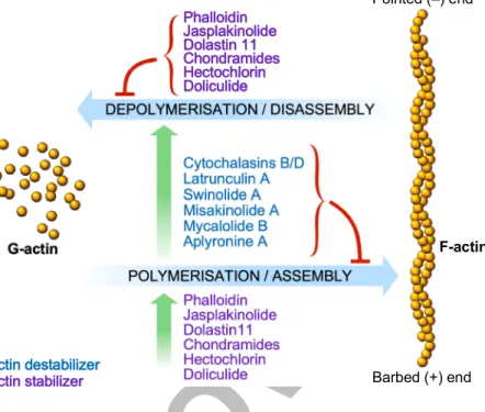
MFs are made of actin polymers and a variety of actin-binding proteins (ABPs). Indeed, in cells actin exists either as a mono-mer (globular, G-actin) or as a polymono-mer (filamentous, F-actin). Monomeric actin that has a molecular weight of approximate-ly 42 kDa is expressed in all eukaryotic cells and is highapproximate-ly conserved throughout evolution. Indeed, six isoforms of actin have been described in mammals: the cytoskeletal and cyto-plasmicβ- and γ-actins, cardiac muscle α-actin, skeletal
muscle α-actin, and the smooth muscle α- and γ-actins [39–41]. MFs are assembled through the polymerization of G-actin monomers into F-actin polymers. This process is highly dependent upon the concentration of free actin mono-mers, which subsequently drives the equilibrium toward po-lymerization that is regulated also by a series of actin-associ-ated proteins [42]. It is the case, for example, of profilin [43, 44] that binds to monomeric actin thereby occupying an actin-actin contact site. Once bound, profilin sequesters actin-actin from the pool of polymerizable“free” monomers, thus impairing polymerization. G-actin binds ATP, which is hydrolyzed to ADP shortly after incorporation in the growing filament [45, 46]. Actin filaments display an inert minus end and a growing plus end to which new monomers are added. Generally, the plus terminus is oriented toward the cell surface. The rapid addition of monomers onto that end promotes the formation of actin surface protrusions for cell migration [17]. Various fac-tors like calcium, ATP, cAMP and actin-binding proteins have been described to regulate the rate of actin polymerization. MFs are often subjected to “treadmilling”, such that mono-mers are continuously added to the plus end and removed from the minus end while the filament maintains the same overall length [9–11]. Actin filaments are present in all cells; however, their proportion is different depending on the cell type. The most abundant and organized system of MFs are striated muscle cells. MFs are ordered and organized by a number of ABPs within the cytoplasm and on the surface of cell membranes [12]. Those proteins have different functions
Table 1 Cytoskeleton components
Structure/composition Diameter Main functions
Microfilaments Twisted double strands of actin, each a polymer of actin monomers, associated to a series of actin-binding proteins [9–12]
7 nm Cell division (cleavage furrow formation) [13] Maintenance of (tension-bearing elements) and
changes in cell shape [5,6] Muscle contraction [14–16] Cell movement/migration [5,17–24] Intracellular transport of organelles [5,25] Microtubules 13 linear protofilaments assembled around a hollow core and arranged
in parallel, composed of a single type of the globular protein tubulin, which is a dimer of 2 closely related polypeptides,α- and β-tubulin [26]
25 nm Maintenance of cell shape by resisting tension (compression-resisting“girders”) [6] Separation of chromosomes during mitosis [8,
27–30]
Intracellular transport of organelles [25,31] Establishment of cell polarity [32] Cell motility (as in cilia et flagella) [22] Intermediate
filaments
Helical subunits of fibrous proteins (varying with function and cell type) supercoiled into thicker cables [33]
8–10 nm Supply of mechanical strength and support necessary to reinforce cells and organize them into tissues [34]
Anchoring of the nucleus and some organelles within the cytoplasm and nuclear lamina formation [35]
Maintenance of cell shape (tension-bearing elements) [6]
FOR
APPROVAL
and act on MFs in various ways [12] (see next sections). The most abundant motor protein associated to MFs ismyosinII, which moves toward the plus end of MFs, this process being driven by ATP hydrolysis. MFs are implicated in the regulation of important functions. A crucial one is to ensure mechanical stability to cells. An example is provided by microvilli on the surface of intestinal epithelial cells or epithelial cells of the kidney tubules. They are stabilized by MF wound around one another to form bundles of filaments [47–49]. MFs also play a crucial role in cell shape changes. For example, during cell division an actin ring that works in concert with myosin constricts the cell under division so that it forms a narrow waist, which finally breaks the connections between the two daughter cells [13]. Moreover, actin, in concert with myosin proteins, promotes cell contraction, as in the contractile apparatus of striated muscle cells [14–16]. Actin MFs are then implicated in several aspects of cell motility, from whole cell migration to the intracellular motility of the different organelles. Even if myosin motor proteins are responsible to generate the force needed for muscle contraction, cytokinesis and vesicle transport, actin MFs are able to generate force by themselves, assembling G-actin monomers into F-actin polymers producing force to deform the cell plasma membrane [50]. This phenomenon occurs for example at lamellipodia at the leading edge of a migrating cell [18,19]. Other important cellular movements in which actin MFs participate are the rocket-like propulsions of pathogens, like the Listeria bacteria, which is responsible of pathogen spread among host cells [20,21], the amoeboid movements observed in some protozoa [22] and the creeping of vertebrate white blood cells [23]. Finally, MFs are arranged in different ways according to the nature of the accessory proteins with which they associate. In contractile assemblies MFs associate with myosin and are antiparallel-oriented, as in the contractile ring responsible for cell division [13] and at the level of stress fibres, through which fibroblasts exert traction on their substratum. In actin-rich finger-like protrusions, like filopodia and other cell projections at the leading edge, MFs arrange as parallel bundles [24], whereas short randomly oriented filaments constitute the gels that are localized in the cortical region of the cell [51].
2.2 Microtubules
MTs are highly dynamic structures playing a crucial role both in cell shape determination and in a variety of cell movements, including some forms of cell locomotion, the intracellular transport of organelles, and the separation of chromosomes during mitosis [27,31]. MTs are composed of a single type of globular protein, named tubulin, which is a dimer of two closely related 55 kDa polypeptides,α-tubulin and β-tubulin. A third type of tubulin,γ-tubulin, has been described which
specifically localizes to the centrosome, where it is critically implicated in the initiation of MT assembly [52]. The poly-merization of head-to-tail arrays of tubulin dimers originate MTs [26], which are generally composed by 13 linear protofilaments assembled around a hollow core and arranged in parallel. Like actin filaments, MTs are polar structures and exhibit two distinct ends: a fast-growing plus end and a slow-growing minus end [53]. This polarity is essential to establish movement direction. MTs are subjected to rapid cycles of assembly and disassembly thanks to continuous cycles of po-lymerization/depolymerization of tubulin dimers. Bothα- and β-tubulin bind to GTP, whose hydrolysis (only when bound toβ-tubulin) weakens tubulin-binding affinity for adjacent molecules, thus promoting their depolymerization. GTP hy-drolysis also determines MT dynamic instability that consists in alternating cycles of growth and shrinkage [54]. The rate of tubulin addition relative to the rate of GTP hydrolysis deter-mines the former or the latter. As long as new GTP-bound tubulin molecules addition is more rapid than GTP hydrolysis, a GTP cap is retained at the plus end and microtubules con-tinue to growth. In contrast, by slowing of the polymerization rate, the GTP at the plus end of the MT will be hydrolyzed to GDP, with subsequent depolymerization and shrinkage of the MT. Like MFs, also MTs undergo “treadmilling” whereby tubulin molecules bound to GDP are lost from the minus end and replaced by GTP-bound molecules at the plus end of the same MT [55]. The rapid turnover of MTs is crucial for the remodelling of the cytoskeleton, for example during cell division, when MTs form the mitotic spindle. As the cell commits to divide, MTs nucleated by the centrosomes become shorter and more dynamic. This causes the disassembly of the interphase MT network [28]. After the breakdown of the nu-clear envelope, MTs reorganize to form the mitotic spindle. Kinetochore MTs are attached to the condensed chromo-somes, polar MTs overlap with each other in the centre of the cell, and astral MTs extend outward to the cell periphery [29]. The local stabilization and organization of those centrosomal and non-centrosomal MTs lead to the assembly of a bipolar spindle that is responsible of chromosome align-ment on the metaphase plate and their segregation into two daughter cells [30]. Each daughter cell then contains one cen-trosome, which nucleates the formation of a new network of interphase MTs. The key protein in the centrosome nucleating MTs assembly isγ-tubulin. Complexes of γ-tubulin originate ring structures that contain from 10 to 13γ-tubulin molecules. Theseγ-tubulin rings function as nucleation sites for the as-sembly of MTs and may remain bound to their minus ends [52,56]. A large number of proteins favour MT stabilization or destabilization [57,58]. Some proteins, for example, are implicated in MT disassembling, either by severing MTs or by increasing tubulin depolymerization rate from MT ends. Other proteins, known as MT-associated proteins (MAPs) bind to MT and increase their stability [59]. Those interactions
FOR
APPROVAL
are crucial to stabilize MTs at the level of specific cell loca-tions and determine cell shape and polarity. Various MAPs have been described, which vary depending on the cell type. The well-characterized ones are MAP-1, MAP-2 and tau, which have been isolated from neuronal cells, and MAP-4, which is found in all non-neuronal vertebrate cell types. The tau protein has been object of great interest because it is the main component of the typical lesions identified in the brains of patients affected by Alzheimer disease [60]. MAP activity is mainly regulated by phosphorylation, which controls MT stability. Finally, MTs are critically involved in establishing cell polarity. It is widely accepted that one common role for MTs in generating polarity is their requirement for delivering positional information necessary to determine the proper site of cortical polarity [32].
2.3 Intermediate filaments
The name IFs derive by their diameter ranging between 8 and 10 nm, which is intermediate in size between the MTs (25 nm) and the MFs (7 nm). IFs are composed of a number of proteins which are expressed in different cell types. Indeed, more than 50 IF proteins have been described and classified into six groups on the basis of their amino acid sequence similarity [33]. Types I and II are two groups of keratins, each including about 15 different proteins expressed in epithelial cells [61]. Vimentin, a protein found in fibroblasts, smooth muscle cells and white blood cells is one of the best-known type III IF proteins [62]. Another type III protein is desmin [63], which is specifically expressed in muscle cells, with the function to connect the Z discs of individual contractile elements. The type IV group of IF proteins include the three neurofilament (NF) proteins NF-L (light), NF-M (medium) and NF-H (heavy), which constitute the major IFs of different types of mature neurons, being particularly abundant in the axons of motor neurons [64]. Type V IF proteins include the nuclear lamins, which are found in most eukaryotic cells as compo-nents of the nuclear envelope and differ from the other IF proteins by their capacity to assemble to form an orthogonal meshwork underlying the nuclear membrane [35]. Finally, the type VI IF group includes nestin, a protein expressed in sev-eral cell types during development, although its expression is usually transient and does not persist into adulthood. The ex-pression of nestin has been well described at the level of neu-ronal precursor cells of the brain subgranular zone [65,66].
The different IF proteins display a similar structural orga-nization. They present a centralα-helical rod domain (around 310 amino acids) flanked by amino- and carboxy-terminal domains (head and tail respectively), varying among the dif-ferent IF proteins in size, sequence and secondary structure [67]. Differently from MFs and MTs, IFs are more stable and are not characterized by the dynamic behaviour typical of those other cytoskeletal components [68]. However,
phosphorylation can regulate their assembly and disassembly within the cells, as in the case of nuclear lamins. Their phos-phorylation results in disassembly of the nuclear lamina and breakdown of the nuclear envelope during mitosis [69,70]. Furthermore, differently from MTs and MFs, assembled IFs have no polarity and association and dissociation of their di-mers can occur all along the filament length.
Finally, IF organization and association with plasma mem-branes suggest that they essentially exert a structural role by providing the mechanical strength necessary to reinforce cells and organize them into tissues [34]. The best example is rep-resented by the mechanical support provided by IFs to the plasma membrane, where it comes in contact with the extra-cellular matrix (ECM) or with other cells.
Importantly, major degenerative diseases of skin ( e p i d e r m o l y s i s b u l l o s a s i m p l e x , E B S ) , m u s c l e (desminopathy), and neurons (amyotrophic lateral sclerosis, ALS) depend on the disruption of the IF cytoskeleton or its connections to other cell structures [71–73]. However, this topic goes beyond the purpose of the present review.
3 The metastasization process
Metastasis is the leading cause of cancer-related deaths. Functionally, metastasis occurs via a series of discrete steps (Fig.1), often termed the“invasion-metastasis cascade” [74]. They include: detachment of cells within the primary tumour; local migration and invasion of stromal tissue; intravasation into and transit through pre-existing and newly formed blood and lymph vessels; extravasation; establishment of dissemi-nated cells (which can stay dormant for a prolonged period of time) at a secondary anatomical site; and outgrowth of micrometastases and macrometastases/secondary tumours, this last step being called “colonization” (Fig.1) [75,76]. Interestingly, recent data have suggested the existence of an additional step at the beginning of the“metastatic cascade”: the creation of a“premetastatic niche” at the target site, before the arrival of the first tumour cells to this distant location [77]. Indeed, Lyden and colleagues have shown that bone marrow-derived cells are mobilized owing to the presence of a distant, intradermal tumour. As a consequence, these cells accumulate as clusters in the lungs, where they change the local microen-vironment into a niche suitable for the establishment of sec-ondary tumours. At a later stage, tumour cells arrive at these sites and co-localize with the bone marrow-derived cell clus-ters [77]. In line with these results, it has been reported that distant tumours induce elevated levels of pro-inflammatory chemokines in the lungs of tumour-bearing mice [78,79] that attract tumour cells to the lungs via a positive feedback loop inducing the expression of nuclear factor-kB (NF-kB) [80]. These findings open the way to new possible therapeutical strategies, as the perturbation of the endocrine and paracrine
FOR
APPROVAL
signalling networks required for the establishment of a premetastatic niche could prevent the establishment and out-growth of distant metastases.
According to the traditional metastasis models, metastatic cells are rare and appear during the late stages of tumour progression. However, expression profiling studies on human cancers, such as breast carcinomas, show that most cells in a primary tumour express the molecular signature that is asso-ciated with metastatic tumours [81]. Indeed, a specific gene-expression profile of primary breast cancers is associated with the development of metastasis and with a poor clinical out-come [82]. Those results suggest that the potential for metas-tasis is determined early in tumorigenesis, challenging the classical models.
To disseminate from a primary tumour to a distant organ, invasive cancer cells must subvert the molecular machinery that under normal conditions enable important physiological functions and facilitate the development and homeostasis of an organism. For example, signalling cascades are required for normal processes such as morphogenesis [83], neurogenesis [84] and angiogenesis [85]. Each of the stages involved in tumour metastasis requires numerous specific molecular inter-actions provided by the tumour cell, the surrounding ECM and the stromal cells (see below) [86]. These interactions are mediated by contact between the cell and the ECM, by direct
cell-cell contact, and by secreted factors. Finally, each step of the metastatic process has one or more physiological barriers that act to inhibit the spread of malignant cells. Thus, to suc-cessfully metastasize, tumour cells need to overcome natural barriers such as the basement membrane and the interstitial matrix of connective tissue.
One important class of molecules that play a crucial role in cell invasion and metastasis are proteases, enzymes that are able to degrade the ECM. Although they are key mediators of many physiological processes, such as embryogenesis, tissue repair and immune response, they are highly expressed during tumour progression and metastasis [87]. Classically, their ac-tivity is associated with the late invasive stage of cancerogenesis, providing cells with the ability to breach bar-riers opposing their movement (such as the basal membrane), to facilitate intra- and extravasation of tumour cells and to promote growth in ectopic environments. However, multiple lines of evidence suggest they have important functional roles at additional stages of neoplastic progression. In pre-malig-nant cancer cells, protease activation favours the onset of an-giogenesis and malignant conversion. In addition, their pro-teolytic activity is required for other functions apart from the local dissolution of basement membrane. Those functions in-clude: the activation of latent proteases, growth factors, and chemotactic agents (and related receptors); the exposure of 1
ECM degradation, invasion and migration
endothelial cells tumour cells stromal cells inflammatory cells blood vessels epithelial cells ECM
Fig. 1 Schematic overview of the essential steps of the metastatic process. Cancer cells detach from the primary tumour, degrade the extracellular matrix (ECM), invade through the basement membrane and migrate through the tumour stroma (1). They then intravasate into
vasculature (2) and transit through pre-existing and newly formed blood vessels (3). After extravasation through the endothelial barrier (4), cancer cells form micrometastasis (5) to finally colonize target organ/s to originate secondary tumour/s (6)
FOR
APPROVAL
integrin-binding sites on the ECM with subsequent transmis-sion from these sites of migratory and survival signals [88, 89]; the release of bioactive fragments of the ECM itself. These in turn are able to enhance protease gene expression or alternatively activate integrins.
During the past decade an important acceleration of the research on cancer cell ability to invade and metastasize has been registered thanks to emerging powerful research tools, novel experimental animal models and the identification by genomic studies of critical regulatory genes implicated in the metastatic cascade. However, major questions still remain unanswered.
An important developmental regulatory program called “epithelial-mesenchymal transition” has emerged as an essen-tial mechanism by which transformed epithelial cells acquire the abilities to invade, to resist apoptosis and to disseminate [90,91]. Carcinoma cells thus co-opt processes implicated in different steps of embryonic morphogenesis and wound healing and in parallel acquire multiple characteristics en-abling them to invade and metastasize. Indeed, transcription factors like Snail, Slug, Twist and Zeb 1–2 orchestrating EMT and related migratory events during embryogenesis are also expressed at various levels and in different combinations in several malignant tumours. They have been demonstrated to be important to direct invasion in experimental models of carcinoma formation and some of them induce metastasis once ectopically overexpressed [92,93]. The biological events triggered by such transcription factors are multiple: loss of adherens junctions with a parallel conversion from a polygo-nal/epithelial to a spindly/fibroblastic morphology, expression of matrix-degrading enzymes, increased migration and aug-mented resistance to apoptosis. Up to date it seems that EMT-inducing transcription factors have the ability to orchestrate most steps of the invasion-metastatic cascade excluding the final step of colonization. Although certain non-epithelial tu-mours, like sarcomas and neuroectodermal tutu-mours, express EMT-inducing transcription factors, their role in triggering the initiation and progression of the metastatic cascade in those tumours is still unclear. Moreover, it remains to be established whether the EMT program is the only one through which carcinoma cells acquire invasive traits or whether other so far unknown regulatory programs cooperate to induce those malignant characteristics. Furthermore, two other distinct modes of invasion have been reported and implicated in can-cer cell invasion [94].“Collective invasion” has been de-scribed as the movement of groups of cancer cells advancing together into adjacent tissues and characterize, for example, squamous cell carcinomas, which are rarely metastatic. This suggests that this type of invasion lacks some characteristics that promote metastasis. The other one is the “amoeboid” invasion [95,96], in which individual cancer cells display a morphological plasticity that allows them to slither through existing interstices in the extracellular matrix, as observed
for both the mesenchymal and the collective forms of invasion.
Finally, it has been demonstrated that a crosstalk between cancer cells and cells of the neoplastic stroma contributes to invasive growth and metastasis [97,98]. An example is pro-vided by mesenchymal stem cells present in the tumour stro-ma which are shown to secrete CCL5/RANTES in response to signals released by cancer cells. CCL5 in turn acts on cancer cells to promote their invasive behaviour [99]. Very interest-ing is also the case of macrophages at the tumour periphery, which stimulate local invasion providing matrix-degrading enzymes [98,100]. In an experimental model of metastatic breast cancer, tumour-associated macrophages provide breast cancer cells with epidermal growth factor (EGF), whereas cancer cells stimulate macrophages via CSF-1 secretion. This concerted action facilitates cancer cells intravasation into the circulatory system and metastatic dissemination [97,101].
3.1 Actin cytoskeleton proteins implicated in cancer
metastasis
3.1.1 Regulatory proteins: Rho small GTPases
Rho small GTPases belong to the Ras superfamily of GTPases. They have emerged as essential players in the reg-ulation of key signalling pathways underlying cell migration, like cytoskeleton dynamics, assembly and disassembly of cell-cell junction, directional sensing and integrin-matrix ad-hesion. Since their discovery as homologous of Ras small GTPases, over 20 mammalian members have been reported and divided into 8 subfamilies [102]. Rac1, Cdc42 and RhoA are the best characterized members of the family. Their crucial role in regulating actin cytoskeleton organization and dynam-ics has been well documented [103]. Rac1, Cdc42 and RhoA were first reported to promote the formation of the following actin-rich structures in fibroblasts, respectively: lamellipodia/ ruffles, filopodia and stress fibres [104]. The formation of lamellipodia/ruffles and filopodia by Rac1 and Cdc42 is a critical prerequisite for actin-driven protrusion at the leading edge of the cell, whereas RhoA-dependent stress fibres forma-tion regulates actomyosin contracforma-tion of the cell body and retraction of the trailing edge. Subsequently, Rho small GTPases have been shown to have a role also in the regulation of key cellular functions, like polarity, survival, cell cycle progression and gene expression [105, 106]. Similarly to Ras small GTPases, Rho small GTPases are molecular switches that cycle between a GTP-bound active form and a GDP-bound inactive one [106]. Rho GTPase activity is regu-lated by 3 main families of regulatory proteins: guanine nu-cleotide exchange factors (GEFs), GTPase-activating proteins (GAPs) and guanine nucleotide dissociation inhibitors (GDIs). GEFs activate Rho GTPases catalysing the exchange of GDP for GTP [107], whereas GAPs inactivate Rho
FOR
APPROVAL
GTPases accelerating their intrinsic GTPase activity [108]. Eighty-two GEFs and 67 GAPs have been described to regu-late GTPase activity downstream of several cell surface tors such as growth factor receptors, integrins, cytokine recep-tors, and cadherins [107]. The role of GDIs is to bind to Rho GTPases in their inactive GDP-bound form and sequester them in the cytosol, thus acting as inhibitors of the Rho GTPase signalling [109]. Indeed, once translated, Rho GTPases are subjected to post-translational modifications like geranylgeranylation, or less commonly farnesylation, on a C-terminal CAAX motif, which promotes anchorage to intracel-lular membranes where they are activated [110]. Rho GTPases once bound to GTP undergo conformational changes and as-sociate to a spectrum of effector proteins to stimulate down-stream signalling pathways [111,112]. The total number of effectors is too large to be discussed here. Excellent reviews containing a detailed description are available [111–113]. Some examples will be briefly reported here. Rac binds to the Wave complex to release active Wave, which induces actin polymerization in lamellipodia through Arp2/3 complex activation. Both Rac and Cdc42 bind and activate the p21-activated (PAK) kinases, which have multiple substrates in-cluding LIM kinase (LIMK) that promotes actin polymeriza-tion. PAK activity controls also myosin phosphorylation and cell contractility through a number of signalling cascades, including myosin light chain kinase (MLCK), myosin regula-tory light chain, myosin heavy chain and caldesmon. Rac and Cdc42 also bind to the actin-binding protein IQGAP, which participates to the regulation of cell-cell adhesion. Moreover, both Rac and Cdc42 bind to and stimulate PI3 kinase (PI3K). The lipids phosphorylated by PI3K bind to and stimulate Rac GEFs in a positive feedback loop that promotes cell motility. Cdc42 mediates the formation of filopodia through WASP, which interacts directly with Arp2/3 complex to promote actin polymerization. Cdc42 is also able to activate the Myotonic dystrophy kinase-related Cdc42-binding kinases (MRCKs), which are linked to Rho kinases and can stimulate myosin phosphorylation. Rho kinases 1 and 2 (ROCK1 and ROCK2) are key Rho effectors. Among the effectors of ROCKs, we can list (a) the myosin-binding subunit of myosin phosphatase (MBS), which causes the inhibition of its phos-phatase activity, the increase of MLCK phosphorylation and a subsequent increase in general tension; (b) LIMK, which is responsible of cofilin phosphorylation and hence actin poly-merization, and (c) myosin regulatory light chain itself that in turn promotes contractility. Finally, phosphatidylinositol 4,5 bisphosphate activates ezrin-radixin-moesin (ERM) proteins and promotes actin polymerization through WASP, profilin and multiple actin-capping proteins.
Rho small GTPases and their regulators and effectors par-ticipate in multiple aspects of cancer progression. Differently from Ras, which has been found to be mutated in around 20– 30% of human cancers, Rho GTPases are rarely mutated.
Recently, it has been reported that proline 29 in Rac1 is mu-tated in a subset of melanomas, breast tumours and head and neck tumours [114] and that RhoA is mutated in some tu-mours, like diffuse gastric cancer and angioimmunoblastic T cell lymphoma [115–118]. However, in the majority of the cases Rho GTPases are upregulated or display an increased activity as a result of altered expression levels of GEF, GAPs and/or GDIs. This is supported by abundant evidence based on in vitro experiments involving manipulation of the expres-sion levels or the activity of Rho GTPases in cancer cell lines by overexpressing constitutive active or dominant-negative forms of Rho GTPases, their regulators or effectors, or exploiting small interfering RNAs (siRNAs) or small mole-cule inhibitors [119]. A series of tissue-specific conditional knock-out mouse models (as direct targeting of Rac1, Cdc42 and RhoA is embryonic lethal) has definitely provided strong evidence for the physiopathological role of Rho small GTPases [120, 121]. Rac1, Cdc42 and RhoA are overexpressed in a variety of malignancies [122–127]. Interfering with their expression and/or activity by knock-down or by expressing dominant-negative mutants or by using small molecule inhibitors (when available, see next sections) has been reported to inhibit lamellipodia/ruffles and filopodia formation, reduce the number of focal contacts, impair migra-tion and invasion of a number of human cancer cell lines, inhibit colony formation in soft agar and decrease tumour metastasis in vivo [128–136].
The evidence that Rho GTPases are frequently overexpressed or hyper-activated but rarely mutated in several malignancies indicates that regulatory proteins like GEFs, GAPs and GDIs play a key role in the dysregulation of sig-nalling pathways implicated in cancer initiation and progres-sion. Indeed, Rho GTPase hyperactivation may occur through (a) their overexpression, (b) loss of GAP- or GDI-mediated inactivation, (c) upstream activation or overexpression of RhoGEFs. Altered expression or mutations of RhoGEFs, RhoGAPs and RhoGDIs have been reported in a variety of human cancers [137,138]. Since GEF activation is the most common mechanism for signal-mediated GTPase activation, the theme that has emerged is that aberrant GEF regulation contributes to Rho GTPase activation in cancer, making Rho GEFs attractive therapeutic targets for cancer [138] (see next section).
Rho GAPs represent the flip-side of the coin to Rho GEFs. Surprisingly, differently from the many Rho GEFs that are altered in human malignancies, except for the deleted in liver cancer (DLC) family [139,140], limited evidence exists for the role of Rho GAPs in cancer. It has been hypothesized that in many cancers GAP activity is normal, but Rho GTPase hyperactivation through GEFs or GTPase overexpression it-self can override normal GAP-mediated inactivation.
Concerning RhoGDIs, even if changes in their expression levels have marked effects on the overall level of activities of
FOR
APPROVAL
Rho GTPases and have been linked to a number of cancers [141–144], little is known about how Rho GDI expression is regulated. Despite the wide variety in the Rho GTPase family, only three genes have been reported to encode RhoGDIs in mammals, implying that the consequences of changes in their expression are manifested through the action on more than 20 Rho GTPases. The effects of the Rho GDIs on malignancies are thus complex and do not fit a simple explanatory model. 3.1.2 Structural proteins: actin-binding proteins
As reported above, actin can exist in 2 forms: G-actin and F-actin. They both interact with a large variety of proteins in the cell. Up to date more than 100 ABPs have been described. They are critically implicated in regulating polymerization and depolymerization of actin filaments, organizing actin fil-aments in higher magnitude superstructures and controlling complex MFs dynamic properties [12]. Through those activ-ities ABPs allow the actin cytoskeleton to rapidly respond to cellular and extracellular signals, being essential for the par-ticipation of the cytoskeleton to several cellular processes, like cell shape and motility, muscle contraction, intracellular traf-ficking, cell pathogenesis. ABPs can be divided into 7 groups according to their specific functions on actin polymers and/or monomers: (1) actin nucleators; (2) regulators of G-actin po-lymerization/F-actin depolymerization; (3) capping and actin severing proteins; (4) bundling and cross-linking proteins; (5) motor proteins; (6) anchoring actin filaments to the plasma membrane; (7) F-actin stabilizing and regulatory proteins (Fig.2).
1- An important set of actin regulators initiates the forma-tion of new actin filaments through a process called nucle-ation. Up to date 3 classes of proteins have been described
that initiate new filament polymerization: the actin-related protein 2/3 (Arp2/3) complex, the formins and spire. Each promotes actin nucleation by a distinct mechanism. We will focus here on the Arp2/3 complex which has been the first to be identified and to critically participate in the formation of branched-actin-filament networks during different cellular processes ranging from cell migration to endocytosis. The Arp2/3 complex is a seven-subunit protein complex. Two of its subunits, the actin-related protein 2 and 3 (ARP2 and ARP3) closely resemble the structure of monomeric actin. Indeed, the complex is thought to mimic an actin dimer or trimer and to serve as a template for the initiation of a new (daughter) filament that branches off of an existing (mother) filament and generates y-branched (with a regular 70° branch angle) actin networks [145,146]. This nucleating/branching action of the Arp2/3 complex is known as autocatalytic branching or dendritic nucleation and is crucial for its in vivo activity. The Arp2/3 complex displays little biochemical ac-tivity per se. However, when engaged by a nucleation promot-ing factor (NPF), it is activated to initiate actin nucleation. NPFs are divided into 2 main groups and are controlled by signalling cascades that coordinate actin polymerization in space and time. Class I NPFs include among others the Wiskott-Aldrich syndrome protein (WASP) and the suppres-sor of cyclic AMP repressuppres-sor (SCAR, also known as WASP-family verprolin homologous protein WAVE) [147–149]. They display a common WCA domain, consisting of WASP-homology-2 (WH2) domain, a central (C; also called cofilin-homology or connector) and an acidic (A) region (both regions together are known as CA region). It has been pro-posed that for class I NPFs the A region mediates the binding to Arp2/3, the central region initiates an activating conforma-tional change in the complex and the WH2 and central regions
Actin nucleators
Arp2/3, Wasp, N-Wasp, Formins
7 F-actin stabilising and regulatory proteins
Tropomyosins ACTIN BINDING PROTEINS M G-actin monomers monomer-sequestering proteins depolymerisation polymerisation 1 2 3 4 5 6 NN N N G-actin polymerisation/
F-actin depolymerisation regulators
Profilin, Thymosin 4, Cofilin
Myosins
Anchoring actin filaments to the plasma membrane
Talin, Kindlin, ERM proteins
Capping and severing proteins
CapZ, Gelsolin
Bundling and crosslinking proteins
Fascin, Filamin, -actinin Tpm
Fig. 2 Overview of the main families of actin-binding proteins. Actin-binding proteins are divided into seven groups according to their specific functions on actin polymers and/ or monomers. They can (1) promote actin nucleation; (2) regulate G-actin polymerization/ F-actin depolymerization; (3) cap or sever F-actin filaments; (4) bundle or crosslink F-actin filaments; (5) anchor F-actin filaments to the plasma
membrane; (6) generate force; (7) stabilize F-actin filaments. Representative proteins are indicated for each group
FOR
APPROVAL
present an actin monomer to the complex, facilitating the for-mation of a nucleus for the polymerization of the daughter filament. The class II NPFs include, among others, cortactin. Like class I NPFs, they display an Arp2/3 binding to the acidic region, but they lack a G-actin– binding region and their mechanism of Arp2/3 activation is still unclear. The altered function of the Arp2/3 complex is linked to cancer metastasis. Indeed, the levels of Arp 2/3 mRNA as well as those of N-WASP (the closely related N-WASP neural isoform) are upreg-ulated in a variety of tumours and invasive cells, being func-tionally associated with cell invasion and metastasis [150, 151]. The critical prerequisite for cancer cell invasion is the formation of actin-rich structures, like podosomes and invadopodia that possess adhesive and protrusive activities with the ability to degrade the ECM. The activation of the Arp2/3 complex is needed for podosome formation [152]. At their actin-reach core the Arp 2/3 complex colocalizes with WASP, N-WASP and cortactin [153,154]. Similarly, the Arp 2/3 complex and N-WASP localize to invadopodia and are critically required for their formation [155]. The Arp2/3 com-plex and their activators are thus crucial for tumour cell inva-sion, being potential interesting target for therapeutic interven-tion (see next secinterven-tion).
Finally, just a few lines on formins. Formins are multi-domain proteins containing a highly conserved actin assembly formin homology 2 FH2 domain, and the associated profilin-binding FH1 domain. FH1 and FH2 domains are flanked on either side by regulatory domains, the function of which is to regulate localization and activation [156,157]. Some formins are activated by Rho GTPases and autoinhibited by associa-tion of their N- and C-terminal regulatory domains [156]. Once activated, the FH2 domain either nucleates actin fila-ment assembly or associates with pexisting filafila-ments, re-maining continuously linked to the elongating actin filament barbed end [158]. Through processive association formins protect barbed ends from capping proteins [159,160] and promote the addition of profilin-actin bound to the FH1 do-main [161,162]. As a result, long-straight filaments often bundled in cells and pulled by myosin motors to generate contraction are produced.
2- The regulators of G-actin polymerization include pro-teins like profilin (see above) and thymosinβ 4, a protein that sequesters actin monomers by forming complexes with G-actin, thus maintaining the pool of unpolymerized actin. Among F-actin depolymerization factors we would like to focus our attention on cofilin. It belongs to a family of related proteins sharing similar biochemical activities, the actin depolymerizing factor (ADF)/cofilin family. Cofilin and its regulatory proteins participate in the early steps of the motility cycle and evidence shows that the expression of a number of genes of the cofilin pathway is altered in invasive tumour cells. Cofilin is a small ubiquitous protein that binds to both G-and F-actin. Studies carried out using light microscopy to
observe interaction between cofilin and actin filaments that are immobilized by cross-linking to a substratum have shown how cofilin can both increase the number of free barbed ends for polymerization and increase the rate of actin depolymeri-zation (thus refilling the cell with new G-actin monomers) [163–166]. Those sets of experiments have also demonstrated that cofilin is a much more powerful severing protein than expected [165,166]. Indeed, the appearance of free barbed filament ends together with further remodelling of the actin cytoskeleton can lead to the formation and retraction of path-finding structures like lamellipodia, invadopodia and filopodia used in chemotaxis, cell migration and invasion of tumour cells. The cofilin pathway has recently emerged as a central player in the generation of free barbed ends and actin filament-turnover in a large number of motile cells, including fibroblasts [163], mammary carcinoma cells [167,168] and glioblastoma cells [169] during the formation of those struc-tures. Furthermore, cofilin can greatly potentiate the dendritic nucleation activity of the Arp2/3 complex (see above), be-cause through the severing of the mother filament it originates polymerized actin filaments that are preferred for the actin nucleation by the Arp 2/3 complex [170]. The cofilin pathway includes a group of kinases and phosphatases that control cofilin activation state in response to different stimuli, like EGF, transforming growth factorβ (TGFβ), stromal cell-de-rived factor 1 (SDF1) and heregulin. The regulation of cofilin is very complex as it involves 4 independent mechanisms: (1) the phosphorylation on Ser 3 by LIMK1, LIMK2 and testic-ular protein kinase 1 and 2 (TESK1 and 2), which causes inhibition of cofilin actin-binding activity [171–173]; (2) the dephosphorylation of Ser 3 by phosphatase types 1, 2A and 2B, slingshot (SSH) and chronophin, resulting in the activa-tion of actin-binding by cofilin [174–176]; (3) the binding to phosphatidylinositol 4,5 bisphosphate (PIP2, whose hydroly-sis depends on phospholipase Cγ, PLC γ) that inhibits actin binding by cofilin [177]; (4) the changes in pH over the phys-iological range (6.8–7.4) by the Na-H exchanger protein, which promote the severing activity of cofilin when it is de-phosphorylated [178,179]. Indeed, it is now clear that a bal-ance of the stimulatory and inhibitory branches of the cofilin pathway is needed to ensure that protrusion, cell migration and chemotaxis occur in an optimal way. Expression profiling data have shown that the cofilin pathway is a major determi-nant of metastasis, several genes whose expression status is upregulated during metastasis being clustered in the cofilin pathway and its downstream effectors [180,181]. This makes the cofilin pathway a potential druggable target for anti-met-astatic therapy (see next section). Although studies of the ef-fect of cofilin activity on metastasis have been carried out only in mammary tumours, it is likely that the cofilin cascade also plays a crucial role in the invasion and metastasis of other types of cancer, as demonstrated by the critical requirement of cofilin for the motility of different cell types and the
FOR
APPROVAL
correlation of cofilin pathway gene expression with the meta-static phenotype in various malignancies [182–186]. In partic-ular, in rat and mouse mammary tumour models, it has been shown that 9 genes of the cofilin pathway are overexpressed, showing that many combinations are possible to increase the output of the cofilin pathway. Only cofilin and LIMK1 have been tested for their effects on the activity of the cofilin path-way, the results of those studies showing that cofilin signalling output is predictive for metastatic potential [187].
3- Capping and actin severing proteins regulate actin fila-ment dynamics by blocking the addition and loss of G-actin monomers to F-actin filaments. Two of those proteins are CapZ, which caps the barbed end of actin filaments in muscle cells, and gelsolin, which controls actin filament length, being one of the most potent members of the actin-severing gelsolin/ villin superfamily.
4- The actin-bundling and cross-linking proteins include among others filamin, a crucial actin crosslinker;α-actinin, an actin-bundling and cross-linking protein thought to anchor actin to a variety of intracellular structures, and the actin-bun-dling protein fascin (FSCN). We will focus our attention on this last protein. Three FSCN isoforms are known in mam-mals: FSCN1, which is widely expressed; FSCN2, which is expressed in retina and ear hair cells; FSCN3, which is spe-cifically expressed in testis. FSCN1 has received considerable attention in recent years as its expression is very low or absent in normal adult epithelia, but it is dramatically upregulated at both the transcript and protein levels in all forms of human carcinomas studied to date [188–190]. FSCN1 crosslinks actin filaments into tightly packed parallel bundles, oriented with their growing ends toward the plasma membrane. It has been reported that its association with actin filaments can be in-creased by interaction with specific matrix [191] or upon cell stimulation by growth factors or cytokines [192]. FSCN1 lo-calizes to actin-rich cytoskeletal structures, like stress fibres [193, 194], protrusive structures like microspikes and filopodia [195] and ECM-degrading structures, like podosomes and invadopodia [196,197]. Cell biology studies have shown that FSCN1 is critically required for the activity of those structures and its expression levels correlate with higher cell motility and invasiveness [190,198]. FSCN1 is mainly regulated by different factors like TGFβ [199], Wnt/ β catenin [200], STAT3/NF-kB [201], microRNAs [202–204] at the transcriptional level, where it has been ob-served to be induced when epithelial tumours start to acquire invasive and migratory properties. FSCN1 can be post-translationally modified by PKC and, once phosphorylated on Ser 39, it shows a reduced actin binding and bundling activity [205,206]. FSCN1 has been demonstrated to be a valuable prognostic biomarker in some epithelial cancers. Indeed, immunohistochemistry detection on conventional sec-tions or tissue microarray (TMA) has revealed that higher FSCN1 expression levels positively correlate with tumour
aggressiveness/malignancy and higher tumour grade/stage in carcinomas [207]. Elevated FSCN1 levels have been detected in advanced stage and lymph node metastasis of head and neck squamous cell and colorectal cancers with a bad progno-sis [208–211]. In breast cancer, FSCN1 expression has been associated with oestrogen receptor-negative (ER2), progester-one receptor-negative (PR2) and basal-like phenotype, being correlated with an increased risk of mortality [212, 213]. Higher FSCN1 expression levels have also been detected in advanced tumour stage pancreatic cancers and lymph node metastasis of lung cancers [214–217]. Remarkably, we have recently shown that FSCN1 immunohistochemical detection is an independent prognostic factor in adrenocortical carcino-ma (ACC), also refining results obtained with staging and Ki67 labelling index [>]]. The robust prognostic power of FSCN1 levels has been further confirmed in two independent ACC cohorts [218]. Moreover, we have provided evidence for a role of FSCN1 in promoting tumour cell invasion in a human ACC cell line that overexpresses the transcription factor ste-roidogenic factor-1 (SF-1) resulting in a more aggressive and invasive phenotype [219, 220]. Indeed, interfering with FSCN1 by gene silencing or chemical inhibition substantially impaired actin cytoskeleton reorganization affecting the for-mation of filopodia and reduced the increased tumour migra-tion and invasiveness [218] observed in that cell line upon increased SF-1 dosage conditions that we had previously dem-onstrated to affect cytoskeleton dynamic and invasive proper-ties through the upregulation of the Rho GEF VAV2 [220] (see next section). Overall, the above evidence indicates that besides being an excellent prognostic marker for a number of malignancies, FSCN1 is an attractive drug target. This is par-ticularly interesting for a type of cancer like ACC, especially in the advanced stages, where therapeutic options are rather disappointing and the need for new and more effective drugs is urgent. Specific small molecule inhibitors for FSCN1 have been developed and shown to antagonize metastatic dissemi-nation in pre-clinical cancer models (see next section).
5- Myosins are a superfamily of actin-dependent molecular motors which upon interaction with MFs are able to convert energy deriving from ATP hydrolysis to mechanical stress [221]. They have a crucial function in several aspects of eu-karyotic motility, like cell movement, cytokinesis, organelle/ particles trafficking, signal transduction and in maintaining cellular morphology. In the human genome around 40 differ-ent myosin-related genes have been iddiffer-entified. They encode 12 classes of myosins [221]. Myosins are composed of three functional subdomains: the NH2-terminal domain, which is required for actin binding and ATP hydrolysis; the neck do-main, which binds calmodulin and light chains; the COOH-terminal domain, which is class-specific and is implicated in the transport of cargo along MFs. This tail region participates also in signal transduction and membrane interaction. Increasing evidence has pointed on myosins as critical actors
FOR
APPROVAL
in the process of tumorigenesis and thus potential druggable target for cancer therapy. Myosins are overexpressed in a va-riety of cancer types, including breast [222,223], ovarian [224,225], prostate [226,227] and colorectal cancer [228, 229], intestinal [230,231], gastric [232,233] and pancreatic cancer [234,235], melanoma [236,237], anaplastic gliomas [238] and acute myeloid leukaemia [239]. In each type of malignancy myosins play different but essential roles in tu-mour progression. Myosin1e, for example, has been implicat-ed in recruiting invadosome components to the plasma mem-brane and transport vesicles to the sites of new invadosome assembly [240]. Myosin II has been reported to drive cell invasion in pancreatic, breast and prostate cancer cells and in anaplastic glioma cells [222,234,238,241,242]. In breast cancer it also maintains cell polarization, stabilizes nascent focal adhesion complexes and mediates efficient integrin-based cell migration [243]. Moreover, it regulates tumour cell migration by interacting with P-cadherin [236]. Blocking of myosin II activity by specific MLCK inhibitors could prevent pancreatic and breast cancer cell adhesion and invasion [222, 234] (see next section). Myosin Va, positively regulated by Snail, seems to be a key factor in cytoskeletal organization and migration of metastatic colorectal cancer cells [244]. Its over-expression promotes actin assembly and cell motility, whereas its depletion impairs cell spreading, cytoskeletal remodelling and cell migration [245]. Myosin Vb has been reported to be a strong prognostic factor for disease recurrence [246]. Myosin VI is an early marker in prostate cancer development [247] and has a role in the dissemination of ovarian cancer cells [224]. Myosin IX has been shown to downregulate Rho ac-tivity [248] and to prevent actin bundle assembly during the nascent formation of cell adhesion, being critically required to sustain the collective migration of epithelial cells [249]. Finally, myosin X localizes at filopodia tips, being actively implicated in their formation [250,251], and promotes metas-tasis development in primary glioblastoma and acute lympho-blastic leukaemia [252,253]. It also appears to play a role in cancer cell protrusion and metastasis development by transporting β1 integrins to the filopodia tips [254]. Moreover, mutant p53-associated myosin X upregulation has been reported to promote breast cancer invasion and me-tastasis [255].
6- The actin-binding proteins which anchor actin filaments to the plasma membrane include among others talin, kindlin, and the three closely related proteins ezrin, radixin and moesin. Here we focus on talin, one of the major components of the focal adhesion complex, mainly acting as an interlink between transmembrane integrin receptors and cytosolic F-actin. Talin, besides actin, is also able to interact with a num-ber of other proteins in the adhesion complex to regulate their functional dynamics. Indeed, 2 talin isoforms have been de-scribed in mammals (talin 1 and talin 2). They are about 270 kDa proteins in size and consist of an N-terminal head
domain and a C-terminal flexible rod domain [256]. The talin head domain is composed of a FERM (4.1, ezrin, radixin, moesin) domain consisting of 3 subdomains (F1, F2 and F3) and an F0 subdomain that has no homology with other already known domains. The F3 subdomain is implicated in the bind-ing to the cytoplasmic tails of integrins [256,257]. The C-terminal rod domain is composed of a series of helical bundles that contain multiple binding sites for the F-actin-binding pro-tein vinculin and a second integrin-binding site [258]. The C-terminal domain includes also a THATCH (talin/HIP1R/ Sla2p actin tethering C-terminal homology) domain mediat-ing the dimerization and providmediat-ing a direct linkage between talin and F-actin [259,260]. Talin is a recruited component of the focal adhesion complex where it functionally interacts with the cytoplasmic tails of integrins. Talin interconnects β-integrin cytoplasmic tails and actin filaments by directly binding both. It has been demonstrated that a single point mutation selectively abrogates the binding of talin head do-main to integrinβ tails, leading to the disruption of integrin localization to talin-rich focal adhesions [261]. The role of talin as a crucial regulator ofβ-integrin affinity for its ligands has been reported and the molecular mechanisms through which talin accomplishes this role have been described [256]. Talin function of regulating cell adhesion to the ECM is essential for both physiological processes during embryonic development, immune response, angiogenesis and pathologi-cal processes, like cell invasion and metastasis [262]. Talin1 has been shown to be implicated in focal adhesion dynamics, cell migration and cell invasion [263–265]. It enhances pros-tate cancer cell adhesion, migration, and invasion through the activation, in response to anoikis, of a FAK–Src complex and an AKT survival signalling leading to enhanced metastasis [265]. Further, talin-1 has been reported to be highly expressed in hepatocellular carcinoma cells relative to non-cancer liver epithelial cells and to promote tumour growth and metastasis [266]. It has also been proposed to be a poten-tial marker for colon cancer and hepatocellular carcinoma di-agnosis because of its high expression levels in serum speci-mens from cancer patients [267,268]. In particular, in hepa-tocellular carcinoma Talin1 has been reported to have a higher sensitivity and specificity compared to the traditional bio-marker alpha-fetoprotein (AFP). Indeed, both talin1 and talin2 expression levels correlate with the malignancy potential of human hepatocellular carcinoma cells [269], whereas talin2 has been implicated in the regulation of breast cancer cell migration and invasion [270]. Recent findings point to the clinical significance of talin’s contribution to the metastasization process, talin1 being significantly upregulated in primary tumours and metastatic prostate cancer respect to the normal prostate gland [265]. Talin1 expression has been shown to be significantly higher in poorly differentiated pros-tate tumours (Gleason > 8) compared to moderately differen-tiated ones (Gleason 6 and 7). The same authors reported a
FOR
APPROVAL
strong inverse correlation between talin1 and E-cadherin ex-pression in human prostate tumours and metastatic lesions. A proteomic analysis-based study showed that high levels of talin1 (> 16-fold) were expressed by highly metastatic cells respect to cells with low metastatic potential [271]. Interestingly, in a transgenic adenocarcinoma mouse prostate (TRAMP) model, talin1 is more highly expressed (more than 2-fold) in advanced disease than in early stage tumours [265]. 7- The F-actin stabilizing and regulatory proteins include among others the tropomyosins (TPMs). They are key actin-binding proteins mainly implicated within the troponin com-plex in the calcium-dependent contraction of skeletal and smooth muscle cells or displaying a functional complexity in non-muscle cells, where they primarily maintain cytoskeleton stability [272]. Four TPM genes have been described in mam-mals: TPM1, TPM2, TPM3 and TPM4. Several isoforms (around 40) arise from those genes through exon splicing and alternate promoter usage [273]. All those isoforms fold into 2 parallel, linear chains of spiralα-helices [274]. It has been reported that the N- and C-termini of adjacent Tpm mol-ecules originate a sort of“overlap complex” through which Tpms form cables along both sides of the helical actin fila-ment. Among the identified isoforms, 3 are striated muscle-specific and constitute part of the actin thin filament of the contractile apparatus, where they are implicated in the control of actin-myosin interactions and in providing strength and stability to the contractile apparatus [275].The resting iso-forms are the “non muscle” or “cytoskeletal” ones. Interestingly, it has been shown that distinct filament popula-tions in various cell types are characterized by specific Tpm and actin isoforms [276–279], providing the opportunity to target well defined actin filament populations in cells based on their Tpm composition. Moreover, it emerges from the current knowledge that Tpms function as the“gatekeepers” of the actin cytoskeleton by virtue of their role as regulators of the interaction of other actin regulatory proteins with the MFs [280]. Functionally, distinct actin filament populations, char-acterized by their specific Tpm isoform composition, control a variety of physiological processes in yeast, insect and mam-malian systems [273,281,282]. TPMs are implicated in the control of many benign myopathies, like myasthenia gravis and familial hypertrophic cardiomyopathy [283], their muta-tions being directly involved in cardiac and skeletal muscle diseases. Evidence has been shown that in malignancy the Tpm expression profile is dramatically altered, with a con-comitant significant reorganization of the cytoskeleton [284], directly affecting cancer spread. TPM1 has been first reported to be downregulated in breast cancer, where it functions as a tumour suppressor gene (TSG) [285]. Later, it has been ob-served that TPM1 acts as a TSG in other types of cancer, like glioma, cholangiocarcinoma and oral squamous cell carcino-ma [286–288]. Furthermore, downregulation of the TPM1 gene, in some cases caused by its deletion, has been shown
to correlate with the development of colorectal cancer [289]. Interestingly, a number of studies have demonstrated that microRNA-21, a well-known oncogene overexpressed in sev-eral solid tumours, decreases TPM1 expression [290,291]. In anti-miR21-treated MCF7 breast cancer cells, where TPM1 has been shown to be upregulated, identification of TPM1 as a miR-21 target gene has provided a possible explanation of the growth-inhibiting properties of this miR [290]. TPM1 has been shown to be significantly downregulated in both renal cell carcinoma tumours and cell lines [292]. In vitro assays demonstrated that TPM1 expression is associated with renal cell carcinoma apoptosis, invasion and migration [292]. Indeed, TPM1 overexpression has been shown to stabilize F-actin and increase the number of cell-cell junctions, thus reducing endothelial cell migration [293]. The low molecular weight isoform Tpm3.1 appears very interesting. Even if cell transformation is characterized by changes in the composition of cytoskeletal Tpm isoforms, this isoform together with Tpm4.2 have been reported to persist in all malignant cell types evaluated up to now, being required for melanoma and neuroblastoma cell survival [294–296]. This is the reason why Tpm3.1 has been considered a potential therapeutic target, small molecule compounds targeting Tpm3.1 being devel-oped as anti-cancer agents [294,297] (see next section).
4 Drugs targeting actin cytoskeleton
organization and function
4.1 Interfering with Rho small GTPase signalling
Similarly to other signalling molecules which propagate the information by intracellular protein-protein interactions and are characterized by large contact surfaces, lacking the grooves and pockets for small molecule interactions, Rho GTPases together with their GEFs and GAPs have been con-sidered for a long time as“undruggable” [138]. The complex picture of Rho small GTPases downstream signalling path-ways (Fig.3) also makes more complicated to achieve the goal of specifically targeting a given cellular process. However, a number of rational strategies have been studied to inhibit the activation of Rho GTPases. Among them, blocking interaction with GEFs or nucleotide binding emerged as the most prom-ising ones to inhibit Rho GTPases in cancer cells and mouse models (Table2and Fig.3). Other strategies to inhibit Rho GTPase activation will be briefly discussed and related small molecule compounds reported (Fig.3and Table2).
& GEF interaction inhibitors
The multiple evidence of aberrant GEF activity in cancer has pointed on those proteins as attractive targets for