HAL Id: hal-01777799
https://hal-amu.archives-ouvertes.fr/hal-01777799
Submitted on 26 Apr 2018
HAL is a multi-disciplinary open access
archive for the deposit and dissemination of
sci-entific research documents, whether they are
pub-lished or not. The documents may come from
teaching and research institutions in France or
abroad, or from public or private research centers.
L’archive ouverte pluridisciplinaire HAL, est
destinée au dépôt et à la diffusion de documents
scientifiques de niveau recherche, publiés ou non,
émanant des établissements d’enseignement et de
recherche français ou étrangers, des laboratoires
publics ou privés.
Distributed under a Creative Commons Attribution - NonCommercial - NoDerivatives| 4.0
International License
Dilyana Todorova, Stephanie Simoncini, Romaric Lacroix, Florence Sabatier,
Francoise Dignat-George
To cite this version:
Dilyana Todorova, Stephanie Simoncini, Romaric Lacroix, Florence Sabatier, Francoise
Dignat-George.
Extracellular Vesicles in Angiogenesis.
2017, pp.1658 - 1673.
1658
A
ngiogenesis, defined as the formation of new bloodves-sels from a pre-existing vascular network, naturally oc-curs in an organism during growth and development and also in response to injury to restore a tissue’s blood supply and pro-mote wound healing. The new vessels can be formed by either sprouting angiogenesis, where endothelial cells (ECs) form sprouts that grow toward an angiogenic stimulus, or intussus-ceptive angiogenesis, where interstitial tissues invade the exist-ing vessels and form transvascular tissue pillars that expand
and split the vessel.1 Sprouting angiogenesis comprises several
steps: enzymatic degradation of the vessel’s basement mem-brane, EC proliferation, migration, sprouting, branching, and tube formation. The stabilization and maturation of the newly formed vascular structures require the recruitment of pericytes, the deposition of extracellular matrix, and mechanical stimu-lation by the shear stress. In healthy tissues, angiogenesis is
tightly regulated by a precise balance between stimulatory
and inhibitory signals.2 Abnormal blood vessel growth occurs
when this balance is disturbed and is a major cause of numer-ous diseases, such as cancer, atherosclerosis, corneal neovas-cularization, rheumatoid arthritis, or ischemic diseases.
During the past decade, the vesicles released by different cell types have been shown to be important mediators dur-ing the process of blood vessel formation and, as such, they have attracted particular interest among researchers from
vari-ous fields of biology and medicine, including angiogenesis.3,4
Longtime considered as inert debris or a hallmark of cell injury, extracellular vesicles (EVs), which include apoptotic bodies, microvesicles, and exosomes, have emerged as an im-portant tool for intercellular communications in normal
physi-ology and in pathophysiological conditions.5,6 Indeed, EVs
function as the carriers of small bioactive molecules, such as
Circulation Research is available at http://circres.ahajournals.org DOI: 10.1161/CIRCRESAHA.117.309681
Abstract:
During the past decade, extracellular vesicles (EVs), which include apoptotic bodies, microvesicles, and exosomes, have emerged as important players in cell-to-cell communication in normal physiology and pathological conditions. EVs encapsulate and convey various bioactive molecules that are further transmitted to neighboring or more distant cells, where they induce various signaling cascades. The message delivered to the target cells is dependent on EV composition, which, in turn, is determined by the cell of origin and the surrounding microenvironment during EV biogenesis. Among their multifaceted role in the modulation of biological responses, the involvement of EVs in vascular development, growth, and maturation has been widely documented and their potential therapeutic application in regenerative medicine or angiogenesis-related diseases is drawing increasing interest. EVs derived from various cell types have the potential to deliver complex information to endothelial cells and to induce either pro- or antiangiogenic signaling. As dynamic systems, in response to changes in the microenvironment, EVs adapt their cargo composition to fine-tune the process of blood vessel formation. This article reviews the current knowledge on the role of microvesicles and exosomes from various cellular origins in angiogenesis, with a particular emphasis on the underlying mechanisms, and discusses the main challenges and prerequisites for their therapeutic applications. (Circ Res. 2017;120:1658-1673. DOI: 10.1161/ CIRCRESAHA.117.309681.)Key Words: cell-derived microparticles ■ endothelial cells ■ exosomes ■ extracellular vesicles
■ regenerative medicine
Extracellular Vesicles in Angiogenesis
Dilyana Todorova, Stéphanie Simoncini, Romaric Lacroix, Florence Sabatier, Françoise Dignat-George
Extracellular Vesicles
Chantal Boulanger, Guest Editor
From the Aix-Marseille Univ, INSERM, VRCM, UMR_S 1076, Marseille, France (D.T., S.S., R.L., F.S., F.D.-G.); APHM, CHU de la Conception, Service d’Hématologie, Marseille, France (R.L., F.D.-G.); and APHM, CHU de la Conception, Laboratoire de Culture et Thérapie Cellulaire, INSERM, UMR_S 1076, CBT1409, Marseille, France (F.S.).
Correspondence to Prof Florence Sabatier, INSERM UMR_S 1076, Faculté de Pharmacie, 27 Bd Jean Moulin 13385, Marseille Cedex 05 France. E-mail [email protected]
© 2017 The Authors. Circulation Research is published on behalf of the American Heart Association, Inc., by Wolters Kluwer Health, Inc. This is an open access article under the terms of the Creative Commons Attribution Non-Commercial-NoDerivs License, which permits use, distribution, and reproduction in any medium, provided that the original work is properly cited, the use is noncommercial, and no modifications or adaptations are made.
by guest on April 25, 2018
http://circres.ahajournals.org/
peptides, proteins, lipids, and nucleic acids, that act as
regula-tors in the recipient cells in a paracrine or endocrine manner.7–9
This article reviews the current knowledge on the role of microvesicles and exosomes from various cellular origins in angiogenesis, with a particular emphasis on the underlying mechanisms, and discusses the main challenges and prerequi-sites for their therapeutic applications.
Biogenesis of EVs
EVs are defined as heterogeneous plasma membrane vesicles released from various types of cells into biological fluids
un-der both normal and stressed conditions.5 According to their
size and biogenesis pathways, EVs can be divided into 3 main types: exosomes, microvesicles (also called microparticles), and apoptotic bodies. Table 1 shows the main characteristics of these different types of EVs.
Exosomes are assumed to represent a homogeneous popu-lation with a size between 30 and 120 nm in diameter, and
they typically display a cup-like shape.10 However, it was
demonstrated that cells release distinct subpopulations of exo-somes with heterogeneous sizes and compositions that elicit
differential molecular and biological properties.11 Exosomes
are derived from the endosomal system and are generated by the intraluminal budding of endosomal compartments, form-ing the intraluminal vesicles (ILVs) in intracellular multive-sicular bodies. ILV formation constitutes the starting point of the exosome biogenesis process. The main mechanism of ILV formation is mediated by the Endosomal Sorting Complexes
Required for Transport machinery.12 However, ILV
invagina-tion and exosome secreinvagina-tion are also regulated in an Endosomal Sorting Complexes Required for Transport–independent
man-ner that includes tetraspanin microdomains and lipid rafts.8,13,14
After multivesicular bodies fuse with the plasma membrane of the parent cell, ILVs are released as exosomes into the extra-cellular environment. Similar to the various mechanisms pro-posed for the biogenesis of exosomes, a set of mechanisms has also been proposed for their release, involving Rab GTPases (Rab11/35, Rab27), the aforementioned tetraspanin and the SNARE (soluble N-ethylmaleimide-sensitive attachment
pro-tein receptor) complex.15–17
In contrast to exosomes, microvesicles, with a size ranging from 100 to 1000 nm in diameter, represent a more heteroge-neous population and are formed by the outward blebbing of the plasma membrane. The release is visible within a few
sec-onds after stimulation.16 In response to stimuli, outward
bleb-bing may be dependent on various enzymes and mitochondrial or calcium signaling. During the blebbing process, a cytoskel-eton reorganization and alterations of phospholipid symmetry occur. These processes may differ significantly between cell
types.18–20 In this way, the modification of membrane
asym-metry promotes the redistribution of aminophospholipids, principally phosphatidylserine, to the outer part of the plasma membrane. Microvesicle formation seems to occur selectively in the lipid-rich microdomains of the membrane, such as in
lipid rafts or caveolae domains.21,22
Interestingly, several studies have reported that changes in the plasma membrane may be independent of asymmetry loss. These processes involve the Endosomal Sorting Complexes Required for Transport pathway or tetraspanin
microdo-mains.14,17 The few studies that have analyzed the molecular
mechanisms of microvesicle production clearly show that multiple complex pathways are involved. Depending on the starting stimulus, these include myosin light chain and Rho-associated kinase I and II, nuclear factor-κB, tumor necrosis factor–related apoptosis-inducing ligand, or p38
mitogen-ac-tivated protein kinase.6
Apoptotic bodies are another type of EVs that are ex-clusively released during the last steps of apoptosis by cas-pase-mediated cleavage and the consequent activation of
Rho-associated kinase I.17,23 They are characterized by the
presence of an externalized phosphatidylserine and a perme-able membrane, and they are larger than exosomes and mi-crovesicles. Several reports indicate that apoptotic bodies contain a variety of cellular materials, such as histones, DNA,
cellular organelles, and membrane/cytosolic components.17,24
However, little is known about their molecular composi-tion, and only a few recent studies have provided proteomic characterization.
Importantly, whether the passive transfer of cargo by apop-totic bodies has a functional involvement in intercellular com-munication is not well known, and it requires more studies.
Molecular Properties of EVs
In the past decades, numerous works have focused on provid-ing an exhaustive and comprehensive characterization of EV content, leading to the creation of specialized online databases,
such as EVpedia and vesiclepedia,25,26 which record the
bioac-tive molecules and markers observed within these vesicles. Vesicle content specifically reflects the vesicle’s localization,
cellular origin, and mechanism of secretion (Table 1).17,27,28
However, a set of proteins is commonly found in EVs. These proteins are associated with vesicle trafficking and biogen-esis, such as tetraspanins (CD81, CD9, and CD63), specific stress proteins (HSP90 [heat shock proteins]), members of the Endosomal Sorting Complexes Required for Transport com-plexes (Tsg101, Alix), proteins involved in membrane fusion
(Rabs, ARF6), and signaling proteins.8,15,29 It is now agreed on
that these proteins, which were considered for years to be spe-cific to exosomes, can also be detected in larger vesicles, such as microvesicles (Table 1). In addition, several recent reports point to a nonuniform distribution of these proteins in all EV
subpopulations.12,30 Importantly, EVs also carry membrane
re-ceptors on their surface that are characteristic of the cells that they have been generated from. In addition to their surface molecules, EVs also carry important soluble mediators, such
as cytokines, growth factors, and transcription factors.8,26 In
addition, their lipid content displays a particular organization
Nonstandard Abbreviations and Acronyms
EC endothelial cells
ECFC endothelial colony-forming cells
EPC endothelial progenitor cells
EVs extracellular vesicles
MSC mesenchymal stem/stromal cells
by guest on April 25, 2018
http://circres.ahajournals.org/
and composition that can be distinct from the parent cell, even
though they share common features.8,31,32
The lipids that are generally enriched in EVs are sphin-gomyelin, cholesterol, phosphatidylserine, ceramide, and gly-cosphingolipids, which confer an EV structure that is close to the raft domains. Phosphatidylserine represents one of the characteristic EV markers that is expressed on their outer sur-face. Interestingly, microvesicles lacking phosphatidylserine
have been recently identified.33–35 This finding suggests that
phosphatidylserine externalization might not be the sole mechanism of EV biogenesis. A lack of phosphatidylserine-positive staining might also be because of very low expression of phosphatidylserine, which is undetectable by the current methods. However, a lack of phosphatidylserine-positive staining might also result from phosphatidylserine engage-ment with lactadherin, Del-1, or protein S, thereby preventing its recognition by labeled Annexin V. Furthermore, lipids are emerging as important factors in EV functions. Several lipids, such as eicosanoids, fatty acids, and cholesterol, have been de-scribed as key mediators that are able to activate signaling in the recipient cells. In addition to proteins and lipids, EVs can also incorporate genetic material, such as small and long, cod-ing and noncodcod-ing RNA (mRNA, miRNA, and lncRNA), and
other cytosolic components and molecules.36–39 Some studies
have also reported the presence of genomic and mitochondrial
DNA in the EVs.40,41 The mechanisms of DNA packaging in
the EVs are still unclear, but it may involve cell apoptosis,42
the existence of plasma membrane associated DNA,43 or the
release of genomic DNA in the cytosol.44–46
Interestingly, the lipid-bilayered EVs encapsulate their
ge-netic cargo and protect it from enzymatic digestion.47 EVs may
therefore serve as nucleic acid delivery vehicles, representing
a new mechanism of genetic exchange between cells.36,48,49 For
example, several studies have demonstrated that encapsulated mRNA can be exchanged between cells and can induce the
reprogramming of hematopoietic progenitors and ECs.3,37 EVs
can directly shuttle RNAs and transcription factors, which are
capable of inducing phenotypic changes in the target cells.50,51
In addition, EVs carry a broad range of miRNAs, which are known to modulate gene expression by translational inhibition
or by promoting the degradation of target mRNAs.52 Of note,
EVs can be packaged with disease- and activation-specific
bio-active molecules.53 Also, the microvesicles released from
leu-kemia cells have been demonstrated to increase the global DNA
methylation levels in recipient cells.54 Thus, the EV-mediated
horizontal transfer of genetic material may directly modulate
the phenotype and behavior of the recipient cells.3,55,56
EV-Mediated Intercellular Communication/
Interaction With Target Cells
Many studies have documented the biological role of EVs as the mediators of cell communication. EVs convey information to the recipient cells that are present in the surrounding extracel-lular environment through several mechanisms. Some EVs can deliver their content through different types of endocytosis, such as clathrin-mediated endocytosis that is dependent or indepen-dent of receptors, macropinocytosis, and raft domain-mediated
endocytosis.57,58 Once absorbed into the endosomal–lysosomal
system, EVs can fuse with the organelle membrane and dump their contents into the cytoplasm. They can also fuse with the membrane of the recipient cell to release their cargo
intracel-lularly, either directly or through specific receptors.59 They may
also release their contents into the extracellular space and
acti-vate a fast response in the neighboring cells.16 Finally, the
mem-brane surfaces of EVs can trigger signaling cascades through
receptor/ligand interactions without internalization.5,16,60 Thus,
EVs have the potential to deliver complex information to mul-tiple cells in their tissue environment. They function as dynamic systems and are capable of adapting their content, and conse-quently their function, depending on both the cellular source and the stimulus that engendered their biogenesis.
EVs and Angiogenesis
EVs are generated from different cell types, and they can modulate angiogenesis by stimulating or inhibiting it. These effects are highly dependent on the EV content and surface
Table 1. Exosomes, Microvesicles, and Apoptotic Bodies: Main Characteristics
Characteristic Exosome Microvesicle Apoptotic Bodies
Size, nm 30–120 100–1000 800–5000*
Morphology Cup-shaped Heterogeneous Heterogeneous
Origin Multivesicular body Plasma membrane Plasma membrane
Formation mechanisms Exocytosis of MVB Budding from plasma membrane Budding from plasma membrane
Pathways ESCRT-dependent Ca2+-dependent Apoptosis-related pathways
Tetraspanin-dependent Stimuli- and cell-dependent (various
pathways) Ceramide-dependent
Timing of release Ten minutes or more A few tenths of a second
Enriched protein marker CD81, CD63, Alix, Tsg101 Selectins, integrin, CD40 Caspase 3, histones
Composition Protein, lipids, coding RNA,
noncoding RNA, DNA*
Protein, lipids, cell organelles, coding RNA, noncoding RNA, DNA*
Cell organelles, proteins, nuclear fractions, coding RNA, noncoding RNA, DNA ESCRT indicates Endosomal Sorting Complexes Required for Transport; MVB, multivesicular body.
*Data that are controversial in the literature (as discussed in Biogenesis of EVs and Molecular Properties of EVs sections of this article).
by guest on April 25, 2018
http://circres.ahajournals.org/
molecule expression, which can be highly modulated by the stimulus used to induce EV production.
Table 2 summarizes the main mechanisms involved in the modulation of angiogenesis by EVs. During the past 10 years, a large amount of studies has documented the role of EVs in angiogenesis and highlighted their therapeutic potential. However, some of the earlier studies need to be revisited as they attributed the biological effect of the EVs without con-firming the purity of the vesicle preparation. Here, we focus on the EVs derived from ECs, endothelial colony-forming cells, mesenchymal stem/stromal cells, platelets, leukocytes, and erythrocytes and discuss the underlying mechanisms. EC-Derived EVs
Angiogenesis is a dynamic and tightly regulated process in which EC are in constant communication with their environment through multiple paracrine factors and cell and cell-to-matrix interactions. Among the extracellular factors, the vesicles released by ECs have been described as important mediators in the formation of blood vessels. Figure 1 illustrates the main mechanisms involved in the modulation of angiogenesis by endothelial EVs. The angiogenic potential of the vesicles shed
by EC was documented for the first time in 2002.82 This study
evidenced that the EVs released by EC contain β1 integrin and the enzymatically active matrix metalloproteinases matrix me-talloproteinase-2 and matrix metalloproteinase-9. The EVs were capable of promoting EC invasion and capillary-like structure
formation in vitro.82 In addition, stimulation with VEGF
(vas-cular endothelial growth factor) and FGF-2 (fibroblast growth factor-2) resulted in an increased amount of both the active and
proenzyme forms of vesicle-associated metalloproteinases.82
Recently, it was shown that the EVs generated in response to interleukin-3 stimulation promoted angiogenesis through the transfer of miR-126-3p and pSTAT5 into the recipient EC,
leading to a lower Spred-1 level and increased ERK1/2
activa-tion and cyclin D1 transcripactiva-tion (Figure 1).61 The involvement of
miR-126 in angiogenesis was previously documented in another study, which showed that the presence of miR-126 in apoptotic bodies induced CXCL12 expression in the target cells and the recruitment of progenitor cells in mice with atherosclerosis and conferred an atheroprotective effect by increasing plaque
stabil-ity.62 In addition, other micro-RNAs present in the EC-derived
exosomes, such as miR-214, was shown to promote angiogen-esis both in vitro and in vivo by repressing ATM expression
and preventing senescence.64 During blood vessel formation,
the selection of tip- and stalk cells depends on δ-like 4/Notch
signaling.97 Interestingly, it was demonstrated that this signaling
does not necessarily require direct cell contact, as δ-like 4 can be incorporated into endothelial exosomes and transferred to the
target EC.98 These δ-like 4 containing exosomes stimulated the
formation of capillary-like structures in vitro and in vivo through the internalization of the Notch receptor, followed by its
degrada-tion.98 Microparticles released from the microvascular EC were
shown to contain a tissue factor and to trigger angiogenesis ex
vivo and postischemic collateral vessel formation in vivo.91 The
proangiogenic effect of the microparticles was mediated by β1 integrin interaction with the target EC, leading to Rac1-ERK1/2-ETS (Ras-related C3 botulinum toxin substrate 1-extracellular signal-related kinase 1 and 2-avian erythroblastosis virus E26
homolog-1) signaling and CCL2 production (Figure 1).91
EC-derived EVs also generate monomeric CRP (C-reactive protein),
which was shown to be highly angiogenic.79–81 In addition, it
was demonstrated that endothelial microparticles play an im-portant role in plasmin generation, which can affect the in vitro
tube formation of endothelial progenitor cells.83 This effect was
dose dependent, as low amounts of endothelial microparticles increased tube formation, whereas a high concentration had an
inhibitory effect.83 In line with this study, an increased
concen-tration of EC-derived microparticles inhibited angiogenesis in the cultured sections of hearts by inducing eNOS (endothelial
nitric oxide synthase) dysfunction.99 Recently, it was shown that
EC-derived EVs inhibit tube-like structure formation by micro-vascular EC in vitro and in vivo. This antiangiogenic effect of EVs was triggered by reactive oxygen species in an NADPH (nicotinamide adenine dinucleotide phosphate) oxidase and Src family kinase-dependent manner and required the expression of
CD36 on the target EC (Figure 1).68 The involvement of
oxi-dative stress in the antiangiogenic effect of EV was also docu-mented by other studies, showing that endothelial microparticles impaired acetylcholine-induced endothelial vasorelaxation and nitric oxide (NO) production in rat aortic rings and inhibited in
vitro angiogenesis by increasing the oxidative stress.70,71
Endothelial Colony-Forming Cell–Derived EVs Since the discovery of endothelial progenitor cells (EPC)
by Asahara et al100 in 1997, there has been a considerable
amount of debate surrounding the EPC identity and their functional role in postnatal neovascularization. Notably, the uptake of platelets microparticles and the subsequent platelet antigen acquisition by ex vivo cultured peripheral blood mononuclear cells has been recognized as a ma-jor confounding factor in the definition of the endothelial
phenotype of circulating EPC.101 By contrast, endothelial
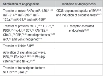
Table 2. Main Mechanisms Involved in the Modulation of Angiogenesis by EVs
Stimulation of Angiogenesis Inhibition of Angiogenesis
Transfer of micro-RNAs: miR-126,61–63
miR-214,64 miR-296,63 miR-
125a,65 miR-31,66 and miR-15067
CD36-dependent uptake of EVs68,69
and induction of oxidative tress68–72
Transfer of proteins: VEGF,73,74 FGF-2,74
PDGF,74,75 c-kit,76 SCF,76 RANTES,77
CD40L,78 CRP,79–81 metalloproteases,76,82
uPA,83 and Sonic hedgehog84–87
LDL receptor-mediated endocytosis88,89
Transfer of lipids: S1P90
Activation of signaling pathways: PI3K,3,90 ERK1/2,61,74,91–93 Wnt4/β-
catenin,94 and NF-κB95,96
Transfer of transcription factors: STAT3,95,96 STAT561
CRP indicates C-reactive protein; ERK1/2, extracellular signal-regulated kinase 1 and 2; EV, extracellular vesicle; FGF, fibroblast growth factor; LDL, low-density lipoprotein; NF-κB, nuclear factor-κB; PDGF, platelet-derived growth factor; PI3K, phosphoinositide 3-kinase; RANTES, regulated on activation, normal T-cell–expressed and secreted; S1P, sphingosine-1-phosphate; SCF, stem cell factor; uPA, urokinase-type plasminogen activator; and VEGF, vascular endothelial growth factor.
by guest on April 25, 2018
http://circres.ahajournals.org/
colony-forming cells (ECFC) have been recognized as genuine EPCs, displaying an unambiguous endothelial dif-ferentiation potential, proliferative activity, and a specific capacity to self-assemble into functional blood vessels in
vivo.102 Because these cells have higher vasculogenic
poten-tial compared with mature EC, EVs originating from ECFC have been proposed as potent mediators of proangiogenic signals. Several studies clearly demonstrated their
capac-ity to stimulate blood vessel formation.63,103,104 For instance,
ECFC-derived EVs were shown to incorporate into EC via interaction with α4 and β1 integrins, and they stimulated angiogenesis both in vitro and in vivo through the deliv-ery of mRNA associated with eNOS and the PI3K/AKT
signaling pathway.3 In addition, ECFC-derived EVs
pre-vented capillary rarefaction and subsequent tissue damage
in a rat model of kidney ischemia-reperfusion injury.63 The
renoprotective action of EVs was abolished by the specific depletion of miR-126 and miR-296, indicating their critical role in the stimulation of angiogenesis. Additional in vitro studies demonstrated that the EV internalization by hypox-ic EC was mainly mediated by L-selectin, and it restored the expression of proangiogeneic and antiapoptotic genes
that were downregulated by hypoxia.63 Similar mechanisms
of miRNA delivery have been involved in the capacity of ECFC-EV to promote neovascularization and limit muscle damage after hindlimb ischemia or to enhance the survival
of human islet transplants.103,104 Recent studies reported that
ECFC-derived exosomes promote cutaneous wound heal-ing in diabetic rats by enhancheal-ing neovascularization in the wound sites. The exosomes mediated their proangiogenic potential by activating Erk1/2 signaling in EC and by
stimu-lating the expression of several angiogenic molecules.92,93
Figure 1. Mechanisms involved in the modulation of Angiogenesis by endothelial cell (EC)–derived extracellular vesicles (EVs). EC
release EVs rich in micro-RNA such as miR-214 and miR-126, that are transferred to recipient EC and induce proangiogenic signaling. EVs contain functional matrix metalloproteinases that facilitate angiogenesis through the degradation of components of the extracellular matrix. Dll4 is transferred to EC by the EVs and induces Notch receptor internalization and tip cell formation. EVs bear, at their surface, a tissue factor that interacts with β1 integrin and induces Rac1-ERK1/2-ETS1 signaling, leading to the increased secretion of CCL2. EVs transport the complex uPA/uPAR, which stimulates angiogenesis through plasmin generation. The phosphatidylserine present on the surface of the EVs interacts with CD36 and induces Fyn kinase signaling, which leads to increased oxidative stress and the inhibition of angiogenesis. ATM indicates ataxia telangiectasia mutated; CCl2, chemokine c-c motif ligand 2; Dll4, Delta-like 4; ECM, extracellular matrix; ERK1/2, extracellular signal-related kinase 1 and 2; ETS1, avian erythroblastosis virus E26 homolog-1; IL-3R, interleukin-3 receptor; MMPs, matrix metalloproteinases; NotchR, Notch receptor; PS, phosphatidylserine; Rac1, Ras-related C3 botulinum toxin substrate 1; ROS, reactive oxygen species; TF, tissue factor; TIMPS, tissue inhibitor of metalloproteinases; uPA, urokinase plasminogen activator; and uPAR, urokinase plasminogen activator receptor .
by guest on April 25, 2018
http://circres.ahajournals.org/
Mesenchymal Stem/Stromal Cell–derived EVs Mesenchymal stem/stromal cells (MSCs) have potent proan-giogenic properties that have been attributed to their secre-tion of paracrine factors. However, it was recently suggested that among soluble factors, EVs play an important role in in-tercellular communication and are involved in angiogenesis. Figure 2 summarizes the main mechanisms involved in the modulation of angiogenesis by MSC-derived EVs. The func-tionality of the MSC secretome is strongly influenced by the microenvironment. For example, on hypoxia stimulation, bone-marrow–derived MSC released EVs that can be incor-porated by EC, stimulated in vitro tube formation, and
pro-moted angiogenesis in a rat myocardial infarction model.105
In addition, exosomes derived from MSC enhanced the tube formation of EC and stimulated neovascularization, lead-ing to improved heart function after ischemic injury in a rat
myocardial infarction model.105 Recently, it was demonstrated
that EVs isolated from bone-marrow–derived MSC increased
postischemic neuroangiogenesis in a way similar to that of
MSC after focal cerebral ischemia in mice.106 In line with this
study, after traumatic brain injury in rats, the administration of exosomes derived from MSC significantly improved
func-tional recovery by promoting endogenous angiogenesis.107
The main mechanisms involved in the proangiogenic effect of MSC-derived exosomes in ischemic conditions involve the activation of the nuclear factor-κB pathway and the transfer
of transcriptionally active STAT3 (Figure 2).95,96 In addition,
exosomes that were isolated from bone marrow MSC were able to reduce infarct size and enhance blood flow recovery by increasing the density of functional capillaries after
myocar-dial infarction in rats.105
Another important source of MSC is the umbilical cord. A hypoxia treatment of umbilical cord–derived MSC increased their production of EVs with proangiogenic potential. These EVs enhanced in vitro capillary-like structure formation and fur-ther improved blood flow recovery in a rat hindlimb ischemia
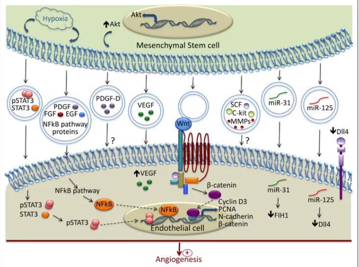
Figure 2. Mechanisms involved in the modulation of Angiogenesis by MSC-derived extracellular vesicles (EVs). In response to hypoxia,
mesenchymal stem/stromal cells release EVs containing active pSTAT3 and nuclear factor (NF)-κB pathway–associated proteins, which are transferred to recipient endothelial cell (EC) and promote the transcription of proangiogenic proteins. EVs contain and transfer several growth factors to EC (PDGF, FGF, EGF, VEGF, SCF, and c-kit). Wnt is present in the EVs and through interaction with its receptor, promotes the transcription of several molecules involved in angiogenesis. EVs stimulate vessel formation through the transfer of various micro-RNA. Among them, miR-31 acts by suppressing the factor inhibiting HIF-1α, and miR-125 promotes tip cell specification by suppressing Delta-like 4 (Dll4). ? indicates that the exact mechanisms of their transfer and the underlying signaling pathways are not completely described; EGF, epidermal growth factor; FGF, fibroblast growth factor; FIH, factor inhibiting HIF-1α; HIF, hypoxia inducible factor; PCNA, proliferating cell nuclear antigen; PDGF, platelet-derived growth factor; SCF, stem cell factor; and VEGF, vascular endothelial growth factor.
by guest on April 25, 2018
http://circres.ahajournals.org/
model.108 After unilateral kidney ischemia in rats, the
intrave-nous injection of EVs derived from umbilical cord MSC had protective effects by increasing the capillary vessel density
through the delivery of VEGF.73 The exosomes released from
Akt-overexpressing MSC derived from umbilical cord im-proved cardiac function after intravenous infusion in a rat model of acute myocardial infarction. In addition, the exosomes iso-lated from Akt-overexpressing MSC stimuiso-lated EC migration, proliferation, and tube-like formation in vitro, but they also in-creased the formation of blood vessels in vivo. This study identi-fied PDGF-D (platelet-derived growth factor-D) as an important
player in the exosome-mediated stimulation of angiogenesis.75
The activation of the Wnt4/β-catenin pathway was reported as another mechanism involved in the proangiogenic effect of
hu-man umbilical cord MSC-derived exosomes (Figure 2).94
Exosomes that were isolated from adipose tissue–derived MSC were also shown to promote angiogenesis in vitro and
in vivo. This proangiogenic effect was because of the trans-fer of miR-125a that promoted tip cell specification through
the direct suppression of δ-like 4 (Figure 2).65 The angiogenic
potential of EVs can be greatly improved by modulating the culture conditions of MSC. Thus, PDGF treatment was shown to modulate EV protein composition, leading to an increased c-kit, SCF (stem cell factor), and metalloprotease content
and, therefore, it enhanced their proangiogenic potential.76
Preconditioning of the adipose-derived stem cells with an endothelial differentiation medium upregulated the release of microvesicles and their proangiogenic properties through the delivery of miR-31 and the suppression of factor inhibiting
HIF-1α (Figure 2).66
Induced pluripotent stem cells present an unlimited cellular source for MSC generation. Interestingly, it was reported that exosomes derived from induced pluripotent stem
cells–differ-entiated MSC promoted angiogenesis in osteoporotic rats.109
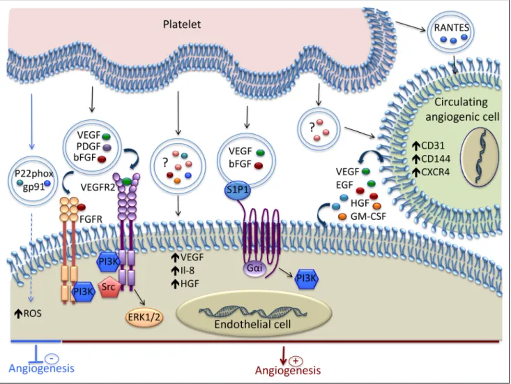
Figure 3. Mechanisms involved in the modulation of Angiogenesis by platelet-derived extracellular vesicles (EVs). Platelet-derived
EVs contain various growth factors and chemokines that induce proangiogenic signaling in endothelial cell (EC). Spingosine-1-phosphate (S1P1), present on the EV surface, induces PI3K activation and, together with VEGF and bFGF, promotes angiogenesis. The EVs released by platelets stimulate the proangiogenic potential of circulating angiogenic cells by increasing their expression of both membrane molecules and soluble factors. Platelet-derived EVs can inhibit angiogenesis by transferring the p22phox and gp91 subunits of NADPH oxidase and increasing the oxidative stress in EC. ? indicates that the exact content of the EVs is not reported; bFGF, basic fibroblast growth factor; EGF, epidermal growth factor; GM-CSF, granulocyte-macrophage colony-stimulating factor; HGF, hepatocyte growth factor; NADPH, nicotinamide adenine dinucleotide phosphate; PI3K, phosphoinositide 3-kinase; RANTES, regulated on activation, normal T-cell–expressed and secreted; ROS, reactive oxygen species; VEGF, vascular endothelial growth factor; and VEGFR, vascular endothelial growth factor receptor.
by guest on April 25, 2018
http://circres.ahajournals.org/
Platelet-Derived EVs
In addition to their key role in hemostasis and thrombosis, platelets are involved in several biological processes such as inflammation, angiogenesis, and tissue regeneration. During the past 12 years, the role of platelet EVs in angiogenesis has been well documented. Figure 3 shows the main mechanisms involved in the modulation of angiogenesis by platelet-derived EVs. The amount of platelet microvesicles and their proteome
are dependent on the stimulus used for their generation.110
Thus, different pathological contexts lead to the release of mi-croparticles with a specific protein content that will further determine the message delivered to the neighboring cells.
The role of platelet microparticles in angiogenesis was
evi-denced for the first time in 2004.90 This study showed that the
platelet microparticles isolated from healthy donors were capable of promoting proliferation, migration, and tube formation of hu-man umbilical vein EC through the cooperative effect of VEGF,
FGF-2, and lipid components, such as spingosine-1-phosphate.90
These effects were essentially mediated via the pertussis toxin-sensitive G protein and the PI3 kinase pathway (Figure 3).
The proangiogenic activity of platelet microparticles was further supported by a study showing that EPC cultures con-tain platelet microparticles with the capacity to stimulate
en-dothelial tube formation in vitro.101 This proangiogenic effect
was attenuated after removing the microparticles by filtration or ultracentrifugation or by specifically inhibiting the forma-tion of the platelet integrin complex αIIbβ3.
In a rat aortic ring model, platelet-derived microparticles exerted proangiogenic effects in a dose-dependent manner through the transfer of cytokines, such as VEGF, FGF-2, and PDGF, and the activation of PI3 kinase, src kinase, and ERK
(Figure 3).74 These results were supported by increased in
vi-tro tube-like formation and in vivo angiogenesis using agarose beads that contained platelet microparticles. In addition, in a model of chronic myocardial ischemia in rats, platelet mic-roparticles were shown to increase the number of functional
capillaries after injection into the myocardium.74 Platelet
mi-crovesicles were also documented as important mediators in tumor progression and angiogenesis in lung cancer through the upregulation of factors involved in tumor vascularization, such
as interleukin-8, VEGF, and HGF (hepatocyte growth factor).111
It was also shown that platelet-derived EVs can have delete-rious effects on EC. The exosomes isolated from septic patients contain the p22phox and gp91 subunits of NADPH oxidase and
were able to produce reactive oxygen species (Figure 3).72 The
incubation of these exosomes with EC enhanced their apoptosis. Further studies showed that platelets exposed to lipopolysaccha-ride or NO, but not thrombin or tumor necrosis factor-α, gener-ated exosomes similar to those recovered from septic patients,
and they induced apoptosis in EC through redox signaling.112
It was also shown that platelet-derived microparticles stimulated endogenous stem cell repair mechanisms after middle cerebral artery occlusion in rats. The delivery of plate-let microparticles increased angiogenesis and neurogenesis at
the infarct zone in a dose-dependent manner.113 These results
were supported by another study that demonstrated the protec-tive effect of platelet-derived microparticles against cerebral
ischemic reperfusion injury.114
Platelet microparticles can also enhance the regenerative capacity of early outgrowth ECs by modulating their pheno-type and secretome, as attested by the increased expression of CD31, VE-cadherin, and CXCR4, and the secretion of VEGF, EGF, HGF, and GM-CSF (granulocyte-macrophage
colony-stimulating factor; Figure 3).115 In addition,
plate-let microparticles increased the ability of early outgrowth ECs to stimulate endothelial migration, proliferation, tube formation, and importantly, endothelial regeneration after injury in vivo. In line with this study, the pretreatment of circulating angiogenic cells with platelet microparticles isolated from peripheral blood of atherosclerotic patients led to increased neovascularization in rats with hindlimb
ischemia.77 This proangiogenic effect was shown to be
de-pendent on the release of RANTES by the platelet micropar-ticles (Figure 3).
Leukocyte-Derived EVs
Leukocyte EVs may originate from lymphocytes, monocyte/ macrophages, or neutrophils, and they have been shown to actively participate in vascular homeostasis. Several studies, mostly focusing on lymphocytes and monocytes, emphasized that leukocyte EVs may also play a role in angiogenesis. Depending on the stimuli used for their generation, the EVs derived from lymphocytes can exert either pro- or antiangio-genic effects. For instance, lymphocytes undergoing activa-tion and apoptosis release microvesicles with proangiogenic properties, whereas microparticles shed from apoptotic
lym-phocytes inhibit angiogenesis.69,84,85
The microparticles generated from T lymphocytes after treatment with phytohemagglutinin, phorbol ester, and acti-nomycin D express Sonic hedgehog and induce the formation of capillary-like structures in vitro through FAK activation and the upregulation of ICAM-1 (intercellular adhesion
mol-ecule-1), RhoA, and VEGF.84 These microparticles expressing
Sonic hedgehog favored neovascularization in a mouse model of hindlimb ischemia by activating the eNOS pathway and NO production in not only the ischemic area but also the aorta
of mice.85 The stimulation of NO production by lymphocyte
microparticles carrying Sonic hedgehog also had a beneficial effect in restoring endothelial function in mouse models of an-giotensin II–induced hypertension and myocardial ischemia/
reperfusion injury.86,87
In contrast, actinomycin D treatment of lymphocytes in the absence of activation signals resulted in a microparticle
release that inhibited angiogenesis.69 Thus, in a model of
in vivo corneal neovascularization and in vitro aortic rings, the microparticles generated after actinomycin D treatment suppressed angiogenesis through increased reactive oxygen species production, upregulation of CD36 on target EC and the consequent reduction of VEGFR2 and phospho-ERK
levels.69 In addition, the antiangiogenic effect of
micropar-ticles was shown to present an interesting therapeutic ap-plication in pathological angiogenesis, such as retinopathies
and tumor angiogenesis.88,89 The inhibition of angiogenesis
by T-lymphocyte–derived microparticles was mediated by low-density lipoprotein receptor, as low-density lipoprotein receptor knockdown in target endothelial and tumor cells re-sulted in decreased microparticle uptake and abrogated the
by guest on April 25, 2018
http://circres.ahajournals.org/
antiangiogenic effect.88,89 In the case of choroidal angiogenesis,
it was demonstrated that the inhibitory effect of microparticles was dependent on the production of pigment epithelium-de-rived growth factor and nerve growth factor by retinal pigment epithelial cells and the activation of p75 neurotrophin receptor
in choroidal microvascular cells.116 Recently, the same group
evidenced that on CD36-dependent uptake, lymphocyte mic-roparticles can exert their antiangiogenic effect by altering the
expression of several angiogenic factors in macrophages.117
The microvesicle derived from monocytes were also shown to promote angiogenesis both in vitro and in vivo through the
transfer of miR-150 to EC.67 THP-1 (human acute monocytic
leukemia cell line)–derived EVs were also shown to generate monomeric CRP, which is highly angiogenic to vascular EC and
therefore might impact on the process of vascularization.79–81 In
addition, CD40 ligand–positive microparticles, which were iso-lated from human atherosclerotic plaques and of mostly macro-phage origin, were able to stimulate endothelial proliferation and angiogenesis, suggesting that microparticles could participate in
intraplaque neovascularization and plaque vulnerability.78
Erythrocyte-Derived EVs
In addition to their vital role in transporting oxygen to organs and tissues, erythrocytes were shown to participate in
angiogen-esis.118 A recent study demonstrated that the production of
sphin-gosine-1-phosphate by erythrocytes is essential for vascular stabilization, maturation, and remodeling during embryonic
de-velopment.118 The process of EV formation occurs during
eryth-rocyte maturation and allows for the selective retention of certain proteins in the plasma membrane and the removal of noxious
molecules, such as oxidized proteins.119 Vesicle release is
accel-erated during erythrocyte senescence and is considered a part
of apoptosis-like form in these cells.120 Even though studies
ad-dressing the specific role of erythrocyte EVs in angiogenesis are still lacking, some studies suggest that erythrocyte-derived EVs may have a deleterious effect on EC, particularly in some patho-logical conditions, such as sickle cell disease and malaria. It was reported that EVs derived from malaria-infected erythrocytes altered EC gene expression and barrier properties through the
transfer of miR-451a.121 In addition, erythrocyte microparticles
were significantly elevated in patients with sickle cell disease, and they expressed more sphingosine-1-phosphate than
eryth-rocyte microparticles from healthy donors.122 Sickle cell disease
erythrocyte-derived MP are internalized by myeloid cells and promote proinflammatory cytokine production and EC adhesion in an ERK ½-ICAM-4–mediated fashion. These results suggest that circulating MP may contribute to the inflammation-rooted
pathogenesis of the disease.122 In addition, it was evidenced that
erythrocyte vesiculation produced microparticles loaded with heme that could be further transferred to EC, inducing
oxida-tive stress and apoptosis.123 Heme-laden microparticles strongly
reduced endothelium-dependent vasodilation and induced
va-soocclusions in kidneys.123 These observations indicate that
mi-croparticles carrying heme may be a source of oxidant stress, contributing to endothelial dysfunction and vascular injury.
Toward EV-Based Therapeutic Angiogenesis
Based on the various studies discussed above, EVs have emerged as a major form of intercellular communication,playing important roles in angiogenesis and cardiovascular homeostasis. Their role in cellular signaling during vascular development, growth, and maturation has been extensively documented and has opened up new perspectives for their ther-apeutic application in regenerative medicine and tissue engi-neering as drug delivery vehicles modulating vascularization. Since the early 2000s, EVs have been tested in clinical
trials involving patients with melanoma,124 nonsmall cell lung
cancers,125,126 colorectal cancer,126a and type I diabetes mellitus
(NCT02138331). Although a small number of patients was in-cluded in the studies, these clinical trials demonstrated the fea-sibility and safety of EV administration in humans. Recently, a prospective clinical trial was initiated to evaluate the effect of autologous plasma-derived exosomes on wound healing in patients with intractable cutaneous ulcers (NCT02565264). This trial will provide important information about the feasi-bility and the efficiency of EV-based therapy in angiogenesis-related disorders.
Although the pro- and antiangiogenic effects of EVs from various sources have been reported, further in vivo studies are required to better understand the specific role of EVs in exchanging molecules between cell types. Several limitations need to be overcome to progress toward the use of EVs in the therapeutic modulation of angiogenesis. In particular, the standardization of the end product, quality control, and the associated costs remain major issues for EV-based therapies. Cellular Source and Conditions for EV Production One of the key points to consider is the choice of the cellu-lar source and the conditions for a suitable quantity of clini-cal grade EV production. Recent studies suggest that several stimuli and culture conditions play an important role in the biogenesis and secretion of EVs with proangiogenic
proper-ties.61,82,96,127 In addition, it is important to evaluate how the
patient’s pathological environment impacts the EV proper-ties and therapeutic potential. Even though most of the pre-clinical works supporting the capacity of EVs for therapeutic angiogenesis have been done with EVs from cells that were isolated from healthy donors, there are emerging data show-ing that the proangiogenic capacity of MSC-derived EVs are
impaired in patients with cardiovascular risk factors.128 Thus,
donor-related variability may be responsible for the thera-peutic differences observed among comparable EV fractions, requiring the definition of donor inclusion/exclusion criteria and potent markers to able to anticipate the therapeutic ef-fects of EVs in vivo.
EV Concentration
One important question is what concentration of EVs should be used for the stimulation of angiogenesis without inducing any side effects? An overview of the literature shows that the effect of EVs on angiogenesis seems to be dose dependent. For
instance, low amounts of endothelial microparticles (2×10³ per
well) were able to increase tube formation in vitro, whereas a
high concentration had an inhibitory effect (2×10⁵ per well).83
Another study reported that endothelial microparticles in-hibited angiogenesis in the cultured sections of hearts when
concentrations higher than 105 microparticles/mL were used.99
Platelet-derived microparticles exerted proangiogenic effects
by guest on April 25, 2018
http://circres.ahajournals.org/
in vitro when doses between 30 and 100 μg of protein/mL were
applied.74 However, when platelet microparticles were used to
stimulate vascularization in vivo, a much higher concentration
(250 μg/mL) was necessary to stimulate postischemic
revas-cularization of the myocardium.74 In addition, it was reported
that MSC-derived exosomes exerted the most potent angio-genic effect in vitro and in vivo at a concentration of 100 μg/
mL.65 However, another study showed that a low concentration
of MSC exosomes (1–10 μg/mL) was more effective for tube
formation than a high concentration (100 μg/mL).95
All these observations demonstrate that the question on EV concentration remains controversial. It is difficult to address this question and compare the results from all these studies, as different culture conditions have been used for EV production and various methods have been used to quantify EVs.
Because of the small size of EVs and the technical limita-tions of nanosized particle analysis, EV quantification has been challenging to scientists. Several techniques have been used to quantify EVs, such as dynamic light scattering, nanoparticle tracking analysis, flow cytometry, and tunable resistive pulse sensing. However, a universal method for standardized EV quantification is still lacking. The studies mentioned above suggest that the optimal EV concentration for the stimulation of angiogenesis will depend on various factors, such as the cel-lular source of EVs, the stimulus used to induce EV release, EV content, the way of EV delivery in vivo, and also the patho-logical microenvironment. Determination of the EV concentra-tion for the stimulaconcentra-tion of angiogenesis is hampered not only by the various methods used for EV quantification but also by the different degrees of purity of the EV preparations.
Purity of EV Preparations
Another important aspect to consider is the purity of the dif-ferent EV populations and the characterization of their content and biological properties. Several techniques have been devel-oped to isolate EVs, such as differential ultracentrifugation, polymer-based precipitation, density gradients,
microfiltra-tion, and size-exclusion-based approaches.129 It is important
to note that compared with ultracentrifugation and polymer-based precipitation, the development of flotation through density gradients, microfiltration, and size-exclusion chroma-tography for EV purification marked significant progress in the field.
A pure isolation of EVs from tissue culture supernatants and body fluids is hampered by the presence of macromolec-ular structures. For instance, the presence of bovine proteins and microvesicles in the culture media can impact the growth and phenotype of cultured cells, thereby influencing the
qual-ity and quantqual-ity of EVs produced in vitro.130 Furthermore, the
RNAs, proteins, and lipids carried by EVs that are present in the bovine serum can also affect the results involving the pro-teomic, transcriptomic, and functional analysis of EVs. Thus, it is necessary to eliminate the EVs carried by the bovine serum before cell culture. Filtration of the culture media using 0.1-µm filters could help to eliminate microparticles, but not all the exosomes. Switching the cells to serum-free conditions dur-ing EV production could allow us to circumvent this problem; however, serum-free conditions may add additional stress to the cells and affect the nature and amount of secreted EVs.
Within the EV population, many distinct subtypes of ves-icles exist. However, the currently used methods do not al-low for their efficient separation and can negatively impact their integrity, function, and therapeutic potential. Therefore, the development of new, more selective isolation techniques is necessary to achieve good purity in each vesicle population and to determine, in a more precise way, the properties and biological functions of each of them. Most of the molecules responsible for the therapeutic effects of EVs still remain to be identified and extensively characterized. To further explore their potential, proteomic and mRNA/miRNA microarray analysis of each vesicle population may provide useful
in-formation.131–133 Table 3 summarizes the main transcriptomic,
proteomic, and lipidomic studies on the EVs that were derived from the cell types discussed in this review. In addition, the methods used for the isolation and purification of EVs var-ies considerably between different research groups and also varies depending on the tissue that they are extracted from. Indeed, considerable heterogeneity among EV preparations has been observed. Therefore, one of the most important chal-lenges before applying EVs in therapeutics is the optimization and standardization of the methods of EV purification, enrich-ment, and characterization, which will allow increasing the homogeneity within EV preparations and will enable
achiev-ing good manufacturachiev-ing practices.145
EV Biodistribution After Administration
A better understanding of EV biodistribution after administra-tion is an essential point for the safe successful clinical appli-cation of EVs in tissue regeneration. Although the substantial proangiogenic effects of EVs have been reported by several preclinical studies, our current knowledge of their biodistri-bution and clearance dynamics still remains limited. Several methods for EV labeling and tracking have been developed,
such as reporter systems,146,147 fluorescent dye labeling, loading
with superparamagnetic iron oxide nanoparticles148 or
radioiso-tope labeling with clinically validated radio tracers.149 Several
studies independently reported the short half-life of exogenous EVs that were artificially introduced in the circulation because of their retention and uptake in organs. In vivo tracking stud-ies showed that intravenously administered EVs derived from kidney embryonic cells are taken up mainly by the kidney, fol-lowed by the liver and lung, whereas some EVs were retained
by the heart, brain and muscle, albeit in low amounts.146 In
addition, EVs derived from red blood cells showed increased uptake by the liver and bone, followed by the skin, muscle, spleen, kidney, and lung, whereas melanoma-derived EVs
were mainly taken up by the lungs and spleen.8 Thus, the
cel-lular source and the route of administration are the key
deter-minants of EV tissue distribution.150 Therefore, establishing the
best route of EV delivery in normal and pathological condi-tions may help improve the therapeutic efficiency of EVs. In addition, the efficiency of EV delivery could be improved by a better understanding of the mechanisms of EV uptake and interaction with the target cells, in particular, with the cells of the cardiovascular systems, as few studies are available.
Finally, further studies are necessary to evaluate the risk of adverse effects or side effects after EV administration. Although the EVs used in preclinical studies clearly lack the potential to
by guest on April 25, 2018
http://circres.ahajournals.org/
directly induce tumor growth, the proproliferative and proan-giogenic effects of EVs evoke the possibility that they could ac-celerate or participate in cancer progression. The procoagulant activity of EVs has been the subject of several studies, suggest-ing that another adverse effect associated with EV adminis-tration could be an increased risk of thrombosis. Moreover, because some EVs can both promote or inhibit angiogenesis,
preclinical models that allow us to study the involvement of en-dogenous EVs, in addition to those that are exogenously admin-istrated, are needed to better delineate the benefit/risk balance.
Conclusion
In addition to their role as promising biomarkers in vari-ous pathological conditions, EVs are important mediators in
Table 3. Transcriptomic, Proteomic, and Lipidomic Studies on EVs
EV source Isolation Analysis Year References
Endothelial cells (HUVEC) 20 min 120g 90 min 16 000g Next-generation sequencing of miRNAs 2012 Diehl et al53 5 min 4000g 90 min 100 000g
Proteomic analysis (LC-MS/MS) 2005 Banfi et al134
4 min 200g 1 h 100 000g
Proteomic analysis (LC-MS/MS) 2008 Peterson et al135
5 min 200g 2 h 100 000g
Proteomic analysis (LC-MS/MS) 2013 Liu et al136
Endothelial progenitor cells 20 min 2000g 1 h 100 000g Transcriptomic analysis (microarray) 2007 Deregibus et al3 15 min 400g 5 min 12 500g 150 min 20 500g
Proteomic analysis (LC-MS/MS) 2009 Prokopi et al101
Mesenchymal stem cells 20 min 2000g 1 h 100 000g Transcriptomic analysis (Microarray) 2009 Bruno et al137 10 min 500g Concentration 1 kDa 1 h 15 000g 18 h 110 000g Next-generation sequencing of miRNAs Proteomic analysis (LC-MS/MS) Lipidomic analysis 2014 Vallabhaneni et al138 10 min 500g 15 min 800g Concentration 100 kDa Sucrose cushion 2 h 100 000g
Proteomic analysis (LC-MS/MS) 2012 Kim et al131
Platelets 15 min 710g
90 min 150 000g
Proteomic analysis (LC-MS/MS) 2005 Garcia et al139
30 min 5000g 130 000g Gel filtration
Proteomic analysis (LC-MS/MS) 2009 Dean et al140
1500g 1 h 100 000g
Proteomic analysis (LC-MS/MS) 2012 Shai et al110
Monocyte (THP-1) 20 min 120g 90 min 16 000g Next-generation sequencing of miRNAs 2012 Diehl et al53 Macrophages 30 min 500g 20 min 16 500g Filtration 0.22 μm 180 min 120 000g Microarray 2011 Yang et al141 Erythrocytes Microvesicles: 1550g; 40 000g Nanovesicles: 1550g; 40 000g; 100 000g
Proteomic analysis (LC-MS/MS) 2008 Bosman et al142
Neutrophils 10 min 3000g
1 h 100 000g
Proteomic analysis (LC-MS/MS) 2013 Dalli et al.143
Atherosclerotic plaques
15 min 500g 5 min 12 500g 150 min 20 500g
Proteomic analysis (LC-MS/MS) 2009 Mayr et al144
EV indicates extracellular vesicle; HUVEC, human umbilical vein endothelial cells; LC-MS/MS, liquid chromatography coupled to tandem mass spectrometry; and THP-1, human acute monocytic leukemia cell line.
by guest on April 25, 2018
http://circres.ahajournals.org/
intercellular communication in normal physiology and dis-ease. An increasing number of studies has documented the ca-pacity of EVs to actively modulate the angiogenic programs in EC by acting on the key steps of vessel formation. Depending on the cellular source, the conditions in which they have been generated and their content, EVs may have either beneficial or detrimental effects on angiogenesis and tissue regeneration. EVs are released by various cell types, such as ECs, endothe-lial colony-forming cells, mesenchymal stem cells, platelets, or leucocytes, and are able to deliver powerful proangiogenic signals with subsequent beneficial effects in tissue repair. Specific conditions, such as hypoxia and angiogenic growth factor stimulation, favor the release of EVs with vasculogenic potential. However, some of the EVs produced by ECs, plate-lets, or lymphocytes can also exert an inhibitory activity on vessel growth, mainly by increasing the oxidative stress in target cells. Although the role of EVs in blood vessel forma-tion is well documented and the main underlying mechanisms are identified, further studies are necessary to determine the precise role of each subpopulation of EVs. Indeed, one of the major challenges that the expanding field of EVs faces is the high diversity of vesicle subtypes and the complexity of specifically studying the characteristics, compositions and biological functions of each of them separately. Importantly, because some EVs can either promote or inhibit angiogenesis, animal models allowing us to study the specific involvement of endogenous EVs are needed to delineate when and how EVs can be protective.
A better understanding of EV biogenesis and the multi-faced functions of EVs that defines the specific phenotypic characteristics and active components carried by each sub-population of EVs will allow us to standardize the process of EV preparation for therapeutic applications. Together with the validation of accurate technologies for the clinical grade manufacturing and quality control of EVs, such advances may provide a better appraisal of the benefit-risk ratio, sustaining the currently emerging therapeutic applications of EVs in re-generative medicine or in angiogenesis-related diseases.
Sources of Funding
This work was supported by the A*MIDEX project (no ANR-11-IDEX-0001-02), funded by the Investissements d’Avenir French Government program, and managed by the French National Research Agency and INSERM.
Disclosures
None.
References
1. Patel-Hett S, D’Amore PA. Signal transduction in vasculogenesis and de-velopmental angiogenesis. Int J Dev Biol. 2011;55:353–363. doi: 10.1387/ ijdb.103213sp.
2. Carmeliet P, Jain RK. Molecular mechanisms and clinical applications of angiogenesis. Nature. 2011;473:298–307. doi: 10.1038/nature10144. 3. Deregibus MC, Cantaluppi V, Calogero R, Lo Iacono M, Tetta C, Biancone
L, Bruno S, Bussolati B, Camussi G. Endothelial progenitor cell derived microvesicles activate an angiogenic program in endothelial cells by a horizontal transfer of mRNA. Blood. 2007;110:2440–2448. doi: 10.1182/ blood-2007-03-078709.
4. Gai C, Carpanetto A, Deregibus MC, Camussi G. Extracellular vesicle-mediated modulation of angiogenesis. Histol Histopathol. 2016;31:379– 391. doi: 10.14670/HH-11-708.
5. Raposo G, Stoorvogel W. Extracellular vesicles: exosomes, mi-crovesicles, and friends. J Cell Biol. 2013;200:373–383. doi: 10.1083/ jcb.201211138.
6. Dignat-George F, Boulanger CM. The many faces of endothelial mic-roparticles. Arterioscler Thromb Vasc Biol. 2011;31:27–33. doi: 10.1161/ ATVBAHA.110.218123.
7. Turturici G, Tinnirello R, Sconzo G, Geraci F. Extracellular membrane vesicles as a mechanism of cell-to-cell communication: advantages and disadvantages. Am J Physiol Cell Physiol. 2014;306:C621–C633. doi: 10.1152/ajpcell.00228.2013.
8. Yáñez-Mó M, Siljander PR, Andreu Z, et al. Biological properties of ex-tracellular vesicles and their physiological functions. J Extracell Vesicles. 2015;4:27066.
9. Théry C. Exosomes: secreted vesicles and intercellular communications.
F1000 Biol Rep. 2011;3:15. doi: 10.3410/B3-15.
10. Théry C, Amigorena S, Raposo G, Clayton A. Isolation and characterization of exosomes from cell culture supernatants and biological fluids. Curr Protoc
Cell Biol. 2006;Chapter 3:Unit 3.22. doi: 10.1002/0471143030.cb0322s30. 11. Willms E, Johansson HJ, Mäger I, Lee Y, Blomberg KE, Sadik M, Alaarg
A, Smith CI, Lehtiö J, El Andaloussi S, Wood MJ, Vader P. Cells release subpopulations of exosomes with distinct molecular and biological prop-erties. Sci Rep. 2016;6:22519. doi: 10.1038/srep22519.
12. Colombo M, Moita C, van Niel G, Kowal J, Vigneron J, Benaroch P, Manel N, Moita LF, Théry C, Raposo G. Analysis of ESCRT functions in exo-some biogenesis, composition and secretion highlights the heterogeneity of extracellular vesicles. J Cell Sci. 2013;126:5553–5565. doi: 10.1242/ jcs.128868.
13. Trajkovic K, Hsu C, Chiantia S, Rajendran L, Wenzel D, Wieland F, Schwille P, Brügger B, Simons M. Ceramide triggers budding of exosome vesicles into multivesicular endosomes. Science. 2008;319:1244–1247. doi: 10.1126/science.1153124.
14. Andreu Z, Yáñez-Mó M. Tetraspanins in extracellular vesicle forma-tion and funcforma-tion. Front Immunol. 2014;5:442. doi: 10.3389/fimmu. 2014.00442.
15. Colombo M, Raposo G, Théry C. Biogenesis, secretion, and intercel-lular interactions of exosomes and other extracelintercel-lular vesicles. Annu
Rev Cell Dev Biol. 2014;30:255–289. doi: 10.1146/annurev-cellbio- 101512-122326.
16. Cocucci E, Meldolesi J. Ectosomes and exosomes: shedding the confusion between extracellular vesicles. Trends Cell Biol. 2015;25:364–372. doi: 10.1016/j.tcb.2015.01.004.
17. Kalra H, Drummen GP, Mathivanan S. Focus on extracellular vesicles: introducing the next small big thing. Int J Mol Sci. 2016;17:170. doi: 10.3390/ijms17020170.
18. Larson MC, Hillery CA, Hogg N. Circulating membrane-derived mi-crovesicles in redox biology. Free Radic Biol Med. 2014;73:214–228. doi: 10.1016/j.freeradbiomed.2014.04.017.
19. Leroyer AS, Anfosso F, Lacroix R, Sabatier F, Simoncini S, Njock SM, Jourde N, Brunet P, Camoin-Jau L, Sampol J, Dignat-George F. Endothelial-derived microparticles: Biological conveyors at the cross-road of inflammation, thrombosis and angiogenesis. Thromb Haemost. 2010;104:456–463. doi: 10.1160/TH10-02-0111.
20. Curtis AM, Edelberg J, Jonas R, Rogers WT, Moore JS, Syed W, Mohler ER 3rd. Endothelial microparticles: sophisticated vesicles modulat-ing vascular function. Vasc Med. 2013;18:204–214. doi: 10.1177/ 1358863X13499773.
21. Morel O, Toti F, Morel N, Freyssinet JM. Microparticles in endothelial cell and vascular homeostasis: are they really noxious? Haematologica. 2009;94:313–317. doi: 10.3324/haematol.2009.003657.
22. Del Conde I, Shrimpton CN, Thiagarajan P, López JA. Tissue-factor-bearing microvesicles arise from lipid rafts and fuse with activated plate-lets to initiate coagulation. Blood. 2005;106:1604–1611. doi: 10.1182/ blood-2004-03-1095.
23. EL Andaloussi S, Mäger I, Breakefield XO, Wood MJA. Extracellular vesicles: biology and emerging therapeutic opportunities. Nat Rev Drug
Discov. 2013;12:347–357.
24. van der Pol E, Böing AN, Harrison P, Sturk A, Nieuwland R. Classification, functions, and clinical relevance of extracellular vesicles. Pharmacol Rev. 2012;64:676–705. doi: 10.1124/pr.112.005983.
25. Kim D-K, Kang B, Kim OY, et al. EVpedia: an integrated database of high-throughput data for systemic analyses of extracellular vesicles.
J Extracell Vesicles. 2013;2.
26. Kalra H, Simpson RJ, Ji H, et al. Vesiclepedia: a compendium for ex-tracellular vesicles with continuous community annotation. PLoS Biol. 2012;10:e1001450. doi: 10.1371/journal.pbio.1001450.
by guest on April 25, 2018
http://circres.ahajournals.org/