HAL Id: inserm-01728520
https://www.hal.inserm.fr/inserm-01728520
Submitted on 11 Mar 2018
HAL is a multi-disciplinary open access
archive for the deposit and dissemination of
sci-entific research documents, whether they are
pub-lished or not. The documents may come from
teaching and research institutions in France or
abroad, or from public or private research centers.
L’archive ouverte pluridisciplinaire HAL, est
destinée au dépôt et à la diffusion de documents
scientifiques de niveau recherche, publiés ou non,
émanant des établissements d’enseignement et de
recherche français ou étrangers, des laboratoires
publics ou privés.
Alain Calender, Pierre Antoine Rollat Farnier, Adrien Buisson, Stéphane
Pinson, Abderrazzaq Bentaher, Serge Lebecque, Harriet Corvol, Rola Abou
Taam, Véronique Houdouin, Claire Bardel, et al.
To cite this version:
Alain Calender, Pierre Antoine Rollat Farnier, Adrien Buisson, Stéphane Pinson, Abderrazzaq
Ben-taher, et al.. Whole exome sequencing in three families segregating a pediatric case of sarcoidosis.
BMC Medical Genomics, BioMed Central, 2018, 11 (1), pp.23. �10.1186/s12920-018-0338-x�.
�inserm-01728520�
R E S E A R C H A R T I C L E
Open Access
Whole exome sequencing in three families
segregating a pediatric case of sarcoidosis
Alain Calender
1,3*, Pierre Antoine Rollat Farnier
2, Adrien Buisson
1, Stéphane Pinson
1, Abderrazzaq Bentaher
3,
Serge Lebecque
4, Harriet Corvol
5, Rola Abou Taam
6, Véronique Houdouin
7, Claire Bardel
2, Pascal Roy
2,
Gilles Devouassoux
8, Vincent Cottin
9, Pascal Seve
10, Jean-François Bernaudin
11, Clarice X. Lim
12,
Thomas Weichhart
12, Dominique Valeyre
13,14, Yves Pacheco
3, Annick Clement
15,
and Nadia Nathan
15in the frame of GSF (Groupe Sarcoïdose France)
Abstract
Background: Sarcoidosis (OMIM 181000) is a multi-systemic granulomatous disorder of unknown origin. Despite multiple genome-wide association (GWAS) studies, no major pathogenic pathways have been identified to date. To find out relevant sarcoidosis predisposing genes, we searched for de novo and recessive mutations in 3 young probands with sarcoidosis and their healthy parents using a whole-exome sequencing (WES) methodology. Methods: From the SARCFAM project based on a national network collecting familial cases of sarcoidosis, we selected three families (trios) in which a child, despite healthy parents, develop the disease before age 15 yr. Each trio was genotyped by WES (Illumina HiSEQ 2500) and we selected the gene variants segregating as 1) new mutations only occurring in affected children and 2) as recessive traits transmitted from each parents. The identified coding variants were compared between the three families. Allelic frequencies and in silico functional results were analyzed using ExAC, SIFT and Polyphenv2 databases. The clinical and genetic studies were registered by the ClinicalTrials.gov - Protocol Registration and Results System (PRS) (https://clinicaltrials.gov) receipt under the referenceNCT02829853and has been approved by the ethical committee (CPP LYON SUD EST– 2 – REF IRB 00009118– September 21, 2016).
Results: We identified 37 genes sharing coding variants occurring either as recessive mutations in at least 2 trios or de novo mutations in one of the three affected children. The genes were classified according to their potential roles in immunity related pathways: 9 to autophagy and intracellular trafficking, 6 to G-proteins regulation, 4 to T-cell activation, 4 to cell cycle and immune synapse, 2 to innate immunity. Ten of the 37 genes were studied in a bibliographic way to evaluate the functional link with sarcoidosis.
Conclusions: Whole exome analysis of case-parent trios is useful for the identification of genes predisposing to complex genetic diseases as sarcoidosis. Our data identified 37 genes that could be putatively linked to a pediatric form of sarcoidosis in three trios. Our in-depth focus on 10 of these 37 genes may suggest that the formation of the characteristic lesion in sarcoidosis, granuloma, results from combined deficits in autophagy and intracellular trafficking (ex: Sec16A, AP5B1 and RREB1), G-proteins regulation (ex: OBSCN, CTTND2 and DNAH11), T-cell activation (ex: IDO2, IGSF3), mitosis and/or immune synapse (ex: SPICE1 and KNL1). The significance of these findings needs to be confirmed by functional tests on selected gene variants.
Keywords: Sarcoidosis, Whole-exome sequencing (WES), Sarcoidosis, Candidate genes
* Correspondence:[email protected]
1Genetics Department, Hospices Civils de LYON (HCL), University Hospital,
East Pathology Center, LYON, B-A3, 59 Bld Pinel, 69677 BRON Cedex, France
3Inflammation & Immunity of the Respiratory Epithelium - EA7426 (PI3)–
South Medical University Hospital– Lyon 1 Claude Bernard University, 165
Chemin du Grand Revoyet, 69310 Pierre-Bénite, France Full list of author information is available at the end of the article
© The Author(s). 2018 Open Access This article is distributed under the terms of the Creative Commons Attribution 4.0 International License (http://creativecommons.org/licenses/by/4.0/), which permits unrestricted use, distribution, and reproduction in any medium, provided you give appropriate credit to the original author(s) and the source, provide a link to the Creative Commons license, and indicate if changes were made. The Creative Commons Public Domain Dedication waiver (http://creativecommons.org/publicdomain/zero/1.0/) applies to the data made available in this article, unless otherwise stated.
Background
Sarcoidosis is an enigmatic multisystem disease character-ized by the development and accumulation of granulomas, a compact collection of macrophages which have differen-tiated into epithelioid and multinucleated giant cells
asso-ciated with lymphocytes [1]. The disease is considered to
be the consequence of granulomatous reaction after ex-posure to various antigens and/or mineral particles in
in-dividuals with a susceptible genetic background [1, 2].
Sarcoidosis may be a chronic, benign or fatal disease af-fecting mainly lungs, skin, eyes and various other organs. This inflammatory disease develops before the age of 50 years and its incidence varies around the world with the highest frequencies being found in Northern European
countries (10 to 40 per 100.000) [1]. In children,
sarcoid-osis is a very rare disease with an estimated prevalence of
0.4 to 1.2 per 100.000 [3]. A recent French study on a
series of 41 children with sarcoidosis provided updated in-formation on the initial presentation, the management
and the follow-up of the disease [4]. It affects mainly
chil-dren older than 10 years, while one third of them are younger. The disease is often severe with general symp-toms at the forefront (asthenia, fever) and multiple organs involved at diagnosis in 85% of the children. The evolution is marked by frequent relapses and at risk to lung fibrosis despite extensive corticosteroid and immunosuppressive
treatments [5]. The relative contribution of genetic and
environmental factors is unknown but an 80-times in-crease in risk for monozygotic twins suggests that genetic factors might account for 50–60% of the disease
suscepti-bility [6]. Sarcoidosis is commonly sporadic with familial
presentation occurring in 3–5% of cases. The pedigree analysis suggests an autosomal dominant mode with in-complete penetrance. The French National SARCFAM study, based on a clinical network including 29 clinical centers, allowed the collection of clinical data and DNA samples of a cohort of 140 families with at least two first-degree relatives affected by sarcoidosis and rare situations
of nuclear families with pediatric cases [7]. We gave
prior-ity consideration to the analysis of pediatric clinical situa-tions in which the child with sarcoidosis has healthy parents, ie trios, expecting that new mutations and /or gene variants transmitted on a recessive pattern may high-light specific genetic pathways involved in the disease. Ini-tially the genetic studies were focused on the genes previously identified by GWAS as putative pathogenic de-terminants in the risk of sarcoidosis. One of candidate gene, BTNL2, codes a cofactor of CD86, a regulator of T-cell activation, and a BTNL2 rs2076530 SNP splicing
vari-ant has been shown to induce an≈ 2.0 fold increase in risk
of occurrence of the disease [7,8]. However, we have
pre-viously shown that this variant did not discriminate the sporadic and familial forms of the disease, thus suggesting BTNL2 is not a major gene predisposing to sarcoidosis
[7]. GWAS studies in several series have identified SNP’s
directly or indirectly implicated in innate immunity, such as HLADRB, CCDC88B, ANNEXINA11 (ANXA11),
XAF1 and other candidate loci [9]. Granuloma formation
is a process with hypothesized immune synapse abnormal-ities, suggesting new trials for treatment that target B and/
or T cells [10]. A remarkable feature of sarcoidosis is the
compartmentalization of CD4 (+) T helper 1 (Th1) lym-phocytes and activated macrophages in the affected organs to initiate the formation and maintenance of granulomas
[11]. Another subset of T-cells, Th17 effector CD4 (+),
which mediate the crosstalk between immune cells and tissues, has been shown to participate in the progression of granulomas and in the fibrotic phase of the disease. A third subgroup of immune cells, the CD4 (+) CD25 (+) regulatory T cell (Treg) showed an abnormal expansion which may be responsible for the paradoxical suppression of the immune response to tuberculin observed in
sar-coidosis [12]. Taken together, the immune pattern in
sar-coidosis is close to that observed in other Th1/Th17 related diseases, as tuberculosis, leprosy, chronic beryllium disease (CBD), ulcerative colitis or Crohn disease but mo-lecular signals that control granuloma genesis are largely undefined. A series of data obtained from animal models suggested a triggering of granuloma formation by mineral
pollutants such as carbon nanotubes [13]. On the
molecu-lar side, several previous biochemical studies have shown
abnormalities in pathways involving NF B [14], STAT1
[15], Gαi [16], and RIP2/IRAK [17], highlighting the
diffi-culty of targeting a specific signalling pathway for thera-peutic purposes. It was recently shown that the constitutive activation of mTORC1 in a mouse model de-ficient for TSC2 promotes granuloma formation through a metabolic reprogramming mediated by CDK4, suggest-ing a crucial role of the TSC1/TSC2 complex and mTor
pathway in granulomatous diseases [18]. Nevertheless,
identifying the genes which might be related to familial predisposition remains challenging as sarcoidosis is con-sidered as a complex multifactorial disease involving a synergistic effect of many genes carrying either rare and more common variants. Our working hypothesis that we have tested here is that a comparative WES analysis of children affected early in life by a serious form of sarcoid-osis and both their unaffected parents (trio analysis) may identify de novo and recessive genetic variants that could potentially point to pathways acting alone or in combin-ation in disease development.
Methods
Patient recruitment, clinical evaluation, and sample collection
We recruited 3 trios, defined by a pediatric case of sar-coidosis and their unaffected parents. The clinical data were retrieved through the reference center for rare lung
diseases (RespiRare) network and samples were collected in the frame of the SARCFAM project which involved 28 French university departments of internal medicine and/or pulmonology. The genetic project has ethical
committee approval (CPP SUD EST– 2 – IRB 00009118
– September 21, 2016) and the clinical and genetic
stud-ies are registered by the ClinicalTrials.gov- Protocol
Registration and Results System (PRRS) Receipt under the reference NCT02829853.
Trio 1 (T1), reported in [3,19]
An 8-year-old girl presented with massive and painful splenomegaly, deep pancytopenia, and mild systemic inflammation. Her parents were healthy and non-consanguineous. A bone marrow biopsy revealed epi-thelioid granulomas, leading to consider the diagnosis of a granulomatous disease. She had an increased plasma concentration of angiotensin converting enzyme (ACE) and bilateral hilar and mediastinal enlarged lymph nodes with several lung nodules on the lung CT scan. Pulmonary function tests showed a decreased dy-namic lung compliance with normal blood gases and CO transfer. The bronchoalveolar lavage (BAL) showed an increased total cell count with 50% lymphocytes with a CD4/CD8 ratio of 5.4. No mutation of the NOD2/CARD15 gene was detected. She was treated by monthly methylprednisolone pulses in association with daily oral prednisone therapy. Her general condition improved and, 6 months later, complete blood count was normal, splenomegaly, hilar lymphadenopathies and the pulmonary nodules had significantly decreased. During the following 7 years, she presented multiple re-lapses that remain corticosteroid-sensitive. A methotrex-ate treatment was started and she is currently under evaluation.
Trio 2 (T2)
The diagnosis of sarcoidosis was made in a 6-year-old boy presenting with fever, weight-loss and tiredness. He also suffered from dyspnea, obstructive sleep snoring and apnea related to laryngeal and tonsil infiltration. Endoscopic examination revealed laryngeal infiltrates and pulmonary CT scan pulmonary infiltrates. Trans-bronchial biopsies allowed the identification of charac-teristic epithelioid granuloma and inflammatory related lesions. He was treated by 3 pulses of methylpredniso-lone followed by a 1 year treatment with oral prednisol-one. The evolution was regressive with the absence of complications or symptomatic expression to date. Re-cent examinations by X-ray, CT-scan and pulmonary function tests were normal. His parents were healthy and a priori non-consanguineous. No mutation of the NOD2/CARD15 gene was detected.
Trio 3 (T3)
Sarcoidosis occurs in a young girl (age 5 yr) expressing a systemic disease with fever and pancytopenia, hepato-megaly and splenohepato-megaly, mediastinal and peritoneal lymphadenopathies. Pulmonary CT-scan revealed ex-tensive bilateral, hilar and mediastinal lymph node en-largement. Bronchoalveolar lavage (BAL) showed an increase of lymphocytes (50%) with more than 90% of CD4+ T-lymphocytes. Salivary glands and medullary bi-opsies allowed the characterization of granulomas with epithelioid and giant cells without necrosis. The patient was treated with methylprednisolone for 1 year with disease recurrence observed 8 months after the end of the treatment. The evolution affected many organs, lungs, liver, spleen and kidneys, confirmed by a renal biopsy showing similar epithelioid and giant cells rich granulomas, without necrosis. Corticoid therapy was initiated, associated to immunosuppressive protocol using mofetilmycophenolate. The treatment has been modified since 1 year with decreasing doses of the im-munosuppressive agent and recently, the patient was considered to be in a remission phase. No mutation of
theNOD2/CARD15 gene was detected.
Targeted exome sequencing
Genomic DNA was captured using Agilent in-solution
enrichment methodology (SureSelect Human Clinical
Research Exome, Agilent) with the supplied biotinylated
oligonucleotides probes library (Human Clinical
Re-search Exome, Agilent), followed by paired-end 75 bases massively parallel sequencing on Illumina HiSEQ 4000
[20]. Sequence capture, enrichment and elution were
performed according to manufacturer’s instruction and protocols (SureSelect, Agilent) without modification ex-cept that library preparation was performed with NEB-Next® Ultra kit (New England Biolabs®). For library preparation 600 ng of each genomic DNA was fragmen-ted by sonication and purified to yield fragments of 150–200 bp. Paired-end adaptor oligonucleotides from the NEB kit were ligated on repaired, A-tailed fragments then purified and enriched by 8 PCR cycles. 1200 ng of these purified libraries were then hybridized to the Sure-Select oligo probe capture library for 72 h. After hybridization, washing, and elution, the eluted fraction was PCR-amplified with 9 cycles, purified and quantified by QPCR to obtain sufficient DNA template for down-stream applications. Each eluted-enriched DNA sample was then sequenced on an Illumina HiSEQ 4000 as paired-end 75b reads. Image analysis and base calling is performed using Illumina Real Time Analysis (RTA 2.1.3) with default parameters. Library preparation, ex-ome capture, sequencing and data analysis have been done by IntegraGen SA (Evry, France).
Bioinformatics
Two independent bioinformatics analyses were con-ducted, for the purpose of reducing the risk of missing a variant of interest. Both used the hg19 assembly version of the human genome.
First, the Integragen© pipeline was used. Reads were mapped using Elandv2e, and duplicates were removed. The variant call was performed using CASAVA1.8. Re-gions with low mappability (QVCutoff < 90) and variants with a weak quality (10 for SNVs, 20 for indels), were fil-tered out.Variants were annotated with population data-bases (1000G, ESP, ExAC, plus an Integragen© in-house database of exomes) and with score predictions (SIFT, Polyphen). Functional consequences of variants were pre-dicted by Variant Effect Predictor. The familial analysis was done on each trio using Eris software (Integragen©).
Second, our in-house pipeline (PASS) was used that
fol-lows the recommendations of the Broad Institute (https://
www.broadinstitute.org/gatk/guide/best-practices). Reads were trimmed with Trimmomatic, and aligned using BWA-MEM. Duplicates were marked using PicardTools, and the alignments were realigned and recalibrated using GATK. The variant call and genotyping were performed using GATK HapoltypeCaller (gVCF mode) and Geno-typeGVCFs. Then, variants were recalibrated using GATK VariantRecalibrator. Variant annotation was done by snpEff/SnpSift, using both population (1000 Genomes, ExAC) and pathogenicity score (SIFT, Polyphenv2, Mu-tation Taster) databases. The familial analysis was per-formed with genoFilter (an in-house script). For this purpose, GATK CalculateGenotypePosteriors was first used with a pedigree file of the trio in order to reassign genotypes and genotype qualities. Only variants with balanced allele frequencies (maximum of imbalance of 0.35/0.65), and a genotype quality superior to 20 were considered.
Statistical and functional evaluation of variants in silico
Gene sequences of the gene including known variations
were downloaded from http://www.ensembl.org/ Homo_
sapiens/Transcript/Variation_Transcript/. Predicted con-sequences of genetic variations were evaluated by using
online bioinformatics tools such as SIFT [21], PolyPhen-2
[22], and EX_Skip [23]. The same information is compiled
in ALAMUT© Visual software (from Interactive Biosoft-ware©) which allows us to know if the variants observed have already been described in recessive or dominant genetic diseases. A statistical analysis has been performed on these scores. VCF files were annotated with the
dbNSFP v3.5 database [24]. CADD (Combined
Annota-tion Dependent DepleAnnota-tion) [25], SIFT and POLYPHEN
ranked scores were recovered for the selected de novo and recessive variants and for all the variants in the same genes and in the same patients, a mean value of the scores
was considered when variants affect several transcripts. The different scores being non-normally distributed, their median was compared in the two groups (selected de novo and recessive variant vs all other variants in the same genes) by a non-parametric Wilcoxon/Mann-Whitney test. To identify which pathways the deleteri-ous variant carrying genes might be enriched in an over-representation gene enrichment KEGG pathway analysis was performed on all the deleterious variant car-rying genes identified from T1, T2 and T3 amongst total
identified variant carrying genes using WebGestalt [26].
Sanger sequencing for variants confirmation
Primer oligonucleotides for polymerase chain reaction and DNA sequencing were located within the reference se-quence for the SPICE1 and CASC5 (Knl1) gene and sur-rounding the c.1912 T > G (SPICE1) and c.3853A > G (CASC5), respectively. For SPICE1 a 698 bp amplicon was produced and sequenced using primers 5′-CCATGGT GGGCTAATGAAATGA-3′ and 5′-GCCAAACATAAGC CATCTAGCC-3′.
For CASC5 a 726 bp amplicon was produced and se-quenced using primers 5′-AGATCACTAGGAGTCA CACAA-3′ and 5′ CTTCCTTCTCTAACAAAGGACA-3′. Agarose gel electrophoresis was used as a PCR qual-ity control with size controls. The PCR products were then purified by Illustra™ ExoProStar™. The sequencing was performed by Big Dye terminator v1.1 after purifi-cation with BigDye® XTerminator™ Purifipurifi-cation Kit. Se-quence delineation and base calling used an automated fluorescent DNA sequencer (Applied Biosystems™, model 3130xl).
Estimation of the inbreeding coefficient (f)
Inbreeding coefficients for each individual of T1, T2 and
T3 were estimated by using the FEstim method [27]
im-plemented in the FSuite software [28]. FEstim is a
maximum-likelihood method that takes marker depend-encies into account through a hidden Markov model. It gives more-specific information about an individual’s genome than the genealogy does, because it better re-flects the true proportion of the individual’s genome that is autozygous. To obtain unbiased estimates of the in-breeding coefficient (f ), a set of markers in minimal link-age disequilibrium should be used. We thus used the set of around 33,000 common polymorphisms in European population recommended by FSuite’s authors and drew 100 subsamples of around 3900 markers. The inbreeding coefficient was then estimated as the median value of the 100 estimates obtained on the different subsamples
[28]. A likelihood ratio test is then performed in FEstim
Results
Inbreeding coefficient (f) of affected children (c) in trios 1, 2 and 3
Inbreeding coefficients were respectively 0 (p =
0.975045) for T1c, 0.007 (p = 0.08312) for T2c and 0 for T3c (p = 0.97502). These data are suggestive of the ab-sence of consanguinity in the three patients: the null hy-pothesis stating that the individuals are outbred is not rejected at the 5% level.
WES sequencing and data selection
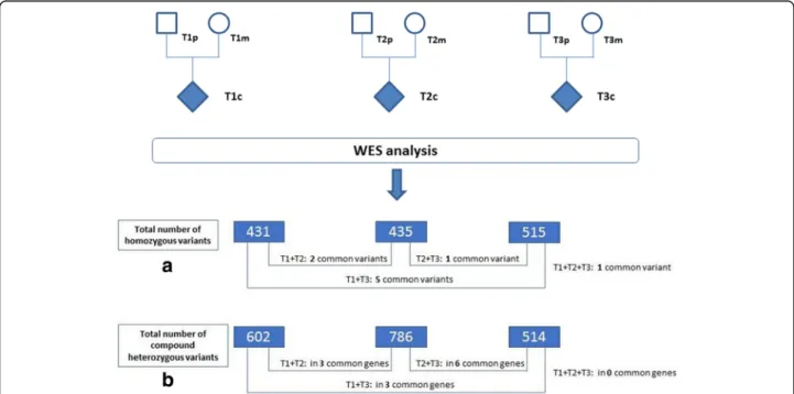
As shown in Fig.1, the total number of variants
inher-ited as homozygous recessive in the affected children represents 431 variants for trio 1 (404 missense, 3 frame-shifts, 6 in frame insertions, 14 splice site, and 4 start or stop codons lost), 435 variants for trio 2 (409 missense, 1 nonsense, 5 frame shift, 7 in frame deletion / insertion, and 2 stop codons lost), and 515 variants for trio 3 (478 missense, 4 nonsense, 8 frame shift, 12 in frame deletion / insertion, 12 splice site and 1 stop lost). We observed only 6 de novo mutations / variants, occurring in 6 genes and respectively 2 for T1 (IGSF3 and SPICE1), 2 for T2 (ZNF717 and CTNND2), and 2
for T3 (NPHS2 and PRSS55) (Table1). We did not find
recurrence of specific de novo mutations between two trios. Taking into account all values of minor allele fre-quency (MAF), we observed 9 variants in 9 different genes expressed as recessive traits in at least 2 trios,
and respectively Sec16A and ADGRV1 (T1 + T2), RHBDL2, ZNF804A, AP5B1, TYR and CPAMD8 (T1 + T3), PRSS48 (T2 + T3) and OR11G2 (T1 + T2 + T3)
(Additional file1: Table S1). We observed also 12 genes
sharing variations in at least two trios, but with differ-ent intragenic locations between the three patidiffer-ents
(Additional file 2: Table S2). This concerns T1 + T2
(ASPN, WFDC3, CCT6B), T1 + T3 (OBSCN, SLC16A8),
T2 + T3 (AFAP1, HSD17B4, DNAH11, CNGB1,
MARCH10) and lastly T1 + T2 + T3 (RHBG, KNL1) for a total of 26 variants. We also took into account the possi-bility of composite heterozygocity which was observed in 11 genes, representing 44 variants and occurring in at least two trios: T1 + T2 (CMYA5, PCDHB16, RREB1), T1 + T3 (AIM1L, IDO2, KIR3DL1 and T2 + T3 (TDRD5,
DNAH11, PIEZO1, FCGBP and PCNT) (Additional file3:
Table S3). By restricting MAFs to values less than 0.01, we identified 9 homozygous recessive variants occurring in a
single trio (Table2). This concerns five different genes for
T1 (ZNF717, WNT2, GOG6, BEGAIN, NDUFV3), two for T2 (SHROOM1 and FMNL1) and two for T3 (DCP1B and CHRNA3).
Statistical evaluation of prioritized variants
We performed a Wilcoxon/Mann-Whitney test to com-pare the deleterious effect of the prioritized variants, i.e., SIFT, PolyPhen, and CADD rank scores, against the
other variants within the same gene [25]. The data
Fig. 1 Flowchart of WES (Whole Exome Sequencing) analysis: T1, T2, T3 design the three nuclear families respectively, p (paternal) for the father, m (maternal) for the maternal and c for the affected child. The number in dark boxes indicate the total number of variants identified respectively in T1, T2 and T3 and inherited in affected children as (a) homozygous and (b) compound heterozygous variants. As mentioned in the text, we selected only those variants which were observed in at least two trios and putatively pathogenic as suggested by SIFT and/or POLYPHENv2 software
Table 1 Possibly pathogenic de novo mutations observed in trios (T). We did not observe any variants common to at least two trios Gene Trios Chr Posi tion Vari ant QUAL Dep th Anno tation Refere nce SIFT Poly phen EX aC IGSF3 T1 1 11 71222 85 I.F. IN S 14,5 80 191 c.312 2_312 3insG GA p.Asp 1040_ Asp104 1insG lu (NM_0 01542 .3) rs1149 15440 IN FRAME INS IN FR AME IN S 0.25 SPIC E1 T1 3 11 31725 43 SN P 3572 291 c.191 2 T > G p.Ser638Ala (NM_1 44718 .3) nd unkno wn unk nown nd ZNF71 7 T2 3 75 78125 7 SN P 106, 499 580 c.293 A > C p.Gl n98Pro (NM_0 01290 210.1) rs1502 24351 0.06 0 nd CTNN D2 T2 5 10 98191 4 SN P 299 111 c.338 8G > A p.Ala1130Thr (NM_0 01332 .3) nd unkno wn unk nown nd NPHS 2 T3 1 17 95263 40 SN P 6106 530 c.560 T > C p.Met1 87Thr (NM_0 14625 .3) nd unkno wn unk nown nd PRSS5 5 T3 8 10 39047 3 SN P 3426 172 c.656 G > C p.Trp 219Ser (NM_1 98464 .3) nd unkno wn unk nown nd Abbreviations: nd not defined, Chr chromosome, SNP single nucleotide polymorphism, QUAL. a quality parameter measuring the probability p that the observation of the variant is due to chance (for ex: QUAL = n, p = 1/n). INS insertion, NM NCBI reference sequence of mRNA, ExAC minor allele frequency as defined in Exome aggregation consortium
Table 2 Possibly pathogenic and rare (MAF < 0.01) recessive variants mutations observed in each single trio (T). We did not observe any variants common to at le ast two trios Trios Gen e Chr. Positio n Vari ant QUAL Dep th An notation Re feren ce SIFT Polyp hen Fu nction EX aC Recess ive Variants – MA F < 0.01 T1 ZNF 717 3 75787 726 SNP 221,913 1345 c.10 48C > T p.A rg350Cy s (NM _0011 28223 .1) nd 0.14 0.689 * Zin c finger prot ein Un known nd T1 WN T2 7 11696 3030 SNP 7083 305 c.14 T > G p.L eu5A rg (NM _0033 91.2) rs1 45839 592 0.39 0.063 Wnt /ß cate nin pathway 0. 0011 T1 COG 6 13 40254 100 SNP 4954 228 c.62 4-3dupT fra me shift (NM _0207 51.2) rs3 97756 552 High impact High impa ct Comp lex Oligo mer Gol gi com plex nd T1 BE GAIN 14 10103 6074 SNP 20,030 273 c.-1 6 + 1G > C Sp lice donor (NM _0208 36.3) rs1 01405 54 SPLIC E DEF ECT SPL ICE DEFE CT Gu anylate kinase-assoc iated prot ein nd T1 NDUF V3 21 44317 156 SNP 8515 341 c.16 8A > C p.L ys56As n (NM _0210 75.3) rs1 41922 962 0.01 * 0.927 * Mi tochon drial com plex I sub unit 0. 0051 T2 SHR OOM1 5 13216 1699 SNP 13,112 130 c.13 4C > A p.Pro 45Gln (NM _0011 72700 .1) rs1 43556 262 0.02 * 0.274 Re gulator o f the microtubule cyt oskeleto n 0. 0065 T2 FMN L1 17 43320 554 SNP 7318 194 c.20 80G > A p.A la694T hr (NM _0058 92.3) rs7 69499 26 1 0.121 Rac1 -medi ated cell mi gration an d div ision 0. 0019 T3 D CP1B 12 20623 23 In frame IN S 65,685 295 c.78 0_782 dupGCA p.Gl n261d up (NM _1526 40.3) rs1 49912 567 nd * nd * Decapping mRNA at the 5' end 0. 00784 T3 CHR NA3 15 78913 067 In frame DEL 27,072 105 c.67 _69del CTG p.L eu23de l (NM _0007 43.4) rs6 67932 22 nd * nd * nic otinic acet ylchol ine rece ptor 0. 00138 Abbreviations: nd not defined, Chr. chromosome, SNP single nucleotide polymorphism, QUAL. a quality parameter measuring the probability p that the observation of the variant is due to chance (for ex: QUAL = n, p = 1/n). INS insertion, DEL deletion, DUP duplication, NM NCBI reference sequence of mRNA, ExAC minor allele frequency as defined in Exome aggregation consortium, SIFT and POLYPHEN scores are indicated in italic bold characters and an asterisk when considered as pathogenic in silico
showed no statistical differences for SIFT (dbNSFP_
SIFT_rankscore:p = 0.4705) and POLYPHEN (dbNSFP_
Polyphen2_HDIV_rankscore: p = 0.9322). We observed
a statistically significant difference for the CADD score
(dbNSFP_CADD_phred:p = 0.008468) (Additional file4:
Figure S1).
Confirmation of variants and sanger sequencing
As these results were only descriptive and not associated to functional tests, all the gene variants were analyzed for their putative functional effect on the encoded pro-tein with the SIFT and PolyPhen-2 online softwares da-tabases. The quality (QUAL) of variants were assessed by a mean depth value of 349 [88–1012] and a mean quality score of 24,521 [299–183,916], suggesting that on average, the probability that the observed variants are due to a random occurrence is less than 0,000042. As
shown in Fig. 2a the variants occurring in SPICE1 and
KNL1 (CASC5) as de novo and recessive respectively, the IGV (Integrative Genomics Profile) confirmed the specificity of these observations according to the paren-tal and child genomic profiles. Lastly, for some of these variants, we performed Sanger sequencing to confirm
the presence of the variants found by WES (Fig.2b).
Functional interpretation of variants related to gene function
We based our in silico functional analysis on the puta-tive pathogenicity of each variants, as suggested by SIFT and Polyphenv2 and then, an exhaustive search in PubMed (National Center for Biotechnology Informa-tion, U.S. National Library of Medicine) screening of published data linking a gene to immunity or related pathways. We performed an over-representation analysis of all deleterious variant carrying genes identified in T1, T2 and T3 probands amongst the list of total variant genes to find KEGG pathways that the deleterious genes might be enriched in. Interestingly, we found that these genes are enriched in pathways related to Antigen pro-cessing and presentation and that of the hematopoietic
cell lineage (Additional file 5: Table S4). As shown in
Table 3, among 37 genes, 10 (Sec16A, AP5B1, RREB-1,
ASPN, RHBDL2, RHBG, KIR3DL1, TDRD5, MARCH10 and AFAP1) can be linked to autophagy and intracellular trafficking involving vesicles, Golgi apparatus and
cyto-skeleton, 6 genes (OBSCN, DNAH11, CTNND2,
CNGB1 and NPHS2) play a direct or indirect role in G-protein regulation, 4 genes (IDO2, IGSF3, PIEZO1 and HSD17B4) may be related to T-cell activation, 6 genes
Fig. 2 Parallel visualization of two gene variants identified in T1 by Integrative Genomics Viewer (IGV) (Broad Institute©) and Sanger Sequencing. a + b Confirmation of the de novo c.1912 T > G (c.1912A > C on reverse strand by WES) variant in the SPICE1 gene inherited by T1c (a + b), either on IGV (a) and Sanger sequence (b). c + d Confirmation of the homozygous c.2806A > G variant in the KNL1 (CASC5) gene inherited by T1c, either on IGV (c) and Sanger sequence (d). The IGV profiles show the absence of c.1912 T > G (SPICE1) in both T1p and T1 m parents (a) and the status of heterozygous carriers of T1p and T1 m for the c.2806A > G (KNL1) variant (c)
Table 3 Functional summary on genes sharing variants in at least two trios Function al path ways Genes Des cription / Interac tion Function Re lated disea ses Add RE F Autophagy an d traffick ing Sec16 A End oplasmic Reticulu m Expo rt Facto r ULK1 , ULK2 (aut ophagy initiat ion) Fam ilial axia l spond yloarthritis [ 35 , 36 , 66 ] AP5B 1 Adap tator protein com plex 5 AP memb rane coat adap tor com plex Atop ic march and psoriasis [ 37 , 67 , 68 ] RREB-1 Ras respo nsive el ement bind ing 1 RalA and Ral B GTPa ses, DNA transcriptiona l regul ator Un know n [ 49 , 69 – 71 ] ASPN Asp orin (PLAP -1), ligan d o f C D 4 4 Regu lation of TLR2 an d TLR4 in macro phages O steoarthritis susceptibility [ 72 – 75 ] RHBDL2 Rhom boid -like-2 serine protease Regu lation of EGF R and anoikis res istance, angiogenes is Un know n [ 76 – 78 ] RHBG Rhe sus-associated (Rh) glycoprot eins Ammon ium trans port in epith elial cells Un know n [ 79 , 80 ] KIR3DL1 killer cell immu noglob ulin like rece ptor HLA inte raction on NK and T-cells Ps oriasis, spond yloarthritis, Be hçet [ 81 – 83 ] TDRD5 Tudor dom ain cont aining 5 Cytop lasmi c RNA processing Sp ermato genes is defec ts [ 84 , 85 ] MARC H10 Mem brane -associated RING -CH 10 Microtubule-associ ated E3 ubiqu itin ligase Un know n [ 86 , 87 ] AFAP1 Acti n filament ass ociated protein 1 Actin -binding protein an d a cSrc-ac tivating protei n Un know n [ 88 – 90 ] G-proteins regul ation OBSCN Obscu rin Rho-g uanine nucle otide exch ange facto r D ilated car diomy opath ies [ 91 , 92 ] DNA H11 Dyne in axon emal heavy chai n 1 1 Compo nent of the ci liary structure Pri mary ci liary dysk inesia [ 93 – 95 ] CTNND 2 δ -caten in/NP RAP/Ne urojungi n Regu lation of Rho-G TPases in cyt oskeleto n Aut ism, compl ex hum an dis orders a [ 47 , 96 ] CNGB 1 (GARP) Cycl ic nuc leoti de gated channe l be ta 1 cGMP signali ng, guan ylate cyclase, exp ressed on T-reg Re tinitis pig mentos a [ 97 – 10 0 ] ADGRV 1 Adhe sion G protein -coupl ed recept or V1 G-protein cou pled rece ptor bind ing calc ium Ushe r syndrom e type II b [ 10 1 , 10 2 ] NPHS 2 Podoc in (spe cific for podoc ytes) Filtration barrier in mamm alian kidn ey (TRPC channe l) Ne phrotic syn dromes [ 10 3 – 105 ] T-cell activation IDO2 Indol eamine 2, 3-dioxygenas e 2 Acts for IDO1 depe ndent induction of T-regulatory cells Coli tis, arthr itis [ 52 , 53 , 10 6 ] IGSF3 (EW I-3) Immu noglob ulin super family member 3 Putative interac tion with CD9 an d CD8 1 (TPSP AN) Un know n [ 10 7 – 110 ] PIEZO1 Piezo type me chanos ensitive ion chann el 1 Mech anotransduction and regul ation of cell morp hology Here ditary Xerocyt osis [ 11 1 – 113 ] HSD 17B4 Hydro xysteroi d 17 -beta deh ydroge nase 4 Beta -oxidation of very long chai n fatty acids PE RRAULT syn drome c [ 11 4 , 11 5 ]
Table 3 Functional summary on genes sharing variants in at least two trios (Co ntinued) Function al path ways Genes Des cription / Interac tion Function Re lated disea ses Add RE F Cell cyc le -im mun e synaps e SPIC E1 Spindl e and centr iole associated prot ein 1 Regu lation of centr ioles el ongat ion wit h CEP120 an d CPA P Un know n [ 61 , 116 ] KNL1 (CAS C5) Kine tocho re scaffol d 1 Stabilization of sister chromat ids to microtubules Un know n [ 64 , 117 – 11 9 ] CCT6 B Chape ronin containi ng TCP1 subuni t 6 B Transcriptional res ponse to st ress in mitotic cell s Un know n [ 12 0 , 12 1 ] PCNT Perice ntri n, phosp horyl ated by PLK1 Ensure prope r cen trosome an d mi totic spindle format ion Pri mordial dwarfism [ 12 2 – 124 ] PCD HB16 Prot ocadherin beta 16 Cell-ce ll adhesion and intracellular signall ing Un know n [ 12 5 , 12 6 ] CMYA 5 (TRIM 76) Card iomyop athy asso ciated 5, myo spryn Muscle targ et ge ne for the MEF2A transcription factor Hyp ertrophi c car diomy opath y d,e [ 12 7 – 130 ] Innat e immu nity CPAM D8 Alp ha-2-mac roglobulin dom ain containi ng 8 Up-regu lated in immu ne stim ulated cells An terior se gment dy sgene sis [ 13 1 , 13 2 ] WFDC3 (WAP 14) WAP four-di sulfide core dom ain 3 Antimi crobial, immu ne, and ti ssue homoe ostasis activ ities Un know n [ 13 3 , 13 4 ] FCGBP Fc fragm ent of IgG binding protei n Regu lator of TGF -1-indu ced EM f trans ition Un know n [ 13 5 , 13 6 ] Unclassified: OR11G2, PRSS48, ZNF804A, ZNF717, TYR, SLC16A8, PRSS55, AIM1L Abbreviations of each gene name are detailed in the text athe CTNND2 gene has been linked to a broad spectrum of human diseases, such as cancer, bipolar disorder, schizophreni a, autism, Cri-du-chat syndrome , myopia, cortical cataract-linked Alzheimer ’s disease, and infectious diseases. bGlycoprotein A repetitions predominant (GARP), a transmembra ne protein containing leucine rich repeats (LRR), has been found to be highly expresse d on the surface of activated T-reg cells. cPatients with Usher syndrome type 2 present hearing loss and develop a visual impairment called Retinitis pigmentosa, a disease not observed in our ca ses, the c.6695A > G variant described in T1 + T2 being not described in this syndrome. dPERRAULT syndrome is a rare recessive disease characterized by hearing loss, ovarian dysgenesis, intellectual disability and ataxia, a condition w hich is not observed in our cases, the c.1606 T > C (T2) and c.392G > A (T3) variants being not linked to this rare disease. ethe clinical screening of T1 and T2 affected children did not identify hypertrophic cardiomyopathy. fEM: epithelial to mesenchymal transition
(SPICE1, KNL1, CCT6B, PCNT, PCDHB16 and CMYA5) as regulation factors of transcription, cell cycle and im-mune synapse, and lastly, three (CPAMD8, WFDC3 and FCGBP) in innate immunity. Eight genes could not be classified. In order to summarize the most relevant data which implicate 4 major pathways that have been related to molecular / cellular defects in granuloma genesis, we presented the data for 10 of the 37 genes identified in this work and served as a basis for discus-sion of possible underlying pathological mechanisms
related to sarcoidosis (Table4).
Discussion
Sarcoidosis is a multifactorial disease that involves envir-onmental pathogenic factors on a genetic background. Two sets of observations indicate a strong genetic het-erogeneity. First, the rate of familial sarcoidosis remains low (3 to 5%) with most of the cases being a priori
spor-adic cases [7]. Second, a wide range of genetic variants
have been previously identified by association studies in genes that may play a role in the pathogenesis of sar-coidosis, such as HLA-DP, BTNL2, Annexin A11, Toll-like receptors, CCDC88B (coiled-coil domain containing protein 88B), Ataxin/SH2B adapter protein 3, IL12B and
NF-kappa-B p105 subunit [9, 29,30]. The present work
focuses on exceptional pediatric forms in terms of fre-quency and severity, with affected children having both parents healthy. We designed a WES genetic analysis on trios since a recent study suggested that for unsolved
clinical exomes, extensive WES analysis of parent–
off-spring trios could identify likely contributory variant in
36% of cases [31]. Such an observation cannot be applied
directly to sarcoidosis even if we might expect that com-paring the exome of a patient and their healthy parents remains the most powerful strategy which has been used in many multifactorial diseases such in autism-spectrum disorders (ASD). As for ASD, both de novo and reces-sive variants must be considered as a powerful approach
of relevant candidate genes [32]. Of the 37 genes
identi-fied in this work, 29 may be related directly or indirectly to pathways linked to immunity and related processes, such as autophagy, intracellular transport, T-cell activa-tion and differentiaactiva-tion, cell cycle and immune synapse. The results obtained were selected according to strict quality criteria, including their reproduction in two inde-pendent analyzes, in-house pipeline PASS filtration, high analysis depth and suggestion of in silico pathogenicity in at least one of the software, SIFT or POLYPHENv2. Comparative statistical analysis of the selected variants versus other variants of the same gene showed no sig-nificant difference for the SIFT and POLYPHEN scores and a significant result for the CADD score (Additional
file 4: Figure S1). The latter integrates several systems
for analyzing the pathogenicity of variants and suggests that the variants observed belong to less than 10% of the most deleterious variants of the genome. However, the data should be analyzed with caution because many fre-quent polymorphisms in the genome have high SIFT and POLYPHEN scores and the non-parametric test per-formed in our study is likely to lack power given the small number of selected variants.
The variants and genes were classified in two ways: First, according to the genetic transmission, we identi-fied 6 de novo variants / mutations in 6 different genes without any overlap between the three affected children
(Table 1). Except for IGSF3 and ZNF717, these variants
were unknown in databases for SPICE1, CTNND2, NPHS2 and PRSS55 genes. All these heterozygous vari-ants might induce pathogenic events and the function of genes was discussed later except those of ZNF717 and PRSS55 which remain unknown to date. Due to the very large number of variants observed by the WES tech-nique in each of the trios, we then focused only on rare (MAF < 0.01) and recessive variations in each of the
chil-dren of the trios (Table2). This concerns 9 variants in 9
genes. A brief functional description for 4 of them (WNT2, COG6, FMNL1, DCP1B) was proposed in rela-tion to immunity related process.
We then focused our attention on more common vari-ants, all MAF values taken together, occurring in at least 2 trios and/or affecting a similar gene between at least two trios. They were classified as true homozygous
re-cessive variants (Additional file 1: Table S1), or
oc-curring in a common gene for at least two trios
(Additional file 2: Table S2), and recessive heterozygous
composites (Additional file 3: Table S3). This represents
a total of 79 coding variants, 58 (73.4%) of them being putatively considered a pathogenic for the encoded pro-tein by either SIFT and/or Polyphenv2. The integration in our work of common variants was absolutely neces-sary because sarcoidosis is probably a highly polygenic disease induced by the combined effect of rare patho-genic mutations and common polymorphisms in genes related to immunity.
In a second part, we have attempted a functional clas-sification based on the fact that sarcoidosis as a multifac-torial environmental and genetic disease may be related to defects in various subcellular, biochemical and im-mune targets susceptible to explain granuloma forma-tion. We focused mainly on genes with recurring variants in at least two trios. The data are summarized
for the whole series of genes in Table 3. We then
de-tailed the functional discussion on 10 genes for which an interesting link has been identified in the bibliography and likely to suggest a link with the pathophysiology of
the granuloma (Table 4). We were able to distinguish 4
Table 4 Functional classification of 10 genes sharing putative pathogenic variants in at least two trios Trios Gen e C h r. Positio n Variant Q UAL Dep th Ann otation Refere nce SIFT Polyp hen Alamut EXaC Autophagy an d Intr acellular Tra fficking T1 + T2 (hom oz) Sec 16A 9 13936 8953 SNP 28 ,526 23 ,786 602 430 c.311 5C > T p.Arg1 039C ys (NM _0148 66.1) rs3812 594 0.14 0.689 * Class3 Unknow n Exon skip ? 0.21 T1 + T3 (hom oz) AP5B 1 11 65,547, 333 SNP 15 ,898 27 ,369 187 219 c.631 C > T p.L eu211Ph e (NM _1383 68.4) rs1214 6493 0 * 0.999 * Class 3 Exon skip 0.29 T1 + T2 (c/het ) RR EB1 6 72318 43 72469 98 72306 80 72473 44 SNP (T1 p) SNP (T1 m) SNP (T2 p) SNP (T2 m) 27 48 61 91 33 ,331 14 ,853 226 202 371 173 c.351 1G > A p.Asp 1171As n (NM _0010 03698 .3) c.415 0G > A p.Gl y1384 Arg (NM _0010 03698 .3) c.234 8G > T p.Gl y783V al (NM _0010 03698 .3) c.449 6C > A p.Se r1499T yr (NM _0010 03698 .3) rs9379 084 rs2281 833 rs9502 564 rs3574 2417 0 * 0.12 0.06 0.01 * 0.999 * 0 0.156 0.025 Class3 Unknow n Exon skip ? nd nd Class3 Unknow n Exon skip ? 0.11 0.27 0.44 0.14 G-Prot eins Regu lation T1 (hom oz) OBS CN 1 22850 5204 SNP 11 ,933 256 c.164 72G > A p.Arg5 491H is (NM _0012 71223 .2) rs4653 942 0 * 0.91 * Class3 Unknow n 0.26 T3 (hom oz) 22849 4790 SNP 39 ,725 479 c.149 86G > A p.Gl y4996 Arg (NM _0012 71223 .2) rs4357 76 0.01 * 0.939 * Class 3 Unknow n 0.41 T2 (de nov o) CT NND2 5 10981 914 SNP 29 9 111 c.338 8G > A p.Ala1 130Thr (NM _0013 32.3) nd nd nd nd nd T2 + T3 (c/het ) DNA H11 7 21893 993 SNP (T2 p) 46 ,459 445 c.111 22G > T p.Val 3708Le u (NM _0012 77115 .1 rs4722 064 0.02 * 0.611 * Class 3 Unknow n 0.43 21584 693 SNP (T2 m) 16 10 159 c.421 G > T p.Asp 141Ty r (NM _0012 77115 .1) rs7265 5969 0 * 0.601 * Class 3 Unknow n 0.012 21,628, 242 SNP (T3 p) 20 ,259 553 c.196 1C > G p.Se r654Cy s (NM _0012 77115 .1) rs6244 1683 0.67 0.02 Class 3 Unknow n 0.13 21678 643 SNP (T3 m) 19 ,593 850 c.490 4A > G p.Asp 1635G ly (NM _0012 77115 .1) rs1714 4835 0 * 0.783 * Class 3 Unknow n Exon skip ? 0.045
Table 4 Functional classification of 10 genes sharing putative pathogenic variants in at least two trios (Co ntinued) Trios Gen e C h r. Positio n Variant Q UAL Dep th Ann otation Refere nce SIFT Polyp hen Alamut EXaC T-Cel l Acti vation and Immu ne Synapse T1 + T3 (c/het ) IDO2 8 39862 881 SNP (T1 p) 14 ,321 289 c.742 C > T p.Arg2 48Tr p (NM _1942 94.2) rs1010 9853 0 * 1 * Class 3 Unknow n 0.48 39873 053 SNP (T1 m) 77 71 184 c.119 5G > A p.Ala3 99Thr (NM _1942 94.2) rs7263 2016 0.57 0.024 Class 3 Unknow n 0.06 39872 935 SNP (T3 p) 21 ,240 451 c.107 7 T > A p.Ty r359 * (NM _1942 94.2) rs4503 083 STOP * STOP * Truncating Polymo rphism 0.226 39862 893 SNP (T3 m) 35 10 154 c.754 T > A p.Se r252Thr (NM _1942 94.2) rs3521 2142 0.02 * 0.444 * Class 3 Unknow n 0.02 T1 (de nov o) IGSF 3 1 11712 2285 In frame INS 14 ,580 191 c.312 2_312 3insG GA p.Asp 1040_ 1041ins Glu (NM _0015 42.3) rs1149 15440 IF IN S IF INS In fra me polymo rphism 0.25 Mitos is and Im mun e Synapse T1 (de nov o) SPIC E1 3 11317 2543 SNP 35 72 291 c.191 2 T > G p.Se r638Ala (NM _1447 18.3) nd nd nd Unknow n nd T1 (hom oz) KNL1 (CAS C5) 15 40916 237 SNP 57 ,541 697 c.385 3A > G p.L ys128 5Glu (NM _1705 89.4) rs1774 7633 0.05 * 0.058 Class 3 Unknow n Exon skip ? 0.32 T2 (hom oz) 40915 190 SNP 18 3,916 368 c.280 6A > G p.Arg9 36Gly (NM 17058 9.4) rs8040 502 1 0 Class 3 Unknow n 0.24 T3 (hom oz) 40914 177 SNP 66 ,997 297 c.179 3 T > C p.Me t598T hr (NM 17058 9.4) rs1185 8113 0.01 * 0.006 Class3 Unkown 0.39 Abbreviations: nd not defined, Chr. chromosome, SNP single nucleotide polymorphism, QUAL. a quality parameter measuring the probability p that the observation of the variant is due to chance (for ex: QUAL = n, p = 1/n). INS insertion, DEL deletion, DUP duplication, NM NCBI reference sequence of mRNA, ExAC minor allele frequency as defined in Exome aggregation consortium, SIFT and POLYPHEN scores are indicated in italic bold characters and an asterisk when considered as pathogenic in silico
Autophagy and intracellular vesicular trafficking
In contrast with Crohn’s disease, very little information is available on constitutive defects in autophagy for sarcoidosis. These processes are involved in the cellular response to metals and nanoparticles in experimental
mice models of granuloma [33,34]. For instance, Sec16A
(affected in T1 + T2) is an interaction partner of ULK1 and ULK2 thought to be essential for initiating
autoph-agy [35]. ULK-mediated phosphorylation of SEC16A
regulated the assembly of endoplasmic reticulum (ER) exit sites and ER-to-Golgi trafficking of specific cargo. In frame deletions of SeC16A have been observed in
fa-milial forms of spondyloarthritis [36]. AP5B1 (Adaptator
protein complex 5), recently implicated in atopic asthma, T1 and T3 share a p.Leu211Phe pathogenic
variant [37]. Adaptator proteins sort cargo into vesicles
for transport one membrane compartment of the cell to another and AP5 is considered as a candidate for a late endosomal coat, regulating endosomal sorting and
vesicle budding from this compartment [38]. COG6,
sharing a recessive frame shift mutation in T1 is a mem-ber of the conserved oligomeric Golgi (COG) complex which has been implicated in the regulation of
endo-some to trans-Golgi network retrograde trafficking [39].
Mutations in the COG genes have been shown to result in the mislocalization of some of the autophagy-related (Atg) proteins, which are critical components involved in autophagosome formation. Interestingly, single nu-cleotide polymorphisms of the COG6 gene have been re-cently shown as shared risk locus for rheumatoid
arthritis and systemic lupus erythematosus [39]. Another
interesting gene is RREB-1 (Ras-responsive element-binding protein 1), a zinc finger transcription factor, which is an effector of the RaIA/B small GTPases, acting as major regulator in exosome release, membrane
traf-ficking, cytokinesis, cell shape and movement [40].
G-proteins regulation
When in the GTP-bound active form, Rho GTPases transduce signals by binding to effector proteins in-volved in many cellular processes including regulation of the actin skeleton, microtubule dynamics, cell division,
migration and adhesion [41]. Such a regulation occurs in
various immune cells: monocyte/macrophage, T and B lymphocytes and neutrophils. The switch between the GDP inactive and GTP active of typical Rho GTPases is regulated by guanine nucleotide exchange factors (GEF) and GTPase-activating proteins (GAP). Rho GTPases are known to exert an effect on the cytoskeleton through
NF B, a major player in inflammatory processes [41].
The present study identified genes encoding GTPases or
GEF, such as OBSCN and CTNND2. OBSCN
(OBSCURIN), mutated in T1 and T3, are giant cytoskel-etal proteins with structural and regulatory roles on
Rho-GEF activity and play a role in the maintenance of cell-cell adhesion and regulate epithelial to mesenchymal
transition [42]. The T1 patient shares a rare and
reces-sive variant in FMNL1, encoding one isoform of the for-min proteins. Macrophages express multiple forfor-mins, including FMNL1, with potential to impact actin
remod-eling involved in migration [43]. Formins interact with
GTP-bound Rho, Rac and Cdc42 GTPases and contrib-ute to macrophage migration activity by stabilizing the lifespan of podosomes, a key actor of actin cytoskeleton dynamics during adhesion and migration within tissues
[44, 45]. DCP1B (decapping enzyme 1), mutated in T3,
interacts with DEF6, a Rho-family guanine nucleotide exchange factor, which contributes to the regulation of the spatiotemporal organization of components of T cell
signaling pathways and Cdc42-dependent actin
polymerization [46]. CTNND2 (Delta Catenin 2),
af-fected by a de novo mutation in T2, is an adhesive junction-associated protein in the delta subfamily of the β-catenin superfamily and functions in Wnt signaling pathways to regulate gene expression and modulate Rho
GTPases in cytoskeletal reorganization [47]. By
regulat-ing the activity of small GTPases and the disruption of E-cadherin based adherens junction, delta catenin 2 pro-motes cell migration and one might expect that such a
process could play a role in macrophage fusion [48]. The
observation of an uncommon recessive variant in the WNT2 gene (T1 patient) may be relevant of a patho-genic role of the canonical Wnt/ß-catenin signaling pathway in part of the inflammatory process occurring in sarcoidosis. Two other genes (DNAH11 and RREB1) present compound heterozygotes variants respectively in (T1 + T2) and (T2 + T3) and have been characterized as
proteins interacting with small GTPases [49,50].
T-cell activation
The immune patterns and T-reg cells disturbances ob-served in sarcoidosis are similar to those obob-served in other
Th1/Th17 related diseases [51]. IDO2 (indoleamine 2,3
dioxygenase) is a particularly interesting gene, as affected
by deleterious variants both in T1 and T3 (Table3). IDO2
is expressed in antigen-presenting cells (APCs), including dendritic cells (DCs) and B cells and deserves special attention by its function in T-cell regulation and
differen-tiation in vitro [52, 53]. IDO2 may contribute to
inflam-matory processes by acting in monocyte-derived DCs to control regulatory T-reg cells, as a putative immunosup-pressive factor. IDO1 and IDO2 are functional interacting partners and have been associated to various autoimmune
states, such as colitis, arthritis and encephalomyelitis [53].
These conflicting results suggest both an
immunosuppres-sive and a positive inflammatory influence [54].
Interest-ingly, the two variants observed respectively in T1
(p.Tyr359* - rs4503083– MAF = 0,226) are two common
inactivating polymorphisms observed in humans [52]. The
first, Arg248Trp, introduces a mutation at a catalytic resi-due that results in > 90% reduction in catalytic activity in
in vitro assays [52, 55]. The second is a STOP codon at
residue 359, leading to a truncated and catalytically in-active protein product. Based on allele frequencies it esti-mated that at least 25% of the population has a functionally inactive IDO2. The two affected children from T1 and T3 share a second allelic variant with a puta-tive deleterious suggesting that expression of IDO2 may be dramatically affected in both cases. IDO2 is essential for the IDO1-dependent induction of T-regulatory cells
[53]. It can be hypothesized that IDO2, in synergy with
IDO1 is both a pro inflammatory factor acting in B lym-phocytes to promote the cross talk between B and T cells, increasing the number of antibody-secreting cells, thus acting as an immunosuppressive agent by the control of T-reg cells. Inactivating mutations may be responsible for a decrease in the activity and / or number of regulatory T-cells, thereby inducing a pro inflammatory situation. These genes are already being studied as potential thera-peutic targets. It has recently been shown that IDO2-specific monoclonal antibodies reduced auto reactive T and B cell activation and alleviated arthritis in two
inde-pendent preclinical arthritis models [56]. A de novo
vari-ant has been observed in T1 for the IGSF3 gene, an in-frame insertion which high frequency (0,25) in control population. The function of IGSF3 may be related to the molecular background of immune synapse, involving members of the immunoglobulin (Ig) superfamily such as
BTNL2 [29]. IGSF3 or EWI-3 belongs to a novel Ig
sub-family containing a Glu-Trp- Ile (EWI) motif not seen in other Ig proteins and has a strong similarity with CD101, a T-cell marker notably expressed on T-REG with potent suppressor activity, and which expression regulates IL-17 production and disease activity in inflammatory bowel
and rheumatoid diseases [57,58].
Cell cycle and immune synapse
At the crosstalk of innate immunity and the toxicity of environment-derived particles, a strong interest has been shown for a series of molecules that might play a role in promoting granuloma formation. However there is con-troversy about the molecular and cellular mechanisms generating polyploidy giant cells found in sarcoidosis granuloma. It was thought that these lesions were gener-ated by a cell fusion mechanism; however evidence from other studies suggests that mitotic defects and DNA
damage may generate polyploidy in macrophages [59].
Previous works have described elevated numbers of cells in the S + G2/ M phase in advanced forms of sarcoidosis, suggesting that such mitotic defects may affect different
immune cell lineages [60]. In connection with these
concepts, we were interested to observe an unknown de novo variant in T1, p.Ser638Ala in the SPICE1 gene which encodes a protein that localizes to spindle micro-tubules in mitosis and to centrioles throughout the cell
cycle [61]. Together with centrosome proteins CEP120
or CPAP, SPICE regulates the elongation of centrioles, which serve as templates for the assembly of centrosome
structures [62]. Interestingly, the centrosome plays also
a crucial role in the assembly and function of immune synapse and cytokine production during inflammation
[63]. We thus might expect that defects in the molecular
interaction that impact on the docking of centrosome at the plasma membrane initiated by T-cell and APC inter-action is a relevant process related to innate immunity. On the other hand, experimental depletion of SPICE1
causes severe mitotic defects [61]. KNL1 (CASC5), a
kinetochore substrate of Aurora-B, share putative
patho-genic variants in the three trios (Table2). This gene has
been involved in the stabilization of sister chromatids to microtubules during mitosis, maintenance of centro-meres cohesion and regulation of mitotic checkpoint
si-lencing [64,65].
Conclusions
Using a WES approach in pediatric forms of sarcoidosis
and parent– offspring trios we have identified a panel of
genes that led us to propose hypotheses about 4 func-tional pathways potentially mediating the underlying mechanism(s) of granuloma formation: autophagy re-lated intracellular vesicular trafficking, G-protein signal-ing and Rho GTPases, immune synapse and T-cell activation, and lastly intimate mechanisms of cell cycle and mitosis regulation. An over-representation gene en-richment KEGG pathway analysis suggests that a signifi-cant number of genes selected belongs to pathways related to Antigen processing and presentation and that of the hematopoietic cell lineage We are aware that this study is of descriptive type and that some of the selected variants may have no direct role in the development of the disease. Nevertheless, these results fit with the fact that sarcoidosis is a multifactorial disease with a com-plex genetic background and agree the hypothesis of multiple and combined mechanisms in the genesis of the granuloma. Clinically, it is possible that the variable expressivity of the disease can be explained by a strong genetic heterogeneity, in particular between the different evolutionary forms. Further studies are awaited to com-pare gene variants identified in pediatric cases and auto-somal dominant family forms. The characterization of a very broad panel of genes associated with the different clinical forms of sarcoidosis will ultimately not only de-fine the relative risk of disease occurrence in families at risk, but also better characterize the evolutionary profiles and help to optimize therapeutic strategies.
Additional files
Additional file 1:Table S1. Recessive variants found in at least two affected children of different trios. Possibly pathogenic recessive variants (polymorphisms) found by whole-exome -sequencing in at least two affected children of the trios (T). Chr., chromosome; SNP, single nucleotide polymorphism; QUAL., a quality parameter measuring the probability p that the observation of the variant is due to chance (for ex: QUAL = n, p = 1/n). As detailed in the text, Alamut® Visual integrates missense variant
pathogenicity prediction tools and in silico study of variants’ effect on
RNA splicing, allowing the assessment of their potential impact on splice junctions and splicing regulatory sequences. Alamut® Visual helped us also to exclude well known mutations identified in recessive diseases for those genes which have been related to known genetic
diseases (as shown in Table3). (DOCX 23 kb)
Additional file 2:Table S2. Recessive variants shared by a common gene in at least two different trios. Possibly pathogenic recessive variants observed at different positions for a single gene in at least two affected
children of the trios (T). Abbreviations are the same as in Tables1,2and
Additional file1: Table S1. (DOCX 31 kb)
Additional file 3:Table S3. Composite heterozygocity observed in a common gene in at least two different trios. Possibly pathogenic compound heterozygous variants (allelic heterogeneity) observed in different positions of a common gene in at least two trios. The origin of either the paternal and maternal allele was detailed for each variant. Abbreviations are the same as
in Tables1,2, Additional files1and2: Tables S1 and S2. (DOCX 51 kb)
Additional file 4:Figure S1. CADD scoring of prioritized variants versus other variants in the selected genes. (PDF 71 kb)
Additional file 5:Table S4. Over-representation gene enrichment KEGG pathway analysis was performed on all the deleterious variant carrying genes. Data of the over-representation gene enrichment KEGG pathway analysis was performed on all the deleterious variant carrying genes in trios 1, 2 and 3. (XLSX 9 kb)
Additional file 6:All recessive variants identified in cases T1, T2 and T3. The de novo dominant variants have been fully described in the manuscript. The datasets of cases T1, T2 and T3 extracted from VCF (Variant call Format) and extracted in XLS file. (XLSX 368 kb)
Abbreviations
1000G:1000 genomes; BTNL2: Butyrophilin like 2; CCDC88B: Coiled-coil
domain containing protein 88b; CPP: Comité de Protection des Personnes; CT: Computed tomography; ESP: Exome sequencing project; ExAC: Exome aggregation consortium; GTP/GDP: guanine tri/diphosphate; GWAS: Genome wide association studies; MAF: Minr allele frequency; mTor: Mammalian target of rapamycin; NOD2: Nucleotide binding oligomerization domain containing 2; Polyphenv2: Polymorphism Phenotyping v2; QPCR: Quantitative polymerase chain reaction; SARCFAM: Familial sarcoidosis; SIFT: Sorting Intolerant From Tolerant; SNP: Single nucleotide polymorphism; STAT1: Signal transducer and activator of transcription 1; TSC1/2: Tuberous sclerosis; XAF1: X inhibitor of apoptosis associated factor 1
Acknowledgments
We thank the families for their kind participation and continued interest in this study. We thank Raphaelle LAMY and Céline AUBOIROUX for expert technical assistance. We thank Mrs. Dr. Janet Hall for reviewing this manuscript. This work has been performed in the frame of a national clinical and research group working on sarcoidosis, Group Sarcoidosis France (GSF).
Funding
This study was performed with the grant named‘High Throughput sequencing
and rare disease’ from the “Fondation Maladies Rares” (FMR - France) and
financial support from the INNOVARC - DGOS ref. 12–027-0309.
Availability of data and materials
The datasets (Variant call Format extracted in XLS file) supporting the
discussion of this article are available are presented in Additional file6.
Authors’ contributions
Conceived and designed the experiments: AC (1), AB (1) and SP. Performed the experiments: cooperative projects with IntegraGen SA © (Evry, France). Analyzed the data: AC, PARF, SP, YP, AB (2), CB, PR, and SL. Wrote the paper: AC, YP and NN. Provided the clinical cases and data: NN, HC, RAT, VH, DV, GD, VC, PS, AC (2) and JFB. All authors read and approved the final manuscript.
Ethics approval and consent to participate
This project has been approved by the ethical committee (CPP LYON SUD
EST– 2 – REF IRB 00009118 – September 21, 2016), according to the Laws
Nos. 94–548 of 1 July 1994, Nos. 94–653 and 94–654 of 29 July 1994, taking
into account the amendments further introduced by the laws n ° 2004–800
of 6 August 2004 and n ° 2011–814 of 7 July 2011, both relating to bioethics.
This approval included an informed consent form and a detailed information
sheet for patients. According to the article R. 1131–4 of the French Public
Health Code, for the participants who are under 18 yr., the consent has been given by both mother and father during clinical and genetic counseling. In the three cases, there were the natural parents which have the parental authority. In addition, the consent of the minor is systematically sought if he is able to express his will and participate in the decision. The clinical and
genetic studies were registered by theClinicalTrials.gov- Protocol Registration
and Results System (PRS) receipt under the reference NCT02829853.
Consent for publication
The SARCFAM protocol was presented to the ethical committee (CPP LYON
SUD EST– 2 – REF IRB 00009118) dated September 21, 2016 and includes
consent for genetic studies and an information document on the objectives of genetic analysis and the scientific SARCFAM project. This document is an informed consent for the publication of scientific and clinical data subject to a complete anonymity of the information. Our study does not present any photographic data. This informed consent is valid for adult patients and for children (< 18 yr) after signature of both legal parents.
Competing interests
The authors declare that they have no competing interests.
Publisher’s Note
Springer Nature remains neutral with regard to jurisdictional claims in published maps and institutional affiliations.
Author details
1Genetics Department, Hospices Civils de LYON (HCL), University Hospital,
East Pathology Center, LYON, B-A3, 59 Bld Pinel, 69677 BRON Cedex, France.
2Department of biostatistics, University Hospital, Hospices Civils de LYON
(HCL), Lyon, France.3Inflammation & Immunity of the Respiratory Epithelium
- EA7426 (PI3)– South Medical University Hospital – Lyon 1 Claude Bernard
University, 165 Chemin du Grand Revoyet, 69310 Pierre-Bénite, France.
4Cancer Research Center, INSERM U-1052, CNRS 5286, 69008 Lyon, France.
5Pediatric pulmonology and Reference Center for rare lung diseases
RespiRare, Hôpital Trousseau, AP-HP, INSERM UMR-S938, Sorbonne University,
Paris, France.6Pediatric pulmonology and Reference Center for rare lung
diseases RespiRare, Hôpital Necker, Paris, France.7Pediatric pulmonology and
Reference Center for rare lung diseases RespiRare, Hôpital Robert Debré,
INSERM U-1142, University Paris Diderot VII, Paris, France.8Department of
Pulmonology, University Hospital, Hôpital Croix Rousse, Lyon, France.
9Department of Pulmonology, University Hospital, Hôpital Louis Pradel, Lyon,
France.10Department of Internal medicine, University Hospital, Hôpital Croix
Rousse, Lyon, France.11Histology and Tumor Biology, ER2 UPMC, Hôpital
Tenon, Paris, France.12Medical University of Vienna, Center for
Pathobiochemistry and Genetics, Institute of Medical Genetics, Währinger
Straße 10, 1090 Vienna, Austria.13EA2363, University Paris 13, COMUE
Sorbonne-Paris-Cité, 74 rue Marcel Cachin, 93009 Bobigny, France. 14
Assistance Publique Hôpitaux de Paris, Department of Pulmonology,
Avicenne University Hospital, 93009 Bobigny, France.15AP-HP Pediatric
pulmonology and Reference Center for rare lung diseases RespiRare, Hôpital Trousseau, INSERM UMR-S933, Sorbonne University, Paris, France.