HAL Id: tel-02292543
https://tel.archives-ouvertes.fr/tel-02292543
Submitted on 20 Sep 2019
HAL is a multi-disciplinary open access archive for the deposit and dissemination of sci-entific research documents, whether they are pub-lished or not. The documents may come from teaching and research institutions in France or abroad, or from public or private research centers.
L’archive ouverte pluridisciplinaire HAL, est destinée au dépôt et à la diffusion de documents scientifiques de niveau recherche, publiés ou non, émanant des établissements d’enseignement et de recherche français ou étrangers, des laboratoires publics ou privés.
Dynactin1 mutations associated with amyotrophic
lateral sclerosis and their effect on axonal transport and
neuromuscular junction formation
Valérie Bercier
To cite this version:
Valérie Bercier. Dynactin1 mutations associated with amyotrophic lateral sclerosis and their effect on axonal transport and neuromuscular junction formation. Neurons and Cognition [q-bio.NC]. Université Pierre et Marie Curie - Paris VI, 2017. English. �NNT : 2017PA066176�. �tel-02292543�
Université Pierre et Marie Curie
École doctorale Cerveau, cognition, comportement (ED 158)
Institut Curie / Laboratoire développement des circuits neuronauxDynactin1 mutations associated with amyotrophic lateral
sclerosis and their effect on axonal transport and
neuromuscular junction formation
Par Valérie Bercier
Thèse de doctorat en Sciences de la vie, spécialité Neurosciences
Dirigée par Filippo Del Bene
Présentée et soutenue publiquement le 18 septembre 2017
Devant un jury composé de :
Dr DEL BENE, Filippo, DR Directeur de thèse
Dr JANKE, Carsten, DR Rapporteur
Dr SCHMID, Bettina, DR Rapporteur
Dr STEVANIN, Giovanni, DR Examinateur
Dr HAZAN, Jamilé, DR Examinatrice
i “It is a mistake to think you can solve any major problems just with potatoes.”
ii
Acknowledgements
First of all I would like to thank my supervisor Filippo Del Bene for the opportunity to work in his lab. It was not an easy start and we had to learn a lot to get this project on the way, as well as butt heads a few times, but in the end, we found a phenotype!!!
Thank you to the Del Bene lab for all the help, the teaching moments, the coffee breaks, the picnics and retreats. You were all there during the triumphs and the unfortunate disasters that also sometimes happen. In particular, thank you to Karine, Thomas and Christoph who were the first ones to very patiently answer all my questions, and to Vincenzo, Flavia, Céline, Shahad, Giulia, Noé, Julie, Juliette, Marion, and Gokul. It is true when they say “it takes a village” and you guys were it! Also thank you to the passing members of the lab, namely Federica, Alessio, Anne, Dina, Natalia, and Katrina who all brought a little bit of sunshine to the back corner of the office.
In addition, a special thanks goes out to the animal facility staff (Olivier, Armelle, Tarek and Cédric) and to the awesome Curie administrative staff, who all made my life easier (Isabelle, Virginie and Déborah). Thank you also to the dwellers of the 3rd floor for your camaraderie, in particular ALL THE ITALIANS who seem to appear from nowhere and aggregate like (very loud) prions (Angelo, Roberta, Sara, Ariana, Manu, Edoardo, Magherita, Irene, etc.) Audrey, Daniel, Michel, Antoine and the rest. Also thank you for bringing sweets and leaving them unattended at the coffee machine…
Of course one must not forget the fellowship that provided me with support, both administrative, and emotional, since before I even moved to Paris: the École des Neurosciences de Paris. The help from Yvette, Laura, Deborah, Laetitia, André, Patricia, Laure and Laurent was invaluable. In addition, thank you to the students who, like me, are part of this fellowship. In particular, I would like to thank Malou and Jaime, as well as Hannah and Darinka who not only were great friends, but somehow got roped into being ENP student representatives with me. Thank you also to Heike, Laurianne, Emma, Maja, Damiano, Sabah, Mariana, Jenna, Urs, Alessandra, Ralitsa, Maria Julia, Kasia and the rest. Even though you came from all over the world, you were like a family to me during my time in France.
iii My gratitude also goes out to Claire Wyart and her team at ICM. Thanks to Kevin and Jeff for tackling the physiology of this project, and to Claire for her sound advice and unfailing enthusiasm.
Finally, thank you to my family for supporting me across an ocean. I’m really grateful for the visits, the skype calls and the packages that brought a bit of home to our apartment. Thank you to my husband, Witek, for his love, care and encouragement, and for going head first into this crazy adventure with me. France has been at times harsh and wonderful to us and I think we will forever keep good memories of it, which brings me to my last acknowledgement: thank you to “croquette” for being my writing partner and for giving me perspective in the end pages of this thesis. Indeed, there are great things still ahead!
iv
Abstract
Amyotrophic lateral sclerosis (ALS) is an adult-onset neurodegenerative disease, which is mainly sporadic in nature. This progressive pathology has an estimated incidence of 1-2 per 100 000 worldwide and generally leads to death within 2-5 years of diagnosis due to muscle wasting and severe motor neuron loss. Over the last years, mutations have been identified in both sporadic and familial ALS patients, interfering with the function of many genes, including DCTN1, which encodes for a subunit of the motor protein complex dynactin. The dynactin complex serves as an adaptor for the dynein motor complex, responsible for retrograde axonal transport, and it is believed to regulate dynein activity and the binding capacity for cargo, however its role in ALS pathogenesis is still unknown.
We set out to characterize a mutant zebrafish line for dynactn1a (named mikre okom632, mokm632), looking specifically at caudal primary motor neurons (CaPs), with regard to axonal development, formation and stability of the neuromuscular junction (NMJ) and the behavioral phenotype produced in embryos, as well as axonal transport metrics.
We observed that homozygous mutant embryos exhibited an abnormal locomotor activity at 48hpf, as assayed by a touch-evoked escape response assay, suggesting a fatiguing phenotype of the neuromuscular junction. These embryos also had an abnormal axonal morphology at 6dpf, with arbors of reduced size and with a lower number of projections. The neuromuscular junction showed defects in structure, with misaligned pre- and postsynaptic sites. However, miniature end-plate currents (mEPC) at the NMJ were normal, suggesting fusion and spontaneous release machinery to be functional in our mutant embryos. Paired-recordings of motor neurons and muscle at 6dpf revealed abnormal firing patterns for both low and high frequency stimulation, where mutant embryos display failures and asynchronous release. Additionally, we found that our mutants have a reduced number of putative synapses, which were also of smaller size and unstable. This instability was postulated to be the cause of the reduced growth in CaPs the NMJ physiological defects, which leads to the behavioral abnormalities. We were able to rescue the mutant CaP morphological phenotype by overexpressing the human Dynactin1-GFP protein, whereas the overexpression had no effect on CaP morphology in wild-type embryos. The human protein was found to localize at synaptic sites, supporting a possible role in synapse stability. Fast axonal transport was quantified in primary motor neurons using the GAL4/UAS bipartite system and fusion protein tracking in vivo by confocal time-lapse microscopy. We quantified the transport dynamics of cargos such as mitochondria and early, late and recycling endosome in the motor neurons of wild-type versus mokm632 mutant embryos in vivo. We did not observe a change in transport states of cargoes (stable, anterograde, retrograde) and while we found few differences in transport behavior, the overall results suggest that dynactin1 is not essential for dynein activity and for axonal transport in general.
We investigated dynactin1’s interaction with the cytoskeleton and did not find defects in microtubule growth in arbors, microtubule stability, or at the level of microtubule capture and anchoring at synapses. In addition, we did not find changes in the filopodia dynamics in CaPs, which could indicate defective trophic signaling, since filopodia dynamics were normal, as was the transport of the p75 low-affinity trophic receptor. Overall, we suggest a role for dynactin1 in synapse stability, where the loss-of-function of this gene leads to growth defects, electrophysiological abnormalities and behavioral deficits. Surprisingly, this role appears to be independent of its known function as a regulator of dynein, its implication in axonal transport, or its regulation of microtubule dynamics.
v
Résumé
La sclérose latérale amyotrophique (SLA) est une pathologie neurodégénerative progressive se déclarant vers 50-60 ans. Elle est majoritairement de nature sporadique son incidence est estimée à 1-2 par 100 000. La SLA mène à une paralysie progressive et entraine généralement à la mort des patients de 2 à 5 ans suivant le diagnostic aux suites d’une fonte musculaire importante liée à la perte des neurones moteurs. Au cours des années, plusieurs mutations ont été identifiées autant chez les patients atteints de SLA sporadique que de SLA familiale. Ces mutations interfèrent avec la fonction de gènes variés, tels que DCTN1, codant pour la protéine dynactine1, sous-unité du complexe multimoléculaire dynactine. Ce complexe sert d’adaptateur au moteur moléculaire dynéine, chargé du transport axonal rétrograde, où sa fonction permettrait de régir l’activité du complexe moteur et sa capacité à lier divers cargos, mais son rôle dans le développement de la SLA demeure spéculatif. Nous avons donc entrepris la caractérisation d’une lignée de poissons zèbre mutants pour dynactin1a (nommés mikre okom632,
mokm632), plus particulière en terme du développement d’un type de neurone moteur primaire
(les CaPs), afin de déterminer l’effet de la perte de fonction de ce gène sur l’axonogenèse, la formation et la stabilisation de la jonction neuromusculaire, sur le comportement de l’embryon, ainsi que sur le transport axonal.
Nous avons observé une activité locomotrice anormale à 48hpf suggérant un phénotype de fatigue de la jonction neuromusculaire. Nous rapportons aussi une morphologie anormale des CaPs chez les embryons homozygotes à 6dpf, qui présentent une arborisation de taille et de complexité réduite. La jonction neuromusculaire a aussi révélé des défauts structuraux, où les sites pré- et post synaptiques sont désalignés. Au niveau fonctionnel toutefois, l’analyse de fusions spontanées de quanta (mEPC) à la jonction neuromusculaire n’a pas révélé d’anomalies, suggérant que les mécanismes de fusion sont fonctionnels chez notre mutant. Des enregistrements couplés neurone moteur-fibre musculaire ont ensuite été effectués à 6dpf, révélant une activité anormale de décharge neuronale suite à des stimulations de haute et basse fréquence, où les embryons mutants démontrent des échecs de réponse au niveau du muscle, ainsi qu’une augmentation de décharges asynchrones. De plus, une étude de la jonction neuromusculaire des embryons mutants a révélé une diminution du nombre de synapses putatives, qui étaient également de taille réduite et à caractère instable. Nous suggérons que cette instabilité mène aux défauts de croissance des CaP, aux anomalies élécrophysiologiques, qui engendrent le comportement anormal. Nous avons obtenu le sauvetage du phénotype morphologique des CaPs mutants par surexpression de la protéine humaine Dynactine1-GFP, alors que cette même surexpression n’a eu aucun effet sur la structure des CaPs chez les embryons de type sauvage. La protéine humaine ainsi surexprimée est localisée au niveau des synapses, suggérant un rôle pour dynactin1 dans la stabilité synaptique.
Le transport axonal a été analysé et quantifié chez les neurones moteurs primaires par usage du système bipartite GAL4/UAS et l’expression de protéines de fusion permettant le suivi in vivo des cargos par imagerie confocale de type time-lapse. Nous avons examiné le transport in vivo de cargos tels que les mitochondries, les endosomes précoces, matures et de recyclage au sein des CaPs d’embryons de type sauvage versus mutants pour mokm632. Nous n’avons cependant
pas distingué de changement dans la cinétique de transport des cargos (stable, antérograde ou rétrograde) et quoique nous ayons noté quelques différences en termes de comportement, la vue d’ensemble des résultats suggère que la dynactine1 n’est pas essentielle à l’activité de la dynéine et au processus de transport axonal. Nous avons ensuite examiné l’interaction de la dynactine1 avec le cytosquelette et n’avons pas observé de défauts au niveau de la croissance
vi des microtubules au sein de l’arborisation axonale, ni au niveau de la stabilité des microtubules ou de leur capture fin d’ancrage synaptique. De plus, aucun changement n’a été détecté chez les CaPs indiquant un potentiel défaut de traitement des signaux trophiques. En effet, la dynamique des filopodes d’actine s’est avérée normale, ainsi que le transport des récepteurs trophiques de faible affinité p75.
Somme toute, nous suggérons que dynactin1 favorise la stabilité synaptique, où une perte de fonction de ce gène entraine des défauts de croissance, des anomalies éléctrophysiologiques et un comportement anormal. Ce rôle semble être indépendant des fonctions connues de régulateur du moteur dynéine, de son implication dans le transport axonal ou de son action sur la dynamique des microtubules.
vii
List of abbreviations
A.a.: Amino acid
AChR: Acetylcholine receptor ALS: Amyotrophic lateral sclerosis AnkB: Ankyrin-B
Arl3: ADP-ribosylation factor-like 3 Bp: Base pairs
BSA: Bovine serum albumin CaP: Caudal primary motor neuron
CAP-Gly: cytoskeletal-associated protein glycine-rich
CHCHD10: Coiled-coil-helix- coiled-coil-helix domain containing protein 10 CLIP-170: Cytoplasmic linker protein-170
CMT2F: Charcot-Marie-Tooth type 2F
CRISPR/Cas9: Clustered regularly interspaced palindromic repeats/CRISPR-associated protein 9
CSF: Cerebrospinal fluid
DDB: Dynein-dynactin-BicD2 complex DIC: Dynein intermediate chain
Dpf: Days post-fertilization DMSO: Dimethyl sulfoxide EB: End-binding proteins ENU: N-ethyl-N-nitrosourea ER: Endoplasmic reticulum fALS: Familial ALS
viii HAP1: Huntingtin-associated protein1
Hpf: Hours post-fertilization
HMN7B: Distal hereditary motor neuropathy type VIIB HSP: Hereditary spastic paraplegia
ICM: Institut du Cerveau et de la Moelle Épinière, Paris, Fance JIP1: C-jun-amino-terminal kinase-interacting protein 1
LC8: Dynein light chain 8
Lis1: Lissencephaly1 or Platelet-activating factor acetylhydrolase IB subunit alpha MAPs: Microtubule-associated proteins
MiP: Middle primary motor neuron MND: Motor neuron disease NGF: Nerve growth factor NMJ: Neuromuscular junction ORF: Open reading frame
ORPL1: Oxysterol-binding protein-related protein 1 PI3P: Phosphatidylinositol 3-phosphate
PBS: Phosphate-buffered saline solution Rab11-FIP3: Rab11 family-interacting protein RILP: Rab7-interacting lysosomal protein ROI: Region of interest
RoP: Rostral primary motor neuron sALS: Sporadic ALS
SBMA: Spinal bulbar muscular atrophy
SNARE: Soluble NSF adaptor proteins (SNAPs) receptors SNX6: Sorting nexin 6
ix SOD1: Superoxide dismutase 1
TBCB: Tubulin-binding cofactor B TEER: Touch-evoked escape response UAS: Upstream activating sequence UPR: Unfolded protein response VNR: Ventral nerve root
x
Table of contents
Acknowledgements ... ii
Abstract ... iv
Résumé ... v
List of abbreviations ... vii
Table of contents ... x
Introduction ... 1
I-ALS ... 1
I.1-Pathophysiology ... 3
I.2-The neuromuscular junction (NMJ) ... 6
II-Dynactin1 ... 7
II.1-The different domains of dynactin1 ... 9
II.2-Disassembly of the dynactin complex from dynein ... 11
II.3-Axonal transport ... 12
II.4-The neuronal cytoskeleton ... 13
II.5-Synapse stability ... 16
II.6-Alternative splicing: p135 versus p150 ... 18
I1.7-Mutations and associations with human disease ... 18
III-The zebrafish animal model ... 22
III.1-The zebrafish spinal cord and NMJ ... 22
III.2-Mutant zebrafish line for dynactin1a ... 23
III.3-Role of dynactin1b ... 24
IV-Aim of the thesis ... 26
Results ... 28
I-Morphological phenotype of the mikre oko (mok) m632-/- embryos ... 28
II-Motor behavior at 48hpf ... 29
III-Axonal morphology and neuromuscular junction structure ... 30
IV-Axonal morphology at 48hpf and 6dpf ... 31
V-Neuromuscular junction structure at 48hpf and 6dpf ... 33
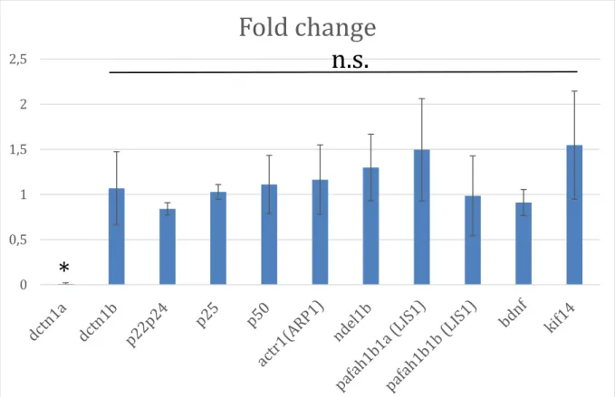
VI-Quantitative RT-PCR profile of 6dpf mok m632-/- embryos ... 35
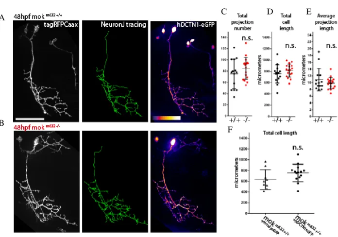
VII-Rescue of the axonal morphology phenotype with human DCTN1 ... 36
VII.1- Axonal morphology at 6dpf ... 37
VII.2-Axonal morphology at 48hpf ... 38
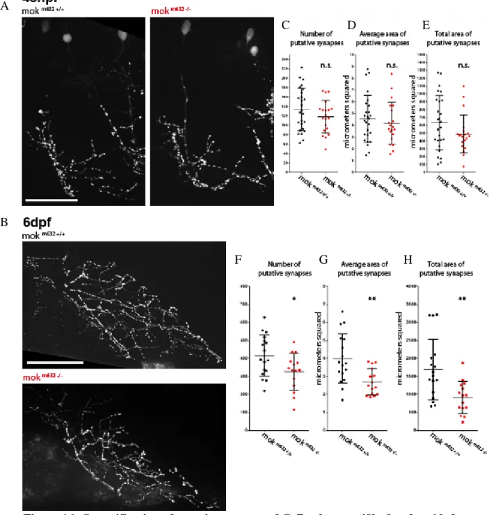
VIII-Putative synapse number at 48hpf and 6dpf ... 39
IX- Putative synapse stability at 48hpf ... 41
X-Spontaneous activity of the CaPs in the spinal cord at 4dpf ... 42
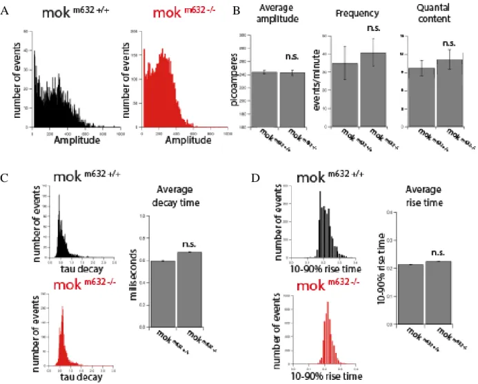
XI-Neuromuscular junction mEPC at 6dpf ... 43
XII- Neuromuscular junction paired-recordings at 6dpf ... 45
XIII- Axonal transport ... 46
XIII.1-Choice of cargo and method of expression ... 47
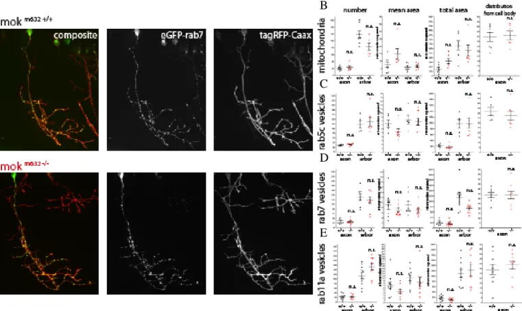
XIII.2-Cargo distribution at 48hpf ... 48
XIII.3-Cargo behavior and run metrics at 48hpf ... 50
XIII.4-Caveats of the expression method ... 57
XIV-Trophic signaling involvement ... 59
XIV.1- Actin filopodia dynamics from 48hpf ... 59
XIV.2-p75 receptor axonal transport ... 61
XV- Microtubule stability at 48hpf and 6dpf ... 63
xi
XVII- Synaptic microtubule capture at 48hpf and 6dpf ... 66
XVIII- Adhesion molecules at the NMJ (N-Cadherin) ... 68
XIX- Controls and validation ... 69
XIX.1-dynactin1b CRISPR/Cas9 mutant generation ... 69
XIX.2-Other dynein/dynactin mutant and DN-dynactin1 axonal morphology ... 73
Discussion ... 74
Conclusion and perspectives ... 87
Materials and Methods ... 88
I-Zebrafish husbandry and transgenic lines ... 88
I.1- list of zebrafish lines ... 88
II-Microinjections ... 88
III-DNA extraction and genotyping ... 88
III.1- mok m632 genotyping ... 89
III.2- dynactin1b *253 genotyping ... 89
IV-Molecular cloning ... 89
IV.1-Gateway cloning ... 90
IV.2-Gibson assembly cloning ... 91
IV.3-List of constructs used ... 92
V-Touch-evoked escape response assay ... 93
VI-Microscopy and image analysis ... 93
VI.1-Morphological images ... 93
VI.2-Spinning disk confocal ... 93
VII-Whole-mount immunohistochemistry ... 94
VII.1-List of antibodies ... 95
VIII-Quantitative RT-PCR ... 95
VIII.1-List of primers ... 96
IX-Calcium imaging of fictive swimming (Kevin Fidelin, Claire Wyart laboratory) ... 96
X-Electrophysiology (Jeffrey M. Hubbard, Claire Wyart laboratory) ... 97
XI-CRISPR/Cas9-mediated knock-in of dynactin1b ... 98
XII-Graph generation and statistical analysis ... 99
References ... 100
Annex ... 114
Annex 1-Research article: Deletion of a kinesin I motor unmasks a mechanism of homeostatic branching control by neurotrophin-3. Auer, Xiao, Bercier et al, eLife, 2015 114 Table of figures ... 141
1
Introduction
I-ALS
Amyotrophic lateral sclerosis (ALS) is a neurodegenerative disease arising in mid-life, which is mainly sporadic in nature (sporadic ALS, sALS, over 90% of patients). World-wide incidence is 1-2 per 100 000, making it the most common adult-onset motor neuron disease, and one of the most common adult-onset neurodegenerative disease. This progressive pathology causes dysfunction leading to cell death of upper and lower motor neurons in a non-cell-autonomous manner, and is characterized by insoluble inclusions of ubiquitinated protein in the cell body, as well as axonal accumulation of neurofilament (Shaw, 2005; Wijesekera & Leigh, 2009).
It generally leads to the demise of patients within 2-5 years of diagnosis owing to respiratory failure caused by muscle wasting and severe motor neuron loss. ALS symptoms vary between patients and is diagnosed based on clinical presentation of typical signs and progression (according to the El Escorial Diagnosis Criteria) as well as on exclusion of other disease with similar symptoms (Wijesekera & Leigh, 2009). Moreover, it is a heterogeneous pathology, where several causative genes and risk factors have been identified in both sALS and familial ALS (fALS) patients. To date, more than 20 different genes have been linked with this disease, leading to ALS subtype classifications, including the well-known SOD1, TARDBP, FUS/TLS, and C9orf72, but also more rare occurrences like UBQLN2 and DCTN1, which were identified in very few patients (Moloney, de Winter, & Verhaagen, 2014). Furthermore, many patients often have mutations in more than one gene at once, which could be explained by low penetrance, and supported by the interindividual variability exhibited in clinical presentation within carriers or the same mutation of the same family (Therrien, Dion, & Rouleau, 2016). Accordingly, the pathophysiology of ALS when considering the disease as a whole is complex. While most ALS cases exhibit the same early signs like electrophysiological abnormalities and degeneration of the connection between motor neuron and muscles cells (Sleigh, Burgess, Gillingater, & Cader, 2014), many mechanisms have been proposed to explain the specific degeneration of motor neurons and could possibly be used to strengthen classification and for the elaboration of targeted therapy in personalized medicine.
2
Figure 1: Pathophysiological mechanisms proposed to underlie ALS motor
neuron degeneration. The mechanisms shown here include 1- changes in transport of mRNAs and RNA-binding
proteins in the cytosol and nucleus, 2-impaired RNA metabolism due to mislocalization of RNA-binding proteins, formation of stress granules, aggregate formation, 3-impaired proteostasis overloading the proteasome with reduced autophagy lead to
protein aggregate accumulation, 4-impaired DNA repair, 5-mitochondrial dysfunction and oxidative stress caused by disruption of the organelle’s normal function, and the accumulation of ROS, 6-glial implication, degeneration and dysfunction leading to reduced support of neurons, 7-neuroinflammation caused by activated astrocytes and microglia, 8-perturbed axonal transport due to disorganization of the cytoskeleton, 9-impaired vesicular transport, 10-escitotoxicity caused
3
I.1-Pathophysiology
Protein misfolding and aggregation has historically been thought to be a major component of the ALS pathogenic mechanism. This is due to the fact that ubiquitinated protein inclusions are a prominent feature of ALS and other neurodegenerative diseases, but also because the insoluble aggregates found in patients’ brain tissue contain misfolded and truncated versions of mutant proteins like TDP-43 (TARDBP), FUS and SOD1. However, multiple other mechanisms of action have been proposed to be involved, perhaps even acting synergistically or in succession, in the pathogenesis of ALS. Notable examples include mitochondrial disturbances, endoplasmic reticulum (ER) and oxidative stress, excitotoxicity and axonal transport defects. In addition, the implication of non-neuronal cells, like glia and muscle fibers, has also been demonstrated to have an essential role in disease progression (Ilieva, Polymenidou, & Cleveland, 2009; Shaw, 2005).
I.1.1-Protein aggregates
The presence of protein aggregates present in motor neuron cytosol of ALS patients is a hallmark of this neurodegenerative disease, but their role is debated. It has been hypothesized that these aggregates could interfere with normal cellular function by sequestering essential proteins, by acting as hindrance in the way of axonal transport or by mobilizing the proteasomal degradation pathway (Atkin et al., 2013). However, whether these defects are causative of degeneration or a way for the cell to manage the chaos by clearing away defective proteins is still uncertain (Wijesekera & Leigh, 2009).
As these aggregates often contains proteins encoded from genes mutated in ALS patients (like TDP-43, FUS OPTN, UBQLN2 and C9ORF72), are found in tissues of fALS and sALS patients alike, as well as in the context of other neurodegenerative disease, this mechanism suggests a convergence to defects in the protein quality control process leading to neurodegeneration (Blokhuis, Groen, Koppers, van den Berg, & Pasterkamp, 2013).
I.1.2-Mitochondrial dysfunction
Mitochondria are essential to proper function of a neuron, as they are the “cell powerhouses”, providing energy by production of ATP, but are also involved in calcium
4 homeostasis and apoptotic mechanisms. ALS patients often have mitochondrial abnormalities, where energy metabolism is impaired (Wijesekera & Leigh, 2009) and present with misregulated intracellular calcium (Jaiswal, 2013). As calcium is a known secondary messenger, with roles in regulating metabolic pathways, cell development, and growth of synapses, many studies have focused on how this could lead to motor neuron degeneration (Aren et al., 2015).
The first gene identified as causative in ALS was superoxide dismutase 1 (SOD1), which encodes a cytoplasmic antioxidant found at the mitochondrial outer membrane. This mutant protein leads to misfolding and ubiquitinated accumulations, but also to mitochondria vacuolation and degeneration (Soo, Farg, & Atkin, 2011). In a mouse model, it was found that misfolded mutant SOD1 formed aggregates inside mitochondria, leading to fragmentation and depolarization of the membrane potential before the onset of symptoms (Luo et al., 2013). A new mutation has also been identified in ALS patients, disrupting the gene encoding coiled-coil-helix- coiled-coil-helix domain containing protein 10 (CHCHD10). The function of this protein is still unknown but it localizes at the mitochondrial inner membrane and is reported to be involved in oxidative phosphorylation (Therrien et al., 2016).
In addition, abnormal mitochondrial dynamics and altered localization were also seen in TDP-43 overexpression and loss-of-function mice models, suggesting that mitochondrial dysfunction could be a common mechanism among multiple disease models and patients alike (Magrané, Cortez, Gan, & Manfredi, 2014; W. Wang et al., 2013).
I.1.3-ER and oxidative stress
Post-mortem studies have revealed evidence for free radical damage in the (cerebro-spinal fluid) and CNS tissue of ALS patients. This is not surprising as reactive oxygen species cause stress, which can lead to cell death, especially in post-mitotic cells like neurons, and as these effects can be cumulative it corroborates the link between ALS and aging. Moreover, mutations in SOD1, coding for superoxide dismutase, an enzyme involved in the clearance of the superoxide radical O2-, supports the involvement of this mechanism in neurodegeneration (Shaw, 2005).
ER stress can be triggered by multiple events in a diseased neuron, like the production of misfolded proteins or defects in trafficking, and can induce a number of pathways of stress
5 response. In many cases, this leads to the activation of the unfolded protein response (UPR), which acts as a quality control for misfolded proteins and helps restore homeostasis by inducing degradation, and when overactivated, it can lead to apoptosis (Matus, Valenzuela, Medinas, & Hetz, 2013). It was also suggested that different types of motor neurons have a variable degree and threshold for ER stress response, which might explain selective vulnerability of certain types of muscle fibers to denervation and degeneration (Matus et al., 2013; Saxena & Caroni, 2011).
I.1.4-Excitotoxicity
Excitotoxicity is a result of the overstimulation of glutamate receptors resulting from defective intracellular calcium homeostasis or free radical abundance. Glutamate is the preponderant excitatory neurotransmitter in human CNS and its role in fast synaptic transmission relies on the cell-surface expression of AMPA receptors found in neurons, and on reuptake mechanisms via glutamate transporters like EEAT2, which are found in neurons and glia. This supports a role for non-cell autonomous mechanisms in ALS pathogenesis (Shaw, 2005). Reduced levels of EEAT2 mRNA have been found in ALS patient CSF, which is thought to lead to the decreased expression of the transporter. Diminished EEAT2 function, and thus excess extracellular glutamate, lead to increased stimulation of glutamate receptors and excess calcium influx into the cell, which impairs calcium signaling and could lead to degeneration (Aren et al., 2015).
Interestingly, the only approved treatment for ALS at this time, riluzole, is known to enhance glutamate clearance.
I.1.5-Axonal transport
Motor neurons have a high metabolic rate and need to extend very long axons to form contact with muscles. They also have a high expression of AMPA receptors and glutamate transporters, as well as low expression of calcium-binding proteins, which are necessary for calcium buffering. In addition, they have a high threshold for the initiation of heat shock response, which lowers their ability to cope with stress (Shaw, 2005). All these characteristics could make motor neurons particularly vulnerable to defects arising from the pathogenic mechanisms described above, but most importantly means that they rely heavily on axonal transport to supply mitochondria as well as newly synthesized proteins and mRNAs to
6 synapses, for the distal clearance of detritus, aging organelles and vesicles, and for relaying back signals from the periphery to the cell body.
It is therefore no surprise that axonal transport defects have been reported in ALS patients and models alike, with hallmark signs like organelle and vesicle accumulation, degradation defects, impaired stress signaling and mitochondria dysfunction (Soo et al., 2011). Some studies investigated transport dynamics in transgenic mice or neuronal cell culture and found a reduction in both slow and fast axonal transport affecting various cargoes such as mitochondria, early and late endosomes, as well as TrkB vesicles (Brady & Morfini, 2017; De Vos & Hafezparast, 2017; Encalada & Goldstein, 2014; Millecamps & Julien, 2013; Eran Perlson, Maday, Fu, Moughamian, & Holzbaur, 2010).
Considering the dynamics of axonal transport, the defects observed in neurodegenerative diseases could have different causes: preferential type of cargo selection for transport, varying load size (possibly due to protein aggregation) or depletion/dilution of the motor protein population, which could affect development, maintenance or survival of the neurons (Mitchell & Lee, 2012). Independently from the motor dynamics defects, there could be other impairments depending on cargo adapter proteins or chemical mediators, like phosphorylation (Blasier et al., 2014; Gibbs, Greensmith, & Schiavo, 2015).
In the context of ALS, whether the observed axonal transport defects are causative of neurodegeneration or consequences of other pathophysiological processes, like mitochondrial disturbances or protein aggregates, is yet to be determined (Millecamps and Julien, 2013). Indeed, impairments at any level of this process could affect cell survival and lead to neurodegeneration, but the identification of particular dysfunction, a disease-specific “pathogenic signature” which might be particular to mutations or pathophysiological mechanism, could open up new therapeutic avenues and warrants investigation.
I.2-The neuromuscular junction (NMJ)
The neuromuscular junction is an early target in ALS, as molecular changes at this level occur before the onset of neurodegeneration (Moloney et al., 2014).
Fast-fatigable, fast-fatigable-resistant, and slow muscle fibers are the three functional types of mammalian muscle units innervated by motor neurons. In ALS mice models, fast-fatigable muscle fibers get denervated first, before onset of clinical symptoms. This is followed
7 by denervation of fast-fatigable resistant fibers in the late symptomatic phase, and finally the slow muscles lose their connections in the end stage (Saxena & Caroni, 2011).
These three functional muscle subtypes express distinct muscle protein isoforms, and were studied in mice to determine their specific synaptic plasticity. Selective synapses were shown to be weakened in various mice models of motor neuron disease, including the SOD1 (G93A) mice, leading to denervation before motor neuron death and onset of clinical symptoms. This weakening was attributed to the intrinsic properties of various types of synapses present at the NMJ, with the most vulnerable ones, present on fast-fatigable muscle fibers, as they were shown to have a low competence for terminal sprouting (Frey et al., 2000). Thus, the specific properties of motor neurons, muscles and NMJ synapses, which could be affected by pre- and postsynaptic environment, are thought to be responsible for the selective vulnerability of fast-fatigable neuromuscular synapses to degeneration in motor neuron disease.
II-Dynactin1
Neurons are highly polarized cells, with a network of dynamic microtubules connecting distal synapses with the soma. These tubules, polymers of tubulin, are composed of a fast growing (+) and a slow growing (-) end; in axons, they are organized with the fast growing, unstable end (+) oriented towards the synapse and the more stable (-) end is oriented towards the nucleus at the level of the soma. This component of the cytoskeleton provides the rails on which intracellular transport is carried out. As the synthesis of new proteins takes place in the cell soma, axonal transport is known to be essential for growth and maintenance of synapses.
During this process, ATP-driven molecular motors that move unidirectionally along the axonal microtubule network transport organelles and vesicles. This transport is mediated by two types of molecular motors: the kinesin superfamily, transporting cargoes towards the synapse (anterograde, towards microtubule fast-growing ends) and the dynein motor protein complex, moving from the synapse to the cell body (retrograde). The 45 diverse kinesin members have different roles and are subdivided into 14 families based on the heterogeneous structure of their motor and tail domains, while the dynein motor, which has one major assembly, relies partly on isoform expression and mostly on adaptor polypeptides, like the dynactin complex, for functional versatility. Many motor proteins are present on cargoes
8 simultaneously, and most cargoes alternate between them by coordinated regulation, allowing for bidirectional transport along microtubule tracks (Akhmanova & Hammer III, 2012).
The dynactin macromolecular complex has been proposed to regulate dynein activity, and to be essential for the motor complex recruitment to microtubules distal ends, which serve as a loading point for the dynein motor and the recruitment of cargo, while providing a supposed “crutch” of extra support for attachment (Chowdhury, Ketcham, Schroer, & Lander, 2015; Waterman-Storer, Karki, & Holzbaur, 1995).
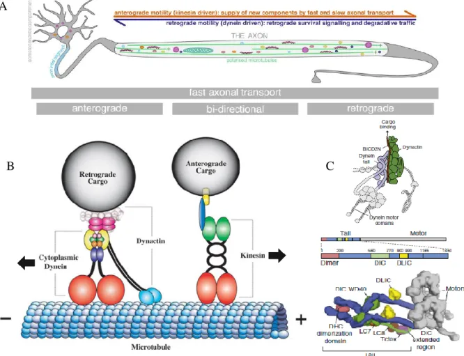
Figure 2: Axonal transport and its molecular motors. A) Representation of axonal transport in a motor
neuron, where a very long axon separates the site of protein synthesis, the soma, from the synapse. This highlights the dependence of those neurons on a functional process, both for signaling between the growing synapse and the cell body, the supply of proteins and organelle for growth, but also for the removal of detritus for degradation. B) Schematic illustration of the anterograde dynein-dynactin motor complex and the retrograde kinesin complex on a microtubule track. The dynactin
complex is an adaptor complex that binds the motor complex of dynein to regulate its activity. The dynactin1 subunit is represented here as a dimer, demonstrating its binding capacity for dynein and the dynactin complex-black stripes, and with
microtubules-blue circles. C) Protein structure with identified structural domains of the dynein motor complex. A reconstruction of the complex shows how the heavy motor chains are assembled with the intermediate and light chains, forming the tail that binds the motor complex to the dynactin complex. (Adapted from Carter, Diamant, & Urnavicius, 2016;
Duncan & Goldstein, 2006; Maday, Twelvetrees, Moughamian, & Holzbaur, 2014)
A
9 It is composed of 11 subunits organized into structural domains: dynactin1 (DCTN1, also called p150), dynamitin (DCTN2, also called p50), and p22/p24 (DCTN3), forming the “projecting arm”, and Arp1 (ACTR1A), CapZ and β-actin, forming the “Arp1 rod”, and Arp11 (ACTR10), p62 (DCTN4), p25 (DCTN5), and p27 (DCTN6) forming the “pointed end complex”. In the projecting arm, subunits are present as self-associated polymers, where four copies of dynamitin, two copies of both dynactin1 and p22/24 are present (Schroer, 2004).
Dynactin1 is the largest subunit of the dynactin adaptor complex (Schnapp & Reese, 1989) and serves as the link between the two heavy chains of the dynein motor and their cargo, where the interaction occurs at the level of the intermediate chains (K. T. Vaughan & Vauee, 1995). In addition to dynein intermediate chains, dynactin1 has been found to bind directly to microtubules and to another subunit of the dynactin complex, namely actin-related protein centractin 1 (Arp-1) (Waterman-Storer et al., 1995).
The dynactin1 subunit is encoded by the DCTN1 gene in humans (ENSG00000204843; OMIM 601143) and is composed of 32 exons, forming a full-sized protein of 150kDa harboring multiple functional domains.
II.1-The different domains of dynactin1
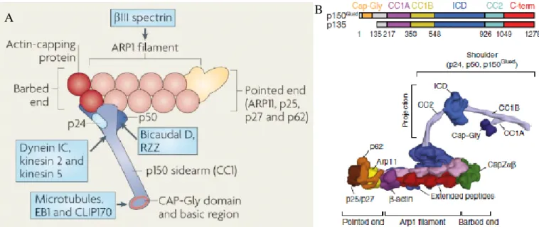
Figure 3: Schematic representation of the dynactin complex. A) The dynactin complex is composed
of 11 subunits that are assembled into three structural domains: the projecting arm domain (dynamitin/p50, dynactin1, and p22/24), the arp rod domain (Arp1, CapZ and β-actin), and the pointed end domain (Arp11, p62, p25 and p27). Many of the
subunits in this complex are known to interfere with other proteins (identified in blue boxes), which could regulate the assembly or function of the complex. B) Protein domains of the dynactin1 subunit with identified functional domains. This
gene produces two isoforms, including a shorter 135kDa isoform lacking the microtubule-binding CAP-Gly domain. The functional domains of dynactin1 gives the subunit an arm-like shape, which allows interaction with other subunits of the
complex, as well as the dynein motor and microtubules. (Adapted from Kardon & Vale, 2009; Sorbara et al., 2014; Urnavicius et al., 2015)
10 The N-terminal cytoskeletal-associated protein glycine-rich domain (CAP-Gly) is responsible for this subunit’s interaction with microtubules, a process which is concentration dependent (Ayloo et al., 2014), and regulated by phosphorylation (Vaughan, Miura, Henderson, Byrne, & Vaughan, 2002). This domain is however not necessary for processivity of dynein, once it is loaded unto microtubule, as a truncated dynactin1 lacking the N-terminus, does not interfere with retrograde transport (Kim et al., 2007; Tripathy et al., 2014). The CAP-Gly domain has however been shown to confer dynactin1 with a “skating” function along microtubules which allows for greater stability in dynein’s binding to microtubules, thus preventing the detachment of the dynein-dynactin-cargo complex during active transport (Culver–Hanlon, Lex, Stephens, Quintyne, & King, 2006). Furthermore, it has been proposed that the CAP-Gly domain’s interaction with microtubule during axonal transport enhances the processivity of dynein under heavy load, also by preventing detachment, meaning it could act as a module necessary for greater force generation by the retrograde motor (Moore, Sept, & Cooper, 2009). This domain has been reported to bind to other +tip microtubule-binding proteins like CLIP-70 and end-binding protein 1 (EB1) (Schroer, 2004), where these interactions have been respectively involved in loading of dynein at microtubule + end and modulation of microtubule anchoring at the centrosome (Askham, Vaughan, Goodson, & Morrison, 2002; Vaughan, Tynan, Faulkner, Echeverri, & Vallee, 1999). Importantly, the interaction of dynactin1 with microtubules is known to be independent of its interaction with dynein.
The basic domain, rich in basic amino acids, immediately follows the CAP-Gly domain, and is also within the microtubule-binding region of dynactin1. It was shown to be present only in neuron-specific splice forms of the protein and could be sufficient for microtubule-binding, but both this domain and the CAP-Gly domain are necessary for microtubule polymerization, and the reduction of microtubule catastrophe (Lazarus, Moughamian, Tokito, & Holzbaur, 2013).
The first coiled-coil domain (CC1) is responsible for binding to dynein, and is split into two separate domains with different functions: CC1A and CC1B. Both of these subdomains were found to bind dynein directly. It was also reported that they both have an effect on dynein processivity, CC1B by promoting it and CC1A by favoring a diffusive state, and that they interact with each other and with the N-terminal portion of dynactin1 to coordinate the different states of dynein motility (Tripathy et al., 2014). Additionally, when expressed by itself, the CC1
11 fragment has been shown to inhibit dynein motility due to its ability to compete with the binding of both a fully assembled dynactin complex and NudE-Lis1, another regulatory complex, to dynein.
The second coiled-coil domain (CC2) is responsible for many protein-protein interactions and contains an actin-binding motif that acts as a link with the Arp-1 subunits, which forms the base of the dynactin complex (Schroer, 2004; Waterman-Storer et al., 1995).
The C-terminus of dynactin1 also has a “cargo-binding” domain known to bind to vesicular adaptors like rab7-interacting lysosomal protein (RIPL) (Johansson et al., 2007), huntingtin-associated protein1 (HAP1), retromer protein sertin nexin 6 (SNX6) and c-Jun regulator JIP1 (MAPK8IP1) (Fu & Holzbaur, 2014).
The existence of an additional dynein-interacting domain of dynactin, located between the CC1 and the CC2 is debated, but known to be involved in the unloading of cargo by interaction with ADP-ribosylation factor-like 3 (Arl3). This function is carried out by dissociation of dynactin from dynein, with the help of LC8 (dynein light chain 8, smallest subunit of the dynein motor) which interacts with DIC (dynein intermediate chain) (Jin, Yamada, Arai, Nagai, & Hirotsune, 2014).
II.2-Disassembly of the dynactin complex from dynein
The disassembly of dynactin from dynein is thought to be part of the mechanism of cargo unloading and relies on the interaction between AnkyrinB (AnkB), which is bound to membrane cargoes, and dynactin1, which is bound to the rest of the dynactin complex as well as dynein (Lorenzo et al., 2014). However, many ways to disrupt the dynein-dynactin interaction have shown to affect the function of this complex.
Dynamitin (DCTN2) is known to be the link between the projecting arm structure and the rest of the dynactin complex via interactions with Arp-1 (Cheong, Feng, Sarkeshik, Yates, & Schroer, 2014) and dynactin1 (Carter et al., 2016) and its overexpression has been found to lead to disassembly of the dynein-dynactin complex, inhibiting minus-end directed movement (Burkhardt, Echeverri, Nilsson, & Vallee, 1997; Echeverri, Paschal, Vaughan, & Vallee, 1996). Interestingly, it was reported that this overexpression led to the loss of the ARP1 portion of the complex from microtubules without affecting microtubule-associated dynactin1 or CLIP-70 (Vaughan et al., 1999).
12 Furthermore, the overexpression of the CC1 domain of dynactin1 has also been shown to interfere with the dynein-dynactin complex assembly, as well as affecting microtubule organization. In this case, the overexpressed fragment is acting in a competitive manner where free CC1 binds to dynein and leaves the rest of dynactin assembled independently of dynein (Quintyne et al., 1999). Many of these methods have been used to probe the function of the dynactin complex and dynactin1 specifically, but the disassembly might also affect regulation or the assembly of different regulatory complexes or disrupt normal interactions.
II.3-Axonal transport
The dynactin complex is thought to be essential for axonal transport via the dynein motor complex and the dynactin1 subunit has a very important role to play in many aspects of this crucial function.
For instance, dynactin1 is known to interact with kinesin motors and has been suggested to be involved in the control of bidirectional transport. As kinesins and dyneins are found bound to the same cargoes, a tug-of-war mechanism was proposed to direct the switching between retrograde and anterograde movement, but it was noted that enhanced recruitment or inhibition of motors could produce a directional bias in the movement of vesicles and organelles (Hendricks et al., 2010).
Dynactin was also reported to interact with kinesin-2 via the dynactin1 subunit, which enhances its processivity by increasing run lengths. This interaction was shown to depend on dynactin1’s binding to microtubules (Berezuk & Schroer, 2007). Another study reported that dynactin1 bound kinesin-2 and dynein’s intermediate chain via the same domain, but not simultaneously (Deacon et al., 2003). This subunit is thus believed to be involved in the regulation of bidirectional transport, and that the inhibition of a motor in one direction can affect a motor in the opposite direction.
Axonal transport-regulating scaffolding proteins also have an important role to play, like C-jun-amino-terminal kinase-interacting protein 1 (JIP1), which also requires binding to dynactin1 to interacts with kinesin heavy chain in order to regulate bidirectional transport of APP in neurons. It was reported that the interaction of JIP1 with dynactin1 and kinesin was mutually exclusive, competitive when bound to dynactin1, and regulated by phosphorylation (Fu & Holzbaur, 2014).
13 Furthermore, interaction of dynactin1’s CAP-Gly domain with EBs has also been shown to be necessary for the retention of this subunit at microtubule distal ends, a necessary process to initiate axonal transport (Moughamian & Holzbaur, 2012). As dynactin1 was first identified as part of the dynactin complex regulating dynein activity, and its most studied domain is the CAP-Gly domain, a major part of this subunit’s function will involve interactions with the neuronal cytoskeleton, an important player in axonal transport.
II.4-The neuronal cytoskeleton
The cytoskeleton is composed of three components: microtubules, actin, and neurofilaments. Microtubules are essential to neuronal polarity, and the polymerization of their tubulin subunits into protofilaments results in the assembly of dynamic fast-growing +ends oriented towards the synapse in axons. Neurofilaments are also part of the axonal architecture, where they provide structure and regulate axonal caliber. Actin filaments constitute the network supporting the growth cone and pre-, as well as postsynaptic region, essential for plasticity, and play an important role in dendrites where they form the spines. Transport on actin is mediated by myosins is essential at the synapse for the recycling of receptors or vesicles, and tethering of synaptic components. The actin and microtubule components of the cytoskeleton can influence each other’s polymerization, and interact with each other for proper guidance during growth cone migration, and for establishment of arborisation, a process which requires reorganization of existing cytoskeletal structures (Dent & Kalil, 2001).
The microtubule network, providing the rails for active axonal transport, is regulated by motor proteins and microtubule-associated proteins (MAPs), which influence both its dynamics and function. End-binding proteins belong to the latter category, and bind to the + ends of microtubules (+ tip proteins) to play a role in stability, but also to regulate interactions with organelles, coordinate the actin cross-talk between axonal microtubule and growth cone actin, as well as mediate downstream signaling events via guidance receptors during migration (Bearce, Erdogan, & Lowery, 2015).
14
Figure 4: The neuronal cytoskeleton during development. A) Neurons have a very complex and
dynamic microtubule cytoskeleton that is subject to many types of post-translational modifications regulating its dynamics and interactions. These modifications vary according to location in the cell, as well as throughout the life of the neuron. B) The growth cone is an interface between microtubules and actin, where different types of cytoskeletal structures participate in
forming the dynamic growth cone. Stable acetylated microtubules form the axon, whereas unstable tyrosinated microtubules are present in the growth cone. At the growth cone, filopodial actin is necessary for sensing of environmental factors that will
guide the axon during migration. Interaction between the two cytoskeletons is essential for axon branching. C) +tip proteins are located at the fast-growing end of microtubules, at the level of the growth cone where they play a role in coupling between the microtubules and the actin cytoskeleton for outgrowth and guidance. (Adapted from Bearce et al., 2015; Dent &
Kalil, 2001; Song & Brady, 2015)
A
15 Dynein, dynactin and CLIP-170 have been found to accumulate at microtubule +ends, where the dynactin complex is necessary for dynein to be present (Vaughan et al., 1999), where CLIP-70’s cargo-binding domain is necessary for dynactin’s recruitment (Valetti et al., 1999), and where EB1 is necessary for the recruitment of CLIP-170 (Moughamian, Osborn, Lazarus, Maday, & Holzbaur, 2013). However, in the absence of dynein, dynactin and CLIP-170 can each be found independently bound to microtubules (Vaughan et al., 1999). It has been demonstrated that the binding of these +tip-associated proteins depends on microtubule tyrosination, where the absence of tubulin-tyrosine ligase in fibroblasts leads to protein mislocalization and cytoskeletal defects (Peris et al., 2006).
The p62 subunit of dynactin is located at the pointed end of the complex and found to interact with the actin cytoskeleton (Garces, Clark, Meyer, & Vallee, 1999). This interaction of this subunit with the membrane could also be the anchor necessary for dynein-dynactin’s role in microtubule sliding involved in cytoskeleton remodeling during growth. This process relies on the action of severing enzymes like spastin and katanin and motor proteins like dynein to reorient small microtubule fragments in a growing cell by interaction with filamentous actin (Baas, Karabay, & Qiang, 2005; Baas, Nadar, & Myers, 2006).
Another role at the level of the actin cytoskeleton is dynactin1’s interaction with MISP (C19ORF21), thought to contribute to the formation of focal adhesion sites by serving as a link between actin and microtubules, although this has not been confirmed in neurons (Maier, Kirsch, Anderhub, Zentgraf, & Krämer, 2013). On another note, it was found in flies expressing glued, a dominant-negative mutant of dynactin1, that a gap was present between the pre- and post-synaptic sites of the neuromuscular junction (NMJ). This detachment was not known to be causative, or a consequence of the synapse disassembly also observed in this model, but supports a role for dynactin at the level of adherens junctions (Eaton, Fetter, & Davis, 2002).
In addition, it has been reported that dynactin1 interacts with tubulin-binding cofactor B (TBCB), a protein involved in microtubule assembly and polymerization. Recruitment of dynactin1 by TBCB was shown and overexpression of TBCB leads to dynactin1 dissociation from the microtubules. However, disruption of this interaction had no apparent effect on the cytoskeleton, synaptogenesis or neuronal maturation (Kuh et al., 2012).
As previously mentioned, dynactin1 was also reported as binding to both microtubules and free, soluble tubulin, to promote polymerization. Both the CAP-Gly domain and the basic
16 domain are required for this anti-catastrophe activity at microtubule +ends (Lazarus et al., 2013).
Dynactin and dynein are also known to have a role in chromosome alignment and spindle organization during mitosis (Crowder et al., 2015; Echeverri et al., 1996) and the CAP-Gly domain of dynacitn1 appears to be essential for the organization of spindle microtubule arrays, suggesting that this particular subunit is necessary for cell division (Kim et al., 2007). New evidence however suggests that the dynactin complex might be dispensable for some mitotic functions of dynein, as it was shown that another dynein regulator, the Lis1/NudE complex, prevented dynein detachment from microtubules and is now thought to regulate spindle dynein. As this complex and dynactin bind competitively to dynein, and as depletion of only components of Lis1/NudE cause severe chromosome alignment defects, it is possible different aspects or dynein function are regulated by different regulating complexes (Wadsworth & Lee, 2013).
II.5-Synapse stability
Synapses are complex structures composed of a pre- and post-synaptic component. The presynaptic side is formed by assembly of necessary components at specialized sites called active zones, which include among others, calcium channels, synaptic vesicles and scaffolding proteins (Petzoldt, Lu, & Sigrist, 2016). Presynaptic vesicles are filled with neurotransmitters that are released into the extracellular space by fusion with the membrane. In order to maintain a ready-pool of vesicle close to the active zones where the capture and release machinery is located, synapses rely on the endosomal system to provide recycling of synaptic vesicles and receptors as well as means for degradation of damaged proteins. As dynactin1 is found accumulating at presynaptic sites, interacting with microtubules and with the actin cytoskeleton via other dynactin subunits, a role at the level of the synapse could be considered.
17
Figure 5: The synapse organization and function. A) The synapse is site dense with protein, organized in
specialized domains called active zones, where the synaptic release machinery is localized adjacent to calcium channels. At these sites, synaptic vesicles fuse with the membrane to release neurotransmitters into the synaptic cleft and need to cycle back to the ready, releasable pool. They do so via the clathrin-dependent endocytosis, which then takes them to be refilled in
the recycling pool before moving back to the ready pool. B) The neuromuscular junction is a special synaptic connection, where a motor neuron is innervating a muscle fiber. The presynaptic site is apposed to acetylcholine receptors organized in end plates, which are located a short distance away from the active zones, across the synaptic cleft. C) Adhesion molecules have a very important role at the synapse, as they organize the active zone proteins and fusion machinery to efficiently recruit
the synaptic vesicle and produce a flow of neurotransmitters. They are also involved in trans-synaptic connectivity, which allows proper alignment of pre- and postsynaptic sites. (Adapted from Dean & Dresbach, 2006; Haucke, Neher, & Sigrist,
2011;Drachman, 1978)
In support of this, dynactin was found to promote synapse stability at the fly neuromuscular junction, where disruption of dynein-dynactin by either expression of the glued dominant-negative dynactin1 subunit, or the inhibition of Arp-1 expression by RNAi caused disruption of the microtubule cytoskeleton, electrophysiological deficits, and synaptic retraction in a cell-autonomous manner (Eaton et al., 2002).
At the level of the NMJ, a role for dynactin1 in synapse growth appears to be mediated via interaction with arfaptin2, a membrane-binding protein localized at the golgi, essential for the dynactin complex binding to membranes in neurons. This interaction was reported to be dispensable for axonal transport and synapse stability, as assayed by the presence of synaptic footprints, a sign of degeneration where post-synaptic sites are left behind without a presynaptic site. It was suggested that this interaction could regulate the sorting and delivery of particular
A B
C B
18 proteins involved in synapse growth, or that it could affect ion channel expression at the membrane (Chang et al., 2013).
Overall, both of these studies suggest a role for dynactin1-mediated axonal transport in synapse growth and stability, but could be due to long-range effects (like improper signaling or delivery of synaptic components) or short-range effects (like cytoskeleton anchoring and synaptic vesicle dynamics).
II.6-Alternative splicing: p135 versus p150
Interestingly, as a result of alternative splicing, the DCTN1 gene produces two major isoforms: a full-size 150kDa protein, and a shorter 135kDa protein lacking the N-terminus region where the microtubule-binding domain is located. Both of these isoforms bind dynein in independent complexes, which indicates a role for dynactin1 independent of microtubule binding (Tokito, Howland, Lee, & Holzbaur, 1996). In addition, both of these isoforms are found in neuronal populations (Carter et al., 2016) and it was suggested that because it can still recruit dynein, the 135kDa isoform dynein-dynactin complex would have a reduced initiation rate and lower affinity for tyrosinated microtubules, due to its lack of CAP-Gly domain (McKenney, Huynh, Vale, & Sirajuddin, 2016).
As the CAP-Gly domain was reported to have a role in regulating the cytoskeleton and be essential for initiation but dispensable to the processivity of axonal transport, the shorter 135kDa isoform could still support active transport, as was observed in drosophila S2 cells (Kim et al., 2007) and in human retinal pigmented epithelial cells (RPE-1) (McKenney, Huynh, Tanenbaum, Bhabha, & Vale, 2014).
I1.7-Mutations and associations with human disease
Defects in both transport of material for development or clearance of detritus in the axon can lead to neuronal stress and culminate in cell death. Predictably, axonal transport deficits have been reported in various neurodegenerative diseases like Huntington’s disease, Alzheimer’s disease, spinal bulbar muscular atrophy (SBMA), Charcot-Marie-Tooth type 2F (CMT2F) and amyotrophic lateral sclerosis (ALS) (Chevalier-Larsen & Holzbaur, 2006; Duncan & Goldstein, 2006; Soo et al., 2011).
19 Additionally, mutations in many components of the axonal transport machinery have been associated with human pathology, for instance KIF5a in Hereditary Spastic Paraplegia (James & Talbot, 2006), DYNC1H1 in CMT2F (Lipka, Kuijpers, Jaworski, & Hoogenraad, 2013) and MND (Hafezparast et al., 2003), and DCTN1 with SBMA and Perry syndrome. Mutations in the gene coding for dynactin1 (DYNACTIN1; DCTN1), have also been reported in motor neuron disease and ALS patients (Table1).
Table 1: Mutations identified in DCTN1 in the context of ALS or MND. Many pathogenic
mutations have been reported within DCTN1, this table displays the reports of cases where patients were diagnosed as having either ALS or MND. The clinical presentation is included, as is the inheritance, when the data was available.
The G59S point mutation is perhaps the most studied and in patients has been linked with an early adulthood onset motor neuron disease (MND), presenting with breathing difficulties, and progressive facial muscle and hands weakening, followed by lower extremities impairment (Puls et al., 2003). In mice, a homozygous knock-in of this mutation leads to embryonic lethality, as does a homozygous deletion of the whole gene, similarly to the loss of dynein heavy chain. Heterozygous carriers however develop progressive motor neuron impairments. This suggests that the dynein-dynactin complex and dynactin1 have an essential
Protein domain
Mutation Associated
disease
Phenotype description Inheritance Reference
Dynactin1 CAP-Gly
G59S Motor neuron
disease
Lower motor neuron disease, no sensory symptoms, slow progression Autosomal dominant (Puls et al., 2003)
Dynactin1 T1249I sALS Middle-age onset; gait
disturbance, distal limbs weakness and muscle atrophy, slow progression
Autosomal dominant ( Münch et al., 2004) Dynactin1 Dynein-binding domain
M571T fALS Middle-age onset; upper
limb onset with bulbar symptoms Autosomal dominant ( Münch et al., 2004) Dynactin1 Dynein-binding domain R785W fALS incomplete penetrance
Middle-age onset; upper limb onset or bulbar onset
Uncertain ( Münch et al., 2004)
Dynactin1 R1101K ALS/FTD Middle-age onset; speech
impairment, upper limb weakening, lower muscle
atrophy Autosomal dominant (Münch et al., 2005) Dynactin1 E34Q, D63Y, I196V and R1049Q
ALS Not reported Not reported;
likely autosomal
dominant
(Stockmann et al., 2013)
20 function in cell division which is not affected by a possible haploinsufficiency or toxic gain of function (Lai et al., 2007). Mice overexpressing the mutant peptide, which is found to form aggregates, exhibit defects in vesicular transport, autophagic cell death, as well as NMJ degeneration and muscle atrophy (Laird et al., 2008).
This mutation has also been linked with SBMA. In a mouse model, a proliferation of lysosomes was seen, as well as a reduction in motor neuron axonal caliber and NMJ disturbances, without any reported effect on retrograde axonal transport (Chevalier-Larsen, Wallace, Pennise, & Holzbaur, 2008). Another disease linked to this mutation is distal hereditary motor neuropathy type VIIB (HMN7B), where G59S appears to have a dominant-negative effect: the produced peptide is not incorporated correctly in the dynactin complex, forms aggregates and was found to disrupt axonal transport of lysosomes in the axon of primary dorsal root ganglion neurons (Moughamian & Holzbaur, 2012).
Other mutations in the CAP-Gly domain of dynactin1 (G71R, Q74P), associated with Perry syndrome (Vilariño-Güell et al., 2009), a disease characterized by parkinsonism and TDP-43 inclusions, cause a phenotype similar to a loss of the whole domain (Moughamian & Holzbaur, 2012). In the fly, a different mutation (G38S) has been linked with Perry syndrome was shown to lead to adult-onset behavioral deficits, disrupted vesicular transport, and impaired neurotransmitter release (Lloyd et al., 2012).
However, many studies have looked at these ALS and MND- linked mutations and aside from the G59S, their role in motor neuron degeneration is not clear. It has been suggested that rather than be causative, that they could act as a risk factor (Vilariño-Güell et al., 2009). In support of this hypothesis, a study has looked at expression of four ALS-associated mutations in dynactin1 (M571T, R785W, R1101K, and T1249I (Levy et al., 2006)) and found that the incorporation of the mutant subunit into the dynactin complex was normal, and that their expression did not lead to aggregate formation or golgi disruption, unlike what is seen for G59S. It was however noted that the regulation of dynactin1 expression is tightly controlled in cells and that mutations altering protein expression or stability might be considered a risk factor in itself (Dixit, Levy, Tokito, Ligon, & Holzbaur, 2008). Another study looked at variants found in ALS patients (particularly E34Q, D63Y, I196V and R1049Q out of 24 identified targets) and characterized their effect on the cell biology of cultured motor neurons. While they report changes like aggregate formation, effect on microtubule network and ubiquitination, most of the variants studied were of too low occurrence to determine if they are rare causes and
21 pathogenic, perhaps with incomplete penetrance, or simply risk factors of ALS (Stockmann et al., 2013).
To support a role for dynactin in ALS, DCTN1 was found to be downregulated in the spinal motor neurons of sALS patients, even in well-preserved populations of neurons. Furthermore, this downregulation was shown to be pre-symptomatic (Tanaka, Ikenaka, Yamamoto, & Sobue, 2012). Another study reported lower levels of DCTN1 mRNA and protein in sALS patient cortex and spinal cords (Kuźma-Kozakiewicz et al., 2013).
Moreover, it was found in mice models with fALS SOD1 mutations (A4V, G85R, and G93A) that the mutant proteins co-localize and interact with the dynein-dynactin complex. This interaction has been shown to be essential for aggregate formation, where disruption of the dynactin complex by overexpression of dynamitin prevented the formation of inclusions and improved cell survival (Ström et al., 2008).
In addition, mice overexpressing dynamitin have been found to exhibit a degenerative phenotype at the level of spinal motor neurons, leading to skeletal muscle atrophy, which was attributed to the loss of retrograde axonal transport (LaMonte et al., 2002). Similarly, SBMA mice with trinucleotide expansions in the androgen receptors (AR) have significantly reduced levels of dynactin1 mRNA as a result of AR-related transcriptional dysregulation, which leads to impaired retrograde transport. The subsequent neuronal dysfunction is however rescued by overexpression of dynactin1 (Katsuno et al., 2006). Mice mutant for another interactor of the dynein-dynactin complex, Bicaudal D2 (BICD2) also show impaired axonal transport and develop motor abnormalities and motor neuron degeneration (Teuling et al., 2008). Hence these studies support a role for dynactin1 and axonal transport in MND pathogenesis.
While it is not clear if dynactin1 is causative of ALS or MND, a role for axonal transport in these diseases is clear, and a role for dynactin1 and the dynactin complex in this process is known to be essential. We have chosen to investigate the loss of dynactin1 function in motor neuron-specific neurodegeneration in order to determine if in vivo compensation mechanisms could underlie defects resulting in pathogenicity.
22
III-The zebrafish animal model
III.1-The zebrafish spinal cord and NMJ
The zebrafish embryo develops quickly, leading to simple stereotyped behavior as early as 17-19 hours post-fertilization (hpf). By 48hpf, the embryos have hatched from their chorion and can swim in reproducible escape bouts. These behaviors rely on the innervation of axial muscle fibers by motor neurons sprouting from the embryonic spinal cord, easily identifiable due to their simple organization and morphological characteristics. The motor neurons are organized in tight bundles of axons called ventral roots, grouping the axons of neurons whose soma is located within one spinal segment, innervating a single muscle segment (Myers, 1985).
Figure 6: The embryonic zebrafish spinal cord, its primary motor neurons, and their
innervation pattern on the ventral musculature. A) 48hpf embryonic zebrafish are already able to
produce complex swimming behavior and rely on their spinal cord motor neurons to do so. B) One somatic segment (delineated in red in A) contains various types of primary motor neurons, with different morphologies, cell body position and innervation patterns, which are repeated on each side of the embryo, and at each somite along the trunk and tail. The primary
motor neurons can be visualized and studied by using a transgenic line where GAL4 is placed under the expression of a specific enhancer called mnx1(shown here in combination with a UAS-driven membrane-bound fluorophore). The caudal primary-CaP- motor neuron we use in this study (at 24hpf, red arrow, at 48hpf, blue arrow). C) The CaPs innervate ventral fast-twitch muscle fibers, located in deeper levels below the slow-twitch muscle layer. (Adapted from Ashworth, Zimprich, &
Bolsover, 2001; Babin, Goizet, & Raldúa, 2014)
Primary motor neurons are the first ones to appear and extend axons out of the spinal cord and are classified in three types (CaP, RoP and MiP, for caudal, rostral and middle primary) depending on the section of muscle where it extends its peripheral arbor. They also exhibit particular morphological features including the size and shape of their soma, and the caliber of their axon, as well as subtype-specific electrical excitability signatures (Moreno &
A
B A