Sébastien Vauchera)
EMPA, Swiss Federal Laboratories for Materials Science and Technology, CH-3602 Thun, Switzerland
Radu Nicula
EMPA, Swiss Federal Laboratories for Materials Science and Technology, CH-3602 Thun, Switzerland; and Institute of Physics, University of Rostock, D-18055 Rostock, Germany
José-Manuel Català-Civera
Polytechnical University of Valencia, School of Telecommunication, Camino de Vera s/n E-46022 Valencia, Spain
Bernd Schmitt and Bruce Patterson
Swiss Light Source, Paul Scherrer Institute, CH-5232 Villigen, Switzerland (Received 8 June 2007; accepted 24 September 2007)
The effect of rapid microwave heating has so far been evaluated mainly by comparing the state of materials before and after microwave exposure. Yet, further progress critically depends on the ability to follow the evolution of materials during ultrafast heating in real time. We describe the first in situ time-resolved monitoring of solid-state phase transitions during microwave heating of metallic powders using wide-angle synchrotron radiation diffraction. Single-phase Al–Cu–Fe quasicrystal powders were obtained by microwave heating of nanocrystalline alloy precursors at 650 °C in <20 s.
I. INTRODUCTION
Ultrafast heating and cooling are essential to the pres-ervation of nanoscale features in bulk nanostructured sol-ids. Both microwave1and spark plasma-sintering2,3 tech-niques offer feasible manufacturing solutions that will soon be able to be used on an industrial scale. Common to these modern technologies is the kinetic suppression of excessive grain growth during densification. During the past few decades, microwave heating has been suc-cessfully used in a broad variety of applications, ranging from rapid food conditioning, to wood processing, ster-ilization, and drying, to the chemical synthesis and sin-tering of new materials.4–7Despite efforts to fine tune the use of microwaves to specific applications, there is still a lack of experimental knowledge on the changes materials undergo in their structure, the constitution of phases, morphology, and properties during their exposure to mi-crowave fields. Modern mimi-crowave-heating technologies still rely solely on the room-temperature examination of the structure, microstructure, and properties of materials before and after microwave heating. The few previous attempts to follow in situ the evolution of materials dur-ing microwave heatdur-ing8–13 were part of the sustained
effort to clarify the occurrence and nature of nonthermal contributions to mass transport [i.e., solely due to the application of high-frequency electromagnetic (EM) fields].7,14–16 The enhancement of atomic diffusion un-der EM field application is known as the “microwave effect.”7 The experimental validation of the microwave effect is a central topic of modern research on the inter-action of materials with microwave fields. Intense efforts were dedicated during the past decade to understanding the role of microwave E and H fields on phase transfor-mations.1,17–20
So far, the few in situ studies8–10of structural transi-tions of materials during microwave heating did almost exclusively rely on neutron scattering. A first in situ x-ray diffraction (XRD) study was performed by Robb et al.,11
using laboratory x-ray equipment. High heating rates of tens of degrees per second and higher are not unusual in microwave heating. Therefore, the use of high-flux synchrotron radiation and of fast x-ray detec-tors is essential to in situ real-time diffraction experi-ments, for which a satisfactory compromise must be reached between the counting statistics and the time reso-lution. We report on the first in situ microwave heating experiments using wide-angle synchrotron radiation powder diffraction. We focus our present discussion on the in situ time-resolved monitoring of solid-state phase transitions during the microwave heating of quasicrystal (QC)-forming Al–Cu–Fe alloys.
a)
Address all correspondence to this author. e-mail: sebastien.vaucher@empa.ch DOI: 10.1557/JMR.2008.0009
Contrary to what was believed for many years, micro-waves may also be used to process metallic fine pow-ders.20 Discovered more than two decades ago in melt-spun Al–Mn alloys,21 QCs were meanwhile shown to form in over 100 alloy systems.22
QCs are nonperiodic, long-range, ordered solids with noncrystallographic ro-tational symmetries,23
and exhibit outstanding properties of major technological interest22–25
(e.g., low friction, adherence or wear, high hardness, high strength, low thermal and electrical conductivity, plastic deformation, or biocompatibility). Due to the wider availability of its constituents, Al–Cu–Fe QCs are nearer to industrial ap-plications,26so that recent research efforts have focused on their synthesis and processing on a larger scale.26–28
II. EXPERIMENTAL
Metallic nanopowders with nominal composition of Al62at.%–Cu25.5at.%–Fe12.5at.% were prepared by the wet milling of high-purity elements using a high-energy planetary ball mill (PM 400; Retsch, Haan, Germany). Suitable precursor powders are readily obtained by short milling for 6–10 h using standard stainless-steel milling equipment.28
Microwave heating was carried out in a WR-340 wave-guide terminated with a sliding short-circuit. The micro-wave generator with a maximum power of 1.2 kW at 2.45 GHz (Dipolar AB, Skellefteå, Sweden) is directly coupled to the waveguide. The incident and reflected microwave power is monitored using a high-power im-pedance analyzer (S-TEAM STHT, Bratislava, Slova-kia). The sliding short-circuit was adjusted to ensure that the samples are located at the maximum of the electrical field of the TE10n mode of the empty cavity. Neverthe-less, it is likely that in the presence of the capillary loaded with the powder specimen, the field distribution will be distorted and so the sample experiences the action of a mixed E/H field.29
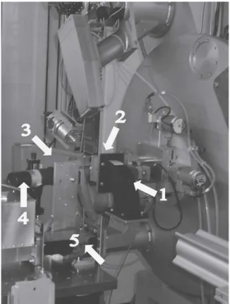
The in situ experiments were performed at the powder-diffraction station of the Materials Science X04SA wiggler beamline30 of the Swiss Light Source (SLS) (Villigen, Switzerland). A photograph of the microwave setup installed at the X04SA beamline is shown in Fig. 1. The beamline optics, comprising two collimation mirrors and a double flat-crystal monochromator, allows for an optimal collimation of the beam to achieve a small beam-spot at the sample location. For our experiment, per-formed using a synchrotron radiation wavelength ⳱ 0.07087 nm, the photon flux was equal to 1012
–1013 photons per second. Conventionally, powder-diffraction measurements are done by scanning a detection system with a few channels over the angular range of interest. Instead, we took advantage of the newly developed MYTHEN microstrip detector,31which employs a mas-sively parallel detection system covering an angular
range of 60°2 with 15,000 independent channels. This reduces the full-spectrum measurement time from hours to seconds and below. The time resolution of the present experiment averaged to 4 s (i.e., fixed exposure time of 1 s, with additional 3–4 s for data transfer).
Dry-powder specimens were mounted into ⭋1-mm alumina capillaries and inserted into the resonant cavity through a nonradiating slot. The sample temperature was monitored by an optical pyrometer (SXACLS; Raytek, GmbH, Berlin, Germany). Diffraction patterns were also collected on empty capillaries and on standard LaB6 (NIST 660a) powders to determine the x-ray wavelength, the zero-shift, and the instrument-resolution function. The empty capillary spectrum was subtracted from all specimen diffraction patterns. The diffraction patterns of the QC-phase were indexed following the formalism of Cahn et al.32using (N,M) indices. The XRD lines of the icosahedral structure are defined by a pair of scattering vectors in the parallel and perpendicular spaces32:
QII= 1 aQ
冑
N + M 2+ = 1 a6D冑
N + M 2共2 + 兲 = 2sin , (1a) Q⊥= 1 aQ冑
共N − M兲 2 + = 1 a6D冑
共N − M兲 2共2 + 兲 , (1b) FIG. 1. Portable microwave setup installed at the X04SA beamline at the SLS: (1) x-ray beam path; (2) sample position; (3) Mythen detec-tor; (4) infrared pyrometer; and (5) microwave waveguide.where is the x-ray wavelength, is the irrational “golden mean” number, a6Dis the lattice constant of the periodic six-dimensional hypercubic lattice, and aQis the
real space quasi-lattice constant.32The quasi-lattice con-stants were determined by a least-squares fitting proce-dure using at least 10 diffraction lines.
III. RESULTS AND DISCUSSION
Over 500 high-resolution XRD patterns were collected during the in situ microwave-heating experiment. The microwave-heating profile is shown in Fig. 2. We have simultaneously recorded the pyrometer temperature, the applied microwave power, and the microwave power ab-sorbed by the Al–Cu–Fe powders.
The Al–Cu–Fe powder specimens undergo structural phase transitions only within the regions of interest (ROIs) given by the time intervals A and B (gray-shaded regions in Fig. 2). Within region A, the applied micro-wave power is raised to 42 W. The input micromicro-wave power was later increased to 70 W (region B). The in-crease in the applied microwave power to 96 W (elapsed time 1200 to 1300 s) does not cause further structural changes. Finally, region C corresponds to the fast speci-men cooling following microwave shut off. We notice that the Al–alloy powders sustain stable microwave heat-ing without thermal runaway or meltheat-ing over the whole duration of the experiment. There is an immediate re-sponse of the specimen to the application of the micro-wave field and to its removal. The specimen-absorbed microwave power needed to complete the transformation to single-phase QCs is <10 W.
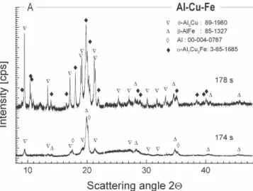
The as-prepared Al–Cu–Fe powders contain a mixture of cubic -Al(Fe) solid solution (PDF 85-1327) and -Al2Cu (PDF 89-1980, tetragonal) phases, together with residual pure aluminum (PDF 4-0787). The first ROI
(region A, elapsed time from 174 to 178 s) comprises a very short time interval of only 4 s, immediately follow-ing microwave application (Fig. 3). Within this ROI, the tetragonal -Al7Cu2Fe phase (PDF 3-65-1685) is formed, still coexisting with the initial -Al(Fe) and -Al2Cu phases.
The next ROI (region B, elapsed time 305 to 319 s) comprises the solid-state phase transformations triggered by the rise of the microwave power to 70 W (Fig. 4). We observe the transformation of the initial -phase (Fe-rich) and -phase (Cu-rich) to the -Al7Cu2Fe phase [Figs. 4(a)–4(d)], which indicates a gradual homogeni-zation of the alloy composition.27The QC-phase starts to form as soon as the tetragonal-Al7Cu2Fe phase be-comes the major constituent of the alloy [Fig. 4(d)]. Alto-gether, the increase of the microwave power to 70 W (region B) triggers the complete transformation to a single-phase icosahedral QC structure [Figs. 4(c)–4(f)]. The -Al7Cu2Fe phase is the main precursor of the quasicrystalline -phase.28 The final → transition [Figs. 4(d)–4(f)] is completed in <10 s, while the ab-sorbed microwave power does not exceed 10 W. The QC -phase is preserved during the rest of the in situ microwave-heating experiment (Fig. 5). The transforma-tion sequence under microwave irradiatransforma-tion may be sum-marized as follows (minor phases in brackets):
-Fe共Al兲 + -Al2Cu共+Al兲 → -Al7Cu2Fe共+兲
→ -Al7Cu2Fe+ → .
The above sequence of phase transformations does not contradict previous findings.27,28 However, the forma-tion of the body-centered cubic␥-phase (Al4Cu9-type) or of the Fe3Al phase was not detected here. The nucleation of these intermediate phases is thus either kinetically suppressed during microwave heating or their transient
FIG. 2. The microwave heating temperature–time profile. Also shown are the applied and the specimen-absorbed microwave power.
FIG. 3. Phase transitions of the as-prepared AlCuFe powders after microwave application (region A).
formation cannot be observed with the present time reso-lution. Another important difference with conventional heating is the spectacular enhancement of the phase-transformation rate upon microwave irradiation. The
ki-netics of the → phase transition upon isothermal conventional heating was recently investigated by time-resolved XRD using synchrotron radiation.33 It was shown that even after 40 min at 700 °C, only 96% of the specimen is transformed to the quasicrystalline-phase. In contrast, the same transition is completed under mi-crowave irradiation in <10 s [Figs. 4(d)–4(f)].
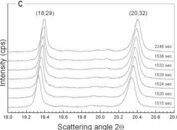
The diffraction patterns of the icosahedral QC-phase were indexed following the formalism of Cahn et al.32 using (N,M) indices. All diffraction lines correspond to an F-type icosahedral QC structure. The well-defined triplet of diffraction lines given by the (6,9), (7,11), and (8,12) Bragg reflections [Fig. 4(f)] gives evidence for the formation of a QC structure25,34and not of large unit-cell approximants.35Single-phase QC powders are therefore obtained (Fig. 5). From the evolution of the quasi-lattice constant aQduring cooling (region C), we could estimate
the maximum temperature attained during microwave exposure. We assume a parabolic temperature depend-ence of the quasi-lattice constant:
aQ= a0+ ␣⭈T + ⭈T 2
, (2)
where T is the absolute temperature,␣ is the coefficient of thermal expansion, and  ⳱ d␣/dT. The coefficients of thermal expansion of the QC -phase are known36,37 ␣ ⳱ 13.18044 × 10⁻⁶
K−1and ⳱ 0.25686 × 10⁻⁸K−2 (measured upon cooling of Al–Cu–Fe powders of similar chemical composition). This in turn allows us to deter-mine the temperature–time evolution of the quasi-lattice parameter aQof the icosahedral-phase during cooling (Fig. 6).
The maximum temperature attained during our in situ experiment (Fig. 2) equals 650 °C (±20 °C), which is in agreement with previous results.28
This further allows for a rough estimation of the cooling rate during microwave treatment, which is found to vary between 45.96 and 23.4 °C/s (Fig. 6).
FIG. 4. Sequence of solid-state transitions leading to the formation of single-phase QCs [up triangles -Fe(Al); down triangles -Al2Cu; diamonds-Al7Cu2Fe; open circles-phase).
FIG. 5. Shift of the XRD patterns of the icosahedral-phase during specimen cooling (region C).
FIG. 6. Quasi-lattice constant of the icosahedral-phase during cool-ing (region C).
IV. CONCLUSIONS
We have demonstrated here the feasibility and poten-tial advantages of using microwave heating for the syn-thesis of alloy powders with a complex atomic structure. Single-phase Al–Cu–Fe QC powders were obtained by microwave heating of metallic precursors in <20 s. High heating (46 °C/s) and cooling (23 °C/s) rates were ob-served. In particular, the high cooling rate is a unique advantage of microwave heating over other powder syn-thesis/processing methods.
Beyond the ultrafast microwave synthesis of quasi-crystalline powders and, even more important, the pres-ent work demonstrates that the combined use of high-brilliance synchrotron radiation sources and of fast x-ray detectors enables the in situ real-time observation of the interaction of materials with microwave fields and of its kinetic features. Information yielded by in situ time-resolved experiments will greatly improve our funda-mental knowledge on the mechanisms of mass transport and structural phase transitions in the presence of EM fields, as well as our means to fine-tune microwave proc-essing for a broad variety of nanomaterials. Furthermore, opportunities for developing new materials will emerge, with potential implications for powder metallurgy, mi-crowave chemistry, catalysis, pharmacy, food proc-essing, biology, or medicine.
ACKNOWLEDGMENTS
Radu Nicula gratefully acknowledges Prof. Dr. Eberhard Burkel and Dr. Manuela Stir (Rostock University, Ger-many) for continuous support.
REFERENCES
1. R. Roy and D. Agrawal: The new science of microwave−materials interactions: The role of separated e and h fields and its real world applications, in Proceedings of the 6th Symposium on Microwave Applications and Related Fields, November 2–4, 2006, Oyaki-City, Japan.
2. S. Seal, S.C. Kuiry, P. Georgieva, and A. Agarwal: Manufacturing nanocomposite parts: Present status and future challenges. MRS Bull. 29(1), 16 (2004).
3. Z. Shen, Z. Zhao, H. Peng, and M. Nygren: Formation of tough interlocking microstructures in silicon nitride ceramics by dy-namic ripening. Nature 417, 266 (2002).
4. J.D. Katz: Microwave sintering of ceramics. Annu. Rev. Mater. Sci. 22, 153 (1992).
5. Materials Research Advisory Board: Microwave Processing of Materials (National Research Council, Publication NMAB-473, National Academy Press, 1994).
6. D.E. Clark, D.C. Folz, C. Folgar, and M. Mahmoud: Microwave Solutions for Ceramic Engineers (The American Ceramics Soci-ety ACerS, Inc., Westerville, OH, 2005).
7. Yu.V. Bykov, K.I. Rybakov, and V.E. Semenov: High-temperature microwave processing of materials. J. Phys. D: Appl. Phys. 34, R55 (2001).
8. A.G. Whittaker, A. Harrison, G.S. Oakley, I.D. Youngson,
R.K. Heenan, and S.M. King: Preliminary experiments on appa-ratus for in situ studies of microwave-driven reactions by small angle neutron scattering. Rev. Sci. Instrum. 72, 173 (2001). 9. A. Harrison, R. Ibberson, G. Robb, G. Whittaker, C. Wilson, and
D. Youngson: In situ neutron diffraction studies of single crystals and powders during microwave irradiation. Faraday Discuss. 122, 363 (2002).
10. M.M. Günter, C. Korte, G. Brunauer, H. Boysen, M. Lerch, and E. Suard: In situ high temperature neutron diffraction study of Sr/Mg-doped lanthanum gallate superionic conductors under mi-crowave irradiation. Z. Anorg. Allg. Chem. 631, 1277 (2005). 11. G.R. Robb, A. Harrison, and A.G. Whittaker:
Temperature-resoved, in-situ powder x-ray diffraction of silver iodide under microwave irradiation. Phys. Chem. Comm. 5, 135 (2002). 12. S. Vaucher, J-M. Catala-Civera, A. Sarua, J. Pomeroy, and
M. Kuball: Phase selectivity of microwave heating evidenced by Raman spectroscopy. J. Appl. Phys. 99, 113505 (2006). 13. A. Nesbitt, P. Navabpour, B. Degamber, C. Nightingale, T. Mann,
G. Fernando, and R.J. Day: Development of a microwave calo-rimeter for simultaneous thermal analysis, infrared spectroscopy and dielectric measurements. Meas. Sci. Technol. 15, 2313 (2004). 14. S.A. Freeman, J.H. Booske, and R.F. Cooper: Microwave field enhancement of charge transport in sodium chloride. Phys. Rev. Lett. 74, 2042 (1995).
15. J.H. Booske, R.F. Cooper, and S.A. Freeman: Microwave en-hanced reaction kinetics in ceramics. Mater. Res. Innovations 1, 77 (1997).
16. K.I. Rybakov and V.E. Semenov: Mass transport in ionic crystals induced by the ponderomotive action of a high-frequency electric field. Phys. Rev. B 52, 3030 (1995).
17. J. Cheng, R. Roy, and D. Agrawal: Experimental proof of major role of magnetic losses in microwave heating of metal and me-tallic composites. J. Mater. Sci. Lett. 20, 1561 (2001).
18. R. Roy, P.D. Peelamedu, J.P. Cheng, C. Grimes, and D. Agrawal: Major phase transformations and magnetic property changes caused by electromagnetic fields at microwave frequencies. J. Mater. Res. 17(12), 3008 (2002).
19. R. Roy, P.D. Peelamedu, L. Hurtt, J.P. Cheng, and D. Agrawal: Definitive experimental evidence for microwave effects: Radi-cally new effects of separated E and H fields, such as decrystal-lization of oxides in seconds. Mater. Res. Innovations 6, 128 (2002).
20. R. Roy, D. Agrawal, J. Cheng, and S. Gedevanshvili: Full sinter-ing of powdered-metal bodies in a microwave field. Nature 399, 668 (1999).
21. D. Shechtman, I. Blech, D. Gratias, and J. Cahn: Metallic phase with long range orientational order and no translational symmetry. Phys. Rev. Lett. 53, 1951 (1984).
22. H-R. Trebin, ed.: Quasi-crystals (Wiley-VCH, Weinheim, 2003). 23. Chr. Janot: The properties and applications of quasi-crystals.
Eu-rophys. News 27, 60 (1996).
24. D.J. Sordelet and J.M. Dubois, ed.: Quasi-crystals. MRS Bull.
22(11), (1997).
25. J.M. Dubois: Useful Quasi-crystals (World Scientific Publishing, Singapore, 2003).
26. E. Huttunen-Saarivirta: Microstructure, fabrication and properties of quasi-crystalline Al–Cu–Fe alloys: A review. J. Alloys Compd.
363,150 (2004).
27. V.V. Tcherdyntsev, S.D. Kaloshkin, A.I. Salimon, E.A. Leonova, J. Eckert, F. Schurack, V.D. Rogozin, S.P. Pisarev, and Y.P. Trykov: Al–Cu–Fe quasi-crystalline phase formation by me-chanical alloying. Mater. Manufact. Proc. 17, 825 (2002). 28. R. Nicula, M. Stir, F. Turquier, and E. Burkel: Single-phase bulk
Al–Cu–Fe quasi-crystals by field-assisted sintering. Mater. Sci. Eng., A (doi:10.1016/j.msea.2007.01.163, in press).
29. D.C. Dube, P.D. Ramesh, J. Cheng, M.T. Lanagan, D. Agrawal, and R. Roy: Experimental evidence of redistribution of fields during processing in a high-power microwave cavity. Appl. Phys. Lett. 85(16), 3632 (2004).
30. B.D. Patterson, Ch. Brönnimann, D. Maden, F. Gozzo, A. Groso, B. Schmitt, M. Stampanoni, and P.R. Wilmott: The materials sci-ence beamline at the Swiss Light Source. Nucl. Instrum. Methods Phys. Rev., Sect. B 238, 224 (2005).
31. B. Schmitt, Ch. Brönnimann, E.F. Eikenberry, F. Gozzo, C. Hörmann, R. Horisberger, and B. Patterson: Mythen detector system. Nucl. Instrum. Methods Phys. Rev., Sect. A 501, 267 (2003).
32. J.W. Cahn, D. Shechtman, and D. Gratias: Indexing of icosahedral quasiperiodic crystals. J. Mater. Res. 1, 13 (1986).
33. E. Otterstein, R. Nicula, J. Bednarcik, M. Stir, and E. Burkel: In situ time-resolved x-ray diffraction investigation of the →
transition in Al–Cu–Fe quasi-crystal-forming alloys. Mater. Sci. Forum 558-559, 943 (2007).
34. Chr. Janot: Quasi-crystals: A Primer (Oxford University Press, New York, 1992).
35. M. Quinquandon, A. Quivy, J. Devaud, F. Faudot, S. Lefebvre, M. Bessiere, and Y. Calvayrac: Quasi-crystal and approximant structures in the Al–Cu–Fe system. J. Phys.: Condens. Matter 8, 2487 (1996).
36. A. Quivy, S. Lefebvre, J.L. Soubeyroux, A. Filhol, and R.M. Ibberson: High-resolution time-of-flight measurements of the lattice parameter and thermal expansion of the icosahedral phase Al62Cu25.5Fe12.5. J. Appl. Crystallogr. 27, 1010 (1994). 37. A.M. Korsunsky, A.I. Salimon, I. Pape, A.M. Polyakov, and
A.N. Fitch: The thermal expansion coefficient of mechanically alloyed Al–Cu–Fe quasi-crystalline powders. Scripta Mater. 44, 217 (2001).