HAL Id: hal-01731575
https://hal-amu.archives-ouvertes.fr/hal-01731575
Submitted on 10 Apr 2018HAL is a multi-disciplinary open access archive for the deposit and dissemination of sci-entific research documents, whether they are pub-lished or not. The documents may come from teaching and research institutions in France or abroad, or from public or private research centers.
L’archive ouverte pluridisciplinaire HAL, est destinée au dépôt et à la diffusion de documents scientifiques de niveau recherche, publiés ou non, émanant des établissements d’enseignement et de recherche français ou étrangers, des laboratoires publics ou privés.
Paraganglioma of the organ of Zuckerkandl associated
with a somatic HIF2α mutation: A case report
Ahmad Esmaeel Abdullah, Carole Guerin, Alessio Imperiale, Anne Barlier,
Stephanie Battini, Morgane Pertuit, Philippe Roche, Wassim Essamet,
Bernard Vaisse, Karel Pacak, et al.
To cite this version:
Ahmad Esmaeel Abdullah, Carole Guerin, Alessio Imperiale, Anne Barlier, Stephanie Battini, et al.. Paraganglioma of the organ of Zuckerkandl associated with a somatic HIF2α mutation: A case report. Oncology Letters, Spandidos Publications, 2017, 13 (3), pp.1083 - 1086. �10.3892/ol.2017.5599�. �hal-01731575�
Abstract. Paragangliomas of the organ of Zuckerkandl
(OZ-PGL) are rare tumors that, in >70% of cases, occur in association with succinate dehydrogenase complex iron sulfur subunit B (SDHB) or SDHD gene mutations. The aim of the current study was to determine whether a somatic genetic defect in the hypoxia-inducible factor 2α (HIF2α) gene was
present in a case of sporadic OZ-PGL. A 32-year-old African female presented with uncontrolled hypertension during the first trimester of pregnancy. A diagnostic hysteroscopy was performed 3 months after delivery, precipitating a hyper-tensive crisis. Thereafter, the patient was diagnosed with noradrenaline-secreting OZ-PGL. A complete blood count identified mild normocytic anemia of an inflammatory origin. Surgical removal of the tumor resulted in normalization of plasma and urinary normetanephrine levels. Genetic testing for germline mutations (including large deletions) in the von Hippel-Lindau tumor suppressor, SDHB, SDHC and SDHD
genes was normal. However, a heterozygous missense muta-tion (c.1589Cys>Tyr) was detected in exon 12 of HIF2α, which
results in a substitution of alanine 530 with valine (Ala530Val) in the HIF2α protein. A germline mutation was excluded based on the negative results of blood DNA testing. A three-dimen-sional homology model of Ala530Val was constructed, which showed impaired HIF2α/VHL interaction and decreased HIF2α ubiquitination. 1H-high-resolution
magic-angle-spin-ning nuclear magnetic resonance spectroscopy detected low succinate levels and high α and β glucose levels. To the best of our knowledge, the present case represents the first of its kind to associate a somatic HIF2α gain-of-function mutation with OZ-PGL. It is therefore recommended that patients without germline SDHx mutations should be tested for HIF2α
muta-tions.
Introduction
In 1901, Emile Zuckerkandl first described the abdominal para-aortic paraganglia in fetal and newborn humans as a paired retroperitoneal organ located laterally to the abdominal aorta at the level of the inferior mesenteric aorta (1). This para-ganglionic complex, known as the organ of Zuckerkandl (OZ), also includes smaller accessory paraganglia located anteriorly to the aorta between the lateral organs or below the aortic bifurcation (2). In 1903, Alfred Kohn established that the OZ commonly originated from chromaffin cells of the adrenal medulla (3), and it has later been established that it constitutes the largest accumulation of extradrenal chromaffin cells in mammals. In humans, the OZ reaches its maximal size at
Paraganglioma of the organ of Zuckerkandl associated
with a somatic HIF2
α
mutation: A case report
AHMAD ESMAEEL ABDULLAH1, CAROLE GUERIN2, ALESSIO IMPERIALE3,4, ANNE BARLIER5, STÉPHANIE BATTINI6,7, MORGANE PERTUIT5, PHILIPPE ROCHE6, WASSIM ESSAMET7,
BERNARD VAISSE8, KAREL PACAK9, FRÉDERIC SEBAG2 and DAVID TAÏEB1,10
1Department of Nuclear Medicine, La Timone University Hospital, European Center for Research in Medical Imaging,
Aix-Marseille University, 13385 Marseille Cedex 5; 2Department of Endocrine Surgery,
Conception Hospital, Aix-Marseille University, 13005 Marseille; 3Department of Biophysics and Nuclear Medicine, Hautepierre Hospital, University Hospitals of Strasbourg, 67200 Strasbourg; 4ICube Joint Research Unit 7357, University of Strasbourg/French National Center for Scientific Research and Federation of Translational Medicine of Strasbourg,
Faculty of Medicine, 67085 Strasbourg; 5Laboratory of Biochemistry and Molecular Biology, Conception Hospital, Aix-Marseille University, 13005 Marseille; 6Integrative Structural and Chemical Biology and Interaction Dynamics and Drug Design Platform, Cancer Research Centre of Marseille, Institut Paoli Calmettes, 13273 Marseille; 7Departments of Neuropathology
and 8Hypertension, La Timone University Hospital, Aix-Marseille University, 13385 Marseille Cedex 5, France;
9Program in Reproductive and Adult Endocrinology, Eunice Kennedy Shriver National Institute of Child Health and
Human Development, National Institutes of Health, Bethesda, MD 20892, USA; 10Cancer Research Centre of Marseille Affiliated to Inserm (UMR1068), Institut Paoli‑Calmettes, 13273 Marseille, France
Received July 24, 2015; Accepted May 10, 2016 DOI: 10.3892/ol.2017.5599
Correspondence to: Dr David Taïeb, Department of Nuclear Medicine, La Timone University Hospital, European Center for Research in Medical Imaging, Aix-Marseille University, 264 Rue Saint-Pierre, 13385 Marseille Cedex 5, France
E-mail: david.taieb@ap-hm.fr
Key words: paraganglioma, nuclear magnetic resonance spectroscopy, endothelial PAS domain-containing protein 1
ABDULLAH et al: HIF2α-RELATED PARAGANGLIOMA
1084
the age of ~3 years and subsequently regresses after reaching its peak by autophagy (4). The OZ is considered to be most important physiologically throughout the early gestational period, during which it secretes catecholamines into the fetal circulation, functioning as a homeostatic regulator of blood pressure (5). The OZ represents a site of origin for paragan-gliomas (PGLs) that preferentially secrete norepinephrine and induce symptoms of catecholamine excess (6). OZ-PGLs are rare tumors typically located close to the origin of the inferior mesenteric artery or between the proximal common iliac arteries (1). These lesions may occur sporadically or, in ~70% of cases, in association with succinate dehydrogenase complex iron sulfur subunit B (SDHB) or, less commonly, SDHD gene mutations (7). In addition, OZ-PGLs are particularly aggres-sive with high rates of metastatic spread (8). At least 150 cases of OZ-PGLs have been reported in the literature. They are strongly associated with an aggressive behavior, likely associ-ated with the SDHB mutation status (7). Due to the rarity of this disease, not much is known about its natural history. A single-center retrospective study of 371 patients with either pheochromocytoma or sympathetic paraganglioma revealed only 21 cases of OZ-PGLs, 14 of which (66%) had metastases at diagnosis (9). To the best of our knowledge, the current case demonstrates that somatic HIF2α [also known as endothelial PAS domain-containing protein 1 (EPAS1)] mutations may be associated with OZ‑PGL for the first time.
Case report
In September 2014, a 32-year-old African woman native to Burkina Faso was referred to the hypertension unit of La Timone University Hospital (Marseille, France) for screening for secondary hypertension. Hypertension was initially noted during the first trimester of pregnancy. The patient went into premature labor at 22 weeks and a cesarean delivery was performed 15 days later; the baby did not survive and succumbed a few minutes after birth. Following delivery, the patient experienced persistent and uncontrolled hypertension despite taking nicardipine (60 mg/day) and labetalol (400 mg/day) for 3 months. A diagnostic hysteroscopy was performed 3 months later, precipitating a hypertensive crisis [systolic blood pressure (BP), 300 mmHg; normal, <140 mmHg]. Thereafter, the patient was referred to the hypertension unit of La Timone University Hospital for secondary hypertension screening in September 2014. There was no known family history of tumors, syncope or sudden death. At admission (weight, 51 kg; height, 163 cm; and body mass index, 19.2), the patient presented with headaches, recurring episodes of palpi-tations and sweating, chest tightness, and polyuria. Treatment with nicardipine and labetalol was replaced with verapamil (240 mg/day). Ambulatory 24-h BP monitoring was performed during treatment with verapamil and demonstrated that the patient maintained a BP of 155/96 mmHg. Prazosin (2.5 mg once per day) was subsequently administered to reduce blood pressure further until surgical intervention.
Additional laboratory tests identified highly elevated 24 h urinary normetanephrine levels [20,140 nmol/24 h; upper reference limit (URL), <1900 nmol/24 h] and normal meta-nephrine levels (380 nmol/24 h; URL, <1600 nmol/24 h). In addition, serum chromogranin A was observed to be elevated
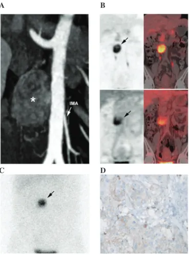
(223 µg/l; URL, <100 µg/l). A complete blood count revealed mild normocytic anemia (hemoglobin count, 108.0 g/l; normal hemoglobin count for female adults, 11.5-15.0 g/dl; mean corpuscular volume, 83.4 fl; normal mean corpuscular volume of adults, 80-100 fl) of an inflammatory origin with normal platelets and leukocytes. Diagnostic computed tomography (CT) revealed a 40-mm hypervascular, heterogeneous, left para-aortic mass located at the level of the inferior mesenteric artery (Fig. 1A). 18Fluorine-L-dihydroxyphenylalanine positron
emission tomography/CT (Fig. 1B) and iodine-123-metaiodo-benzylguanidine scintigraphy (Fig. 1C) confirmed the diagnosis of OZ-PGL without multifocal disease. The tumor also exhibited moderate heterogeneous 18F‑fluorodeoxyglucose
uptake (Fig. 1B). In October 2014, complete surgical resection was performed. Histopathological analysis of the tumor tissue revealed typical PGL features, including a low Ki-67 index (<1%) (monoclonal mouse antibody; clone, MIB-1; catalogue no., M7240; dilution, 1:100: Dako, Glostrup, Denmark). Genetic testing for germline mutations (including large deletions) in the von Hippel-Lindau tumor suppressor (VHL), succinate dehydrogenase complex iron sulfur subunit B (SDHB), SDHC
Figure 1. Imaging and pathological features of the OZ-PGL. (A) Contrast-enhanced CT (arterial phase) showing a 40-mm hypervas-cular and heterogeneous left para-aortic mass located at the level of the IMA (asterisk). (B) 18F-FDOPA (upper image) and 18F-FDG PET/CT (lower
image) imaging showing a single tumor. (C) Iodine-123-metaiodobenzylgua nidine scintigraphy also positively located the mass (planar anterior view). (D) Immunohistochemical analysis of the tumor demonstrated positive glu-cose transporter-1 immunostaining (~10%). CT, computed tomography; IMA, inferior mesenteric artery; 18F-FDOPA, 18
fluorine‑L‑dihydroxyphenylala-nine; 18F-FDG, 18F‑fluorodeoxyglucose; PET, positron emission tomography.
A B
and SDHD genes was normal. Immunostaining demonstrated that the tumor cells were positive for SDHB. Further genetic testing revealed a heterozygous cysteine to tyrosine substitution at base 1589 (c.1589Cys>Tyr) in the HIF2α coding sequence of the OZ-PGL, resulting in the replacement of alanine with valine at amino acid position 530 (Ala530Val). This leads to HIF2α stabilization as described by a previous in vitro experi-ment (10). A germline HIF2α mutation was excluded based on the negative results of blood DNA testing.
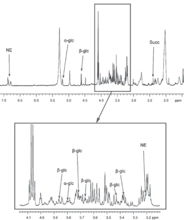
In order to assess the metabolic properties of the tumor, the present study performed 1H-high-resolution
magic-angle-spinning (HRMAS) nuclear magnetic resonance spectroscopy‑based global metabolomic profiling on tumor samples. A one-dimensional proton spectrum (1.5-7.2 ppm) using a Carr-Purcell-Meiboom-Gill pulse sequence with water presaturation was acquired from each intact tissue sample Low levels of succinate were detected, and according to our previous study (11) this excludes a SDH deficiency. Notably, the tumor also exhibited abnormally high levels of
α- and β‑glucose isomers as identified by HRMAS (Fig. 2). The patient is currently in remission, and regular clinical follow up occured every 6 months with normal metanephrines. Conventional radiological imaging (MRI) and functional
imaging (PET-FDOPA) at 1 year post-intervention were also normal.
Written informed consent was obtained from the patient for publication of the present case report and any accompa-nying images.
Discussion
To the best of our knowledge, the present case demonstrates, for the first time, that patients with somatic HIF2α mutations may present with OZ-PGL.
Germline mutations in the HIF2α/EPAS1 gene have been previously associated with congenital polycythemia (12). A syndromic association has been reported between somatic gain-of-function mutations in HIF2α and congenital
Figure 2. Results of HRMAS NMR spectroscopy (500 MHz) performed on tumor samples from the organ of Zuckerkandl paraganglioma. Partial metabolite assignment is indicated. The metabolic content may be directly compared as the spectrum intensity was normalized with respect to the weight of each examined sample. For display purposes, the amplitude of the lactate peak at 4.09 ppm has been cut out. The top image shows a repre-sentative spectrum with highly elevated levels of α-glc and β-glc. Spectrum regions ranging from 3.10‑4.15 ppm are magnified in the lower box. Amongst catecholamines, only an NE signal was detected in all the examined tissue samples. Finally, the level of Succ was low, which confirmed the absence of a succinate dehydrogenase complex deficiency. HRMAS, 1H-high-resolution
magic-angle-spinning; NMR, nuclear magnetic resonance; α-glc, α-glucose; β-glc, β-glucose; NE, norepinephrine; Succ, succinate.
Figure 3. Representation of human HIF2α in the presence of its binding partners EGLN1 and VHL. (A) WT HIF2α (A560) interacting with EGLN1; (B) mutant Val560 interacting with EGLN1; (C) WT HIF2α interacting with VHL; and (D) mutant Val560 interacting with VHL. HIF2α (16 residues) is represented in red and yellow, and the interactive partners (EGLN1 or VHL) are represented in blue and green. (B and D) The inserts present a closer view of Pro531 and Ala530 (or Val530) from HIF2α in the ball‑and‑stick represen-tation to show the atomic details, while HIF-2α partners, (A and B) EGLN1 or (C and D) VHL are shown as a grey surface showing that residues 530 and 531 bind to small pockets at the surface of the protein partner. Residue Ala530 is located in close proximity to residue Pro531, which is hydroxyl-ated by EGLN1 and at the interface with the binding partners EGLN1 and VHL. Hydroxylation of Pro531 is required for interaction with VHL. It is anticipated that valine, which is a larger residue than alanine, increases steric hindrance at Pro531, resulting in a reduction in its accessibility to EGLN1 by inhibition of Pro531 hydroxylation; therefore, interaction with VHL and subsequent ubiquitination is prevented. Panel D is presented as a model, but VHL interaction should not occur in the Val530 mutant. HIF2α, hypoxia inducible factor 2α; EGLN1, Egl-9 family hypoxia-inducible factor 1; VHL, von Hippel-Lindau tumor suppressor; WT, wild-type.
A B
ABDULLAH et al: HIF2α-RELATED PARAGANGLIOMA
1086
polycythemia, multiple PGL, duodenal somatostatinoma and ocular vascular abnormalities (for example, Pacak-Zhuang syndrome) (10,13-16). Mutations in HIF2α have also been observed in apparently sporadic pheochromocytomas (PHEOs)/PGLs without polycythemia (17-19). In one study, mutations (exon 12) were identified in 2 cases of solitary PHEO and 1 para-adrenal PGL (18). In an additional study, 6/42 cases of apparently sporadic PHEOs were identified to have HIF2α mutations (3 in exon 9 and 3 in exon 12) (17). HIF2α protein stability is dependent on the hydroxylation of two specific proline residues (Pro405 and Pro531) located in the O2-dependent degradation domain (10). Until present, all
mutations described were known to be located in hot spots adjacent to hydroxylation sites (16). These specific mutations disturb HIF2α prolyl hydroxylation and subsequent recogni-tion by the VHL protein, resulting in the failure of HIF2α
degradation via ubiquitination (16). As mutant HIF2α protein has a longer half-life compared with the wild-type protein, it has a targeted effect downstream of HIF2α (10).
The mutation identified in the present study had previously been reported in a case of apparently sporadic PHEO/PGL (18). The mutation involved Ala530, which is located in close proximity to the second hydroxylation site (Pro531) and at the interface with VHL and Egl-9 family hypoxia-inducible factor 1 (EGLN1) client-proteins. Homology modeling was performed to outline the biological properties of the Val530 mutant (Fig. 3). These three-dimensional models were gener-ated with IBM SPSS Modeler v14 (IBM SPSS, Armonk, NY, USA) using the crystal structures of HIF1α in interaction with EGLN1 or VHL as templates. HIF1α and HIF2α exhibit a sequence identity of 65% in the region modeled, which guar-antees (>50% identity) that the models are of a high quality. The model anticipates that valine, a larger residue than alanine, increases steric hindrance at Pro531, leading to: i) A reduction in its accessibility to EGLN1 by inhibition of Pro531 hydroxylation; and ii) impairment of HIF2α/VHL interaction with decreased HIF2α ubiquitination. The present study also identified a novel metabolomic pattern with low succinate and high glucose levels associated with HIF2α mutation.
Abnor-mally high levels of glucose may be explained by increased glucose uptake induced by HIF2α stabilization (20).
In conclusion, to the best of our knowledge, the current study identified, for the first time, an association between somatic HIF2α mutations and OZ-PGL. It is therefore recom-mended that patients with OZ-PGL in the absence of germline
SDHx mutations should undergo testing for HIF2α mutations.
References
1. Zuckerkandl E: About sympathetic paraganglions in the retroperitoneal space of man. Verh Anat Ges 15: 95-107, 1901 (In German).
2. Unsicker K, Huber K, Schütz G and Kalcheim C: The chromaffin cell and its development. Neurochem Res 30: 921-925, 2005.
3. Kohn A: The paraganglia. Arch Mikrosk Anat 52: 262-265, 1903 (In German).
4. Schober A, Parlato R, Huber K, Kinscherf R, Hartleben B, Huber TB, Schütz G and Unsicker K: Cell loss and autophagy in the extra‑adrenal chromaffin organ of Zuckerkandl are regulated by glucocorticoid signalling. J Neuroendocrinol 25: 34-47, 2013. 5. West GB, Shepherd DM, Hunter RB and McGregor AR: The
function of the organs of Zuckerkandl. Clin Sci 12: 317-325, 1953. 6. Martucci VL and Pacak K: Pheochromocytoma and paragan-glioma: Diagnosis, genetics, management, and treatment. Curr Probl Cancer 38: 7-41, 2014.
7. Lodish MB, Adams KT, Huynh TT, Prodanov T, Ling A, Chen C, Shusterman S, Jimenez C, Merino M, Hughes M, et al: Succinate dehydrogenase gene mutations are strongly associated with para-ganglioma of the organ of Zuckerkandl. Endocr Relat Cancer 17: 581-588, 2010.
8. Subramanian A and Maker VK: Organs of Zuckerkandl: Their surgical significance and a review of a century of literature. Am J Surg 192: 224-234, 2006.
9. Ayala-Ramirez M, Feng L, Johnson MM, Ejaz S, Habra MA, Rich T, Busaidy N, Cote GJ, Perrier N, Phan A, et al: Clinical risk factors for malignancy and overall survival in patients with pheochromocytomas and sympathetic paragangliomas: Primary tumor size and primary tumor location as prognostic indicators. J Clin Endocrinol Metab 96: 717-725, 2011.
10. Zhuang Z, Yang C, Lorenzo F, Merino M, Fojo T, Kebebew E, Popovic V, Stratakis CA, Prchal JT and Pacak K: Somatic HIF2A gain-of-function mutations in paraganglioma with polycythemia. N Engl J Med 367: 922-930, 2012.
11. Imperiale A, Moussallieh FM, Sebag F, Brunaud L, Barlier A, Elbayed K, Bachellier P, Goichot B, Pacak K, Namer IJ and Taïeb D: A new specific succinate-glutamate metabolomic hallmark in SDHx-related paragangliomas. PLoS One 8: e80539, 2013.
12. Percy MJ, Furlow PW, Lucas GS, Li X, Lappin TR, McMullin MF and Lee FS: A gain-of-function mutation in the HIF2A gene in familial erythrocytosis. N Engl J Med 358: 162-168, 2008. 13. Taïeb D, Yang C, Delenne B, Zhuang Z, Barlier A, Sebag F
and Pacak K: First report of bilateral pheochromocytoma in the clinical spectrum of HIF2A-related polycythemia-paragan-glioma syndrome. J Clin Endocrinol Metab 98: E908-E913, 2013. 14. Yang C, Sun MG, Matro J, Huynh TT, Rahimpour S, Prchal JT,
Lechan R, Lonser R, Pacak K and Zhuang Z: Novel HIF2A mutations disrupt oxygen sensing, leading to polycythemia, paragangliomas, and somatostatinomas. Blood 121: 2563-2566, 2013.
15. Pacak K, Chew EY, Pappo AS, Yang C, Lorenzo FR, Wilson MW, Aronow MB, Young JA, Popovic V and Zhuang Z: Ocular manifestations of hypoxia-inducible factor-2α paraganglioma-somatostatinoma-polycythemia syndrome. Ophthalmology 121: 2291-2293, 2014.
16. Pacak K, Jochmanova I, Prodanov T, Yang C, Merino MJ, Fojo T, Prchal JT, Tischler AS, Lechan RM and Zhuang Z: New syndrome of paraganglioma and somatostatinoma associated with polycythemia. J Clin Oncol 31: 1690-1698, 2013.
17. Welander J, Andreasson A, Brauckhoff M, Bäckdahl M, Larsson C, Gimm O and Söderkvist P: Frequent EPAS1/HIF2α exons 9 and 12 mutations in non-familial pheochromocytoma. Endocr Relat Cancer 21: 495-504, 2014.
18. Comino-Méndez I, de Cubas AA, Bernal C, Álvarez-Escolá C, Sánchez-Malo C, Ramírez-Tortosa CL, Pedrinaci S, Rapizzi E, Ercolino T, Bernini G, et al: Tumoral EPAS1 (HIF2A) mutations explain sporadic pheochromocytoma and paraganglioma in the absence of erythrocytosis. Hum Mol Genet 22: 2169-2176, 2013. 19. Favier J, Buffet A and Gimenez-Roqueplo AP: HIF2A mutations
in paraganglioma with polycythemia. N Engl J Med 367: 2161-2162, 2012.
20. Keith B, Johnson RS and Simon MC: HIF1α and HIF2α: Sibling rivalry in hypoxic tumour growth and progression. Nat Rev Cancer 12: 9-22, 2011.