RECOMMENDATIONS
The use of pocket-size imaging devices: a position
statement of the European Association
of Echocardiography
Rosa Sicari
*
, Maurizio Galderisi, Jens-Uwe Voigt, Gilbert Habib, Jose L. Zamorano,
Patrizio Lancellotti, and Luigi P. Badano
CNR, Institute of Clinical Physiology, ItalyReceived 27 November 2010; accepted after revision 6 December 2010; online publish-ahead-of-print 7 January 2011
Pocket-size imaging devices are a completely new type of echo machines which have recently reached the market. They are very cheap, smartphone-size hand-held echo machines with limited technical capabilities. The aim of this European Association of Echocardiography (EAE) position paper is to provide recommendations on the use of pocket-size imaging devices in the clinical arena by profiling the edu-cational needs of potential users other than cardiologists experts in echo. EAE recommendations about pocket-size imaging devices can be summarized in: (1) pocket-size imaging devices do not provide a complete diagnostic echocardiographic examination. The range of indi-cations for their use is therefore limited. (2) Imaging assessment with pocket-size imaging devices should be reported as part of the physical examination of the patient. Image data should be stored according to the applicable national rules for technical examinations. (3) With the exception of cardiologists who are certified for transthoracic echocardiography according to national legislation, specific training and certi-fication is recommended for all users. The certicerti-fication should be limited to the clinical questions that can potentially be answered by pocket-size devices. (4) The patient has to be informed that an examination with the current generation of pocket-pocket-size imaging devices does not replace a complete echocardiogram.
-Keywords Echocardiography † Hand-held † Pocket size † Screening † Recommendations † Workflow † Teaching
In the last few years, the miniaturization of echo machines has made screening of patients at the bedside possible. A wide variety of machines have been developed: from small, but complete echo machines with all conventional tools providing two-dimensional (2D), colour Doppler, and TEE modalities to hand-held imaging devices with limited technical and/or functional capabilities.1,2 Recent technological advances and price abatement particularly in the latter type of machines have raised the need for a re-assessment of their clinical use by the reference community. The aim of this Euro-pean Association of Echocardiography (EAE) position paper is to provide recommendations on the use of pocket-size imaging devices in the clinical arena by profiling the educational needs of potential users other than cardiologists experts in echo.
The machines
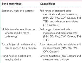
At present, a variety of small echo machines offer features which make them very similar to the most advanced end, high-performance echo machines. On the other hand, others have only
essential diagnostic modalities, with the advantage of excellent trans-portability and low cost. Currently available echo machines may be classified into four main categories (Table 1)3–8: (1) stationary high-end systems (i.e. fully equipped systems with 3D and other advanced modalities). Despite having wheels, they are quite heavy and not supposed to be moved. (2) Mobile systems, equipped with standard modalities, but not necessarily all the advanced modalities. They are smaller than machines in category 1, on wheels, and can be easily transported inside health-care facilities. (3) Portable machines that can be carried by a person and usually offer all essential ities to perform a complete echo study but not all advanced modal-ities. (4) Pocket-size, hand-held imaging devices.
The following text exclusively refers to pocket-size imaging devices (category 4) and their current state of capabilities. Rec-ommendations will be subject to adaptations depending on tech-nological progress.
Current pocket-size imaging devices offer diagnostic quality 2D and—in part—colour Doppler imaging in real-time. They come with a broad-bandwidth, phased array probe (1.7 – 3.8 MHz). The
*Corresponding author. Tel:+39 0503152397; fax: +39 0503152355, Email: [email protected]
Published on behalf of the European Society of Cardiology. All rights reserved.&The Author 2011. For permissions please email: [email protected].
European Journal of Echocardiography (2011) 12, 85–87 doi:10.1093/ejechocard/jeq184
by guest on January 22, 2012
http://ehjcimaging.oxfordjournals.org/
flow sector represents blood flow within an angle of 308. Images and videos (automatic autocycle without the need for ECG trace) can be stored in examination folders, recalled via a gallery function, and transferred to PC or USB throughout a docking station.
Pocket-size imaging devices are not echo machines and have been equipped with just 2D and colour Doppler modalities on purpose. Their technical characteristics may be summarized as: (i) grey-scale images have a 2D sector angle ,758, depth ,25 cm; (ii) colour flow imaging (available in one product only) has a fixed colour box size and a fixed pulse repetition frequency; (iii) measurements are restricted to distances and areas; (iv) options for patient identification are limited; (v) connectivity requires dedicated software tools.
Therefore, technical characteristics and image quality are usually sufficient for the qualitative evaluation of: left and right ventricular function, pericardial and/or pleural effusion, B-lines evaluated by lung ultrasound as a sign of extravascular lung water, size and res-piratory changes of inferior vena cava, and extent of calcification and motion of aortic cusps.9,10If available, valve regurgitation can be assessed by colour Doppler.10
Workflow
Current pocket-size imaging devices should only be considered as screening tools or used to complement the physical examination since they do not allow the performance of a complete echocar-diographic examination.7 This has the potential to deliver a marked change in cardiac care. Pocket-size devices should comp-lement the physical examination in outreach clinics, coronary and intensive care units, and may serve as a tool for fast initial cardiac assessment in emergency units, during cardiologic counsel-ling in- or outside health-care facilities and hospitals, for first cardiac evaluation in ambulances and for screening programmes in schools, industries, and other community activities.11Moreover,
pocket-size imaging devices may be used for the triage of the patient in need of a complete echocardiographic examination. Pocket-size imaging devices may further become a valuable teach-ing tool in medical schools (Table2).
All these applications would allow faster and more accurate clinical diagnoses, save health-care resources, reduce waiting lists for inpatient echocardiographic examinations, and improve teaching by allowing the immediate check of physical signs and auscultatory findings.12 However, since such devices are so powerful and are supposed to be used not only by cardiology-echocardiographists but also by general medicine practitioners, anesthesiologists, emergency medicine specialists, and internists, the cost/effectiveness of their clinical use is necessarily linked to proper training and education of users.11
Training and quality control
Expert (accredited) echocardiographers do not need any training for the use of pocket-size imaging devices. Conversely, specific training is recommended for cardiologists not fully conversant with echocardiography.13 For non-cardiologists and/or other medical professionals, a dedicated training and revision of basic cardiac physiology and pathology knowledge should be mandatory. This appears to be the only way to avoid abuse and potential harm to patients due to both over- and under-diagnosis of serious heart diseases.
The EAE promotes the idea of a training specifically tailored to the information that can be obtained from this new class of devices as mandatory part of a certification process.14,15This would ensure a widespread use of this new technology with certified compe-tence, avoiding abuse and potential misuse.
Reimbursement in EU countries
Health-care providers and controllers should take into account this rapid development in ultrasounds technology. Although pol-icies may differ in certain EU member countries, the position of the EAE is that current-generation pocket-size imaging devices do not allow for a complete diagnostic examination and should
. . . .
Table 1 Classification of currently available echo
machines according to their size and functions
Echo machines Capabilities
Stationary high-end systems Full range of standard echo modalities and measurements (MM, 2D, PW, CW, Colour, TVI, TEE), and advances modalities (3D, contrast)
Mobile (smaller machines on wheels, middle range technology)
Full range of standard echo modalities and measurements (MM, 2D, PW, CW, Colour, TVI, TEE)
Portable (small machines that can be carried by a person)
Basic, standard echo modalities and measurements (MM, 2D, PW, CW, Colour)
Hand-held or pocket-size imaging devices
Limited functions (2D, Colour) and measurement package
2D, two-dimensional; 3D, three-dimensional; Colour, colour Doppler, CW, continuous Doppler; MM, M-mode, PW, pulsed Doppler; TVI, tissue velocity imaging.
Table 2 Summary of indications for pocket-size
devices
1. Complement to a physical examination in the coronary and intensive care unit
2. Tool for a fast initial screening in an emergency setting 3. Cardiologic counselling in- or outside health-care facilities and
hospitals
4. First cardiac evaluation in ambulances
5. Screening programmes in schools, industry, and community activities
6. Triaging candidates for a complete echocardiographic examination 7. Teaching tool
8. Semi-quantification of extravascular lung water
R. Sicariet et al.
86
by guest on January 22, 2012
http://ehjcimaging.oxfordjournals.org/
be rather regarded as a tool to complement a physical examin-ation. Therefore, no reimbursement should be warranted.
EAE recommendations on the use
of pocket-size echo devices
Recommendation 1. Pocket-size imaging devices (category 4 of the present classification) do not provide a complete diagnostic echocardiographic examination. The range of indications for their use is therefore limited as specified in Table2.
Recommendation 2. Imaging assessment with pocket-size imaging devices should be reported as part of the physical examination of the patient. Image data should be stored according to the applicable national rules for technical examinations.
Recommendation 3. With the exception of cardiologists who are certified for transthoracic echocardiography according to national legislation, specific training and certification is rec-ommended for all users. The certification should be limited to the clinical questions that can potentially be answered by pocket-size devices.
Recommendation 4. The patient has to be informed that an examin-ation with the current generexamin-ation of pocket-size imaging devices does not replace a complete echocardiogram.
Conflict of interest: J.-U.V. has received research grants from GE, Siemens and Philips. L.B. has received research grants from GE.
References
1. Roelandt JRTC, Wladimiroff JW, Baars AM. Ultrasonic real time imaging with a hand-held scanner. Part II—Initial clinical experience. Ultrasound Med Biol 1978; 4:93 – 7.
2. Marwick TH. The future of echocardiography. Eur J Echocardiogr 2009;10: 594 – 601.
3. Vourvouri EC, Poldermans D, De Sutter J, Sozzi FB, Izzo P, Roelandt JR. Experi-ence with an ultrasound stethoscope. J Am Soc Echocardiogr 2002;15:80 – 5. 4. Mondillo S, Giannotti G, Innelli P, Ballo PC, Galderisi M. Hand-held
echocardio-graphy: its use and usefulness. Int J Cardiol 2006;111:1 – 5.
5. Fukuda S, Shimada K, Kawasaki T, Fujimoto H, Maeda K, Inanami H, Yoshida K et al. Pocket-sized transthoracic echocardiography device for the measurement of cardiac chamber size and function. Circ J 2009;73:1092 – 6.
6. Senior R, Galasko G, Hickman M, Jeetly P, Lahiri A. Community screening for left ventricular hypertrophy in patients with hypertension using hand-held echocar-diography. J Am Soc Echocardiogr 2004;17:56 – 61.
7. Egan M, Ionescu A. The pocket echocardiograph: a useful new tool? Eur J Echocar-diogr 2008;9:721 – 5.
8. Cardim N, Fernandez Golfin C, Ferreira D, Aubele A, Toste J, Cobos M et al. Use-fulness of a new miniaturized echocardiographic system in outpatient cardiology consultations as an extension of physical examination. J Am Soc Echocardiogr 2010. Published online ahead of print November 11.
9. Bedetti G, Gargani L, Corbisiero A, Frassi F, Poggianti E, Mottola G. Evaluation of ultrasound lung comets by hand-held echocardiography. Cardiovasc Ultrasound 2006;4:34.
10. Prinz C, Voigt JU. Diagnostic accuracy of a hand-held ultrasound scanner in routine patients referred for echocardiography. J Am Soc Echocardiogr. Published online ahead of print 1 December 2010.
11. Badano LP, Nucifora G, Stacul S, Gianfagna P, Pericoli M, Del Mestre L et al. Improved workflow, sonographer productivity, and cost-effectiveness of echocar-diographic service for inpatients by using miniaturized systems. Eur J Echocardiogr 2009;10:537 – 42.
12. Galderisi M, Santoro A, Versiero M, Lomoriello VS, Esposito R, Raia R et al.. Improved cardiovascular diagnostic accuracy by pocket size imaging device in non-cardiologic outpatients: the NaUSiCa (Naples Ultrasound Stethoscope in Cardiology) study. Cardiovasc Ultrasound 2010;8:51.
13. Zamorano JL, Moreno R, Alburquerque C. Echocardiography performed by phys-icians outside of echo-labs—is it possible? Eur Heart J 2002;23:908 – 9. 14. Popescu BA, Andrade MJ, Badano LP, Fox KF, Flachskampf FA, Lancellotti P et al.,
European Association of Echocardiography. European Association of Echocardio-graphy recommendations for training, competence, and quality improvement in echocardiography. Eur J Echocardiogr 2009;10:893 – 905.
15. Nihoyannopoulos P, Fox K, Fraser A, Pinto F. Laboratory Accreditation Commit-tee of the EAE. EAE laboratory standards and accreditation. Eur J Echocardiogr 2007;8:80 – 7.
Use of pocket-size imaging devices
87
by guest on January 22, 2012
http://ehjcimaging.oxfordjournals.org/