HAL Id: hal-03224348
https://hal.archives-ouvertes.fr/hal-03224348
Submitted on 11 May 2021
HAL is a multi-disciplinary open access
archive for the deposit and dissemination of sci-entific research documents, whether they are pub-lished or not. The documents may come from
L’archive ouverte pluridisciplinaire HAL, est destinée au dépôt et à la diffusion de documents scientifiques de niveau recherche, publiés ou non, émanant des établissements d’enseignement et de
Mutation of Tp53 in a Patient with Aggressive Central
Nervous System Solitary Fibrous
Tumor/Hemangiopericytoma
Axel de Bernardi, Maureen Bernadach, Angeline Ginzac, Florence Mishellany,
Tressie Herrmann, Xavier Durando
To cite this version:
Axel de Bernardi, Maureen Bernadach, Angeline Ginzac, Florence Mishellany, Tressie Herrmann, et al.. Mutation of Tp53 in a Patient with Aggressive Central Nervous System Solitary Fibrous Tumor/Hemangiopericytoma. Annals of case reports, Gavin Publishers, 2020, �10.29011/2574-7754/100375�. �hal-03224348�
Annals of Case Reports
Case Study
de Bernardi A, et al. Ann Case Report 14: 375.Mutation of Tp53 in a Patient with Aggressive Central Nervous System
Solitary Fibrous Tumor/Hemangiopericytoma
Axel de Bernardi1,2, Maureen Bernadach1,3,4,5, Angeline Ginzac3,4,5*, Florence Mishellany3,6, Tressie Herrmann1,2, Xavier Durando1,2,3,4,5
1Département d’Oncologie Médicale, Centre Jean PERRIN, Clermont-Ferrand, France 2Université Clermont Auvergne, UFR Médecine, Clermont-Ferrand, France
3Université Clermont Auvergne, INSERM, U1240 Imagerie Moléculaire et Stratégies Théranostiques, Centre Jean PERRIN,
Clermont-Ferrand, France
4Division de Recherche Clinique, Délégation Recherche Clinique et Innovation, Centre de Lutte contre le Cancer, Centre Jean PERRIN,
58, rue Montalembert, Clermont-Ferrand Cedex 1, France
5Centre d’Investigation Clinique, UMR501, Clermont-Ferrand, France
6Département d’Anatomo-Pathologie, Centre Jean PERRIN, Clermont-Ferrand, France
*Corresponding author: Angeline Ginzac, Division de recherche Clinique, Centre Jean PERRIN, 58, rue Montalembert, 63011
Clermont-Ferrand, France. Tel: +33-0463663337; Email: Angeline.ginzac@clermont.unicancer.fr
Citation: de Bernardi A, Bernadach M, Ginzac A, Mishellany F, Herrmann T, et al. (2020) Mutation of Tp53 in a Patient with
Ag-gressive Central Nervous System Solitary Fibrous Tumor/Hemangiopericytoma. Ann Case Report 14: 375. DOI: 10.29011/2574-7754/100375
Received Date: 17 April, 2020; Accepted Date: 28 May, 2020; Published Date: 11 June, 2020
DOI: 10.29011/2574-7754/100375
Abstract
Introduction: Central Nervous System (CNS) Solitary Fibrous Tumor (SFT) / Hemangiopericytoma (HPC) is a fibroblastic
mesenchymal tumor of the meninges that accounts for < 1% of intracranial tumours. The natural history of CNS SFT / HPC is characterized by a high rate of local (60%) and distant (20%) failure following gross total resection (GTR), and treatment options are limited. The identification of new genomic markers could be of interest to improve the understanding and management of CNS SFT / HPC. We present the results of a search for molecular alterations by next-generation sequencing (NGS) in a patient diagnosed with CNS SFT / HPC who experienced rapid distant failure.
Case Description: A 64-year-old Caucasian man was diagnosed with a right hemisphere extra-axial frontal tumor after being
referred for persistent headaches in October 2017. The patient underwent surgical resection followed by adjuvant radiation therapy of the operative cavity. A diagnosis of grade III HPC of the CNS was established. A diffuse metastatic recurrence with multiple bone lesions occurred rapidly after the initiation of radiation therapy. In July 2018, a CT-guided biopsy of the left iliac crest confirmed a highly proliferative metastatic recurrence and a FoundationOne CDxTM test was performed, which showed a
somatic mutation of the tumor suppressor gene TP53 (Tumor Protein p53) (R175H). The microsatellite status was stable and the Tumor Mutational Burden (TMB) was low (1 Muts/Mb). A massive disease evolution of the disease occurred in September 2018. The patient died in November 2018 after neurological decline.
Conclusion: This case report shows that an anatomopathological examination alone is insufficient to correctly classify these
rare tumors and predict their aggressiveness. In this case report, the somatic mutation of TP53 (R175H) was associated with a very poor prognosis (survival of 13 months). Further studies including systematic NGS of CNS SFT / HPC are warranted to investigate the role of TP53 in prognostic assessment to adapt future treatment strategies.
Citation: de Bernardi A, Bernadach M, Ginzac A, Mishellany F, Herrmann T, et al. (2020) Mutation of Tp53 in a Patient with Aggressive Central Nervous System Solitary
Fibrous Tumor/Hemangiopericytoma. Ann Case Report 14: 375. DOI: 10.29011/2574-7754/100375
Keywords:
Central nervous system; Genetic screening;Hemangiopericytoma; High throughput sequencing Individualized medicine; Molecular profiling; Targeted therapy; TP53 mutation
Abbreviations
α-IFN: α-interferon; CAV: Cyclophosphamide, doxorubicin, vincristine; CNS: Central nervous system; CT-scan: Computed tomography scan; FFPE: Formalin-fixed paraffin embedded; GTR: Gross total resection; HPC: Hemangiopericytoma; ICE: Isofamide, cisplatin, etoposide; IHC: Immunohistochemistry; MRI: Magnetic resonance imaging; NAB2-STAT6 NGFIA-A: binding protein 2 - Signal transduction and activator of transcription 5; NCDB: National cancer database; NGS: Next-Generation Sequencing; ORR: Overall response rate; OS: Overall survival; SFT: Solitary fibrosis tumor; SRS: Stereotactic radio-surgery; SRT: Stereotactic radiation therapy; STR: Subtotal resection; T1-WI: T1-weighted imaging; T2-WI: T2-weighted imaging; WHO: World health organization
Introduction
Solitary Fibrous Tumor (SFT) was initially described as a locally invasive mesothelial tumor of the pleura representing <5% of all pleural neoplasms [1-3]. SFT is now defined as an indolent fibroblastic mesenchymal tumor [4] that rarely recurs after surgical resection [5-7]. In 1996 [8], Carneiro et al. reported a different form of extra-pleural SFT arising from the Central Nervous System (CNS), which was given the name “Hemangiopericytoma” (HPC). HPC is a high-grade fibroblastic mesenchymal tumor that originates from smooth muscle perivascular cells surrounding the intra-parenchymal microvasculature [9] and accounts for < 1% of all intracranial tumours [10-12]. Despite their apparently contrasted biological behaviour, SFT and HPC share overlapping histopathological features [7] common to spindle cells tumors [13-15]. SFT and HPC also share a specific cytogenetic signature associated with soft tissue SFTs: the NAB2-STAT6 (NGFIA-A binding protein 2 - Signal transduction and activator of transcription
5) fusion oncogene [16-18]. Given these similarities, the 2016 WHO classification of tumors of the CNS introduced a new tumor group named “Solitary Fibrous Tumor / Hemangiopericytoma” (SFT / HPC) [19]. The new classification was subdivided into three histological grades: grade I is highly collagenous with spindle cells; grade II (HPC) harbours plump cells and a “Staghorn” vascular pattern; grade III is defined on mitotic count (≥ 5 mitoses per 10 high-power fields) [19] However, higher-grade CNS SFT / HPCs are associated with a poor prognosis due to frequent recurrence and limited treatment options. The identification of predictive molecular markers in this group of patients could substantially improve their management. This report presents the case of a patient diagnosed with a grade III CNS SFT / HPC and the results of the Next-Generation Sequencing (NGS) assay that was performed at the time of relapse.
Case Report
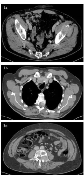
A 64 years-old Caucasian man without personal or familial history of cancer consulted in October 2017 for a 1-month episode of headaches. A brain MRI revealed a right hemisphere extra-axial dura-based tumor of the frontal lobe. The tumor was isointense on T1-Weighted Imaging (T1-WI) and T2-Weighted Imaging (T2-WI), surrounded by vasogenic oedema on T2 FLAIR imaging and strongly enhanced after gadolinium injection. Brain imagery was compatible with a meningioma. The tumor was resected in November 2017 and a histopathological examination concluded to grade-III “HPC of the CNS”. Post-operative MRI showed a residual contrast-enhancement of the operative cavity, which suggested Subtotal Resection (STR). The patient underwent a second brain surgical intervention in April 2018 to achieve Gross Total Resection (GTR), which confirmed residual grade-III HPC with a high mitotic count (23 mitoses per 10 hpf) and intra-tumoral necrosis. In May 2018, adjuvant Stereotactic Radiation Therapy (SRT) of the operative cavity (60 Gy, 30 fractions of 2 Gy) was administered. In June 2018, the patient experienced a distant failure with multiple osteolytic bone lesions (Figure 1, Figure 2).
Figure 1: June 2018 CT-scan in axial section showing multiple
metastatic recurrences. 1a. Massive tumoral infiltration of the left ilio-psoas and minimus gluteal muscle with bilateral osteolytic crest iliac lesions. 1b. Left T5 epiduritis and stenosis of the medullary canal. 1c. Left L4 epiduritis and stenosis of the medullary canal.
Figure 2: June 2018 MRI in sagittal section confirming epiduritis
caused by metastatic recurrences. 2a. MRI with gadolinium injection showing a hypersignal t1 in T5, revealing a stenosis of the medullary canal. 2b. MRI with gadolinium injection showing a hypersignal t1 in L4, revealing a stenosis of the medullary canal.
Citation: de Bernardi A, Bernadach M, Ginzac A, Mishellany F, Herrmann T, et al. (2020) Mutation of Tp53 in a Patient with Aggressive Central Nervous System Solitary
Fibrous Tumor/Hemangiopericytoma. Ann Case Report 14: 375. DOI: 10.29011/2574-7754/100375
A whole-spine MRI showed a metastatic infiltration of T6 and a left-handstenosis of the spinal canal with spinal cord ischemia (Figure 2a, Figure 2b). Decompressive radiation therapies were performed. A CT-guided biopsy of the left-hand iliac crest lesion was carried out in July 2018. The metastasis was highly cellular with short fusiform cells surrounded by a “Hemangiopericytoma-like” vasculature, a Ki-67 of 40% and a lower mitotic count (4 per 10 hpf). Immunohistochemistry (IHC) staining showed a loss of CD34 expression (Figure A), a heterogeneous expression of p53 (Figure C) and strongly positive STAT6 and p16 expressions (Figure B, Figure D). An NGS assay (FoundationOne CDxTM test)
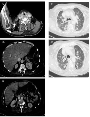
tested a panel of mutations in more than 300 cancer-related genes. A mutation of TP53 (R175H) was detected in the tumor DNA, the microsatellite status (MS) was stable and the Tumor Mutational Burden (TMB) was low (1 Muts/Mb). In September 2018, the patient presented with a left hemiplegia and left upper limb paraesthesia. The CT-scan showed diffuse metastatic progression (Figure 3) with liver metastases (Figure 3b, Figure 3c), bilateral lung metastases (Figure 3d, Figure 3e) and bone lesions increased in size and number. Palliative chemotherapy with dose-dense Paclitaxel (80 mg/m2) and Gemcitabine (800 mg/m2) was started in
October 2018 but discontinued after 2 infusions because of rapid neurological decline. The patient died in November 2018.
Figure A: Negative CD34 staining (x10).
Figure B: Strong p16 cytoplasmic staining (x20).
Figure C: Heterogeneous p53 nuclear staining (x20).
Figure D: Strong STAT6 nuclear staining (x10).
Figure 3: September 2018 CT-scan in axial section revealing global
progression with new osteolytic and visceral lesions. 3a. Massive osteolytic tissue lesion infiltrating vertebral body of C4, with global left vertebral artery entrapment. 3b, c. CT-scan in the portal phase revealing
Discussion
CNS SFT / HPC is more aggressive than other SFTs and has a tendency to recur and to metastasize to distant sites with a 60% local recurrence rate after GTR and extra-cranial metastases in 20% of cases [5,20]. The median time to the development of extra-cranial metastases was 7.5 years in the HPC case series (n=38) published by Ecker et al. [21,22]. A cohort of HPCs (n = 39) published by Schiariti et al. reported a mean overall survival (OS) of 18 years [23]. In this case report, both the time to distant failure (6 months) and OS (13 months) were much shorter than expected.
The management of CNS SFT / HPC is complex and there are currently no therapeutic guidelines. When the disease is localized, the standard care is GTR [20]. Single-institution studies reported an independent role for adjuvant radiation therapy in improving local control, disease-free survival, and OS [20,23-25]. According to the largest cohort of patients with HPC (n=981) treated either with surgery and radiation therapy (47,8%) or surgery alone (37,2%), adjuvant or primary radiation therapy was not associated with improved OS [12]. Stereotactic radio-surgery (SRS) can relieve patients from disabling neurological symptoms by improving local control [26-28]. For patients with recurrent HPC, Chamberlain et al. assessed the sequential administration of anthracyclin-based chemotherapies. Patients (n=6) treated with CAV (Cyclophosphamide, Doxorubicin, Vincristine) had an improved median response duration from onset (4 months) which doubled (8 months) among patients (n=9) additionally treated with α-IFN (α-interferon). Patients (n=5) that received ICE (Ifosfamide, Cisplatin, Etoposide) progressed within 1 or 2 months. The median OS of patients treated with CAV, α-IFN and ICE was significantly longer (20 months) than patients among (n = 6) treated with CAV alone (9.5 months) [29]. Anti-angiogenic therapies show promising extra-cranial response in recurrent CNS SFT / HPC [30,31]. In a retrospective cohort of patients (n=14) with recurrent CNS SFT / HPC treated with Temozolomide (150 mg/m2) and
Bevacizumab (5 mg/kg), the Overall Response Rate (ORR) and the median OS rose to 79% and 24.3 months respectively [32]. More recently, Apra et al. reported good radiological response to Pazopanib among patients (n=2) with recurrent CNS SFT / HPC with respective decreases of 84% and 43% after 3 months at doses of 800 and 600 mg [33].
Regarding tumor profile, pathological patterns in high-grade sarcoma, such as high mitotic rates and necrosis, were found at the time of relapse. These are independent patterns predictive of aggressiveness in CNS SFT / HPC [21,34]. It is noteworthy that the metastatic lesion did not correspond to grade-III CNS SFT / HPC according to the 2016 WHO classification (4 mitoses per 10 hpf). In addition, the Ki-67 index was higher (40%) than the reported median range in HPC (4%) [6]. These atypical histopathological
features could reflect an evolution similar to dedifferentiated extra-CNS SFTs described by Mosquera and Fletcher in 2009, with an abrupt transition from a well-differentiated SFT into a high-grade tumor with anaplastic areas [35-38]. The loss of CD34 expression at recurrence has also been described in dedifferentiated SFT [39] and in higher-grade tumors [4,40,41]. Even if the WHO grade remains the main prognostic factor in CNS SFT / HPC, histopathology alone is not sufficient to consistently classify tumors and predict their evolution. At molecular level, the NAB2-STAT6 fusion oncogene is a specific cytogenetic signature associated with soft tissue SFT [16-18].
The presence of a NAB2-STAT6 fusion oncogene in the tumor confirms the diagnosis of CNS SFT / HPC, and differentiates it from other intracranial bone or soft tissue tumors. The NAB2-STAT6 fusion protein acts as a nuclear transcriptional regulator of STAT6-dependent genes involved in cancer cell proliferation [16]. The downstream aberrant accumulation of STAT6 in tumor cell nuclei makes it detectable by IHC, which is a surrogate marker of NAB2-STAT6 fusion [13,19,42]. In addition to the diagnostic value of NAB2-STAT6, Barthelmeß et al. reported two NAB2-STAT6 fusion subtypes associated with contrasting clinical characteristics [43]. The NAB2ex6-STAT6ex16/17 fusion variant was found in indolent SFT whereas the NAB2ex4-STAT6ex2/3 fusion gene was predominantly detected in aggressive phenotypes closer to HPC [43] In this case report, the FoundationOne CDxTM test performed on
the bone metastasis showed a mutation of TP53. TP53 (13.1p17q) is a tumor suppressor gene coding for p53, a protein involved in the regulation of cell proliferation, differentiation and apoptosis. A loss of heterozygosity in TP53 is an oncogenic driver in many cancer types. In primary tumors of the CNS, the overexpression of p53 detected by IHC nuclear staining is a good surrogate marker for TP53 mutations [44]. For CNS SFT / HPC, the series reported by Oghaki et al. included patients with various types of neoplasms of the CNS among which seven were HPC [45]. A miscoding point mutation of TP53 was detected in one 49 year-old man out of seven cases of HPC (14%) (codon 238; TGl-TlT, Cys-Phe).
The mutation affected a highly preserved region (exon 7) of TP53 corresponding to the central part of p53 that determines the conformation of the protein. Because of the low incidence of TP53 mutations observed in HPC, the authors concluded that TP53 may not play a crucial role in the development of HPC. In 1995, Wang et al. published a series of 30 patients with meningeal tumors that included 3 cases of HPC (a 63-year-old woman, a 40-year-old woman and a 49-year-old man) [46]. Intermediate IHC p53nuclear staining was performed (++, 5-10%) but a PCR of exons 5 to 8 did not detect TP53 mutations. In 2002, Verheijen et al published a case series of meningeal tumors (n=17) [47]. One patient had HPC with 2 single nucleotide polymorphisms on exon 4 (codon 36 and 72) of TP5347. The molecular and IHC features were not correlated
Citation: de Bernardi A, Bernadach M, Ginzac A, Mishellany F, Herrmann T, et al. (2020) Mutation of Tp53 in a Patient with Aggressive Central Nervous System Solitary
Fibrous Tumor/Hemangiopericytoma. Ann Case Report 14: 375. DOI: 10.29011/2574-7754/100375
Conclusion
To conclude, the morphological spectrum of CNS SFT / HPC is broad, and there is still controversy about whether SFT and HPC are distinct entities at opposite ends of the same disease spectrum or different pathologies that share overlapping features. The diagnosis of CNS SFT / HPC has been simplified by the detection of a specific NAB2-STAT6 cytogenetic signature. However, STAT6 nuclear staining is not predictive, and the genetic screening of fusion gene variants such as NAB2ex4-STAT6ex2/3 has not yet been adopted in routine practice. Despite recent efforts by the WHO to produce a uniform classification of CNS SFT and HPC, this case report illustrates the temporal tumor heterogeneity of CNS SFT / HPC and highlights the limits of anatomo-pathology to classify these tumors and predict their aggressiveness. In this case report, the mutation of TP53 (R175H) detected with using the FoundationOne CDxTM test was associated with a rapidly
progressive clinical state. The overall survival was one of the shortest described in the literature. This report is insufficient to conclude on the predictive value of TP53 mutations in CNS SFT / HPC. Further studies including molecular profiling of a larger number of cases are warranted to provide a framework for CNS SFT / HPC prognostic assessment in order to intensify adjuvant therapies among high-risk patients and to identify new therapeutic targets.
Acknowledgements
None Conflict of Interest None to declareReferences
Klemperer P, Rabin CB (1992) Primary Neoplasms of the pleura. A 1.
report of five cases. Am J Ind Med 22: 4-31.
DERVAN PA, TOBIN B, O’CONNOR M (1986) Solitary (localized) fi-2.
brous mesothelioma: evidence against mesothelial cell origin. Histo-pathology 10: 867-875.
Furukawa N, Hansky B, Niedermeyer J, Gummert J, Renner AA (2011) 3.
silent gigantic solitary fibrous tumor of the pleura: case report. J Car-diothorac Surg 6: 122.
Yokoi, Tsuzuki, Yatabe, Suzuki, Kurumaya, et al. (1998) Solitary fi-4.
brous tumour: significance of p53 and CD34 immunoreactivity in its malignant transformation. Histopathology 32: 423-432.
Tihan T, Viglione M, Rosenblum MK, Olivi A, Burger PC (2003) Soli-5.
tary Fibrous Tumors in the Central Nervous System. Arch Pathol Lab Med 127: 432-439.
Fargen KM, Opalach KJ, Wakefield D, Jacob RP, Yachnis AT, et al. 6.
(2011) The central nervous system solitary fibrous tumor: A review
Bisceglia, M, Galliani C, Giannatempo G, Lauriola W, Bianco M, et 7.
al. (2011) Solitary Fibrous Tumor of the Central Nervous System: A 15-year Literature Survey of 220 Cases (August 1996-July 2011). Adv Anat Pathol 18.
Carneiro SS, Scheithauer BW, Nascimento AG, Hirose T, Davis DH 8.
(1996) Solitary Fibrous Tumor of the Meninges: A Lesion Distinct from Fibrous Meningioma: A Clinicopathologic and Immunohistochemical Study Am J Clin Pathol 106: 217-224.
Stout AP, Murray MR (1942) HEMANGIOPERICYTOMA A VASCULAR 9.
TUMOR FEATURING ZIMMERMANNʼS PERICYTES: Ann Surg 116: 26-33.
Dufour H, Métellus P, Fuentes S, Murracciole X, Régis J, et al. (2001) 10.
Meningeal Hemangiopericytoma: A Retrospective Study of 21 Patients with Special Review of Postoperative External Radiotherapy. Neuro-surgery 48: 756-763.
Guthrie BL, Ebersold MJ, Scheithauer BW, Shaw EG (1989) Meninge-11.
al Hemangiopericytoma: Histopathological Features, Treatment, and Long-Term Follow-up of 44 Cases: Neurosurgery 25: 514-522. Trifiletti DM, Mehta GU, Grover S, Sheehan JP (2017) Clinical man-12.
agement and survival of patients with central nervous system heman-giopericytoma in the National Cancer Database. J Clin Neurosci 44: 169-174.
Doyle LA (2014) Sarcoma classification: An update based on the 2013 13.
World Health Organization Classification of Tumors of Soft Tissue and Bone: WHO Update of Sarcoma Classification. Cancer 120: 1763-1774.
Fletcher CDM (1994) Haemangiopericytoma - A dying breed? Reap-14.
praisal of an ‘entity’ and its variants: a hypothesis. Curr Diagn Pathol 1: 19-23.
Gengler C, Guillou L (2006) Solitary fibrous tumour and haemangio-15.
pericytoma: evolution of a concept. Histopathology 48: 63-74. Chmielecki J, Crago AM, Rosenberg M, O’Connor R, Walker SR, 16.
et al. (2013) Whole-exome sequencing identifies a recurrent NAB2-STAT6 fusion in solitary fibrous tumors. Nat Genet 45: 131-132. Levine DA (2013) Integrated genomic characterization of endometrial 17.
carcinoma. Nature 497: 67.
Mohajeri A, Tayebwa J, Collin A, Nilsson J, Magnusson L, et al. 18.
(2013) Comprehensive genetic analysis identifies a pathognomon-ic NAB2/STAT6 fusion gene, nonrandom secondary genompathognomon-ic imbal-ances, and a characteristic gene expression profile in solitary fibrous tumor: NAB2/STAT6 Fusion Gene in Solitary Fibrous Tumor. Genes Chromosomes Cancer 52: 873-886.
Louis DN, Perry A, Reifenberger G, Deimling AV, et al. (2016) The 2016 19.
World Health Organization Classification of Tumors of the Central Ner-vous System: a summary. Acta Neuropathol. (Berl) 131: 803-820. Rutkowski MJ, Jian BJ, Bloch O, Chen C, Sughrue ME, et al. (2012) 20.
Intracranial hemangiopericytoma: Clinical experience and treatment considerations in a modern series of 40 adult patients. Cancer 118: 1628-1636.
Mena H, Ribas JL, Pezeshkpour GH, Cowan DN, Parisi JE (1991) 21.
Hemangiopericytoma of the central nervous system: A review of 94 cases. Hum Pathol 22: 84-91.
Ecker RD, Marsh WR, Pollock BE, Kurtkaya-Yapicier O, McClelland R, 22.
Schiariti M, Goetz P, El-Maghraby H, Tailor J, Kitchen N (2011) Heman-23.
giopericytoma: long-term outcome revisited. J Neurosurg JNS 114: 747-755.
Soyuer S, Chang EL, Selek U, McCutcheon IE, Maor MH (2004) 24.
Intracranial meningeal hemangiopericytoma: The role of radiothera-py. Cancer 100: 1491-1497.
Kano H, Niranjan A, Kondziolka D, Flickinger J, Lunsford LD (2008) 25.
Adjuvant Stereotactic Radiosurgery After Resection of Intracranial He-mangiopericytomas. Int J Radiat Oncol Biol Phys 72: 1333-1339. Ghia AJ, Allen PK, Mahajan A, Penas-Prado M, McCutcheon IE, et 26.
al. (2012) Intracranial Hemangiopericytoma and the Role of Radiation Therapy a Population Based Analysis. Neurosurgery 72: 203-209. Ghia AJ, Chang EL, Allen PK, Majaran A, Penas-Prado M, et al. 27.
(2013) Intracranial Hemangiopericytoma: Patterns of Failure and the Role of Radiation Therapy. Neurosurgery 73: 624-631.
Cohen-Inbar O, Lee CC, Mousavi SH, Kano H, Mathieu D, et al. Stere-28.
otactic radiosurgery for intracranial hemangiopericytomas: a multicent-er study. J Neurosurg JNS 126: 744-754.
Chamberlain MC, Glantz MJ (2008) SEQUENTIAL SALVAGE CHEM-29.
OTHERAPY FOR RECURRENT INTRACRANIAL HEMANGIOPERI-CYTOMA. Neurosurgery 63: 720-727.
Park MS, Araujo DM (2009) New insights into the hemangiopericy-30.
toma/solitary fibrous tumor spectrum of tumors. Curr Opin Oncol 21: 327-331.
Ebata T, Shimoi T, Bun S, Miyake M, Yoshida A, et al. (2018) Efficacy 31.
and Safety of Pazopanib for Recurrent or Metastatic Solitary Fibrous Tumor. Oncology 94: 340-344.
Park MS, Patel SR, Ludwig JA, Trent JC, Conrad CA, et al. (2011) Ac-32.
tivity of temozolomide and bevacizumab in the treatment of locally ad-vanced, recurrent, and metastatic hemangiopericytoma and malignant solitary fibrous tumor: Temozolomide/Bevacizumab Therapy in HPC/ SFT. Cancer 117: 4939-4947.
Apra C, Alentorn A, Mokhtari K, Kalamarides M, Sanson M (2018) Pa-33.
zopanib efficacy in recurrent central nervous system hemangiopericy-tomas. J Neurooncol 139: 369-372.
Louis DN, Ohgaki H, Wiestler OD, Cavenee WK, Burger PC, et al. 34.
(2007) The 2007 WHO classification of tumours of the central nervous system. Acta Neuropathol. (Berl) 114: 97-109.
Evans H (2007) Atypical lipomatous tumor, its variants, and its com-35.
bined forms: A study of 61 cases, with a minimum follow-up of 10 years. Am J Surg Pathol 31: 1-14.
Evans HL (1979) Liposarcoma A study of 55 cases with a reassess-36.
ment of its classification. Am. J. Surg. Pathol 3: 507-523.
Henricks WH, Chu YC, Goldblum JR, Weiss SW (1997) Dedifferenti-37.
ated Liposarcoma: A Clinicopathological Analysis of 155 Cases with a Proposal for an Expanded Definition of Dedifferentiation. Am J Surg Pathol 21: 271-281.
McCormick D, Mentzel T, Beham A, Fletcher CDM (1994) Dedifferenti-38.
ated Liposarcoma Clinicopathologic Analysis of 32 Cases Suggesting a Better Prognostic Subgroup Among Pleomorphic Sarcomas Am J Surg Pathol 18: 1213-1223.
Mosquera JM, Fletcher CD (2009) Expanding the Spectrum of Malig-39.
nant Progression in Solitary Fibrous Tumors: A Study of 8 Cases with a Discrete Anaplastic Component-Is This Dedifferentiated SFT? Am. J Surg Pathol 33: 1314-1321.
Fritchie KJ, Jin L, Rubin BP, Burger PC, Jenkins SM, Barthelmeß S, et 40.
al. (2016) NAB2-STAT6 Gene Fusion in Meningeal Hemangiopericy-toma and Solitary Fibrous Tumor. J Neuropathol Exp Neurol 75: 263-271.
Bertero, L, Anfossi V, Osella-Abate S, Disanto MG, Mantovani C, et 41.
al. (2018) Pathological prognostic markers in central nervous system solitary fibrous tumour/hemangiopericytoma: Evidence from a small series. PloS One 13: e0203570-e0203570.
Schweizer L, Koelsche C, Sahm F, Piro R, Capper D, et al. (2013) 42.
Meningeal hemangiopericytoma and solitary fibrous tumors carry the NAB2-STAT6 fusion and can be diagnosed by nuclear expression of STAT6 protein. Acta Neuropathol (Berl) 125: 651-658 (2013). Barthelmeß S, Geddert H, Boltze C, Moskalev EA, et al. (2014) Solitary 43.
Fibrous Tumors/Hemangiopericytomas with Different Variants of the NAB2-STAT6 Gene Fusion Are Characterized by Specific Histomor-phology and Distinct Clinicopathological Features. Am J Pathol 184: 1209-1218.
Fulci G, Van Meir EG (1999) p53 and the CNS. Mol
44. Neurobiol 19 :
61-77.
Ohgaki H, Eibl RH, Schwab M, Reichel MB, Mariani L, et al. (1993)
45.
Mu-tations of the p53 tumor suppressor gene in neoplasms of the human nervous system. Mol Carcinog 8: 74-80.
Wang JL, Zhang ZJ, Harman M, Smits A, Westermark B, et al. (1995) 46.
Detection ofTP53 gene mutation in human meningiomas: A study us-ing immunohistochemistry, polymerase chain reaction/sus-ingle-strand conformation polymorphism and dna sequencing techniques on paraf-fin-embedded samples. Int J Cancer 64: 223-228.
Verheijen FM, Sprong M, Kloosterman JME, Blaauw G, Thijssen JHH, 47.
et al. (2002) TP53 Mutations in Human Meningiomas. Int J Biol Mark-ers 17: 42-48.