HAL Id: hal-03026551
https://hal.archives-ouvertes.fr/hal-03026551
Submitted on 26 Nov 2020
HAL is a multi-disciplinary open access
archive for the deposit and dissemination of
sci-entific research documents, whether they are
pub-lished or not. The documents may come from
teaching and research institutions in France or
abroad, or from public or private research centers.
L’archive ouverte pluridisciplinaire HAL, est
destinée au dépôt et à la diffusion de documents
scientifiques de niveau recherche, publiés ou non,
émanant des établissements d’enseignement et de
recherche français ou étrangers, des laboratoires
publics ou privés.
Distributed under a Creative Commons Attribution - NonCommercial - ShareAlike| 4.0
In Situ Optical Monitoring of the Electrochemical
Conversion of Dielectric Nanoparticles: From Multistep
Charge Injection to Nanoparticle Motion
Jean-François Lemineur, Jean-Marc Noël, Alexa Courty, Dominique Ausserré,
Catherine Combellas, Frédéric Kanoufi
To cite this version:
Jean-François Lemineur, Jean-Marc Noël, Alexa Courty, Dominique Ausserré, Catherine Combellas,
et al.. In Situ Optical Monitoring of the Electrochemical Conversion of Dielectric Nanoparticles:
From Multistep Charge Injection to Nanoparticle Motion. Journal of the American Chemical Society,
American Chemical Society, 2020, 142 (17), pp.7937-7946. �10.1021/jacs.0c02071�. �hal-03026551�
In Situ Optical Monitoring of the Electrochemical Conversion of
Die-lectric Nanoparticles: from Multi-Step Charge Injection to
Nanopar-ticle Motion
Jean-François Lemineur,
1,2Jean-Marc Noël,
1Alexa Courty,
2Dominique Ausserré,
3Catherine
Combel-las,
1Frédéric Kanoufi
1,*
1 Université de Paris, ITODYS, CNRS-UMR 7086, 15 rue Jean-Antoine de Baïf, 75013 Paris, France. 2 Sorbonne Université, MONARIS, CNRS-UMR 8233, 4 Place Jussieu, 75005 Paris, France.
3 Université du Maine, Institut des Matériaux et Molécules du Mans, CNRS-UMR 6283, Avenue O. Messiaen, 72000 Le Mans, France.
ABSTRACT: By shortening solid-state diffusion times, the nanoscale size reduction of dielectric materials -such as ionic crystals-
has fueled synthetic efforts towards their use as nanoparticles, NPs, in electrochemical storage and conversion cells. Meanwhile, there is a lack of strategies able to image the dynamics of such conversion, operando and at the single NP level. It is achieved here by optical microscopy for a model dielectric ionic nanocrystal, a silver halide NP. Rather than the classical core-shrinking mecha-nism often used to rationalize the complete electrochemical conversion and charge storage in NPs, an alternative mechamecha-nism is proposed here. Owing to its poor conductivity, the NP conversion proceeds to completion through the formation of multiple inclu-sions. The super-localization of NP during such heterogeneous multiple-step conversion suggests the local release of ions, which propels the NP towards reacting sites enabling its full conversion.
INTRODUCTION
Nanoparticles, NPs, are key components in many electro-chemical devices and applications related to energy conver-sion or storage,1 such as photovoltaics,2 batteries,3 fuel cells or
(photo)electrolyzers for the electrocatalytic activation of small molecules (from water splitting to CO2 reduction).4
Ionic crystals, metal oxides or polymers, which are ditrics in the visible range, have long been discarded from elec-trochemical cells owing to their poor electronic and/or ionic conductivities. However, these materials have also benefited from nanoscale size reduction, which predominantly shortens solid-state ion diffusion times, allowing charge transfer.5 In
batteries, this strategy is used to reach ultimate charge storage capacity, as illustrated from the promises of LiFePO4
nano-crystals as a cathode material.6 Such opportunity fuels
synthet-ic efforts towards new dielectrsynthet-ic nanomaterials able to meet the needs of electrochemical storage or conversion cells with higher capacity conversion and faster rates.
The performances of nanomaterials in electrochemical cells are usually probed within an ensemble, electrically connected through additives to a current collector. However, a rational consideration of the intrinsic performances of NPs for energy storage and conversion applications requires probing and imaging intrinsic electrochemical activity or conversion, at the single NP level, particularly under operating conditions. Dif-ferent single NP electroanalytical strategies have been pro-posed,7,8 although rarely for the electrochemical study of
die-lectric materials.5,9-13 A strategy consists in catching the
elec-trochemical activity of a polarized ultramicroelectrode (UME,
a microsized current collector) during the stochastic collision of dispersed colloidal NPs.9,14,15 The current transient recorded
for each stochastic event reflects either the adsorption dynam-ics of inactive NPs,9 or the storage capacity associated to the
electrochemical conversion of electroactive NPs.10 However,
for colloids in solution, the current transients are convoluted to their intrinsic Brownian motion, resulting in multiple discrete sub-events difficult to interpret -if not- by the partial transfor-mation and discrete residence of the NP onto the UME.16,17
Although insightful, such situation drastically differs from the behavior of NPs immobilized on or confined at an elec-trode for which electrochemical imaging strategies are more appropriate. Scanning electrochemical nanoprobes (nanoelec-trodes or nano-electrochemical cells) are able to produce high resolution mapping of individual nanomaterials18 or to address
the charge storage capacity of battery material by inspecting transient (de)intercalation processes at the single particle lev-el.11,12 These processes are often associated to longer
charac-teristic times which are delicate to track from current transi-ents, unless by confining the event in a microreactor (such as emulsion droplets)19 or a sub-micrometric electrochemical
cell.11 Moreover, these intrinsic electrochemical activities are
often associated to structural dynamic changes, such as defor-mation, volume expansion, dissolution, etc…, difficult to visualize without a complementary microscopic observation. Apart from AFM imaging,20 or in situ TEM,21 often restrictive
owing to the slowness of the scanning imaging process or to e-beam dose perturbations, high resolution optical microscopies offer high throughput, quantitative and dynamic analysis of single NPs electrochemistry.22 They afford complementary in
chemical transformations (double layer charging,23-25 NP
con-version,13,22,26-33 nanobubbles electrogeneration34) to
mo-tion,26c,27 or local polarization due to structural changes24,25,32,33
associated to the electrochemical actuation of NPs. Although powerful these strategies have been mostly applied to plas-monic nanomaterials and more rarely employed to depict the electrochemistry of dielectric NPs.13
Herein, the dynamics of the charge storage by electrochemi-cal conversion of individual surface-confined ionic nanocrys-tals is monitored optically. Ionic nanocrysnanocrys-tals are poor elec-tronic conductors, and their electrochemical conversion, into a more conducting material, can be a source of irreversibility during the cycling process. Indeed, as illustrated in Figure 1, if conducting NPs (or NPs capped with a conducting binder) are expected to convert through a spherically symmetric core-shrinking mechanism, dielectric NPs will follow a strongly anisotropic conversion route: the conversion would start from the contact with a current collector through an inclusion for-mation. This is devised from a simulation study in the first part of this work, showing that, from an electrochemical point of view, if both situations may present different simulated char-acteristic transformation rates, they are difficult to differentiate experimentally. However, based on the differing symmetry of each transformation scenario, the intermediates involved, and therefore the conversion mechanism, should be discriminated from their optical response. To decipher such mechanistic scenarii, it is pertinent monitoring in real time and in situ, by high resolution optical microscopy, the electrochemical con-version of dielectric NPs. This relies on the choice of both a model dielectric NP and a highly sensitive label free optical microscopy able to image in situ these dielectric NPs. The latter is afforded by interferometric-based techniques.29b,c,35
The Backside Absorbing Layer Microscopy (BALM)29b,c is
chosen as it uses ultrathin Au coated glass sample surfaces, which are, as presented in Figure 1, both an optical and elec-trochemical sensor of NPs (experimental details given in Sup-porting Information, SI, Section SI1). As for the former model dielectric material, a colloidal silver halide, AgX nanocrystal (here either X = Cl or Br, see SI, Section SI1), was chosen because both AgX and its reduction product, Ag, have well documented optical properties,36,37 which are more appropriate
to devise, from optical response prediction, the different sce-narii occurring during the solid-state conversion:
AgX + eˉ
→
Ag + Xˉ (1)The understanding of the solid-state conversion of silver-ion based dielectric nanocrystals is of interest for energy conver-sion or storage.38 The partially converted AgX/Ag hybrid NPs
are attracting photocatalysts of CO2 reduction. Moreover Ag2O
NPs analogues are commonly used in Zn/Ag high density and high power batteries. The strategy developed herein will di-rectly apply to apprehend such systems, and should be extend-ed, in future works, to less optically explored dielectric NPs.
This work proposes to unravel optically the storage of charge associated to the conversion of AgX into metallic Ag by investigating if it proceeds by a spherical symmetric, radial core-shrinking, or through the formation of inclusions. By tracking the NP optical response and super-localizing its posi-tion during its electrochemical conversion at the BALM elec-trode, we are able to decipher, operando, the dynamics of the solid-state transformation of individual dielectric NPs. The NP conversion is also associated with the production of local ion
fluxes, which are probed from the motion of the NP during its conversion.
Figure 1. Monitoring the dynamics of the electrochemical
version of individual dielectric NPs. The different ways of con-version of a model dielectric material, here AgX which converts into metallic Ag, by radial core-shrinking or formation of hetero-geneous inclusions.
RESULTS AND DISCUSSION
Modelling Single NP Electrochemical Conversion Mechanims. The dynamics of the electrochemical conversion
of a single NP contacting a planar electrode was simulated as a diffusion reaction process described in SI, section SI2, by finite element method (COMSOL®) in Figure 2. Two cases, usually encountered during the conversion of battery-active NPs,39 are considered (Figures 1 and 2).
In the first one (the “radial core-shrinking model”), the outer region of the AgX NP is reduced into metallic Ag through a shell formation while the AgX core shrinks with time (Figure 2a). This corresponds to the case of a perfectly conducting NP whose external surface is equipotential, usually depicted in batteries owing to the presence of conductive binder coating the nanostructured electroactive materials.
The second one relies on the anisotropic growth of an Ag “inclusion” at the NP-electrode contact (Figure 2b) producing a Janus type NP.
Figure 2a and b presents the simulated AgX concentration (black region), and equivalently Ag concentration (white re-gion), profiles in the NP at different conversion times accord-ing to either the core-shrinkaccord-ing or the saccord-ingle-inclusion Janus model. As expected, each mechanism yields intermediates that present either a spherical or an axial symmetry. The different mechanistic situations were then compared based on their respective characteristic conversion time, namely the time needed to convert half the NP material. As sketched in Figure 2, from geometric consideration, the half-conversion, ≈50%, of a NP of diameter d0 corresponds to transport through either a shell of thickness l1/2,CS≈0.15xd0 in the core-shrinking model or l1/2,J≈0.8xd0 for the inclusion formation.
In silver-based crystals thin films, despite great variability in values, the diffusion of Ag+ is mostly limiting, with D
val-ues at maximum in the 10-11cm2/s,40 comparatively to halide
ions, which diffuses about 10 times more slowly than in solu-tion.41 For a purely diffusion limited process, with
concentra-tion profiles given in SI, Figure SI2.1, the core-shrinking conversion half-time, 1/2=l1/22/D would take about 1/2,CS≈0.2s
and would be faster by more than 20-fold than that implying the formation of a single inclusion (1/2,J ≈6s).
As seen in Figure 2, the model, described in SI, Section SI2, also takes into account a propagating interface within the NP between the unconverted and converted materials and the extent of ion expulsion in solution. For this purpose, the model considers not only the diffusion of ions in the NP but also the occurrence of electron hopping. Electron hopping, often sug-gested for explaining conduction in polymer electrolytes and other percolated systems,42 allows accounting for the
produc-tion of a conducting material, which modifies the contact-line between unconverted and converted materials within the NP volume. It is described by a homogeneous chemical reaction between the converted and unconverted materials within the NP, characterized by a rate kex.
Figure 2. Comsol® simulation of the conversion dynamics of a
100nm AgCl NP (half of it is represented) contacting an electrode (lower plane) according to (a) the core-shrinking mechanism or (b) the formation of a single-inclusion Janus NP. Examples of successive concentration profiles (time in ms and conversion, , given) of AgCl in the NP and Cl- ions expelled in the surrounding
solution during the conversion. Simulations were obtained for solid phase diffusion D= 10-11 cm2/s and a characteristic rate for
electron hopping kex=300s-1.
The electron hopping produces much faster conversion than if only the solid-state diffusion limitation was considered,43
and it therefore particularly accelerates the single inclusion conversion. Indeed, as shown in Figure 2, implementing the interface propagation reduces the conversion half-times for both mechanisms to the sub-100ms range (1/2,CS=14ms and 1/2,J=60ms for the core-shell and single-inclusion mechanisms respectively with kex=300s-1). A perfect charge percolation
would engage a hopping rate limited by physical diffusion within adjacent centers, separated by a=3.5Å, the interatomic distance, for example, in AgCl. This upper rate is given by Smoluchovsky theory as kex,lim=8DaNA/Vm,AgCl=1.4x105 s-1
with NA the Avogadro number and Vm,AgCl the molar volume of
AgCl crystal. Extrapolating the simulations of Figure 2 to such limit yields 1/2,CS=0.5 and 1/2,J=3ms for the fastest conversion half-times for each mechanism (see SI, section SI2).
Overall, except likely for the >1s long diffusion limited conversion in the absence of electron hopping, the half-time conversion values are within reach of state-of-the-art electro-chemical single entity studies. It should be interesting design-ing sdesign-ingle entity electrochemical studies to allow differentiat-ing all those mechanisms based on conversion current transi-ents or 1/2 values (see SI, section SI2). However, owing to the
strong variability in the solid state ionic diffusion coefficients, and to the similarities in simulated features (see SI, Section SI2), we believe it is currently difficult to differentiate both mechanistic routes from single NP electrochemical measure-ments. We then propose to recourse to a complementary opti-cal monitoring.
Optical Differentiation of AgX and Ag NPs. Colloidal
so-lutions containing a 3:2 mixture of d0 ~ 100 nm diameter CTAB capped AgCl and AgBr NPs were prepared38,44 as
de-scribed in SI (Figure S1.1 in Section SI1 in SI). The unimodal size distribution suggests AgCl and AgBr NPs cannot be dif-ferentiated by SEM images. The comparable values of AgCl and AgBr refractive indexes also suggest (see discussion in SI, Section SI1) they cannot be differentiated optically. All NPs detected optically were shown to present the same behaviors, suggesting that the processes described apply statistically equivalently to either AgCl or AgBr. Indeed, the conversion was studied here at sufficiently negative potential to ensure mass transfer limited conversion of either material, preventing also an electrochemical differentiation. Therefore, the NPs are denoted AgX throughout the manuscript.
To resolve the dynamics of the electrochemical conversion of individual AgX NPs, the latter should first be adsorbed on the Au BALM electrode. To favor the NP adsorption, the AgX NP solution was diluted 100 times in a NaCl solution (0.05M). It resulted in the destabilization of the colloidal solution, lead-ing to the irreversible adsorption of the NPs on the electrode surface.45
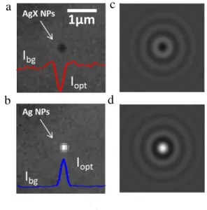
Figure 3. (a,b) BALM images of an adsorbed AgX NP and once
reduced into an Ag NP. (c,d) Simulated 4x4µm2 interferometric
optical images at =510nm of a 100nm AgX NP and a fully con-verted 73nm Ag NP.
An example of monitoring of NP adsorption/conversion is given in the Supplementary Video (SI) summarized in Figure 3a and b. The AgX NP adsorptive impact on the electrode is
5 19% 10 34% 15.9 52% 20 64% 28.2 83% 0.1 ms 1.8% Cl -(mM)AgCl(M) 3.5 39 0 0 10 1% 20 4% 39 19% 63.1 55% 79.4 82% 0.1 ms (mM)Cl- AgCl(M) 2 39 0 0
1µm
b
a
c
d
d0 l1/2 l1/2a
b
X- AgX (mM) (M) X- AgX (mM) (M)detected optically (Figure 3a) thanks to the appearance of a diffraction-limited dark contrast feature, Iopt–Ibg<0, where Iopt is the optical intensity of the contrast feature and Ibg the back-ground level. Even though the BALM electrode is biased at a sufficiently negative potential to allow, when the AgX NP hits the surface, its reduction into Ag, the NP spot, remains dark for ~4s before a change to a bright contrast feature is detected (Iopt–Ibg>0, Figure 3b).
Such optical contrasts are predicted from an optical model adapted from that developed by the Ünlü’s group,35c,46 using
the MNPBEM toolbox46 (details in SI, Section SI3), for
quan-titatively interpreting single NP images obtained by interfero-metric-based microscopies. Figure 2c and d presents the simu-lated optical interferometric images for two NPs adsorbed on an Au BALM electrode: a 100nm AgX NP and a 73nm Ag NP (considering the NP volume change upon full AgX to Ag conversion). The model predicts with a good quantitative agreement in Iopt–Ibg values that dielectric AgX NPs adsorbed on the optical sensor appear as dark contrast features while metallic Ag NPs appear as bright contrast features.
NP Conversion from Simulated Optical Responses.
Dif-ferentiating optically the fully unconverted and fully converted NPs is fruitful, the dynamics of the conversion may also be reached through such complementary monitoring. Owing to the difference in composition and symmetry, the AgX@Ag core-shell and the Janus-like Ag-inclusion in AgX intermedi-ate NPs are expected to present different electronic and there-fore optical properties. Differences in the localized surface plasmon resonance modes or their localization are expected. These differences should be unravelled from changes in scat-tering spectra,37 estimate of the absolute NP polarizability, for
example from complementary measurement of optical intensi-ty and phase,47 or super-localizing displacement of the
scatter-ing centroid.24,25,32 The potentiality of an interferometric
prob-ing is evaluated from the simulated optical responses, Iopt, of the scattering properties of single NPs at a BALM electrode, which are compared to experimental data. The mechanistic strategy proposed here would apply identically to other com-plementary or richer optical information (such as spectroscop-ic or phase and amplitude).
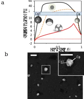
The Iopt response of AgX@Ag core-shell and of a Ag-inclusion in AgX NPs were simulated for different NP conver-sions (see Section SI3 in SI). The NP with Ag inclusion is modeled overall as a sphere, as depicted in Figure 4a, in which the Ag inclusion is the assembly of two identical sphere-caps facing the gold electrode. The volume of Ag material, either as an inclusion or as a shell, was estimated from .
The simulated variations of Iopt with for the core-shell or single inclusion Janus models are presented as the black and orange curves, respectively plain and dashed, in Figure 4a. Both models show that upon conversion the optical contrast,
Iopt-Ibg, of the NP is changed from dark (negative contrast) to bright (positive contrast) feature and with an overall increase in absolute value of the contrast. The core-shell model sug-gests a strong (>20x) contrast enhancement from the early beginning (>10%) of the NP conversion. If a strong contrast enhancement is also observed for the Janus model, it is reached more progressively and for larger values (>25%). As it will be discussed later on, the super-localization of the NP scattering centroid allows probing the NP displacement during its electrochemical transformation. The rotational mo-tion of the axisymmetric Janus NP is expected to affect its
optical response, Iopt, while not affecting that of the spherical symmetric core-shell NP (see discussion and simulated results for 90° or 180° rotated Janus NP in SI, Section SI3). Indeed, upon rotation, the Ag inclusion is pulled away from the Au electrode, lowering their coupling. Then a rotation of the Janus NP would tend to decrease Iopt. However such rotation also makes the overall situation more complex: once the inclusion is rotated, a new unconverted (AgX) part of the NP is now facing the electrode allowing the electrochemical formation of a new inclusion.
The formation of multiple inclusions is often suggested48 for
charge propagation in poorly conducting AgX materials. It invokes heterogeneous localized transformations similar to those observed during the reverse solid-state oxidation of Ag but into Ag oxides.32 It is supported from SEM inspection, in
Figure 4b, of AgX NPs adsorbed on the gold surface and sub-mitted to a 4s short reduction pulse. As seen clearly on the magnification of Figure 4b and confirmed for >90% NPs (n=30 NPs), different brighter regions (metallic inclusions) are present on the NPs. Interestingly, their location opposite to the electrode surface as in the magnification of Figure 4b, sug-gests that the NP has rotated during the electrochemical pro-cess.
Figure 4. Towards optical quantitative imaging. (a) The simulated
Iopt variations with the AgX NP conversion into Ag, assuming
the formation of a core-shell structure (black), or of a single sion Janus-type NP (dashed orange), or multiple mean-field inclu-sions (red); background level: dotted red line. (b) SEM images of AgX NPs adsorbed on the gold electrode surface after a 4s partial reduction: multiple Ag inclusions appear over the AgX NPs sug-gesting rotational motion of the NPs during the reduction (a 180° rotation for the large one shown in the magnified dashed region); scale bars = 200nm.
Simulating the optical response of such multiple inclusions situation is quite speculative without prior knowledge of the number and distribution of the mosaic of inclusions during the NP conversion. An alternative is to consider it in a mean field approach, which can be simulated by considering a plain com-posite Ag/AgCl NP characterized by an apparent refractive index estimated, as a first guess, from the effective medium
-2 0 2 4 6 8 10 12 14 16 18 0 0.5 1 o p ti ca le n h an cm en t conversion (d) 60 40 20
a
b
AgX Ag AgX Agtheory (see discussion in SI, Section SI3).49 The red line in
Figure 4a shows the resulting simulated Iopt variations with : the contrast enhancement increases more progressively and to a much lower maximum value (7x times at 85% conversion). It is further confirmed experimentally from the dynamic op-tical monitoring of the electrochemical conversion of individ-ual AgX NPs: the experimental Iopt and Ibg intensities were measured over the reduction time, and compared to the optical simulations. A typical example of Iopt transient is given in Figure 5, while other examples are given in SI, Section SI4.
First, the experimental intensities at the beginning, = 0, or end, = 1, of the process are in good agreement, compared to the background level (experimental: blue line, simulated: red dashed line) with the simulated one (red solid line). Then, the maximum optical contrast enhancement (approx. 6x at the plateau in Figure 5) never exceeded an 8x factor for n>20 NPs, which disqualifies the core-shell mechanism or single-inclusion models and supports the mechanism based on multi-ple silver inclusions.
Using the simulated Iopt– variations from the mosaic inclu-sion model, a quantitative assignment of the dynamics of the AgX NP conversion, (t), is at hand for interpreting the exper-imental Iopt transients. An illustration is given in Figure 5a (symbols) showing an overall almost complete conversion of the NP, with a first fast stage (up to 80% conversion) with approx. first order transformation rate of 0.8 s-1. Later on, the
full NP conversion is reached with a ten times slower kinetics.
Figure 5. Optical transient, Iopt–t, recorded during the reduction of
a single AgX NP adsorbed on a BALM electrode; experimental background in blue. Simulated optical contrast changes of the NPs (red lines) for = 0, AgX, and = 1, Ag, compared to the simu-lated background (dashed red line) and the conversion kinetics extracted from the Ag inclusion simulation ().
Resolving a Conversion in Multiple Steps. The
transfor-mation of the NPs adsorbed on the surface is close to comple-tion but is surprisingly slow (>3s). This value disqualifies the core-shrinking mechanism. It seems consistent with the simu-lated value of 1/2,J ≈6s for the diffusion limited growth of a single inclusion (see SI, Figures SI2.1 and SI2.2) with limited influence of electron hopping. However the distinct separation between the regions corresponding to either the Ag inclusion or the AgX material in the SEM image of Figure 4b rather supports the propagation of a front of converted material, in line with the electron hopping scenario. The observation in Figure 4b of multiple inclusions and their location far from the electrode contact, suggests the importance of the NPs/surface interaction and of the relative motion of the NP on the
elec-trode that result in the formation of multiple electrochemical steps during conversion.
Such multiple steps transformation is becoming a key fea-ture in single NP electrochemistry, revealing the complexity of NP motion during their electrochemistry. It was evidenced in electrochemical nanoimpact studies of the oxidation of Brownian Ag NPs.16,17 By increasing the experiment time
resolution, closely separated successive sub-ms current spikes were resolved corresponding to successive partial electrodisso-lution steps of a single NP.
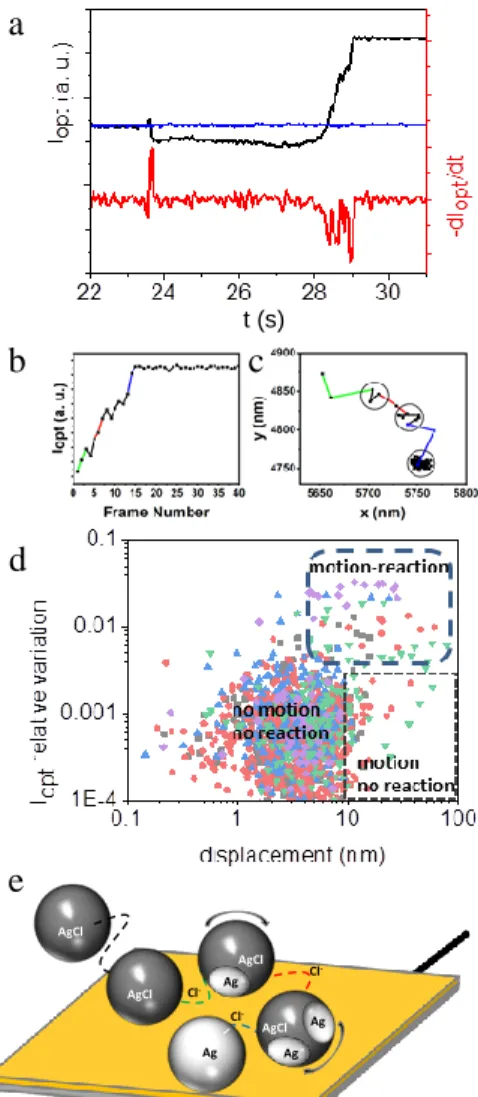
Figure 6. (a) Resolving multistep reductive conversion of surface
confined AgX NPs and derivative of the Iopt transient, showing the
discrete NP conversion (background in blue). (b) Zoom in the Iopt
transient presented in (a) during the time when the NP appears as bright contrast feature. (c) Motion of the NP tracked by super-localization and correlated to the intensity transient in (b). (d) Scatter plot representing the relative variation of Iopt and
dis-placement recorded during the conversion of 5 AgX NPs. The limit for reaction occurrence is 0.5% variation in Iopt; motion is
assigned to NP displacement >7nm. (e) Scheme of the multi-step path of a NP fueled by halide ion release.
Multiple step electrochemical process was also suggested for NPs adsorbed on electrodes and for slower processes such as those involved in corrosion or amorphous solid phase trans-formation. The dynamics of the slow transformation of indi-vidual adsorbed NPs is difficult to cope by direct electrochem-ical techniques. The slow electrochemelectrochem-ical conversion of redox active components inside >400nm attolitter emulsion droplets
Cl -Cl -X -X -X -Cl -Cl -AgCl AgCl AgCl Ag Ag Ag AgCl Ag AgX Ag
a
c
b
d
e
t (s)could be tracked by electrochemical current transients.19,43
However for the smallest converted material, comparable to that corresponding to the full conversion of a 100nm AgX NP (ca. 2pC), the electrochemical current sensitivity and baseline drift have not allowed depicting multi-step conversion. The use of confined electrochemical cell may allow increasing the electrochemical signal to noise ratio. An alternative consists in using a complementary signature of the conversion. The dy-namics of slow conversion processes is within reach of optical microscopy.26c,27 It was demonstrated during the dissolution of
silver pseudo-halides (thiocyanates) NPs28b or during the
oxi-dation of Ag into Ag2O.32
Increasing the optical signal-to-noise ratio, Iopt transients re-veal such dynamics, as in Figure 6a: a NP hits the polarized electrode at t=23.8s, then begins to react after ~4s delay time.
Iopt transients present a continuous increase >1s and further levelling showing a fast conversion of the NP but halting at ca. 80%, based on the optical simulation. The first time-derivative of Iopt (Figure 6a, red curve) mimics qualitatively the evolution of the electrochemical current that should be detected. It re-veals several close-packed optical spikes (between 28 and 29s), suggesting multiple partial reduction events. It is also clear that the stepwise reactions are not due to “one-by-one” reduction of individual NPs trapped in agglomerate. Indeed, agglomerates can be optically dismissed as they are character-ized by an abnormal intensity in comparison with those of single AgX NPs, which always possess similar intensities when hitting the electrode surface.
Identifying NP Motion during its Conversion. This
hy-pothesis was tested by scrutinizing the displacement of the scattering centroid of the surface-confined NP during its trans-formation. The position of the centroid of the NP (regardless of its contrast) was super-localized and tracked in two dimen-sions with sub-pixel accuracy by using the Crocker and Grier’s centroid finding algorithm (see Figure S5.1 in SI, Section SI5).50 The resulting mean square displacement before
and after the NP transformation is included in Figure S5.2. Before the contrast modification (from 23.8 to 28s, Figure 6a), the NP moves over the surface with a restricted diffusion coefficient, DNP = 5x10²nm²/s, 4 orders of magnitude lower than in solution. It cannot be ascribed to solution diffusion; indeed even when considering the transport dampening within the viscous layers near the electrode surface, the diffusion of a 100nm NPs is expected to be dampened by no more than 25 times.51 The hindered motion detected here rather shows the
surface diffusion of the NP, by rotation or translation. During this motion the NP should find a position allowing its conver-sion by charge transfer, resulting in variations in Iopt. At the end of the reaction (>29s), the NP is not diffusing anymore, within the tracking precision, ±7nm. It corroborates that NP transformation is associated to its motion near the electrode surface.26c
Closer examination of the motion of the surface-confined NP during the Iopt variation, which corresponds to its electro-chemical transformation, is also instructive. Figure 6b and c presents, respectively, the variation of the Iopt and NP position when the NP is detected as a bright contrast feature (higher signal to noise ratio).
During this period of time the centroid of the NP has moved by 160nm (it is by >200nm during its whole conversion, see SI, Figure S5.4 in Section SI5). The overall travelling distance larger than the NP dimension cannot be explained by the
prob-ing of a local polarization change of the NP, such as those expected during the formation of an asymmetric NP struc-ture.24,25,32 Indeed the optical modeling of such situation
con-firms that it would induce a relocalization of the scattering centroid by no more than the NP dimension (see SI, section SI3). It rather suggests the motion of the NP over the elec-trode. The rotation of the NP is the most likely, and the l > 200nm overall motion length of the centroid suggests a rota-tion of the NP by an angle of lx360°/xd0 > 230°.
The dynamics of this motion is also accessible. The colored segments in Figure 6c are related to time durations when the highest variations in Iopt are detected (dIopt/dt spikes in Figure 6a red curve), corresponding to the highest probable transfor-mation current, while the black segments are assigned to ap-prox. unchanged Iopt values.
Noteworthy, in Figure 6b, when the NP shows the lowest
Iopt variations, the NP position is almost unchanged within the precision of the super-localization (±7nm, represented as the dark circles in Figure 6c). Such correlation between Iopt varia-tions and NP motion is found for all adsorbed NPs experienc-ing a conversion (n>10 NPs, see other examples in Figure S5.3, SI, Section SI5).
This is summarized for n=5 NPs in the scatter plot showing, in the zone diagram of Figure 6d, the instant Iopt relative varia-tion and displacement of individual NPs. Different zones are clearly identified depending on whether reaction and/or mo-tion are detected. These results allow us proposing a mecha-nism for the dynamics of the NP transformation (Figure 6e). The NP is pinned within an electrochemically inactive position or configuration from which it is either trapped (the most probable situation in the no-motion/no-reaction zone) or may diffuse in search for active regions (motion/no-reaction zone). Once charge transfer occurs, which is probed by Iopt variations, the NP is submitted to >7nm motion over the electrode (mo-tion/reaction zone), suggesting that the NP electrochemical transformation is associated to a propulsion force.
The motion of single NPs during electrochemical actuation was also suspected in earlier stochastic electrochemical stud-ies;9d,28b,52 the extent of such motion was also demonstrated
from the sub-100nm displacement of catalytic NPs probed ex
situ by same location SEM images.53 The authors also imply
and simulate different phoretic mechanisms to explain the dynamic changes of electrodes made of assemblies of NPs. Here, the motion is extended to transient processes: a catalytic reaction at a NP can be sustained for much longer times than the (irreversible) conversion of a NP material. Noteworthy optical microscopy provides a unique means to probe such transient motion in real time and in situ with 10nm spatial resolution through optically correlated transformation/motion events. As for the explanation of the motion, the simulation of the conversion processes given in Figure 3 also suggests a local gradient of X- ions. Whatever the transformation
mecha-nism explored in Figure 3 >2mM ions are expelled within the NP-electrode contact region at the onset of the conversion. As the transformation is highly dynamic and likely does not imply a single inclusion, simulating such effect seems speculative. However, the local release of ions during the chemical trans-formation of a particle is commonly observed in na-nomachines and used for their propulsion.54 It has been
recent-ly detected in a closerecent-ly related AgCl-coated Janus porecent-lymer microbeads: upon photoreduction of the AgCl layer, Cl- anions
with velocities in the µm/s range.55 The lower velocities
de-tected here (>0.2 µm/s) are consistent with a drag force exert-ed by the interaction of the NP with the substrate. By analogy, we suggest that the local electrochemical reduction of the AgX NP actuated at the electrode-NP interface locally releases X
-anions enabling the diffusiophoretic NP motion. The motion temporization is again detected as the NP is pinned to a new electrochemically inactive region, searching again for a better electrochemically active configuration. Successive phases of electrochemical activations and pinning halts are experienced until the NP transformation is completed. Noteworthy the NP-electrode interaction is stronger than the phoretic NP desorp-tion and irreversible loss in soludesorp-tion.
The multi-step partial oxidation of Ag colloids16,17 was
as-signed to their increased mobility owing to a decrease in size upon oxidative dissolution. The decrease in size is compara-tively marginal during the AgX to Ag reduction. Partial reduc-tion events have an alternative origin related to the poor con-ductivity of the AgX material. It is consistent with the poor cycling and storage capacity decay usually reported for battery cathodes made of poorly conducting metal oxide nanostruc-tures. In line with the optical model in favor of a heterogene-ous conversion through inclusions, it suggests the loss of con-ductive contacts within the NP. The duration of the multiple reduction steps may then be related to the occurrence of elec-trical connections between the NP and the electrode:26c for
Brownian and highly mobile NPs, the probability of NP multi-ple contacts is much higher than for surface-confined NPs.
CONCLUSION
The electrochemical conversion of NPs made of dielectric or poorly conducting materials is of increasing relevance in energy storage devices. The dynamics of such electrochemical transformation is studied here operando at the single NP level, for a model system: colloidal AgX NPs adsorbed on a highly sensitive BALM opto-electrochemical sensor. The simulated transport reaction processes within the NP suggests that the NP conversion would be difficult to differentiate from state-of-the-art single NP electrochemical study. It is suggested here that an optical monitoring supported by optical models allows differentiating the conversion mechanisms and quantifying instantaneously the transformation rate of AgX NPs.
The reduction of AgX NPs proceeds through the formation of multiple Ag inclusions rather than of the homogeneous growth of a conductive Ag shell and shrinking of the AgX core. Although this demonstrates the difficult formation of conductive paths within dielectric NPs in the absence of the conductive binder used in battery electrode preparations, the reduction still proceeds up to the full NP conversion, but through multiple contacts with the electrode, i.e. multiple reduction steps. This suggests a relocalization of the NP dur-ing its conversion and explains the relatively long conversion time, which scales as several seconds. It contrasts with the usually much faster (by 3 orders of magnitude) electrochemi-cal transients coped in studies relying on the electrochemielectrochemi-cal collisions of single colloidal NPs on an electrode. It suggests that the latter may not suitably picture the electrochemistry of poorly conducting NPs under real conditions, particularly those where NPs are connected to a current collector.
Finally, super-localizing the NP during its conversion al-lows evidencing the origin of the multiple reduction steps. Its
motion on the electrode is associated to its electrochemical conversion. Such NP motion results from a local formation of an Ag inclusion, due to the poor AgX material conduction, fueled by local release of halide ions.
On one hand, the uncontrolled NP motion associated to the anisotropic electrochemical conversion of poorly conducting NPs may be a source of deformation of NP-based electrode materials. On the other hand, such motion enables multiple NP-electrode connections allowing to exploit the whole con-version and charge storage capacity (higher energy) of dielec-tric nanomaterials even in the absence of binder.
ASSOCIATED CONTENT
Supporting Information. The Supporting Information is
available free of charge on the ACS Publications website. Video showing a single AgX NP landing at a BALM elec-trode and its partial conversion into Ag NP. (AVI)
Additional information related to experimental details (NP synthesis, experimental setups), Comsol® EC simulation, optical model, complementary optical transients, EC nanoimpact experiments, NP motion analysis. (PDF)
AUTHOR INFORMATION Corresponding Author
*e-mail: frederic.kanoufi@univ-paris-diderot.fr
Funding Sources
This work was financially supported by ANR NEOCASTIP ANR-15-CE09-0015-02 project, by Labex MiChem, part of the French state funds managed by the Agence Nationale pour la Recherche, ANR, within the Investissement d’Avenir program under reference ANR-11-IDEX-004-02, and through the CNRS and Paris Diderot University.
ACKNOWLEDGMENT
Nicolas Pageaud is warmly thanked for the artistic version of the graphical abstract.
REFERENCES
1 Zhang, Q.; Uchaker, E.; Candelaria, S. L.; Cao G. Nanomaterials
for energy conversion and storage. Chem. Soc. Rev. 2013, 42, 3127-3171.
2 Huynh, W. U.; Dittmer, J. J.; Alivisatos, A. P. Hybrid
Nanorod-Polymer Solar Cells. Science 2002, 295, 2425-2427.
3
Sun, Y;. Liu, N.; Cui Y. Promises and challenges of nanomateri-als for lithium-based rechargeable batteries Nat. Energy 2016, 1, 16071.
4
Seh, Z. W.; Kibsgaard, J.; Dickens, C. F.; Chorkendorff, I.; Nør-skov, J. K.; Jaramillo, T. F. Combining theory and experiment in electrocatalysis: Insights into materials design. Science 2017, 355, 1-12.
5 Velmurugan, J. ; Zhan, D. ; Mirkin, M.V. Electrochemistry
through glass. Nat. Chem. 2010, 2, 498-502.
6
Gibot, P.; Casas-Cabanas, M.; Laffont, L.; Levasseur, S.; Carlach, P.; Hamelet, S.; Tarascon, J-M.; Masquelier, C. Room-temperature single-phase Li insertion/extraction in nanoscale LixFePO4. Nat.
7 Hui, J.; Gossage, Z. T.; Sarbapalli, D.; Hernández-Burgos, K.;
Rodriguez-López, J. Advanced electrochemical analysis for energy storage interfaces, Anal. Chem. 2019, 91, 60-83.
8 Patrice, F. T.; Qiu, K.; Ying, Y.-L.; Long Y.T. Single
nanoparti-cle electrochemistry. Annu. Rev. Anal. Chem. 2019, 12, 347-370.
9
(a) Quinn, B. M.; Van’t Hof, P. G.; Lemay S. G. Time-resolved electrochemical detection of discrete adsorption events. J. Am. Chem.
Soc. 2004, 126, 8360-8361; (b) Fodsick, S.E.; Anderson, M. J.;
Net-tleton, E. G.; Crooks, R. M. Correlated electrochemical and optical tracking of discrete collision events. J. Am. Chem. Soc. 2013, 135, 5994-5997; (c) Dick, J. E.; Renault, C.; Bard, A. J. Observation of single-protein and DNA macromolecule collisions on ultramicroelec-trodes. J. Am. Chem. Soc. 2015, 137, 8376-8379; (d) Boika, A.; Thor-gaard, S. N.; Bard, A. J. Monitoring the electrophoretic migration and adsorption of single insulating nanoparticles at ultramicroelectrodes.
J. Phys. Chem. B 2013, 117, 4371-4380.
10
(a) Cheng, W.; Zhou, X.-F.; Compton, R. G. Electrochemical sizing of organic nanoparticles. Angew. Chem. Int. Ed. 2013, 52, 12980-12982; (b) Stuart, E.J.E.; Tschulik, K.; Batchelor-McAuley, C.; Compton, R.G. Electrochemical observation of single collision events: fullerene nanoparticles. ACS Nano 2014, 8, 7648-7654; (c) Zampardi, G.; Batchelor-McAuley, C.; Käthelhön, E.; Compton, R.G. Lithium-ion-transfer kinetics of single LiMn2O4 particles. Angew.
Chem. Int. Ed. 2017, 56, 641-644.
11 (a) Takahashi, Y.; Kumatani, A.; Munakata, H.; Inomata, H.; Ito,
K.; Shiku, H.; Unwin, P. R.; Korchev, Y. E.; Kanamura, K.; Matsue, T. Nanoscale visualization of redox activity at lithium-ion battery cathodes. Nat. Commun. 2014, 5, 5450; (b) Tao, B.; Yule, L. C.; Daviddi, E.; Bentley, C. L.; Unwin, P. R. Correlative electrochemical microscopy of Li-ion (de)intercalation at a series of individual LiMn2O4 particles. Angew. Chem. Int. Ed. 2019, 58, 4606-4611.
12
(a) Snowden, M.E.; Dayeh, M.; Payne, N. A.; Gervais, S.; Mau-zeroll, J.; Schougaard, S. B. Measurement on isolated lithium iron phosphate particles reveals heterogeneity in material properties distri-bution. J. Power Sources 2016, 325, 682-689; (b) Dayeh, M.; Ghadivel, M. Z.; Mauzeroll, J.; Schougaard, S. B. Micropipette con-tact method to investigate high-energy cathode materials by using an ionic liquid. ChemElectroChem 2019, 6, 195-201.
13 (a) Jiang, D.; Jiang, Y.; Li, Z.; Liu, T.; Wo, X.; Fang, Y.; Tao,
N.; Wang, W.; Chen, H.-Y. Optical imaging of phase transition and Li-ion diffusion kinetics of single LiCoO2 nanoparticles during
elec-trochemical cycling. J. Am. Chem. Soc. 2017, 139, 186-192; (b) Linlin, S.; Jiang, D.; Li, M.; Liu, T.; Yuan, L.; Wang, W.; Chen, H.-Y. Collision and oxidation of single LiCoO2 nanoparticles studied by
correlated optical imaging and electrochemical recording. Anal.
Chem. 2017, 89, 6050-6055;
14
Xiao, X.; Bard. A. J. Observing single nanoparticle collisions at an ultramicroelectrode by electrocatalytic amplification. J. Am. Chem.
Soc. 2007, 129, 9610-9612.
15
Zhou, Y.-G.; Rees, N. V.; Compton. R. G. The electrochemical detection and characterization of silver nanoparticles in aqueous solution. Angew. Chem. Int. Ed. 2011, 50, 4219-4221.
16
(a) Oja, S. M.; Robinson, D. A.; Vitti, N. J.; Edwards, M. A.; Liu, Y.; White, H. S.; Zhang. B. Observation of Multipeak collision behavior during the electro-oxidation of single Ag nanoparticles. J.
Am. Chem. Soc. 2017, 139, 708-718; (b) Robinson, D.; Liu, Y.;
Ed-wards, M. A.; Vitti, N. J.; Oja, S. M.; Zhang, B.; White. H. S. Colli-sion dynamics during the electrooxidation of individual silver nano-particles. J. Am. Chem. Soc. 2017, 139, 16923-16931.
17 (a) Ustarroz, J.; Kang, M.; Bullions, E.; Unwin. P. R. Impact and
oxidation of single silver nanoparticles at electrode surfaces: one shot
versus multiple events. Chem. Sci. 2017, 8, 1841-1853; (b) Ying,
Y.-L.; Ding, Z.; Zhan, D.; Long. Y.-T. Advanced electroanalytical chem-istry at nanoelectrodes. Chem. Sci. 2017, 8, 3338-3348.
18 Sun, T.; Wang, D.C.; Mirkin, M. V. Tunneling mode of scanning
electrochemical microscopy: probing electrochemical processes at single nanoparticles. Angew. Chem. Int. Ed. 2018, 57, 7463-7467.
19 (a) Glasscott, M.W.; Dick, J.E. Fine-tuning porosity and
time-resolved observation of the nucleation and growth of single platinum nanoparticles. ACS Nano 2019, 13, 4572-4581; (b) Glasscott, M.W.; Pendergast, A.D.;Dick, J.E. A universal platform for the electrodepo-sition of ligand-free meatl nanoparticles from a water-in-oil emulsion system. ACS Appl. Nano Mater. 2018, 1, 5702-5711; (c) Li, Y.; Deng, H.; Dick, J.E.; Bard, A.J. Analyzing benzene and cyclohexane emul-sion droplet colliemul-sions on ultramicroelectrodes. Anal. Chem. 2015, 87, 11013-11021.
20 Cresce, A. V.; Russell, S.; Baker, D. R.; Gaskell, K.; Xu, K. In
situ and quantitative characterization of solid electrolyte interphases.
Nano Lett. 2014, 14, 1406-1412.
21 Lutz, L.; Dachraoui, W.; Demortière, A.; Johnson, L. R.; Bruce,
P. G.; Grimaud, A.; Tarascon, J.-M. Operando monitoring of the solution mediated discharge and charge processes in a Na-O2 battery using liquid-electrochemical transmission electron microscopy. Nano
Lett. 2018, 18, 1280-1289.
22 Wang, W. Imaging the chemical activity of single nanoparticles
with optical microscopy. Chem. Soc. Rev. 2018, 47, 2485-2508
23
Byers, C. P.; Hoener, B. S.; Chang, W.-S.; Link, S.; Landes, C. F. Single-Particle Plasmon Voltammetry (spPV) for Detecting Anion Adsorption. Nano Lett. 2016, 16, 2314-2321.
24
Liu, T.; Liu, S.; Jiang, W.; Wang, W. Tracking sub-nanometer shift in the scattering centroid of single gold nanorods during electro-chemical charging. ACS Nano 2019, 13, 6279-6286.
25
Willets, K. A. Supercharging superlocalization microscopy: how electrochemical charging of plasmonic nanostructures uncovers hidden heterogeneity. ACS Nano 2019, 13, 6145-6150.
26
(a) Fang, Y.; Wang, W.; Wo, X.; Luo, Y.; Yin, S.; Wang, Y.; Shan, X.; Tao, N. Plasmonic imaging of electrochemical oxidation of single nanoparticles. J. Am. Chem. Soc. 2014, 136, 12584-12587; (b) Linlin, S.; Fang, Y.; Li, Z.; Wang, W.; Chen, H.-Y. Simultaneous optical and electrochemical recording of single nanoparticle electro-chemistry. Nano Res. 2017, 5, 1740-1748. (c) Linlin, S.; Wang, W.; Chen, H.-Y. Dynamic nanoparticle-substrate contacts regulate multi-peak behavior of single silver nanoparticle collisions.
ChemElectro-Chem 2018, 5, 2995-2999;
27
Hao, R.; Fan, Y.; Zhang. B. Imaging dynamic collision and oxi-dation of single silver nanoparticles at the electrode/solution interface.
J. Am. Chem. Soc. 2017, 139, 12274-12282.
28
(a) Brasiliense, V.; Patel, A. N.; Martinez-Marrades, A.; Shi, J.; Chen, Y.; Combellas, C.; Tessier, G.; Kanoufi, F.Correlated electro-chemical and optical detection reveals the electro-chemical reactivity of individual silver nanoparticles. J. Am. Chem. Soc. 2016, 138, 3478-3483; (b) Patel, A. N.; Martinez-Marrades, A.; Brasiliense, V.; Ko-shelev, D.; Besbes, M.; Kuszelewicz, R.; Combellas, C.; Tessier, G.; Kanoufi, F. Deciphering elementary steps of transport-reaction pro-cesses at individual Ag nanoparticles by 3D superlocalization micros-copy. Nano Lett. 2015, 15, 6454-6463; (c) Lemineur, J.-F.; Stock-mann, T. J.; Médard, J.; Smadja, C.; Combellas, C.; Kanoufi, F. Optical nanoimpacts of dielectric and metallic nanoparticles on gold surface by reflectance microscopy: adsorption or bouncing? J. Anal.
Test. 2019, 3, 175-188.
29 (a) Hill, C.; Pan, S. A dark-field scattering
spectroelectrochemi-cal technique for tracking the electrodeposition of single silver nano-particles. J. Am. Chem. Soc. 2013, 135, 17250-17253; (b) Lemineur, J.-F.; Noël, J.-M.; Ausserré, D.; Combellas, C.; Kanoufi, F. Combin-ing electrodeposition and optical Microscopy for probCombin-ing size-dependent single-nanoparticle electrochemistry. Angew. Chem. Int.
Ed. 2018, 57, 11998-12002; (c) Lemineur, J.-F.; Noël, J.-M.;
Combel-las, C.; Ausserré, D.; Kanoufi, F. The promise of antireflective gold electrodes for optically monitoring the electro-deposition of single silver nanoparticles. Faraday Discuss. 2018, 210, 381-395.
30 (a) Wonner, K.; Evers, M. V.; Tschulik K. Simultaneous opto-
and spectro-electrochemistry: reactions of individual nanoparticles uncovered by dark-field microscopy. J. Am. Chem. Soc. 2018, 140, 12658-12661; (b) Brasiliense, V.; Berto, P.; Combellas, C.; Kuszelewicz, R.; Tessier, G.; Kanoufi, F. Electrochemical transfor-mation of individual nanoparticles revealed by coupling microscopy and spectroscopy. Faraday Discuss. 2016, 193, 339-352; (c) Wonner, K.; Evers, M. V.; Tschulik, K. The electrochemical dissolution of single silver nanoparticles enlightened by hyperspectral dark-field microscopy. Electrochim. Acta, 2019, 301, 458-464.
31
(a) Brasiliense, V.; Clausmeyer, J.; Dauphin, A. L.; Noël, J.-M.; Berto, P.; Tessier, G.; Schuhmann, W.; Kanoufi, F. Opto-electrochemical In Situ Monitoring of the Cathodic Formation of Single Cobalt Nanoparticles. Angew. Chem. Int. Ed. 2017, 56, 10598-10601; (b) Brasiliense, V.; Clausmeyer, J.; Berto, P., Tessier, G.; Combellas, C.; Schuhmann, W.; Kanoufi, F. Monitoring Cobalt-Oxide Particle Electrochemistry with Subdiffraction Accuracy. Anal.
Chem. 2018, 90, 7341-7348.
32 Sundaresan, V.; Monaghan, J. W.; Willets. K. A. Visualizing the
effect of partial oxide formation on single silver nanoparticle electro-dissolution. J. Phys. Chem. C 2018, 122, 3138-3145.
33 (a) Jing, C.; Rawson, F. J.; Zhou, H.; Shi, X.; Li, W.-H.; Li,
D.-W.; Long, Y.-T. New insights into electrocatalysis based on plasmon resonance for the real-time monitoring of catalytic events on single gold nanorods. Anal. Chem. 2014, 86, 5513-5518; (b) Jing, C.; Gu, Z.; Long, Y.-T. Imaging electrocatalytic processes on single gold nano-rods. Faraday Discuss. 2016, 193, 371-385.
34 Hao, R.; Fan, Y.; Howard, M. D.; Vaughan, J. C.; Zhang, B.
Im-aging nanobubbles nucleation and hydrogen spillover during electro-catalytic water splitting. Proc. Natl. Acad. Sci. U.S.A. 2018, 115, 5878-5883.
35
(a) van Dijk, M. A.; Lippitz, M.; Orrit, M. Far-field optical mi-croscopy of single metal nanoparticles. Acc. Chem. Res. 2005, 38, 594-601; (b) Lindfors, K.; Kalkbrenner, T.; Stoller, P.; Sandoghdar, V. Detection and spectroscopy of gold nanoparticles using supercon-tinuum white light confocal microscopy. Phys. Rev. Lett. 2004, 93, 037401; (c) Sevenler, D.; Avci, O.; Ünlü, M. S. Quantitative interfer-ometric reflectance imaging for the detection and measurement of biological nanoparticles. Biomed. Opt. Express 2017, 8, 2976-2989; (d) Boccara, M.; Fedala, Y.; Bryan, C. V.; Bailly-Bechet, M.; Bowler, C.; Boccara, A. C. Full-field interferometry for counting and differen-tiating aquatic biotic nanoparticles: from laboratory to Tara Oceans.
Biomed. Opt. Express 2016, 7, 3736-3746.
36
See https://refractiveindex.info/ last accessed 12-20-2018.
37 Byers, C. P.; Zhang, H.; Swearer, D. F.; Yorulmas, M.; Hoener,
B. S.; Huang, D.; Hoggard, A.; Chang, W.-S; Mulvaney, P.; Ringe, E.; Halas, N. J.; Nordlander, P.; Link, S.; Landes, C. F. From tunable core-shell nanoaprticles to plasmonic drawbridges: active control of nanoparticle optical properties. Sci. Adv. 2015, 1, e1500988.
38
Abeyweera, S. C.; Rasamani, K. D.; Sun, Y. Ternary Silver Hal-ide Nanocrystals. Acc. Chem. Res. 2017, 50, 1754-1761.
39 Andersson, A.; Thomas, J. The source of first-cycle capacity loss
in LiFePO4, J. Power Sources 2001, 97–98, 498-502.
40 Alonso, C.; Salvarezza, R. C.; Vara, J.M.; Arvia, A.J. The
mech-anism of silver(I) oxide formation on polycrystalline silver in alkaline solution. Determination of nucleation and growth rates. Electrochim.
Acta 1990, 35, 489-496.
41 Mirkin, M. V.; Arca, M.; Bard A. J. Scanning electrochemical
microcopy. 22. Examination of thin solid silver(I) bromide films : ion diffusion in the film and heterogenous kinetics at the film/solution itnerface. J Phys Chem. 1993, 97, 10790-10795.
42
(a) Savéant, J.-M. Molecular catalysis of electrochemical reac-tions. Mechanistic aspects. Chem. Rev. 2008, 108, 2348-2378 ; (b) Andrieux, C. P.; Dumas-Bouchiat, J.M.; Savéant, J.-M. Catalysis of
electrochemical reactions at redox polymer electrodes. Effect of the film thickness. J. Electroanal. Chem. 1980, 114, 159-163 ; (c) Anson, F.C. Kinetic behavior to be expectedfrom outer-sphere redox catalysts confined within polymeric films on electrode surfaces. J. Phys. Chem.
1980, 84, 3336-3338.
43
Le, H.; Lin, C.; Kätelhön, E.; Compton, R. G. Single-entity elec-trochemistry: Diffusion controlled transport of an analyte inside a particle. Electrochim. Acta 2019, 298, 778-787.
44 Li, Z.; Sun, Y. Silver chlorobromide nanoparticles with highly
pure phases: synthesis and characterization. J. Mater. Chem. A 2013, 1, 6786-6793.
45
Krause, K. J.; Brings, F.; Schnitker, J.; Kätelhön, E.; Rinkin, P.; Mayer, D.; Compton, R. G.; Lemay, S. G.; Offenhäusser, A.; Wolfrum, B. The influence of supporting ions on the electrochemical detection of individual silver nanoparticles: understanding the shape and frequency of current transients in nano-impacts. Chem. Eur. J.
2017, 23, 4638-4643.
46 (a) The codes developed by Ünlü35c for SP-IRIS are available
online at https://github.com/derinsevenler/SP-IRIS-BEM last accessed 12-20-2018; they use the MNPBEM-Matlab toolbox available online at: http://physik.uni-graz.at/mnpbem/ last accessed 12-20-2018 devel-opped by Trügler et al.4646 b,c (b) Hohenester, U.; Trügler, A.
MNPBEM – A Matlab toolbox for the simulation of plasmonic nano-particles. Comput. Phys. Commun. 2012, 183, 370-381; (c) Waxeneg-ger, J.; Hohenester, U.; Trügler, A. Plasmonics simulations with the MNPBEM toolbox : Consideration of substrates and layer structures.
Comput. Phys. Commun., 2015, 193, 138-150.
47 Baffou, G.; Bon, P.; Savatier, J.; Polleux, J.; Zhu, M.; Mendin,
M.; Rigneault, H.; Monneret, H. Thermal imaging of nanostructures by quantitative optical phase analysis. ACS Nano 2012, 6, 2452-2458.
48 (a) Yin, B; Huang, X.; Mishra, R.; Sadtler, B.
Compositionally-induced twin defects control the shape of ternary silver halide nano-crystals. Chem. Mater. 2017, 29, 1014-1021; (b) Chen, S.; Carey, J. L.; Whitcomb, D. R.; Bühlmann, P.; Penn, R. L. Elucidating the role of AgCl in the nucleation and growth of silver nanoparticles in eth-ylene glycol. Cryst. Growth Des. 2018, 18, 324-330; (c) Zhang, Y.; Zhang, W.; Wang, L.; Wang, F.; Yang, H. Facile synthesis of AgCl/polydopamine/Ag nanoparticles with in-situ laser improving Raman scattering effect. Appl. Surf. Sci. 2017, 392, 642-648.
49 Markel, V. A. Introduction to the Maxwell Garnett
approxima-tion: tutorial. J. Opt. Soc. Am. A 2016, 33, 1244-1256.
50 (a) Crocker, J. C.; Grier, D. G. Methods of Digital Video
Mi-croscopy for Colloidal Studies. J. Colloid Interface Sci. 1996, 179, 298-310; (b) Van der Wel, C.; Kraft, D. J. Automated tracking of colloidal clusters with sub-pixel accuracy and precision. J. Phys.:
Condens. Matter, 2017, 29, 044001.
51
(a) Eloul, S.; Kätelhön, E.; Batchelor-McAuley, C.; Tschulik, K.; Compton, R. G. Diffusional nanoimpacts: the stochastic limit. J. Phys.
Chem. C 2015, 119, 14400-14410; (b) Kätelhön, E.; Compton, R. G.
Understanding nano-impacts: impact times and near-wall hindered diffusion. Chem. Sci. 2014, 5, 4592-4598.
52 Kang, M.; Perry, D.; Kim, Y.-R.; Colburn, A. W.; Lazenby, R.
A.; Unwin, P. R. Time-resolved detection and analysis of single nanoparticle electrocatalytic impacts. J. Am. Chem. Soc, 2015, 137, 10902-10905.
53
Ustarroz, J.; Ornelas, I. M.; Zhang, G.; Perry, D.; Kang, M.; Bentley, C. L.; Walker, M.; Unwin, P. R. Mobility and Poisoning of Mass-Selected Platinum Nanoclusters during the Oxygen Reduction Reaction. ACS Catal. 2018, 8, 6775-6790.
54 Wang, J. Ed. In Nanomachines: fundamentals and applications,
Wiley-VCH, Weinheim, 2013.
55
Zhou, C.; Chen, X.; Han, Z.; Wang, W. Photochemically excited, pulsating Janus colloidal motors of tunable dynamics. ACS Nano