HAL Id: hal-03218281
https://hal.archives-ouvertes.fr/hal-03218281
Submitted on 5 May 2021HAL is a multi-disciplinary open access archive for the deposit and dissemination of sci-entific research documents, whether they are pub-lished or not. The documents may come from teaching and research institutions in France or abroad, or from public or private research centers.
L’archive ouverte pluridisciplinaire HAL, est destinée au dépôt et à la diffusion de documents scientifiques de niveau recherche, publiés ou non, émanant des établissements d’enseignement et de recherche français ou étrangers, des laboratoires publics ou privés.
Effects of electro-conductive, biomaterial-based tissue
scaffolds on stem cells and transdifferentiation-derived
somatic cells
Jolanta Hybiak, Marine Pivet, Fabienne Fasani, Andrzej Hudecki, Catherine
Grillon, Marek Łos
To cite this version:
Jolanta Hybiak, Marine Pivet, Fabienne Fasani, Andrzej Hudecki, Catherine Grillon, et al.. Effects of electro-conductive, biomaterial-based tissue scaffolds on stem cells and transdifferentiation-derived somatic cells. 2018, pp.34-41. �10.34846/Le-Studium.164.02.FR.10-2018�. �hal-03218281�
LE STUDIUM Multidisciplinary Journal
www.lestudium-ias.comHybiak, J.; Pivet, M.; Fasani, F.; Hudecki, A.; Grillon, C.; Łos, M. J. Effects of electro-conductive,
biomaterial-FELLOWSHIP FINAL REPORT
Effects of electro-conductive, biomaterial-based tissue
scaffolds on stem cells and transdifferentiation-derived somatic
cells
Jolanta Hybiak
1, Marine Pivet
2, Fabienne Fasani
2, Andrzej Hudecki
3, Catherine Grillon
2and Marek J. Łos
2,4,51 Department of Pathology, Pomeranian Medical University, Szczecin, Poland 2 Center for Molecular Biophysics, UPR4301 CNRS, Orléans, France 3 Institute of Non-FerrousMetals, Gliwice, Poland
4 Center of Biotechnology, Silesian University of Technology, 44-100 Gliwice, Poland 5 LE STUDIUM Institute for Advanced Studies, 45000 Orléans, France
REPORT INFO
Fellow: Marek Jan Łos
From Biotechnology Center, Jagiellonian University, Kraków, Poland.
Host laboratory in region Centre-Val de Loire: Center for Molecular Biophysics, UPR4301 CNRS, Orléans Host scientist: Catherine Grillon Period of residence in region Centre-Val de Loire: 2.10.2017 – 1.10.2018
ABSTRACT
The combination of stem cell therapy with a supportive scaffold is a promising approach to improving tissue engineering. We aim producing novel material composites that may serve as artificial Extracellular Matrix (ECM). The natural ECM is composed of an organic (protein, polysaccharide) and inorganic (i.e. hydroxy-apatite) components that when combined with the cells form a tissue. ECM is an integral part of every tissue that besides providing the environment for cells to grow, it also improves tissue’s mechanical properties. It provides elasticity, flexibility and durability for the tissue. Tissue engineering approaches utilize artificial materials (biomaterials) as a substitute of natural ECM. The process of producing tissue scaffolds obtained from biodegradable polymers has become a very intensively researched area for the past several years. Most of the current work focuses on the design and preparation of scaffolds with use of various production technologies and different natural materials like chitosan, collagen, elastin and different synthetic ones, like polymer polycaprolactone (PCL), poly(lactic acid) (PLA), poly(ethylene oxide) (PEO). The objective of this study was to check the impact of the biomaterials on various cell types, and compare their growth pattern. Biodegradable PCL, and five of its hybrids: PCL+SHAP (SHAP, synthetic hydroxyapatite), PCL+NHAP (NHAP, natural hydroxyapatite), PCL+PLGA (PLGA, poly(lactide-co-glycolide), PCL+CaCO3, PCL+SHAP+NHAP+CaCO3 as well as one non degradable
biomaterial: polyacrylonitryl (PAN), were tested. For the experiments four different cell types were used: human dermal skin fibroblasts, B16F10 (mouse melanoma cells), HSkMEC (Human Skin Microvascular Endothelial Cells) and HEPC-CB1 (Human Endothelial Progenitor Cells –Cord Blood 1). Impacts of the biomaterials on cells were assessed: 1) by measuring cytotoxic effect of the biomaterials liquid extracts and 2) by direct contact test. The ability of cells to attach to the biomaterials was tested as well as cells’ potential to growth and proliferate on the surface of the biomaterials. None of the tested biomaterials was cytotoxic towards the tested cells, making them a potential valuable raw ingredient for 3D scaffold development that would find its applications in tissue engineering. The differences in efficiency of cells attachment and proliferation between tested biomaterials and cells lines were observed. In addition, a stimulating effect of the biomaterials on cells growth was also
Keywords :
Extracellular Matrix, ECM, biomaterials, electrospinning, polycaprolactone, PCL, , poly(ethylene oxide), PEO, synthetic hydroxyapatite, natural hydoxyapatite
Hybiak, J.; Pivet, M.; Fasani, F.; Hudecki, A.; Grillon, C.; Łos, M. J. Effects of electro-conductive, biomaterial-based tissue scaffolds on stem cells and transdifferentiation-derived somatic cells, LE STUDIUM Multidisciplinary Journal, 2018, 2, 34-41
1- Introduction
Regenerative medicine aims to develop novel tissue and organ replacements by combination of cells with a supportive scaffold. We aim producing novel composite materials that may serve as artificial Extracellular Matrix (ECM). The natural ECM is composed of an organic (protein, polysaccharide) and inorganic (i.e. hydroxy-apatite) components that when combined with the cells form a tissue. ECM is an integral part of every tissue. ECM besides providing the environment for cells to grow, also improves tissue’s mechanical properties. It provides elasticity, flexibility and durability for the tissues. Tissue engineering approaches utilize artificial materials (biomaterials) as a substitute of natural ECM. The process of producing tissue scaffolds obtained from biodegradable polymers has become a very intensively researched area for the past several years. The purpose of the study was to evaluate the biocompatibility of the selected biomaterials, as a potential source for artificial tissue scaffolds that may act as an artificial ECM in regenerative medicine. In particular, the work presented here focusses on the development and evaluation of properties of
tissue scaffolds with a matrix of synthetic (SHAP) and natural (NHAP) hydroxyapatite, embedded into biodegradable and non-biodegradable nanofibers. The addition of hydroxyapatite alters biologic, mechanical, and electroconductive properties of the bioscaffolds.
2- Experimental details
The following cell lines were used in the biologic assays: human dermal skin fibroblasts (NHDF), B16F10 (mouse melanoma cells), HSkMEC (Human Skin Microvascular Endothelial Cells) HEPC-CB1 (Human Endothelial Progenitor Cells Cord Blood. Fibroblasts were used as they are common
source for reprogramming and
transdifferentiation for regenerative medicine, while HEPC-CB1 represents cell line sensitive to toxins.
Production of biomaterials by
electrospinning: Electrospinnig is the most common method for the preparation of nanofibers from the solution. The electrospinning equipment is shown in figure 1.
After passing the nozzle a double Tylor cone was created. The shape and properties of Taylor cones depend on the characteristic of the respected polymer-forming solution. The shell solution is stretched by electrostatic field within the zone of straight-forward flow. The flow of the core-forming solution is also stretched due
to the interactions and friction imposed on it by the coating solution. The shaping (morphology) of nanofibers of the core-shell polymer-type is also strongly influenced by the evaporation rate of solvents used in the process [1]. The following biomaterials were prepared for
biocompatibility evaluation: PCL
(polycaprolactone), PCL+SHAP (SHAP, synthetic hydroxyapatite), PCL+NHAP (NHAP, natural hydroxyapatite), PCL+PLGA (PLGA, poly(lactide-co-glycolide),
PCL+CaCO3, PCL+SHAP+NHAP+CaCO3
PAN (polyacrylonitryl). PCL is a biodegradable material, while PAN is nondegradable, and so are their respective derivates.
Biocompatibility testing by MTT assay: biocompatibility of the developed biomaterials was assessed by effects of their liquid extracts on cell survival and growth, by using 3-(4,5-dimethyl-2-thiazolyl) 2,5-diphenyl-2H tetrazolium bromide (MTT; Sigma Aldrich) assay. The tested biomaterials were extracted in DMEM-F12 medium containing 10% FBS, for 24 hours. Such DMEM-F12 medium solutions were used for MTT assay either without dilution (100%) or upon 1:1 dilution in fresh complete DMEM-F12 medium containing 10% FBS (referred further to as “50%”). The DMEM-F12 media (100% or 50% diluted) that was previously in contact with the tested biomaterials was then used for toxicity assessment using NHDF or HEPC-CB1 as test cells. The cells were seeded at 104 cells/well in
a flat-bottom 96-wells plate in fresh DMEM-F12 medium, supplemented with 10% FBS, 24 h prior the assay. The samples of the DMEM-F12 media (100% or 50% diluted) that was previously in contact with the tested biomaterials were then incubated for next 72 h without further dilutions with NHDF or HEPC-CB1 to assess any possible toxicity (the inhibition of proliferation). Additional samples were incubated in full-supplemented DMEM-F12 medium (“control-medium”). Next, the DMEM-F12 media samples were removed from, and the cells were washed with PBS solution. Then, each well was filled with 20 µl (5 mg/mL) of MTT solution, and incubated in the cell culture incubator for three hours. Next, the plates were centrifuged and supernatant was discarded. Formazon crystals were dissolved in
isopropanol-HCl solution (1:1 ratio). The readings were performed at 570 nm and 630 nm using a spectrophotometer (Epoch, TKBiotek). Each sample was evaluated three to six times, all of the measurements were presented as mean +/- SD, and finally, the absorbance values were expressed as a percentage change in the viability of the tested cells, relative to the “control-medium” cells [%]. The significance of any changes, according to the control and untreated cells, was calculated with Student’s t test with p value <0.001. The statistically-significant changes presented in the figures were indicated by an asterisk (*). The results were analyzed using MS Office version 2.5.0 and MS Excel 2007.
Biocompatibility testing – direct contact test and proliferation on biomaterials
10mm2 pieces of each of the biomaterial were
prepared. One piece of the biomaterial was placed per well in the 24-well plates. Biomaterials were sterilized under the hood with 70% ethanol for 1h before cell seeding. Ethanol was dried-out, and biomaterials were subsequently washed with PBS and media. Then 104 cells in 30
µ
l of cells suspension wereapplied on biomaterials and incubated for 2h under standard cell culture conditions. Then 1,5 ml of the cell culture media was added to each well and cells were incubated under standard cell culture conditions for 24 hours. FDA (Sigma), alamarBlue (Invitrogen) and DiD (Invitrogen) staining were performed according to manufacturer’s instructions.
3- Results and discussion
All tested biomaterials are to various degree suitable to act as bioscaffolds. Their ultrastructure resembles natural ECM (Fig 2). The nanofiber network resembling network of fibrous proteins within ECM is clearly visible even under relatively low magnification. The porosity of the produced biomaterials offers suitable growth conditions for cells (tested, please see below).
Hybiak, J.; Pivet, M.; Fasani, F.; Hudecki, A.; Grillon, C.; Łos, M. J. Effects of electro-conductive, biomaterial-based tissue scaffolds on stem cells and transdifferentiation-derived somatic cells, LE STUDIUM Multidisciplinary Journal, 2018, 2, 34-41
The PCL and its derivates show hydrophobic properties whereas PAN is hydrophilic. Tissue environment is profoundly hydrophilic. To improve biocompatibility, the hydrophobic properties of PCL could be reversed by short pretreatment of biomaterial surface by i.e. ‘cold plasma’ [2].
The biocompatibility of the obtained biomaterials was tested by MTT-assay while
using four different cell types as indicators (Fig. 3). As evident form the conducted experiments developed and tested biomaterials are biocompatible, show no cytotoxicity and some of them even exhibit growth-promoting properties (Fig. 3). This growth promoting properties were most pronounced towards HSkMEC and HEPC-CB1 cells, and were most prominently displayed by NHAP, PCL-SHAP, and PCL-PLGA biomaterials.
Fig. 3: Biocompatibility of the obtained biomaterials tested by MTT-assay on four indicator cell lines (please see the method section for details).
1:1 dilution of the tested cell culture medium that was previously incubated with biomaterials, with the regular cell culture medium decreased the observed cell
growth-promoting effects of biomaterials, hence indicating that such effects are dose-dependent. Next, we have investigated if the cells are able to grow directly on the obtained biomaterials.
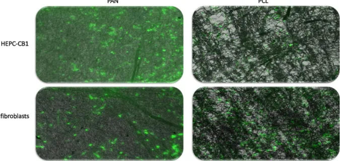
Fig. 4: Biocompatibility of the PAN- and PCL-based biomaterials, as tested by direct seeding of two indicator cell lines on the tested nanofibers. Cells were seeded on the indicated biomaterials and incubated for 26 hours, then stained with FDA and observed under fluorescence microscope (please see the method section for details).
As shown in figure 4, both HEPC-CB1 and NHDF (normal human dermal fibroblasts readily grow on both PAN (non biodegradable) and PCL (biodegradable) nanofibers. We have next compared cells’ ability to grow on the biomaterials developed at our lab, and under standard cell culture conditions, using normal primary human fibroblasts as indicators. alamarBlue reduction was used as an indirect indicator of cell numbers. The results of those
experiments are visualized in figure 5 and summarized in Table 1. The observed growth promoting properties of the tested biomaterials may be based on the following: (i) the decay products of the tested biodegradable biomaterials may be utilized by the cells as “food/energy source” as they may enter the Krebs-cycle for further metabolism, (ii) most of the presented biomaterials are doped with a some form of calcium carbonate.
Hybiak, J.; Pivet, M.; Fasani, F.; Hudecki, A.; Grillon, C.; Łos, M. J. Effects of electro-conductive, biomaterial-based tissue scaffolds on stem cells and transdifferentiation-derived somatic cells, LE STUDIUM Multidisciplinary Journal, 2018, 2, 34-41
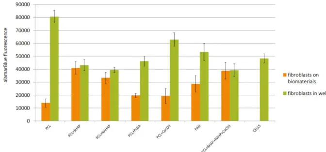
Fig. 5: Determination of cells ability to survive and proliferate on the tested biomaterials. Cellular content was assessed by alamarBlue staining (please consult the method section for experimental details).
Table 1: Determination of cells’ survival and proliferation on the tested biomaterials (data also visualized in figure 5 (above).
Since calcium ions are important for both cell adhesion and cell signaling, a calcium-enriched environment may better support cell proliferation. (iii) the solvents used for the preparation for the biofiber-substrates used during the electrospinning process [1] could also be metabolized, and ultimately enter the Krebs-cycle, hence serving as a “food/energy source”.
4- Conclusion
The biomaterials produced and tested in the course of the project are biocompatible, and some, not only support cell growth, but furthermore, they also (strongly) facilitate/stimulate their growth. Total fluorescence of the alamar Blue was up to twice higher in wells with biomaterials when
compared to the well with cells without any biomaterial. That indicated, that the presence of the biomaterial triggered cell proliferation and growth. The same trend was observed for HEPC –CB1 cells (data not shown). Moreover, cells stained with DiD were incubated for 96h and pictures were taken every 4h. Results indicated cells ability to growth and proliferate on the biomaterials with efficiency that vary between cells type and biomaterial type (data not presented).
5- Perspectives of future collaborations with the host laboratory
The completed Le Studium fellowship allowed for setup and expansion of collaboration between CBM-CNRS Orleans and my research groups at Małopolska Center of Biotechnology, Jagiellonian University, and the Department of
Pathomorfology, Pomeranian Medical University (PUM) in Szczecin. The additional experience gained at CBM-CNRS was also helpful with my own career development, especially with my successful application for the directorship of the Biotechnology Center (http://www.cb.polsl.pl/index.php/en/ ) of the Silesian University of Technology in Gliwice. The initial contact with the CBM-CNRS Orléans, facilitated by my Le Studium stipend was instrumental for the organization of research visits to various units of CNRS Orléans by 3 of my trainees/lab associates from my labs at PUM, in 2018.
For 2019 it is PLANNED: (1.) sending for 2-3 month research visit to CBM-CNRS Dr. Jolanta Hybiak from PUM (pending financial support for the visit); (2.) inviting my host Dr. Catherine Grillon (and partially finance the trip) to a yearly international conference “Gliwice Science Meetings” (http://gsn.io.gliwice.pl/ ) held between 21-23.11.2019; (3.) send at least 1 delegate for one of the Le Studium conferences organized in Orléans, in association with a brief visit to CBM-CNRS Orléans; (4.) attempt to prepare common grant application involving labs form CNRS Orléans, the Univ. Orléans and other research- or commercial entities from the region. The above actions (some en course) aim to develop and foster common collaborative research projects.
For 2020 it is PLANNED: (1.) my application for Le Studium Professorship. Such stipend (if successful) would provide a broad collaborative platform between labs from CNRS Orléans, the Univ. Orléans and other research- or commercial entities form the region, and Biotechnology Center of the Silesian University of Technology in Gliwice, as well as other scientific- and commercial entities from the region. The overreaching aim of the above is to develop a firm foundation (and dedicated, and qualified team) for large international grant applications; (2.) Other actions aimed to foster collaboration.
6- Articles published in the framework of the fellowship
1 Skubis A, Gola J, Sikora B, Hybiak J, Mazurek U, M.J Łos, (2017)
Impact of antibiotics on the proliferation and differentiation of human adipose-derived mesenhymal stem cells
Int. J. Mol. Sci., 18:2522, doi:10.3390/ijms18122522 2 Hudecki A, Skonieczna M, Gola J,
Markowski J, Małecki A, Ghavami S, Maziarz W, and Łos M.J, (2017)
The core-shell PCL/PCL/Ag nanofibers derived by coaxial electrospinning as
slowresorbing biodegradable tissue scaffolds
PeerJ, 8;5:e4125. doi: 10.7717/peerj.4125. 3 Hashemi M, Bahari G, Markowski J, Małecki
A, Łos MJ, Ghavami S.,
Association of PDCD6 polymorphisms with the risk of cancer: Evidence from a meta-analysis,
Oncotarget. 2018; 9:24857-24868. doi: 10.18632/oncotarget.25324.
4 Hashemi M, Bahari G, Tabasi F, Markowski J, Małecki A, Ghavami S, Łos MJ.,
LAPTM4B gene polymorphism augments the risk of cancer: Evidence from an updated meta-analysis,
J Cell Mol Med. 2018; 22:6396-6400. doi: 10.1111/jcmm.13896.
5 Wcisło-Dziadecka D, Simka K, Kaźmierczak A, Kruszniewska-Rajs C, Gola J, Grabarek B, Hybiak J, Grillon C, Mazurek U, Łos MJ.
Psoriasis Treatment Changes the Expression Profile of Selected Caspases and their Regulatory MicroRNAs
Cell Physiol Biochem. 2018; 50:525-537. doi: 10.1159/000494166.
6 Alizadeh J, Shojaei S, da Silva Rosa S, Rezaei Moghadam A, Zeki AA, Hashemi M, Los MJ, Gordon JW, Ghavami S.
Detection of Small GTPase Prenylation and GTP Binding Using Membrane Fractionation and GTPase-linked Immunosorbent Assay,
J Vis Exp. 2018 Nov 11;(141). doi: 10.3791/57646.
7 Kucharzewski M, Rojczyk E, Wilemska-Kucharzewska K, Wilk R, Hudecki J, Los MJ
Novel trends in application of stem cells in skin wound healing.
Eur J Pharmacol. 2019; 843:307-315. doi: 10.1016/j.ejphar.2018.12.012.
8 Hudecki A, Łyko-Morawska D, Likus W, Skonieczna M, Markowski J, Wilk R, Kolano-Burian A, Maziarz W, Adamska J, Łos MJ.
Hybiak, J.; Pivet, M.; Fasani, F.; Hudecki, A.; Grillon, C.; Łos, M. J. Effects of electro-conductive, biomaterial-based tissue scaffolds on stem cells and transdifferentiation-derived somatic cells, LE STUDIUM Multidisciplinary Journal, 2018, 2, 34-41
Composite Nanofibers Containing Multiwall Carbon Nanotubes as Biodegradable Membranes in Reconstructive Medicine,
Nanomaterials (Basel). 2019 ;9 (in press). pii: E63. doi: 10.3390/nano9010063. 9 Kitala D, Klama-Baryła A, Misiuga M,
Łabuś W, Kraut M, Szapski M, Lesiak M, Krakowian D, Sieroń AL, Łos MJ, Kucharzewski M,
Heterogeneous Mixture of Amniotic Cells is Likely a Better Source of Stem Cells than Adipose Tissue
Arch Immunol Ther Exp., 2019; 67: (AITE-D-18-00079, in press).
Books or book chapters:
10 Book: “Stem Cells and Biomaterialsfor Regenerative Medicine“ 1st Edition,
edited by M. J. Łos, A. Hudecki, E. Wiechec;
Elsevier Academic Press, (2018), ISBN: 9780128122587.
11 Łos M.J., Skubis, A., and, S. Ghavami #2:“Stem cells”, in “Biomaterials, Stem Cells
& Regenerative Medicine – in Brief”, Eds.:
Hudecki A, Wiecheć E, and M. J. Łos, (2018), Elsevier Academic Press, (2018), ISBN: 9780128122587.
12 Wiechec E., Hybiak J., Kieda C., #6:
"Introduction to Transplantology" in “Stem Cells and Biomaterials for Regenerative Medicine“ 1st Edition, edited by M. J.
Łos, A. Hudecki, E.
Wiechec; Elsevier Academic Press, (2018), ISBN: 9780128122587.
13 Hudecki A, Kiryczyński G, and M. J. Łos, #7: "Biomaterials, Definition,
Overview” in “Stem Cells and Biomaterials for Regenerative Medicine“ 1st
Edition, edited by M. J. Łos, A. Hudecki, E. Wiechec; Elsevier Academic Press, (2018), ISBN: 9780128122587
14 Łos M.J., Panigrahi S., Sielatycka K. and C. Grillon #13: "Successful
Biomaterial-Based Artificial Organ—Updates on Artificial Blood Vessels" in “Stem Cells and Biomaterials for Regenerative Medicine“ 1st
Edition, edited by M. J. Łos, A. Hudecki, E. Wiechec; Elsevier Academic Press, (2018), ISBN: 9780128122587.
7- Acknowledgements
MJŁ kindly acknowledges the support from LE STUDIUM Institute for Advanced Studies (region Centre-Val de Loire, France) through its Smart Loire Valley General Program, co-funded by the Marie Sklodowska-Curie Actions, grant # 665790. We thank you the staff and researchers of the Grillon Lab for an exemplary hospitality, help with experimental tasks, and assistance with daily life tasks (esp. to Ms Fabienne Fasani and Ms Marine Pivet). We would like also to extend our special thanks to the staff and a management of the Le Studium organization for making the scientific visit, possible, productive and comfortable (esp. to: Sophie Gabillet, Aurélien Montagu, Marie-Frédérique Pellerin, Amélie Schneuwly, and Maurine Villiers). We are also very thankful to Professor Laurent Cherlonneix, for presenting an excellent and deeply intellectual public lecture during the scientific conference organized in Orleans in mid-June 2018.
8- References
[1] Hudecki A, Gola J, Ghavami S et al. Structure and properties of slow-resorbing nanofibers obtained by (co-axial) electrospinning as tissue scaffolds in regenerative medicine PeerJ. 2017; 5:e4125 [2] Atyabi SM, Irani S, Sharifi F, et al. Cell attachment and viability study of PCL nano-fiber modified by cold atmospheric plasma. J Cell Biochem Biophys. 2016; 74:181–190.