HAL Id: hal-03031117
https://hal.archives-ouvertes.fr/hal-03031117
Preprint submitted on 30 Nov 2020HAL is a multi-disciplinary open access archive for the deposit and dissemination of sci-entific research documents, whether they are pub-lished or not. The documents may come from teaching and research institutions in France or abroad, or from public or private research centers.
L’archive ouverte pluridisciplinaire HAL, est destinée au dépôt et à la diffusion de documents scientifiques de niveau recherche, publiés ou non, émanant des établissements d’enseignement et de recherche français ou étrangers, des laboratoires publics ou privés.
The pneumococcal alternative sigma factor σ X mediates
competence shut-off at the cell pole
Calum Johnston, Anne-Lise Soulet, Matthieu Berge, Marc Prudhomme, David
de Lemos, Patrice Polard
To cite this version:
Calum Johnston, Anne-Lise Soulet, Matthieu Berge, Marc Prudhomme, David de Lemos, et al.. The pneumococcal alternative sigma factor σ X mediates competence shut-off at the cell pole. 2020. �hal-03031117�
The pneumococcal alternative sigma factor σ
Xmediates competence shut-off
1
at the cell pole
2
Calum JOHNSTON1,2, Anne-Lise SOULET1,2, Matthieu BERGE1,2,3, Marc PRUDHOMME1,2, David DE 3
LEMOS1,2, Patrice POLARD1,2. 4
1. Laboratoire de Microbiologie et Génétique Moléculaires, UMR5100, Centre de Biologie Intégrative (CBI), Centre Nationale
5
de la Recherche Scientifique (CNRS), Toulouse, France.
6
2. Université Paul Sabatier (Toulouse III), Toulouse, France.
7
3. Dept. Microbiology and Molecular Medicine, Institute of Genetics & Genomics in Geneva (iGE3), Faculty of Medicine,
8
University of Geneva, Geneva, Switzerland.
9 10
Keywords 11
Competence regulation, competence shut-off, alternative sigma factor, σX, DprA. 12 13 14 15 16 17 18 19 20 21 22
Summary 23
Bacterial competence for genetic transformation is a well-known species-specific differentiation 24
program driving genome plasticity, antibiotic resistance and virulence in many pathogens. How 25
competence regulation is spatiotemporally integrated in the cell is ill-defined. Here, we unraveled the 26
localization dynamics of the key regulators that master the two intertwined transcription waves 27
controlling competence in Streptococcus pneumoniae. The first wave relies on a stress-inducible 28
phosphorelay system, made up of the ComD and ComE proteins, and the second is directed by an 29
alternative sigma factor, σX, which includes in its regulon the DprA protein that turns off competence
30
through interaction with phosphorylated ComE. Remarkably, we found that ComD, σX and DprA stably
31
co-localize at a single cell pole over the competence period. Furthermore, we provide evidence that σX
32
is necessary and sufficient to mediate DprA polar accumulation next to ComD. Finally, we show that 33
through this protein targeting function, σX is actively involved in the timely shut-off of the competence
34
cycle, hence preserving cell fitness. Altogether, this study unveils an unprecedented role for a bacterial 35
transcription σ factor in spatially coordinating the negative feedback loop of its own genetic circuit. 36 37 38 39 40 41 42 43 44 45
Introduction 46
In bacteria, sigma (σ) factors are essential transcription effectors that direct the RNA polymerase 47
to and activate RNA synthesis at specific genes promoters. All bacterial species encode a single, highly 48
conserved σ factor that drives the expression of house-keeping genes essential for vegetative growth 49
and cell homeostasis. In addition, many bacteria encode a variable set of alternative σ factors that 50
control specific regulons, providing appropriate properties to the cells in response to various stimuli. 51
These alternative σ factors play pivotal roles in the multifaceted lifestyles of bacteria. They trigger 52
specific developmental programs, such as sporulation or biofilm formation, as well as adapted 53
responses to multiple types of stress and virulence in some pathogenic species (Kazmierczak et al., 54
2005). How these alternative σ factors are activated in the cell has been extensively studied, revealing 55
multiple mechanisms underlying their finely tuned regulation(Österberg et al., 2011a). However, how 56
these mechanisms are orchestrated spatiotemporally within the cell remains poorly understood. 57
The human pathogen Streptococcus pneumoniae (the pneumococcus) possesses a unique 58
alternative σ factor σX (Lee and Morrison, 1999). Itis key to the regulatory circuit controlling the 59
transient differentiation state of competence. Pneumococcal competence is induced in response to 60
multiple types of stresses, such as antibiotic exposure (Prudhomme et al., 2006; Slager et al., 2014). 61
This induction modifies the transcriptional expression of up to 17% of genes (Aprianto et al., 2018a; 62
Dagkessamanskaia et al., 2004a; Peterson et al., 2004; Slager et al., 2019). Competence is a key feature 63
in the lifestyle of pneumococci as it promotes natural transformation, a horizontal gene transfer 64
process widespread in bacteria that facilitates adaptation by acquisition of new genetic traits (Johnston 65
et al., 2014). In addition, pneumococcal competence development provides the cells with the ability 66
to attack non-competent cells, a scavenging property defined as fratricide (Claverys and Håvarstein, 67
2007), is involved in biofilm formation (Aggarwal et al., 2018; Vidal et al., 2013) and virulence (Johnston 68
et al., 2018; Lin et al., 2016; Lin and Lau, 2019; Zhu et al., 2015). 69
Pneumococcal competence induction is primarily regulated by a positive feedback loop 70
involving the genes encoded by the comAB and comCDE operons (Figure 1A). The comC gene codes for 71
a peptide pheromone coordinating competence development within the growing cell population. This 72
peptide, accordingly named CSP (Competence Stimulating Peptide), is secreted by the dedicated 73
ComAB transporter (Hui et al., 1995). After export, it promotes autophosphorylation of the membrane-74
bound two-component system (TCS) histidine kinase (HK) ComD, which in turn phosphorylates its 75
cognate intracellular response regulator (RR) ComE (Figure 1A). Phosphorylated ComE (ComE~P) 76
specifically induces the expression of 25 genes, which include the comAB and comCDE operons, 77
generating a positive feedback loop that controls competence development. Conversely, 78
unphosphorylated ComE acts as repressor of its own regulon, the expression of which is thus 79
modulated by the ComE/ComE~P ratio (Martin et al., 2013). The ComE regulon includes two identical 80
genes encoding σX, named comX1 and comX2 (Lee and Morrison, 1999). The σX regulon comprises ~60 81
genes, with ~20 involved in natural transformation (Claverys et al., 2006; Peterson et al., 2004), 5 in 82
fratricide (Claverys and Håvarstein, 2007) but the majority having undefined roles. The reason why the 83
σX-encoding gene is duplicated is unknown, the inactivation of one of them having no impact on 84
transformation (Lee and Morrison, 1999). To fully activate transcription, σX needs to be assisted by 85
ComW, another protein whose production is controlled by ComE~P (Luo et al., 2004). ComW is 86
proposed to help σX association with the RNA polymerase at promoter sequences presenting the 87
consensual 8 bp cin box motif (Peterson et al., 2004; Sung and Morrison, 2005). Altogether, ComE~P 88
and σX trigger two successive waves of competence (com) gene transcription, commonly referred to 89
as early and late, respectively. Importantly, competence shut-off is mediated by the late com protein 90
DprA (Mirouze et al., 2013; Weng et al., 2013), which directly interacts with ComE~P to turn-off 91
ComE~P-dependent transcription (Mirouze et al., 2013). In addition to defining the negative feedback 92
loop of the pneumococcal competence regulatory circuit, DprA also plays a crucial, conserved role in 93
transformation by mediating RecA polymerization onto transforming ssDNA to facilitate homologous 94
recombination (Mortier-Barrière et al., 2007; Cheruel et al., 2012) (Figure 1A). Over 8,000 molecules 95
of DprA are produced per competent cell (Mirouze et al., 2013). Although only ~300-600 molecules 96
were required for optimal transformation, full expression of dprA was required for optimal 97
competence shut-off (Johnston et al., 2018). Uncontrolled competence induction in cells lacking DprA 98
results in a large in vitro growth defect, and high cellular levels of DprA thus maintain the fitness of the 99
competent population and of resulting transformants (Johnston et al., 2018). In addition, inactivation 100
of dprA was shown to be highly detrimental for development of pneumococcal infection, dependent 101
on the ability of cells to develop competence (Lin and Lau, 2019; Zhu et al., 2015). Together, these 102
studies showed that the DprA-mediated shut-off of pneumococcal competence is key for 103
pneumococcal cell fitness. 104
A hallmark of pneumococcal competence is its tight temporal window, which lasts less than 30 105
minutes in actively dividing cells (Alloing et al., 1998; Håvarstein et al., 1995). How this regulation is 106
coordinated within the cell remains unknown. Here, we studied the choreography of pneumococcal 107
competence induction and shut-off at the single cell level by tracking the spatiotemporal localization 108
of the main effectors of these processes, DprA, σX, ComW, ComD, ComE and exogenous CSP. 109
Remarkably, DprA, σX, ComD, CSP and to some extent ComE were found to colocalize at a single cell 110
pole during competence. This study revealed that the entire pneumococcal competence cycle occurs 111
at cell pole, from its induction triggered by ComD, ComE and CSP to its shut-off mediated by DprA and 112
assisted by σX. In this regulatory mechanism, σX is found to exert an unprecedented role for a σ factor. 113
In addition to directing the transcription of the dprA gene, σX associates with and anchors DprA at the 114
same cellular pole where ComD and CSP are located, allowing this repressor to interact with newly 115
activated ComE~P and promoting timely extinction of the whole transcriptional regulatory circuit of 116 competence. 117 118 Results 119
DprA displays a polar localization in competent cells, which correlates with competence shut-off.
120
To investigate the localization of DprA during competence in live cells, we used a fluorescent 121
fusion protein DprA-GFP, produced from the native dprA locus (Figure S1A). DprA-GFP was synthesized 122
during competence and remained stable up to 90 minutes after induction (Figure 1B), similarly to 123
wildtype DprA (Mirouze et al., 2013), without degradation (Figure S1B). A strain possessing DprA-GFP 124
was almost fully functional in transformation (Figure S1C), but partially altered in competence shut-off 125
(Figure S1D). In pneumococcal cells induced with exogenous CSP, the peak of competence induction 126
occurs 15-20 minutes after induction. 15 minutes after competence induction, DprA-GFP showed a 127
diffuse cytoplasmic localization, punctuated by discrete foci of varying intensity (Figure 1C). The 128
distribution and localization of DprA-GFP foci was analyzed by MicrobeJ (Ducret et al., 2016), with 129
results presented as focus density maps ordered by cell length. Spots represent the localization of a 130
DprA-GFP focus on a representative half pneumococcal cell, while spot colour represents density of 131
foci at a particular cellular location. DprA-GFP foci were present in 70% of cells, predominantly at a 132
single cell pole (Figure 1DE). To ascertain whether the polar localization of DprA-GFP was due to the 133
GFP tag or represented functional localization, we carried out immunofluorescence microscopy with 134
competent cells possessing wildtype DprA using α–DprA antibodies. Results showed that native DprA 135
exhibited a similar accumulation pattern as the DprA-GFP foci upon competence induction (Figure 1F). 136
Next, to explore the relation between these foci and the dual role of DprA in transformation 137
and competence regulation, we investigated focus formation in cells possessing a previously published 138
mutation in DprA impairing its dimerization (DprAAR)(Quevillon-Cheruel et al., 2012). This mutant 139
strongly affected both transformation and competence shut-off (Mirouze et al., 2013; Quevillon-140
Cheruel et al., 2012). Results showed that 15 minutes after competence induction, DprAAR-GFP did not 141
form foci, despite being produced at wildtype levels (Figure S1E). DprA thus accumulates at the cell 142
pole during competence, dependent on its ability to dimerize. To explore whether the polar foci of 143
DprA were involved in its role in transformation, or competence shut-off, we began by investigating 144
how a dprAQNQ mutation, specifically abrogating the interaction between DprA and RecA and thus
145
affecting transformation (Quevillon-Cheruel et al., 2012), affected the localization of DprA-GFP. 15 146
minutes after competence induction, the DprAQNQ-GFP mutant formed polar foci at wildtype levels 147
(Figure S1F). In addition, the inactivation of comEC, encoding for an essential protein of the 148
transmembrane DNA entry pore (Pestova and Morrison, 1998), or recA, encoding the recombinase 149
with which DprA interacts during transformation (Mortier-Barrière et al., 2007), did not alter the 150
frequency or localization of DprA-GFP foci (Figure S1GH). Altogether, these results suggested that the 151
polar foci of DprA-GFP were not related to the DNA entry or recombination steps of transformation 152
but could be linked to competence shut-off. 153
We recently reported that optimal competence shut-off relies on the maximal cellular 154
concentration of DprA (~8000 molecules), but this level could be reduced by 10 fold while still 155
maintaining wildtype frequency of transformation (Johnston et al., 2018). This conclusion was 156
obtained by expressing dprA under the control of the IPTG-inducible Plac promoter (CEPlac-dprA), which
157
enables the modulation of the cellular concentration of DprA by varying IPTG concentration in the 158
growth medium. Here, we reproduced these experiments with the DprA-GFP fusion, to test whether 159
its concentration correlates with the formation of polar foci in competent cells. The expression, 160
transformation and competence profiles of a dprA- mutant strain harbouring the ectopic CEP
lac
-dprA-161
gfp construct in varying concentrations of IPTG (Figure S2ABC) were equivalent to those reported 162
previously for CEPlac-dprA (Johnston et al., 2018). Notably, a steady decrease in DprA-GFP foci was
163
observed as IPTG was reduced (Figure 2AB). When comparing the cellular localization of DprA-GFP foci, 164
a sharp reduction in the proportion of polar foci was observed as IPTG was reduced, with most of the 165
remaining foci observed at midcell and appearing weaker in intensity (Figure 2AC). This shift correlated 166
with a progressive loss of competence shut-off (Figure S2C), presenting a strong link between the 167
presence of polar DprA-GFP foci and the shut-off of pneumococcal competence. Altogether, these 168
results strongly support the notion that the polar foci of DprA-GFP represent the subcellular site where 169
DprA mediates competence shut-off. 170
Finally, to further explore the temporal dynamics of these DprA-GFP foci, their distribution 171
within competent cells was analyzed over the competence period and beyond. The results are 172
presented in Figure 3A as focus density maps ordered by cell length. The number of cells with foci, as 173
well as their intensity, was found to increase gradually to reach a maximum of 74% at 30 minutes after 174
competence induction (Figure 3A), with the majority of cells possessing a single focus that persisted 175
long after induction (Figure 3B). Notably, the DprA-GFP foci localization pattern rapidly evolved from 176
a central position to a single cell pole (Figure 3C). DprA-GFP foci were not observed in a particular cell 177
type, with found in small, large or constricted cells throughout competence (Figure 3C). Finally, 178
tracking DprA-GFP foci formed in the cells after 10 minutes of competence induction by time-lapse 179
microscopy showed that once generated, they remained static over 20 minutes (Figure S1IJ, Movie 1). 180
In conclusion, DprA-GFP forms discrete and static polar foci during competence, with most cells 181
possessing a single focus. This polar localization of DprA correlates with its regulatory role in 182
competent shut-off. 183
184
The polar localization of DprA-GFP requires induction of the late com genes
185
Transcriptional expression of dprA is only detected during competence (Aprianto et al., 2018a; 186
Dagkessamanskaia et al., 2004a; Peterson et al., 2004). To explore whether a competence-specific 187
factor was required for the formation of polar DprA-GFP foci during competence, dprA-gfp was 188
ectopically expressed from a promoter inducible by the BIP peptide in dprA- cells. This BIP-derived
189
induction mimics rapid, strong induction by CSP during competence (Johnston et al., 2016). These cells 190
were found to produce stable DprA-GFP after exposure to BIP, and upon addition of CSP to the growth 191
medium, to transform at wild-type levels and to partially shut-off competence (Figure S3). However, 192
BIP-induced production of DprA-GFP in the absence of CSP resulted in the formation of weak, barely 193
detectable polar foci in only 10% of non-competent cells (Figure 4AB). In comparison, 47% of cells 194
producing DprA-GFP during competence formed bright polar foci (Figure 4AB). This stark increase in 195
DprA-GFP foci showed that a competence-specific factor was crucial for their formation and anchoring 196
at the cell pole. To explore whether the competence-specific factor needed for the polar localization 197
of DprA-GFP in competent cells was part of the early or late com regulons, we generated two 198
constructs allowing us to artificially control DprA-GFP expression and either one or the other of these 199
two connected regulons (Figure 4CD and Supplementary methods). Observation of DprA-GFP in 200
conditions where only late com genes were induced revealed the presence of polar foci at wildtype 201
levels (Figure 4EF). Conversely, no foci were observed when DprA-GFP was ectopically expressed with 202
only the early com genes (Figure 4E), showing that late com regulon expression was required for the 203
polar accumulation of DprA-GFP. 204
205
The polar localization of DprA-GFP depends on the alternative sigma factor σX 206
The late com regulon is comprised of 62 genes, organized in 18 operons (Claverys et al., 2006; 207
Dagkessamanskaia et al., 2004b; Peterson et al., 2004). To identify the hypothetical late com gene 208
product needed for DprA localization at the cell pole, the cin boxes that define the late com promoters 209
were individually mutated, generating a panel of 18 mutant strains, each lacking the ability to induce 210
a specific late com operon. The inactivation was validated by comparing transformation efficiency in 211
three strains, where cin box inactivation mirrored gene knockout levels (Table S1, and Supplementary 212
methods). Visualization of the red fluorescent fusion DprA-mKate2 showed that in all 18 mutants, 213
DprA-mKate2 formed foci at levels and localization comparable to wildtype (Table S2). This result 214
contrasted with our previous result (Figure 4E), which suggested that expression of the late com 215
regulon was required for formation of polar DprA-GFP foci, causing us to revisit our interpretation of 216
Figure 4C-F. In fact, to express only the early com regulon, we inactivated comX1, comX2 and comW 217
(Figure 4C), so this strain produced only early com proteins, except σX and ComW, and lacked DprA-218
GFP foci (Figure 4E). Conversely, to express only the late com regulon, we ectopically expressed comX 219
and comW (Figure 4D), so this strain produced the late com regulon but also the early com proteins σX 220
and ComW and displayed polar DprA-GFP foci at wildtype levels (Figure 4EF). This led us to consider 221
that the only proteins whose presence correlated directly with the presence of polar DprA-GFP foci 222
were thus σX and ComW. 223
To first investigate whether ComW played a role in the formation of polar DprA-GFP foci, the 224
comW gene was inactivated in a strain possessing a rpoDA171V mutation, enabling σX-RNA polymerase 225
interaction and resulting in late com regulon expression in the absence of ComW (Tovpeko et al., 2016). 226
DprA-GFP expressed from the native locus still formed polar foci in this strain at levels comparable to 227
the wildtype strain (Figure S4A). ComW was thus not required for the formation of DprA-GFP foci. In 228
light of this, the only remaining candidate whose presence in competent cells correlated directly with 229
formation of polar DprA-GFP foci was σX. Thus, to determine if σX alone was necessary and sufficient 230
to localize DprA-GFP to the cell poles, both comX and dprA-gfp or dprA-gfp alone were expressed in 231
non-competent rpoDwt cells. Western blot analysis using α-SsbB antibodies indicated that the late com
232
regulon was weakly induced when σX was ectopically produced in the absence of comW (Figure S4B). 233
Cells producing DprA-GFP alone showed polar DprA-GFP foci in 10% of cells (Figure 5A). In contrast, 234
DprA-GFP foci were formed in 37% of cells when σX was also produced in non-competent cells (Figure 235
5A). Importantly, induction of competence in both of these strains resulted in similar foci numbers 236
(Figure S4D). Altogether, this result suggested that σX alone was sufficient to stimulate polar foci of 237
DprA-GFP, highlighting an unexpected role for this early competence σ factor only known to act in 238
concert with ComW to induce late competence gene transcription. 239
240
σX mediates the localization of DprA at the cell pole of competent cells 241
To investigate how σX could be involved in the polar localization of DprA-GFP, we explored how 242
it localized in competent cells. To this end, we generated a comX1-gfp construct at the native comX1 243
locus combined with a wildtype comX2 gene (Figure S5 and Supplementary results). Remarkably, σX -244
GFP formed bright polar foci 15 minutes after competence induction (Figure 5A), reminiscent of those 245
formed by DprA-GFP (Figure 1C). A time-course experiment after competence induction showed that 246
σX-GFP localized to the cell pole as soon as 4 minutes after CSP addition (Figure 5B), when DprA-GFP 247
forms weak foci at midcell (Figure 3A). In contrast, a ComW-GFP fusion protein displayed a diffuse 248
cytoplasmic localization in the majority of competent cells, with only 7% of cells possessing weak foci 249
at the cell poles (Figure S5A and Supplementary results). Thus, σX-GFP localizes to the cell pole without 250
its partner in transcriptional activation ComW, despite the fact that σX is an alternative sigma factor 251
directing RNA polymerase to specific promoters on the chromosome. 252
Analysis of σX-GFP foci distribution showed that they are detected in up to 51% of cells 10 253
minutes after competence induction, with most cells possessing a single focus (Figure 5BC). The 254
number of cells with foci decreased steadily after this point (Figure 5BCD), contrasting with polar DprA-255
GFP which remained stable over 60 minutes after induction. Importantly, σX-GFP continued to form 256
polar foci in a strain lacking dprA (Figure 5E), showing that σX does not depend on DprA for its 257
localization. Together, these results strongly supported the notion that σX promotes the targeting and 258
assembly of GFP foci at the cell pole. To further explore this hypothesis, we co-expressed DprA-259
mTurquoise and σX-YFP fluorescent fusions in the same cells and found that 86% of DprA-mTurquoise 260
foci colocalized with σX-YFP foci (Figure 5F). DprA and σX are thus present at the same pole of the cell 261
at the same time and the polar accumulation of DprA molecules in competent cells depends on σX. 262
These results suggested that σX could interact with DprA to anchor it to the pole of competent cells. 263
The potential interaction between σX and DprA was tested in live competent pneumococcal cells in 264
pull-down experiments. To achieve this, we used GFP-TRAP magnetic beads (Chromotek) to purify σX -265
GFP from competent cells expressing either wildtype DprA or, as a control, the DprAAR mutant that did 266
not accumulate at the cell pole (Figure S1). Results showed that wildtype DprA co-purified with σX-GFP 267
from competent cells extracts, but DprAAR did not, revealing that σX and DprA interact in live competent 268
pneumococci (Figure 5H). Taken together, these findings reveal that σX is responsible for the 269
accumulation of DprA at the cell pole during competence. 270
271
Pneumococcal competence induction occurs at the cell pole
We have shown that DprA localization at a single cell pole correlates with the shut-off of 273
competence (Figure 2). Since DprA interacts directly with ComE~P to mediate competence shut-off 274
(Mirouze et al., 2013), this raised the question of the subcellular localization of ComD and ComE, which 275
define a TCS controlling competence regulation. To first explore the localization of ComE in competent 276
cells, a strain was generated expressing a functional comE-gfp fluorescent fusion at the native comE 277
locus (Figure S6 and Supplementary results). In competent cells, ComE-GFP formed patches around 278
the periphery of the cell, often at the cell pole (Figure 6AB and S7A). A time-lapse experiment showed 279
that these patches were dynamic, navigating around the cell membrane over time (Movie 2). Next, to 280
explore the sub-cellular localization of ComD, we generated a strain expressing gfp-comD at the native 281
comD locus (Figure S6AC and Supplementary results). The resulting GFP-ComD fusion displayed partial 282
functionality in competence induction and transformation (Figure S6DE). In contrast to ComE-GFP, 283
GFP-ComD formed distinct polar foci of varying intensity in 57% of cells (Figure 6AB), a localization 284
pattern reminiscent of those observed with DprA-GFP and σX-GFP (Figures 1and 5, respectively). Since 285
GFP-ComD was not fully functional, we also analyzed the localization of a synthetic fluorescent 286
exogenous CSP peptide (CSP-HF, Figure S6F) in parallel, to track its interaction with ComD at the cell 287
surface. This fluorescent peptide was found to accumulate at a single cell pole in the majority of 288
competent, wildtype cells (Figure 6AB). In addition, this accumulation was dependent on the presence 289
of ComD (Figure 6A), showing that the polar accumulation of the partially functional GFP-ComD fusion 290
represented a functional subcellular localization during competence. In addition, most cells with 291
ComD-GFP foci possessed a single focus (Figure 6C), which persisted after the shut-off of competence 292
(Figure S7B). Altogether, these findings revealed that activation of the positive feedback loop of 293
competence triggered by CSP interaction with ComD to phosphorylate ComE occurs at the cell pole. 294
Since a single pole is mainly targeted by ComD, this also raised the question of its co-localization with 295
ComX and DprA. 296
DprA colocalizes with ComD and CSP during competence
298
To explore the hypothesis that DprA accumulates at the same pole as ComD to mediate 299
competence shut-off, we performed colocalization analyses of DprA with ComE, ComD or CSP 300
respectively, in the same competent cells. Although ComE-YFP foci were dynamic and their localization 301
difficult to analyze, 20% of DprA-mTurquoise foci were nonetheless found to colocalize with ComE-YFP 302
at the cell pole, showing that these proteins can be found at the same pole in the same cells (Figure 303
6D). In contrast, 76% of DprA-mTurquoise foci colocalized with YFP-ComD (Figure 6E), showing that 304
these foci form at the same pole in the majority of cells. In addition, 73% of DprA-GFP foci colocalized 305
with CSP-HF (Figure 6F). Taken together, these results showed that DprA colocalizes strongly with 306
ComD, generally at one cell pole during pneumococcal competence, further suggesting that the 307
localization of DprA to this cellular location, mediated by σX, facilitates pneumococcal competence 308
shut-off. The localization of DprA at the cell pole where ComD appears to interact with CSP means that 309
DprA is present at the time and place that neophosphorylated ComE~P is produced, allowing DprA to 310
interact with the activated regulator at the cell pole and prevent it from accessing its genomic targets, 311
facilitating shut-off. 312
313
Two copies of comX are required for optimal competence shut-off
314
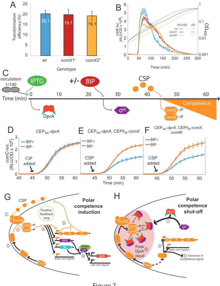
A distinct hallmark of the single pneumococcal alternative sigma factor σX of S. pneumoniae is 315
that it is produced from two distinct and strictly identical genes, at two distinct loci in the 316
pneumococcal genome, known as comX1 and comX2 (Lee and Morrison, 1999). However, inactivation 317
of either of these genes had no effect on the efficiency of transformation as previously reported (Lee 318
and Morrison, 1999) (Figure 7A), suggesting that the expression of either comX1 or comX2 is sufficient 319
to induce the late com regulon to a level ensuring optimal transformation. Having established that σX 320
plays a second key role in competence shut-off, we explored whether this role required both comX 321
genes. Inactivation of either comX gene not only slightly reduced the peak of late com gene expression 322
but also markedly delayed the rate of competence shut-off (Figure 7B), revealing that reducing the 323
cellular level of σX impacts competence shut-off efficiency, presumably because less σX is present to 324
promote accumulation of DprA at the cell poles. In addition, this unregulated competence shut-off of 325
single comX mutants is accompanied by a reduced growth rate of the cell population, consistent with 326
an alteration of cell fitness linked to altered competence shut-off (Johnston et al., 2018). In conclusion, 327
this finding suggests that pneumococci possess two copies of comX to optimize DprA-mediated 328
competence shut-off and maintain the fitness of competent cells. 329
330
Pre-competence expression of DprA and σX antagonizes competence induction 331
This study has uncovered a new functional role of pneumococcal σX in facilitating DprA-332
mediated inactivation of ComE-P at the cell pole. To obtain further proof that polar DprA plays a role 333
in competence shut-off, we reasoned that if σX localizes DprA to the cell pole to allow it to interact 334
with neophosphorylated ComE~P, then early ectopic expression of DprA and σX should antagonize 335
competence induction by interfering with early com regulon induction. To test this hypothesis, we 336
expressed DprA alone or both DprA and σX prior to CSP addition to the growth medium (Figure 7C), 337
referred hereafter as a pre-competence expression. Pre-competence production of DprA alone did not 338
affect competence induction as shown by monitoring luciferase controlled by an early com promoter 339
(Figure 7D). In these conditions, minimal amounts of DprA accumulated at the cell pole (Figure 5A). 340
However, pre-competence expression of both DprA and σX resulted in a significantly slower induction 341
of competence (Figure 7E). This suggested that σX-mediated pre-localization of DprA to the cell poles 342
in pre-competent cells,as shown in Figure 4G, markedly antagonized CSP induction of competence by 343
allowing DprA to interact with and inactivate neophosphorylated ComE~P. Inactivation of comW in this 344
strain to minimize late com gene expression did not alter the profiles (Figure 7F), showing that the 345
observed effect was directly attributable to the role of σX in localizing DprA to the cell poles. Altogether, 346
these findings further prove that in live cells the targeting of pneumococcal DprA to the cell pole, 347
promoted by σX anchored at this cellular location, mediates the timely antagonization of the 348
competence induction signal. 349
350
Discussion 351
The cell pole defines a competence regulation hub in S. pneumoniae
352
We report here a spatiotemporal analysis of competence regulation in live pneumococcal cells. 353
We found that the positive regulators ComD, ComE and σX, which control the early and late 354
competence expression waves, and the negative regulator DprA colocalize during competence at one 355
cell pole to temporally coordinate its development and shut-off. We have shown that the initial stages 356
of competence induction, relying on the CSP-induced phosphorelay between ComD and ComE, occur 357
at the cell pole (Figure 7G). Most importantly, this study uncovered an unexpected second role for σX 358
in competence regulation. In addition to controlling the transcription of the late com gene dprA, σX 359
also mediates the accumulation of DprA molecules mostly at the same pole as ComD, a mechanism 360
that facilitates competence shut-off. σX thus controls both induction of the late com regulon and the 361
shut-off of the early com regulon, including thereby its own expression and by consequence that of its 362
regulon. We found that the σX-directed polar localization of DprA in the vicinity of ComD correlates 363
with its role in competence shut-off. We propose that this targeting favours DprA interaction between 364
DprA and neosynthesized ComE~P to promote efficient shut-off (Figure 7H). Importantly, we 365
previously reported that DprA-mediated competence shut-off is crucial to the fitness of competent 366
cells (Johnston et al., 2018; Mirouze et al., 2013), and we revealed here that this vital role of DprA in 367
actively limiting the competence window is orchestrated at the cell pole, which defines a coordination 368
hub of competence regulation. Unexpectedly, DprA is targeted to this hub by σX, describing an 369
unprecedented role for an alternative sigma factor. 370
The pneumococcal polar competence regulation hub is focused around the histidine kinase ComD
372
An important finding is the discrete accumulation of the pneumococcal HK ComD at the cell 373
pole during competence (Figure 6AB). The localization of HKs has not been extensively studied, but 374
few HKs of two-component signaling systems (TCS) have been found to accumulate at the cell pole. 375
One well-documented example is the Escherichia coli HK CheA, which is involved in chemotaxis. CheA 376
localizes along with its cognate RR CheY to a single cell pole, forming a chemotactic cluster with a 377
variety of chemoreceptors. CheY stimulates the production of flagellae at the opposite cell pole, driving 378
chemotaxis (Baker et al., 2006). Another example is the HK PilS in Pseudomonas aeruginosa, which also 379
accumulates at the cell pole, and along with its cognate cytoplasmic RR PilR, regulates expression of 380
polar pili (Boyd, 2000). Although ComD tethering at one cell pole is similar to CheA and PilS, major 381
differences exist between these three TCS system. Firstly, regarding how their RRs localize, ComE 382
presents a different localization pattern than CheY and PilR, assembling into patches close to the inner 383
side of the cell membrane and focusing dynamically on the cell pole (Figure 6A and Movie 2). ComE 384
phosphorylation by ComD promotes its dimerization and switch into a transcriptional activator (Martin 385
et al., 2013). ComE~P dimers should then leave the cell pole to interact with genomic targets and turn 386
on early com gene expression, producing σX and ComW, which then cooperate to turn on late com gene 387
expression. Secondly, the ComDE TCS induces its own expression, generating a positive feedback loop 388
(Claverys et al., 2006; Martin et al., 2013), and leading to the strong, rapid induction of competence 389
(Dagkessamanskaia et al., 2004b; Peterson et al., 2004). As found here, this leads to the accumulation 390
of the regulatory proteins at the cell pole hub. In addition, the ComDE TCS controls a complex, multi-391
faceted genetic program involving the altered expression of 17% of the genome (Aprianto et al., 2018b) 392
and is present at a unique cell pole, while the actors of transformation are present at midcell (Bergé 393
et al., 2013). Finally, it is possible that the polar localization of the competence regulatory hub is 394
dictated by the very nature of the competence regulation mechanism itself. This localization could be 395
linked to the transient nature of pneumococcal competence. A spatiotemporally controlled localization 396
for this process would provide a mechanism to allow initial induction followed by repression, thus 397
preventing toxicity. It is also possible that the purpose of polar accumulation of σX is to limit the 398
circulating levels of σX. Polar accumulation of σX may thus protect the cell from a potentially dangerous 399
hyper-competent state at two levels, firstly by promoting polar DprA accumulation to facilitate shut-400
off, and secondly by sequestering σX itself to prevent over-induction of the late com regulon. Although 401
the factor localizing σX tothe cell pole is unknown, it could be considered an anti-sigma factor in this 402
light. Anti-sigma factors can control alternative sigma factors by interacting directly with them to 403
sequester them to prevent activity until a specific signal is received (Österberg et al., 2011b). Although 404
ComD goes to the same pole as σX, it is not the anchor directing σX to the pole, since DprA (and thus 405
σX) still localizes to the cell pole in the absence of early com genes (Figure 4EF). 406
407
Two copies of comX ensure optimal fitness of competent cells
408
This study has uncovered a second role for σX in the shut-off of pneumococcal competence, 409
besides its transcriptional role with ComW, which is to associate with the RNA polymerase and direct 410
the expression of the late com regulon. This second role is independent of ComW. It promotes 411
accumulation of DprA at the cell pole to facilitate competence shut-off. Pneumococci possess two 412
identical copies of comX at distinct locations within the genome, called comX1 and comX2 (Lee and 413
Morrison, 1999). It has remained unclear why two copies exist, since inactivation of a single copy of 414
comX does not affect transformation efficiency (Figure 7A) (Lee and Morrison, 1999). The finding of a 415
second role for σX in the shut-off of competence suggests a different reason for this duplication, since 416
both copies of comX are required for optimal competence shut-off (Figure 7B). We thus propose that 417
two copies of comX are maintained within the genome to optimize the shut-off of competence. Since 418
unregulated competence is toxic for the cell (Johnston et al., 2018), two copies of comX provide a fail-419
safe in case of loss or inactivation of one comX gene by transformation or spontaneous mutation, 420
allowing the cell to nonetheless exit the competent state. In light of the second role of σX uncovered 421
here, two copies of comX provide a simple yet elegant means for S. pneumoniae and close relatives to 422
ensure they are always equipped to survive competence, allowing cells to reap the potential benefits 423
of competence without the fitness cost associated with unregulated competence. 424
425
Polar accumulation of DprA and ComD and heterogneity in a post-competence cell population
426
The σX-mediated accumulation of DprA in foci at the cell pole depends on a high concentration 427
of DprA molecules in the cell, resulting from the σX-driven transcription of the dprA gene (Figure 2). 428
This dual feature implies that polar DprA foci are not formed immediately during competence, in line 429
with our findings (Figure 3). We suggest that this short delay provides the opportunity for ComD to 430
phosphorylate ComE and induce competence, before DprA arrives at the cell pole to facilitate shut-off. 431
In addition, the foci formation is dependent on the ability of DprA to dimerize, which alters its both its 432
role in transformation via interaction with RecA and its role in competence shut-off via interaction with 433
ComE (Mirouze et al., 2013; Quevillon-Cheruel et al., 2012). However, DprA polar foci formation is 434
independent of the presence of the transformation pore protein ComEC or the recombinase RecA, 435
suggesting that the observed foci are not linked to the conserved role of DprA in transformation (Figure 436
S1). In addition, the loss of polar foci when reducing cellular levels of DprA-GFP correlates with the loss 437
of competence shut-off, strongly supporting the proposal that DprA accumulation at the cell pole 438
underpins its negative feedback role in competence regulation (Figure 2 and S2). 439
In a competent population, the presence and intensity of DprA-GFP foci varies from cell to cell 440
(Figure 3). The same heterogeneity is observed with GFP-ComD foci (Fig. S7). Indeed, at the peak of 441
CSP-induced competence, a quarter of cells do not present detectable DprA-GFP or GFP-ComD foci 442
and, in the other cells, the foci exhibit different level of brightness (Figures 3 and S7). In addition, foci 443
are observed in all cell types, meaning that their formation is not determined by a particular stage of 444
the cell-cycle. This highlights a heterogeneity in the pneumococcal competent cell population. 445
Furthermore, DprA-GFP and GFP-ComD foci persist in many cells at least 30 minutes after the shut-off 446
of competence (Figure 3 and S7). It has been shown previously that post-competent cells are unable 447
to respond to CSP for a period of time after they shut-off competence, a phenomenon known as ‘blind 448
to CSP’ (Chen and Morrison, 1987; Fox and Hotchkiss, 1957). We suggest that the heterogeneity of 449
polar DprA and ComD accumulation could play a role in this phenomenon. Polar DprA accumulation in 450
a majority of cells could prevent cells from responding to CSP by immediately antagonizing 451
neosynthesized ComE~P, while cells lacking ComD foci may not respond optimally to the competence 452
signal. Our findings explain the suggestion made previously based on a mathematical model simulating 453
competence regulation that a high amount of DprA played a role in this phenomenon (Weyder et al., 454
2018). This notion is supported by the fact that co-expression of DprA and σX prior to CSP addition to 455
the cell culture antagonized competence development (Figure 7C-F). Furthermore, since not all post-456
competent cells possess detectable polar foci of DprA-GFP or GFP-ComD, we suggest that whether 457
sufficient DprA or ComD has accumulated at the pole of a particular cell should govern whether this 458
cell can respond to an external CSP signal and is thus receptive to a second wave of competence. This 459
produces a mixture of competent and non-competent cells, which may maximize the potential survival 460 of a pneumococcal population. 461 462 Concluding remarks 463
In this study, we have shown that the entire pneumococcal competence regulatory cycle 464
occurs at a single cell pole. This generates an asymmetry at the poles of a competent cell, which can 465
be transmitted to future generations and impact the ability to respond to subsequent competence 466
signals. In addition, we have uncovered a key second role for the competence-dedicated alternative 467
sigma factor σX that actively localizes DprA to the polar competence regulatory hub to facilitate 468
competence shut-off. This regulatory mechanism, involving two proteins with other conserved roles in 469
competence and transformation respectively, is pivotal to optimal competence shut-off and maintains 470
the fitness of competent cells. This finding represents the first example of an alternative sigma factor 471
playing a central role in the extinction of the signal on which its own production depends and broadens 472
our knowledge of the regulatory roles played by bacterial alternative sigma factors. 473
474
Materials and Methods 475
Bacterial strains, transformation and competence
476
The pneumococcal strains, primers and plasmids used in this study can be found in Table S3. 477
Standard procedures for transformation and growth media were used (Martin et al., 2000). In this 478
study, cells were rendered unable to spontaneously develop competence either by deletion of the 479
comC gene (comC0) (Dagkessamanskaia et al., 2004a) or by replacing the comC gene which encodes
480
CSP1 with an allelic variant encoding CSP2 (Pozzi et al., 1996), since ComD1 is unable to respond to 481
CSP2 (Johnsborg et al., 2006; Weyder et al., 2018). Both of these alterations render cells unable to 482
produce CSP. Unless described, pre-competent cultures were prepared by growing cells to an OD550 of 483
0.1 in C+Y medium (pH 7) before 10-fold concentration and storage at –80°C as 100 μL aliquots. 484
Antibiotic concentrations (μg mL–1) used for the selection of S. pneumoniae transformants were: 485
chloramphenicol (Cm), 4.5; erythromycin, 0.05; kanamycin (Kan), 250; spectinomycin (Spc), 100; 486
streptomycin (Sm), 200; trimethoprim (Trim), 20. For the monitoring of growth and luc expression, 487
precultures were gently thawed and aliquots were inoculated (1 in 100) in luciferin-containing 488
(Prudhomme and Claverys, 2007) C+Y medium and distributed (300 ml per well) into a 96-well white 489
microplate with clear bottom. Transformation was carried out as previously described (Martin et al., 490
2000). 100 μL aliquots of pre-competent cells were resuspended in 900 μL fresh C+Y medium with 100 491
ng mL–1 CSP and appropriate IPTG concentrations and incubated at 37°C for 10 min. Transforming DNA 492
was then added to a 100 μL aliquot of this culture, followed by incubation at 30°C for 20 min. Cells 493
were then diluted and plated on 10 mL CAT agar with 5% horse blood and appropriate concentrations 494
of IPTG before incubation at 37°C for 2 h. A second 10 mL layer of CAT agar with appropriate antibiotic 495
was added to plates to select transformants, and plates without antibiotic were used as comparison 496
to calculate transformation efficiency where appropriate. Plates were incubated overnight at 37°C. To 497
compare transformation efficiencies, transforming DNA was either R304 (Mortier-Barrière et al., 1998, 498
p.) genomic DNA or a 3,434 bp PCR fragment amplified with primer pair MB117-MB120 as noted (Marie 499
et al., 2017), both possessing an rpsL41 point mutation conferring streptomycin resistance. To track 500
competence profiles, a previously described protocol was used (Prudhomme and Claverys, 2007). 501
Relative luminescence unit (RLU) and OD values were recorded throughout incubation at 37°C in a 502
Varioskan luminometer (ThermoFisher). The comC-luc and ssbB-luc reporter genes were transferred 503
from R825 or R895 as previously described (Bergé et al., 2002; Chastanet et al., 2001). CEPlac-dprA-gfp
504
strains were grown in varying concentrations of IPTG from the beginning of growth, as previously 505
described (Johnston et al., 2018). Detailed information regarding the construction of new plasmids and 506
strains can be found in the Supplementary Information. 507
508
Fluorescence microscopy and image analysis
509
Pneumococcal precultures grown in C+Y medium at 37°C to an OD550 of 0.1 were induced with 510
either CSP (100 ng mL-1) or BIP (250 ng mL-1) peptide. At indicated times post induction, 1 mL samples 511
were collected, cooled down by addition of 500 mL cold medium, pelleted (3 min, 3,000 g) and 512
resuspended in 1 mL C+Y medium. 2 µL of this suspension were spotted on a microscope slide 513
containing a slab of 1.2% C+Y agarose as previously described (Bergé et al., 2013) before imaging. 514
Unless stated, images were visualized 15 minutes after competence induction, at the peak of 515
competence gene expression. To generate movies, images were taken of the same fields of vision at 516
varying time points during incubation at 37°C. Images were captured and processed using the Nis-517
Elements AR software (Nikon). Images were analyzed using MicrobeJ, a plug-in of ImageJ (Ducret et 518
al., 2016). Data was analyzed in R and unless stated, represented as focus density maps plotted on the 519
longitudinal axis of half cells ordered by cell length. Each spot represents the localization of an 520
individual focus, and spot colour represents focus density at a specific location on the half cell. Cells 521
with >0 foci shown for each time point. In cells possessing >1 foci, foci were represented adjacently on 522
cells of the same length. 523
524
Western blots
525
To compare the expression profiles of competence proteins after competence induction, time 526
course Western blots were carried out. Cells were diluted 100-fold in 10 mL C+Y medium pH 7 and 527
grown to OD 0.1. Where appropriate, cells were induced with CSP (100 ng mL-1) or BIP (250 ng mL-1) 528
peptide. At indicated time points, OD550 measurements were taken and 500 µL of culture was 529
recovered. Samples were centrifuged (3 min, 3,000 g) and pellets were resuspended in 40 µL of TE 1x 530
supplemented with 0.01% DOC and 0.02% SDS. Samples were then incubated for 10 minutes at 37°C 531
before addition of 40 µL 2x sample buffer with 10% β-mercaptoethanol, followed by incubation at 85°C 532
for 10 minutes. Samples were then normalized compared to the initial OD550 reading, and loaded onto 533
SDS-PAGE gels (BIORAD). Samples were migrated for 30 min at 200V, and transferred onto 534
nitrocellulose membrane using a Transblot Turbo (BIORAD). Membranes were blocked for 1h at room 535
temperature in 1x TBS with 0.1% Tween20 and 10% milk, before two washes in 1x TBS with 0.1% 536
Tween20 and probing with primary antibodies (1/10,000 as noted) in 1x TBS with 0.1% Tween20 and 537
5% milk overnight at 4°C. After a further four washes in 1x TBS with 0.1% Tween20, membranes were 538
probes with anti-rabbit secondary antibody (1/10,000) for 1h 30 min, followed by another four washes 539
in 1x TBS with 0.1% Tween20. Membranes were activated using Clarity Max ECL (BIORAD) and 540
visualized in a ChemiDoc Touch (BIORAD). 541
542
Co-Immunoprecipitation
543
Co-immunoprecipitation was done using magnetic GFP-Trap beads as per manufacturer’s 544
instructions (Chromotek). Briefly, cells were inoculated 1/100 in 25 mL of C+Y medium pH 7 and grown 545
to OD550 0.1. Competence was induced by addition of 100 ng mL-1 CSP, and cells were incubated for 10 546
min at 37°C. Cultures were mixed with 25 mL cold buffer A (10 mM Tris pH 7.5, 150 mM NaCl) and 547
centrifuged for 15 min at 5,000 g. Pellets were washed twice with 10 mL cold buffer A, and stored at -548
80°C until use. After defrosting, pellets were resuspended in 1 mL buffer B (10 mM Tris pH 7.5, 150 549
mM NaCl, 0.2mM EDTA, 0.1% TritonX100, 1 M DTT) and incubated for 10 minutes on ice, followed by 550
10 min at 37°C, and a further 10 min on ice. Samples were then sonicated (2 x 30s with 10s pause) and 551
centrifuged for 30 min at 4°C and 16,000 g. After normalizing the protein concentrations in the 552
samples, 20 µg mL-1 RNAse A and 50 µg mL-1 DNAse I were added and samples were tumbled end over 553
end at 4°C for 30 min. 75 µl of GFP-Trap beads were added to the samples, which were then tumbled 554
end over end at 4°C for 2 h 30 min. GFP-Trap beads were purified by magnetism and washed twice in 555
500 µL ice cold dilution buffer (10 mM Tris pH 7.5, 150 mM NaCl, 0.5mM EDTA), before being 556
resuspended in 2x sample buffer + 10% β-mercaptoethanol and incubated at 95°C for 10 minutes. 557
Samples were then run on SDS-PAGE gel and Western blots carried out as described above. 558
559
Pre-competence expression of DprA and σX.
560
Cells (R4500, CEPlac-dprA, dprA::spc, CEPIIR- R4509, R4511, ) were grown to OD492 0.2 in 2 561
mL C+Y medium (pH7.6) with 50 µM IPTG. After centrifugation for 5 minutes at 5,000 rpm, cells were 562
resuspended in 1 mL C+Y medium and stored at -80°C in 100 µL aliquots until required. Aliquots were 563
resuspended in 900 µL fresh C+Y medium (pH7.6) and diluted 1/10 in a 96-ell plate in C+Y medium (pH 564
7.6) with luciferin and 50 µM IPTG to induce DprA expression. After 25 minutes, BIP (250ng µL-1) was 565
added where noted to induce σX. 20 minutes later, CSP (100ng µL-1) was added to induce competence 566
for 20 minutes. Luminometric and photometric reading were taken every 2 minutes during this time 567
to report induction of comC-luc. 568
569
Acknowledgements 570
We thank Nathalie Campo and Mathieu Bergé for critical reading of the manuscript, and the 571
rest of the Polard lab for helpful discussions. We thank Jérôme Rech for help creating Movies. We 572
thank Jan-Willem Veening for kind donation of the pMK111 plasmid. This work was funded by the 573
Agence Nationale de la Recherche (Grants ANR-10-BLAN-1331 and ANR-13-BSV8-0022). 574
575
Author Contributions 576
Conceptualization, C. J., M. P., P. P.; Methodology, C. J., P. P.; Investigation, C. J., A. L. S., M. B., 577
M. P., D. D. L.; Writing – Original draft, C. J.; Writing – Review and editing, C. J., P. P.; Funding 578 acquisition, P. P. 579 580 References 581
Aggarwal, S.D., Eutsey, R., West-Roberts, J., Domenech, A., Xu, W., Abdullah, I.T., Mitchell, A.P., 582
Veening, J.-W., Yesilkaya, H., Hiller, N.L., 2018. Function of BriC peptide in the pneumococcal 583
competence and virulence portfolio. PLoS Pathog. 14, e1007328. 584
https://doi.org/10.1371/journal.ppat.1007328 585
Akerley, B.J., Rubin, E.J., Camilli, A., Lampe, D.J., Robertson, H.M., Mekalanos, J.J., 1998. Systematic 586
identification of essential genes by in vitro mariner mutagenesis. Proc. Natl. Acad. Sci. U. S. A. 587
95, 8927–8932. https://doi.org/10.1073/pnas.95.15.8927 588
Alloing, G., Martin, B., Granadel, C., Claverys, J.P., 1998. Development of competence in 589
Streptococcus pneumonaie: pheromone autoinduction and control of quorum sensing by the 590
oligopeptide permease. Mol. Microbiol. 29, 75–83. https://doi.org/10.1046/j.1365-591
2958.1998.00904.x 592
Aprianto, R., Slager, J., Holsappel, S., Veening, J.-W., 2018a. High-resolution analysis of the 593
pneumococcal transcriptome under a wide range of infection-relevant conditions. Nucleic 594
Acids Res. 46, 9990–10006. https://doi.org/10.1093/nar/gky750 595
Aprianto, R., Slager, J., Holsappel, S., Veening, J.-W., 2018b. High-resolution analysis of the 596
pneumococcal transcriptome under a wide range of infection-relevant conditions. Nucleic 597
Acids Res. 46, 9990–10006. https://doi.org/10.1093/nar/gky750 598
Baker, M.D., Wolanin, P.M., Stock, J.B., 2006. Signal transduction in bacterial chemotaxis. BioEssays 599
News Rev. Mol. Cell. Dev. Biol. 28, 9–22. https://doi.org/10.1002/bies.20343 600
Bergé, M., Moscoso, M., Prudhomme, M., Martin, B., Claverys, J.-P., 2002. Uptake of transforming 601
DNA in Gram-positive bacteria: a view from Streptococcus pneumoniae. Mol. Microbiol. 45, 602
411–421. 603
Bergé, M.J., Kamgoué, A., Martin, B., Polard, P., Campo, N., Claverys, J.-P., 2013. Midcell recruitment 604
of the DNA uptake and virulence nuclease, EndA, for pneumococcal transformation. PLoS 605
Pathog. 9, e1003596. https://doi.org/10.1371/journal.ppat.1003596 606
Bergé, M.J., Mercy, C., Mortier-Barrière, I., VanNieuwenhze, M.S., Brun, Y.V., Grangeasse, C., Polard, 607
P., Campo, N., 2017. A programmed cell division delay preserves genome integrity during 608
natural genetic transformation in Streptococcus pneumoniae. Nat. Commun. 8. 609
https://doi.org/10.1038/s41467-017-01716-9 610
Boyd, J.M., 2000. Localization of the histidine kinase PilS to the poles of Pseudomonas aeruginosa 611
and identification of a localization domain. Mol. Microbiol. 36, 153–162. 612
https://doi.org/10.1046/j.1365-2958.2000.01836.x 613
Campbell, E.A., Choi, S.Y., Masure, H.R., 1998. A competence regulon in Streptococcus pneumoniae 614
revealed by genomic analysis. Mol. Microbiol. 27, 929–939. https://doi.org/10.1046/j.1365-615
2958.1998.00737.x 616
Caymaris, S., Bootsma, H.J., Martin, B., Hermans, P.W.M., Prudhomme, M., Claverys, J.-P., 2010. The 617
global nutritional regulator CodY is an essential protein in the human pathogen 618
Streptococcus pneumoniae. Mol. Microbiol. 78, 344–360. 619
Chastanet, A., Prudhomme, M., Claverys, J.P., Msadek, T., 2001. Regulation of Streptococcus 620
pneumoniae clp genes and their role in competence development and stress survival. J. 621
Bacteriol. 183, 7295–7307. https://doi.org/10.1128/JB.183.24.7295-7307.2001 622
Chen, J.D., Morrison, D.A., 1987. Modulation of competence for genetic transformation in 623
Streptococcus pneumoniae. J. Gen. Microbiol. 133, 1959–1967. 624
https://doi.org/10.1099/00221287-133-7-1959 625
Claverys, J.-P., Håvarstein, L.S., 2007. Cannibalism and fratricide: mechanisms and raisons d’être. Nat. 626
Rev. Microbiol. 5, 219–229. https://doi.org/10.1038/nrmicro1613 627
Claverys, J.-P., Prudhomme, M., Martin, B., 2006. Induction of competence regulons as a general 628
response to stress in gram-positive bacteria. Annu. Rev. Microbiol. 60, 451–475. 629
https://doi.org/10.1146/annurev.micro.60.080805.142139 630
Dagkessamanskaia, A., Moscoso, M., Hénard, V., Guiral, S., Overweg, K., Reuter, M., Martin, B., Wells, 631
J., Claverys, J.-P., 2004a. Interconnection of competence, stress and CiaR regulons in 632
Streptococcus pneumoniae: competence triggers stationary phase autolysis of ciaR mutant 633
cells. Mol. Microbiol. 51, 1071–1086. 634
Dagkessamanskaia, A., Moscoso, M., Hénard, V., Guiral, S., Overweg, K., Reuter, M., Martin, B., Wells, 635
J., Claverys, J.-P., 2004b. Interconnection of competence, stress and CiaR regulons in 636
Streptococcus pneumoniae: competence triggers stationary phase autolysis of ciaR mutant 637
cells. Mol. Microbiol. 51, 1071–1086. 638
Ducret, A., Quardokus, E.M., Brun, Y.V., 2016. MicrobeJ, a tool for high throughput bacterial cell 639
detection and quantitative analysis. Nat. Microbiol. 1, 16077. 640
https://doi.org/10.1038/nmicrobiol.2016.77 641
Fox, M.S., Hotchkiss, R.D., 1957. Initiation of bacterial transformation. Nature 179, 1322–1325. 642
https://doi.org/10.1038/1791322a0 643
Guiral, S., Mitchell, T.J., Martin, B., Claverys, J.-P., 2005. Competence-programmed predation of 644
noncompetent cells in the human pathogen Streptococcus pneumoniae: genetic 645
requirements. Proc. Natl. Acad. Sci. U. S. A. 102, 8710–8715. 646
https://doi.org/10.1073/pnas.0500879102 647
Håvarstein, L.S., Coomaraswamy, G., Morrison, D.A., 1995. An unmodified heptadecapeptide 648
pheromone induces competence for genetic transformation in Streptococcus pneumoniae. 649
Proc. Natl. Acad. Sci. U. S. A. 92, 11140–11144. https://doi.org/10.1073/pnas.92.24.11140 650
Hui, F.M., Zhou, L., Morrison, D.A., 1995. Competence for genetic transformation in Streptococcus 651
pneumoniae: organization of a regulatory locus with homology to two lactococcin A 652
secretion genes. Gene 153, 25–31. https://doi.org/10.1016/0378-1119(94)00841-f 653
Johnsborg, O., Kristiansen, P.E., Blomqvist, T., Håvarstein, L.S., 2006. A hydrophobic patch in the 654
competence-stimulating Peptide, a pneumococcal competence pheromone, is essential for 655
specificity and biological activity. J. Bacteriol. 188, 1744–1749. 656
https://doi.org/10.1128/JB.188.5.1744-1749.2006 657
Johnston, C., Hauser, C., Hermans, P.W.M., Martin, B., Polard, P., Bootsma, H.J., Claverys, J.-P., 2016. 658
Fine-tuning of choline metabolism is important for pneumococcal colonization. Mol. 659
Microbiol. 100, 972–988. https://doi.org/10.1111/mmi.13360 660
Johnston, C., Martin, B., Fichant, G., Polard, P., Claverys, J.-P., 2014. Bacterial transformation: 661
distribution, shared mechanisms and divergent control. Nat. Rev. Microbiol. 12, 181–196. 662
https://doi.org/10.1038/nrmicro3199 663
Johnston, C., Mortier-Barriere, I., Khemici, V., Polard, P., 2018. Fine-tuning cellular levels of DprA 664
ensures transformant fitness in the human pathogen Streptococcus pneumoniae. Mol. 665
Microbiol. 109, 663–675. https://doi.org/10.1111/mmi.14068 666
Kazmierczak, M.J., Wiedmann, M., Boor, K.J., 2005. Alternative sigma factors and their roles in 667
bacterial virulence. Microbiol. Mol. Biol. Rev. MMBR 69, 527–543. 668
https://doi.org/10.1128/MMBR.69.4.527-543.2005 669
Kloosterman, T.G., Hendriksen, W.T., Bijlsma, J.J.E., Bootsma, H.J., van Hijum, S.A.F.T., Kok, J., 670
Hermans, P.W.M., Kuipers, O.P., 2006. Regulation of glutamine and glutamate metabolism by 671
GlnR and GlnA in Streptococcus pneumoniae. J. Biol. Chem. 281, 25097–25109. 672
https://doi.org/10.1074/jbc.M601661200 673
Lee, M.S., Morrison, D.A., 1999. Identification of a new regulator in Streptococcus pneumoniae 674
linking quorum sensing to competence for genetic transformation. J. Bacteriol. 181, 5004– 675
5016. 676
Lin, J., Lau, G.W., 2019. DprA-dependent exit from the competent state regulates multifaceted 677
Streptococcus pneumoniae virulence. Infect. Immun. https://doi.org/10.1128/IAI.00349-19 678
Lin, J., Zhu, L., Lau, G.W., 2016. Disentangling competence for genetic transformation and virulence in 679
Streptococcus pneumoniae. Curr. Genet. 62, 97–103. https://doi.org/10.1007/s00294-015-680
0520-z 681
Luo, P., Li, H., Morrison, D.A., 2004. Identification of ComW as a new component in the regulation of 682
genetic transformation in Streptococcus pneumoniae. Mol. Microbiol. 54, 172–183. 683
https://doi.org/10.1111/j.1365-2958.2004.04254.x 684
Marie, L., Rapisarda, C., Morales, V., Bergé, M., Perry, T., Soulet, A.-L., Gruget, C., Remaut, H., 685
Fronzes, R., Polard, P., 2017. Bacterial RadA is a DnaB-type helicase interacting with RecA to 686
promote bidirectional D-loop extension. Nat. Commun. 8, 15638. 687
https://doi.org/10.1038/ncomms15638 688
Martin, B., García, P., Castanié, M.P., Claverys, J.P., 1995. The recA gene of Streptococcus 689
pneumoniae is part of a competence-induced operon and controls lysogenic induction. Mol. 690
Microbiol. 15, 367–379. 691
Martin, B., Prats, H., Claverys, J.P., 1985. Cloning of the hexA mismatch-repair gene of Streptococcus 692
pneumoniae and identification of the product. Gene 34, 293–303. 693
https://doi.org/10.1016/0378-1119(85)90138-6 694
Martin, B., Prudhomme, M., Alloing, G., Granadel, C., Claverys, J.P., 2000. Cross-regulation of 695
competence pheromone production and export in the early control of transformation in 696
Streptococcus pneumoniae. Mol. Microbiol. 38, 867–878. 697
Martin, B., Soulet, A.-L., Mirouze, N., Prudhomme, M., Mortier-Barrière, I., Granadel, C., Noirot-Gros, 698
M.-F., Noirot, P., Polard, P., Claverys, J.-P., 2013. ComE/ComE~P interplay dictates activation 699
or extinction status of pneumococcal X-state (competence). Mol. Microbiol. 87, 394–411. 700
https://doi.org/10.1111/mmi.12104 701
Mirouze, N., Bergé, M.A., Soulet, A.-L., Mortier-Barrière, I., Quentin, Y., Fichant, G., Granadel, C., 702
Noirot-Gros, M.-F., Noirot, P., Polard, P., Martin, B., Claverys, J.-P., 2013. Direct involvement 703
of DprA, the transformation-dedicated RecA loader, in the shut-off of pneumococcal 704
competence. Proc. Natl. Acad. Sci. U. S. A. 110, E1035-1044. 705
https://doi.org/10.1073/pnas.1219868110 706
Mortier-Barrière, I., Campo, N., Bergé, M.A., Prudhomme, M., Polard, P., 2019. Natural Genetic 707
Transformation: A Direct Route to Easy Insertion of Chimeric Genes into the Pneumococcal 708
Chromosome. Methods Mol. Biol. Clifton NJ 1968, 63–78. https://doi.org/10.1007/978-1-709
4939-9199-0_6 710
Mortier-Barrière, I., de Saizieu, A., Claverys, J.P., Martin, B., 1998. Competence-specific induction of 711
recA is required for full recombination proficiency during transformation in Streptococcus 712
pneumoniae. Mol. Microbiol. 27, 159–170. 713
Mortier-Barrière, I., Velten, M., Dupaigne, P., Mirouze, N., Piétrement, O., McGovern, S., Fichant, G., 714
Martin, B., Noirot, P., Le Cam, E., Polard, P., Claverys, J.-P., 2007. A key presynaptic role in 715