Functional changes of the coronary microvasculature
with aging regarding glucose tolerance, energy metabolism,
and oxidative stress
Evangelia Mourmoura&Karine Couturier&Isabelle Hininger-Favier&
Corinne Malpuech-Brugère&Kasra Azarnoush&Melanie Richardson&Luc Demaison
Received: 16 September 2013 / Accepted: 18 June 2014 / Published online: 4 July 2014 # American Aging Association 2014
Abstract This study was aimed at characterizing the functional progression of the endothelial (ECs) and smooth muscle cells (SMCs) of the coronary micro-vasculature between youth and old age, as well as at determining the mechanisms of the observed chang-es on the basis of the glucose tolerance, mitochon-drial energy metabolism, and oxidative stress. Male rats were divided into four age groups (3, 6, 11, and 17 months for the young (Y), young adult (YA), middle-aged (MA), and old (O) animals). The car-diac mechanical function, endothelial-dependent di-latation (EDD) and endothelial-independent dilata-tion (EID) of the coronary microvasculature were determined in a Langendorff preparation. The
mitochondrial respiration and H2O2production were
evaluated and completed by ex vivo measurements of oxidative stress. EDD progressively decreased from youth to old age. The relaxation properties of the SMCs, although high in the Y rats, decreased drastically between youth and young adulthood and stabilized thereafter, paralleling the reduction of mi-tochondrial oxidative phosphorylation. The ECs di-latation activity, low at youth, was stimulated in YA animals and returned to their initial level at middle age. That parameter followed faithfully the progres-sion of the amount of active cardiac endothelial nitric oxide synthase and whole body glucose intol-erance. In conclusion, the progressive decrease in EDD occurring with aging is due to different func-tional behaviors of the ECs and SMCs, which ap-pear to be associated with the systemic glucose intolerance and cardiac energy metabolism.
Keywords Endothelial-dependent dilatation . Langendorff preparation . Phosphorylation . Smooth muscle cells
Introduction
Vascular aging has been extensively studied with a particular focus on the large conduit arteries such as the aorta. Indeed, normal aging is characterized by intima-media thickness increase (Liviakis et al. 2010), aortic dilatation (Sawabe et al.2011), calcification of the aortic smooth muscle cells (SMCs) (Takemura et al.
DOI 10.1007/s11357-014-9670-z
E. Mourmoura
:
K. Couturier:
I. Hininger-Favier:
L. Demaison (*)Laboratoire de Bioénergétique Fondamentale et Appliquée, INSERM U1055, Université Joseph Fourier,
BP 53, 38041 Grenoble cedex 09, France e-mail: luc.demaison@clermont.inra.fr M. Richardson
Department of Population Health Sciences, School of Medicine and Public Health, University of Wisconsin, Madison, WI 53705, USA
C. Malpuech-Brugère
:
L. DemaisonUnité de Nutrition Humaine, INRA, UMR 1019, Clermont Université, Université d’Auvergne,
BP 10448, 63000 Clermont-Ferrand, France K. Azarnoush
Clermont-Ferrand University Hospital, Heart Surgery Department, G. Montpied Hospital,
2010), disruption of elastin fibers (Sawabe2010), and stiffness of the vascular wall (Lee and Oh2010). Asso-ciated with other lifestyle-related abnormalities (lack of physical training, insulin resistance, dyslipidemia, etc.), aging progressively contributes to the occurrence of cardiovascular diseases including stroke, myocardi-al ischemia, arrhythmias, and cardiac failure (Takemura et al. 2010).
Aging is also characterized by progressive reduc-tion of the physical capacities (Charansonney2011) of individuals, which is partly due to a decline in cardiac function (Effron et al.1987). Aside from the pathological events such as atherosclerosis and wall thickening (Scott et al.2011) of the large and medi-um coronary conductance arteries, this can be relat-ed to the coronary microvasculature. Inderelat-ed, the resistance arteries of the coronary network (arteri-oles and capillaries) finely regulate the blood flow of the heart, and thus the maximal capacity of that organ to pump blood through the body. The effect of aging on that microvasculature is poorly described in the literature. A morphological study performed in the senescent beagle (Tomanek et al. 1991) indi-cates lower capillary length density and capillary numerical density without change in volume density due to the enlargement of the capillary diameter. Two physiological studies in humans (Egashira et al.1993; Chauhan et al.1996) and one in guinea pigs (Toma et al. 1985) emphasize the gradual de-crease of the endothelial-dependent dilation (EDD) from youth to senescence.
However, the relative contributions of endothelial cells (ECs) and SMCs in that phenomenon have not been well documented. The aging-induced decrease in EDD is completely reversed by L-arginine infu-sion in the coronary network (Chauhan et al.1996), highlighting the importance of the bioavailability of this endothelial nitric oxide synthase (eNOS) sub-strate in the age-related disturbance of the coronary microvasculature. After L-arginine utilization by the eNOS enzyme, replenishment of its EC pool de-pends on two pathways. The first one is based on the uptake of circulating L-arginine through the sys-tem y+ transporter (Wyatt et al. 2004), whereas the second one is a rescue pathway which regeneratesL -arginine from citrulline, the product of the eNOS reaction, through an energy-dependent process (Wijnands et al.2012). This emphasizes the importance of the energy metabolism whenL-arginine transport is
poorly functioning. However, the energy metabolism appears profoundly affected by aging as evidenced by the decline in oxygen consumption (Castelluccio et al. 1994; Delaval et al.2004) or oxidation phosphorylation rates (Fannin et al.1999) in aged cardiac mitochondria. The oxidative stress known to be increased by aging (Labunskyy and Gladyshev2014) can also influence the energy metabolism, mainly through inhibition of the energy transfer. Indeed, the creatine kinase system is sensitive to the redox potential and its activity is reduced by the oxidative stress (Venkataraman et al.2009).
This study was aimed at determining the influence of aging from youth to old age on the functional behavior of ECs and SMCs in the vasodilatation of the coronary microvasculature. The effect of aging on the ECs and SMCs functions was studied in the ex vivo Langendorff-perfused heart derived from four batches of Wistar rats displaying different ages (3, 6, 11, and 17 months for youth, young adulthood, middle age, and old age). Oral glucose tolerance tests were performed, and the cardiac mechanical activity was also deter-mined. The results were associated with the state of oxidative stress of various biological compartments (-in vivo plasma and ex vivo cardiac cytosol and mito-chondria), as well as the mitochondrial function (oxida-tive phosphorylation and H2O2production).
Materials and methods
Experimental animals and diet
All experiments followed the European Union recom-mendations concerning the care and use of laboratory animals for experimental and scientific purposes. All animal work was approved by the local board of ethics for animal experimentation (Cometh, authorization number 152_LBFA-U1055-LD-03).
Twenty-eight male Wistar rats from an inbred colony were housed two per cage in our animal facility at 3 months of age. After, they were divided in four age groups of seven animals; the young (Y), young adult (YA), middle-aged (MA), and old age (O) rats, which were killed at the age of 3, 6, 11, and 17 months, respectively. They were fed a standard carbohydrate diet (A04, Safe, France) ad libitum, had free access to water, and their body weight was recorded twice per week. On the day of the experiment, the rats were weighed and heparinized via the saphenous vein (1,000 I.U./kg)
before their killing. Blood samples were collected for further biochemical analysis and their adipose tissue was quantified for determination of the abdominal fat mass.
Oral glucose tolerance test
An oral glucose tolerance test OGTT was performed 2 weeks before the killing. Blood samples were collect-ed from the tail vein immcollect-ediately before and at 5, 25, 40, 60, and 180 min after glucose administration to deter-mine the plasma glucose and insulin concentrations as described previously (Mourmoura et al.2014). The area between the plasma glucose curve (AUC) and the basal glycemia was then calculated in order to evaluate the glucose tolerance as previously used by Cortez et al. (1991). However, the insulin samples of groups had an important degree of hemolysis, which made the detec-tion of the insulin levels impossible.
Heart perfusion
A rapid thoracotomy was performed, and the heart was immediately collected in Krebs–Heinselet solution maintained at 4 °C. It was then rapidly (during the first minute following thorax opening) perfused at a constant pressure (59 mmHg) according to the Langendorff mode (Skrzypiec-Spring et al. 2007). The Krebs– Heinselett buffer used contained (mM) NaCl, 119; MgSO4, 1.2; KCl, 4.8; NaHCO3, 25; KH2PO4, 1.2;
CaCl2, 1.2; and glucose, 11 mM as energy substrate. It
was maintained at 37 °C and continuously oxygenated with carbogen (95 % O2/5 % CO2). A latex balloon
connected to a pressure probe was inserted into the left ventricle and filled until the diastolic pressure reached a value of 7–8 mmHg. It allowed the evaluation of heart rate, systolic, diastolic, and left ventricle-developed pressures throughout the perfusion protocol. The heart was perfused at constant pressure for 30 min with a pressure gauge inserted into the perfusion circuit just upstream the aortic cannula. This allowed the estimation of the coronary flow for each heart, which was evaluated by weight determination of 1-min collected samples at the 25th minute of perfusion. After the 30-min perfusion at constant pressure, the heart was perfused with a peristaltic pump (Gilson, Villiers-Le-Bel, France) at the coronary flow previously determined during the perfusion at constant pressure. The systolic, diastolic, and left ventricle-developed pressures as well as the
heart rates were determined after 10 min of perfusion at forced flow in order to allow the heart a satisfying stabilization. The left ventricle-developed pressure was calculated by subtracting the diastolic pressure to the systolic pressure. The rate pressure product (RPP) was defined as the product between the left ventricle-developed pressure and heart rate. It was used as indi-cator of the cardiac mechanical work (Gobel et al. 1978). All the parameters were recorded and analyzed with a computer using the HSE IsoHeart software (Hugo Sachs Elektronik). The perfusion at constant flow was continued until the end of the measurement of the cor-onary reactivity.
Coronary reactivity
After the 10-min equilibration period at constant flow, the coronary tone was raised via the constant infusion of the thromboxane analog U46619 (30 nM) that was continuously infused into the perfusion system near the aortic cannula at a rate never exceeding 1.5 % of the coronary flow. This allowed the obtainment of a coronary pressure between 110 and 140 mmHg. In our perfusion model at forced flow, the aortic pressure equaled the coronary pressure and changes in the coro-nary tone triggered modifications of this parameter. Changes in aortic perfusion pressure were thus used to monitor changes in coronary tone. Relaxation responses to acetylcholine (Ach; 4, 10, 20, 40, 60, 80, and 100 pmol) and sodium nitroprusside (SNP; 100, 200, 400, 600, 800, and 1,000 pmol) injections were then determined reflecting the endothelial-dependent vasodi-latation (EDD) and endothelial-independent vasodilata-tion (EID), respectively.
The dilatation amplitude was calculated as the ratio between the maximal decrease in the coronary pressure and the coronary pressure just before the injection of the dilatation agents. As the heart weight and coronary volume were subjected to intra- and intergroup varia-tions, a correction was performed to normalise the input function of the vasodilatation agents according to the coronary flow. The dose–response curve between the amount of vasodilatation agent injected and the maximal vasodilatation was then fitted to a logarithm function for each heart, which allowed the fulfillment of statistical analyses. Moreover, the vasodilatation activities of the ECs were also estimated from the corrected EDD and EID curves. For each heart and each injected Ach dose, the amount of SNP (reflecting the amount of dilatation
agent) necessary to obtain the same percentage of Ach-induced vasodilatation was extracted from the EID curve according to the formula: EC vasodilatation ac-tivity (ECVA)=e[(% Ach-induced dilatation−b)/a], where a and b are the coefficients of the theoretical EID curve. The results were expressed in picomole-equivalents of SNP. At the end of the perfusion protocol, a piece of myocar-dium (about 200 mg) from the apex of the heart was immediately freeze-clamped and stored at −80°C for further analysis. The other part of the myocardium was immediately used for isolated mitochondria preparation.
Mitochondria preparation
After the perfusion, atria and the remaining aorta were cut off from the heart, and cardiac mitochon-dria were prepared as described previously (Mourmoura et al.2011).
Mitochondrial respiration measurements
The rate of mitochondrial oxygen consumption was measured as previously described (Mourmoura et al. 2011) at 30 °C in an incubation chamber with a Clarke-type O2electrode filled with 1 ml of incubation
medium (KCl, 125 mM; Tris–Base, 20 mM; EDTA, 5 μM; CaCl2, 10 μM; KH2PO4, 3 mM; pH 7.2; and
fatty acid-free bovine serum albumin, 0.15 %). A low amount of CaCl2was added to the medium in order to
stimulate the oxidative phosphorylation. The presence of calcium activates several Krebs cycle dehydroge-nases (Cox and Matlib 1993), leading to maximally stimulated mitochondrial respirations in states II, III, and IV and allowing the emphasis of differences which does not exist in the absence of the divalent cation. All measurements were performed using mitochondria (0.2 mg mitochondrial protein/ml) incubated with the following substrates: (i) glutamate (5.5 mM)/malate (2.5 mM); (ii) succinate (5.5 mM) plus rotenone (2.5 μM); and (iii) glutamate (5.5 mM)/malate (2.5 mM)/succinate (5.5 mM) in the presence of ADP at 1 mM (state 3) and after addition of oligomycin (0.5μg/ml, state 4). The incubation medium was con-stantly stirred with a built-in electromagnetic stirrer and bar flea. Coupling of the mitochondrial oxidative phos-phorylation was assessed by the state 3/state 4 ratio (respiratory control ratio (RCR)). When glutamate/ malate was used as a substrate, the RCR averaged 6.5 ±0.3, 6.6±0.9, 7.3±0.5, and 7.4±0.1 in the Y, YA, MA,
and O groups, indicating a satisfying integrity of our mitochondrial preparations.
Mitochondrial reactive oxygen species release
The rate of mitochondrial H2O2 production was
measured at 30 °C on a F-2500 Hitachi spectro-fluorimeter. It followed the linear increase in fluo-rescence (excitation at 560 nm and emission at 584 nm) due to enzymatic oxidation of amplex red by H2O2 in the presence of horseradish
perox-idase. Reaction conditions were 0.25 mg of mito-chondrial protein/ml, 5 U/ml of horseradish perox-idase, 1 μM of amplex red, with glutamate/malate, succinate without rotenone, or glutamate/malate/ succinate (in the same concentrations as in the respiration measurements). They were added in order to start the reaction in the same incubation buffer with that used for measurements of mito-chondrial oxygen consumption. Mitomito-chondrial ROS were measured in the absence of ADP. Rotenone (1 μM) and antimycin A (0.5 μM) were sequen-tially added to determine, respectively, the maxi-mum rate of H2O2 production of complexes 1 and
3 of the respiratory chain.
Oxidative stress measurements
Plasma oxidative stress
Protein oxidation in the plasma was evaluated by the disappearance of protein thiol groups (Favier1995). The antioxidant status of the plasma was evaluated using ferric-reducing antioxidant power (FRAP) as-say as a global marker of the antioxidant power as described previously (Mourmoura et al. 2013). GPx activity was evaluated by the modified method of Günzler et al. (1974) using terbutyl hydroperoxide (Sigma Chemical Co, Via Coger, Paris, France) as a substrate (Mourmoura et al.2013).
Cytosolic redox potential
Lactate and pyruvate released in the coronary efflu-ents were spectrophotometrically assayed according to Bergmeyer (1974). The lactate to pyruvate ratio was calculated to estimate the cytosolic redox po-tential (Nuutinen1984).
Mitochondrial oxidative stress
The ratio between the activities of aconitase and fuma-rase of the myocardium was calculated as an indicator of ex vivo mitochondrial ROS production. Aconitase and fumarase activities were determined according to Gardner et al. (1994).
Activities of the respiratory chain complexes and citrate synthase
Activities of the NADH-ubiquinone oxydo-reductase (complex 1), succinate-ubiquinone oxydo-reductase (complex 2), ubiquinol cytochrome c reductase (com-plex 3), and cytochrome c oxidase (com(com-plex 4) were determined as previously described (Mourmoura et al. 2011) in isolated mitochondria. The citrate synthase activity was determined in cardiac homogenates accord-ing to Faloona and Srere (1969).
Western blot
The expression of total eNOS and phosphorylated eNOS Ser1177 was evaluated by Western blot in cardiac homogenates prepared as described previ-ously (Mourmoura et al. 2013). The antibodies used were rabbit antibodies against total eNOS (1:150; Thermoscientific, Illkirch, France), phosphospecific mouse antibodies against eNOS Ser1177 (1:1,000; BD Biosciences Pharmingen, Le Pont de Claix, France), horseradish peroxidase-conjugated anti-mouse IgG for eNOS Ser1177 (1:3,000; Jackson Immunoresearch, Montluçon, France), and anti-rabbit IgG for total eNOS (1:20,000; Jackson Immunoresearch, Montluçon, France). PAN-Actin (1:1,000, Cell Signaling Technology, St-Quentin-en-Yvelines, France) was used as a loading control.
Other biochemical determinations
Proteins were measured using the bicinchoninic acid method with a commercially available kit (Thermo Sci-entific, Rockford, IL).
Statistical analysis
Results are presented as mean ± SEM. Animal weight, glycemia, activity of respiratory chain complexes, and data describing the oxidative stress,
the mitochondrial function, the cardiac mechanical, and vascular function (developed pressure, heart rate, RPP, and coronary pressure) were contrasted across the four groups by one-way analysis of variance (ANOVA). Measures related to the action of the vasodilatation agents were treated with repeated-measures ANOVA to test the effect of the age (external factor), that of the amount of dilatation agent (internal factor) and their interactions. When required, group means were contrasted with a Fisher’s LSD test. A probability (p) of less than 0.05 was considered significant. Statistical analysis was performed using the NCSS 2004 software.
Results
General data
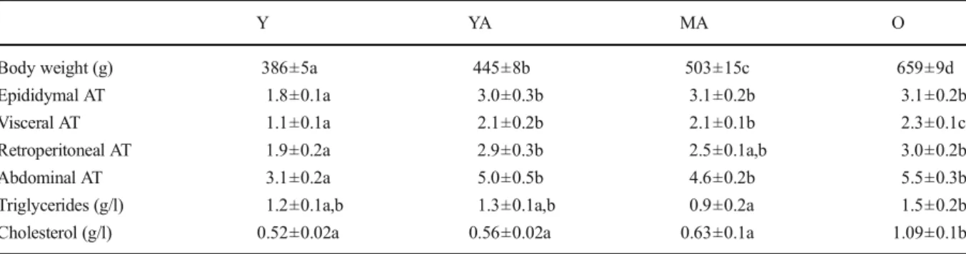
As shown in Table 1, the body weight followed a progressive increase with age. The YA rats had a 15.3 % increase in their body weight, which was followed by a further increase of 13.3 and 48.1 % for the MA and O animals. This was not the case for the adipose tissue which did not follow the same pattern. More specifically, all types of adipose tissue were significantly increased between 3 and 6 months of age (+67, +91, and +53 % for the epididymal, visceral, and retroperitoneal adipose tissues) and stabilized thereafter until the old age. Consequently, the abdominal adipose tissue estimated as the sum of the retroperitoneal and visceral adipose tissues was similarly increased (+61 %) between 3 and 6 months of age. Triglycerides and cholesterol concentrations in the plasma at the moment of the killing were moderately modified until the middle age but in-creased thereafter (+67 and +73 %, respectively).
There was a modest but significant impact of aging on the basal glycemia. Compared with the value mea-sured in the youngest group, the one meamea-sured at 6 months of age was higher (+9 %, Fig.1a). Between 6 and 11 months, it decreased (−20 %) and increased again thereafter (+18 %). The results of the oral glucose tolerance test were different (Fig. 1b). The area under the curve increased strongly between 3 and 6 months (+121 %). Thereafter, that parameter progressively returned to the value determined in the youngest animals.
Cardiac function studied ex vivo
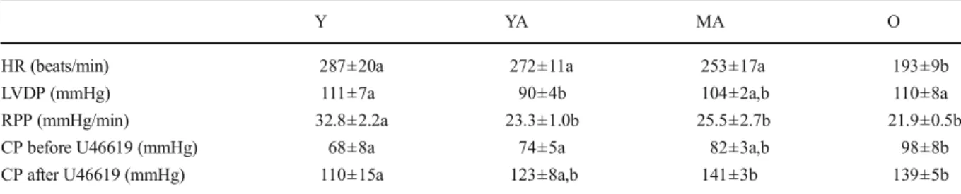
The mechanical function of the perfused isolated hearts was monitored before and 20 min after the infusion of U46619 (Table2). Aging had no significant effects on the heart rate until the age of 11 months, but decreased it
thereafter (−24 %). LVDP was first decreased (−19 % between 3 and 6 months), and then progressively re-stored until the old age. Finally, RPP inversely followed the increase in the adipose tissue mass with a reduction between 3 and 6 months (−29 %) and a stabilization thereafter. The infusion of U46619 raised the coronary pressure to values ranging from 110 to 141 mmHg but did not modify the parameters of the cardiac function.
Coronary reactivity
The responses to Ach of the coronary microvasculature were significantly affected by aging as shown in Fig.2a. Indeed, aging triggered a progressive decrease in the EDD, first between 3 and 6 months of age (−23 % at 60 pmol of injected Ach), and then between 6 and 11 months (−38.5 % at 60 pmol of injected Ach). Thereafter, between 11 and 17 months, the observed decrease was not significant. The calculated vasodilata-tion activities of the ECs are presented in Fig.2b. That parameter was increased between 3 and 6 months (+ 280 % at 60 pmol of injected Ach) but was decreased thereafter reaching the initial level observed at the young age. Fig.2cdepicts the SNP-induced vasodilata-tion in the three age groups. Interestingly, the EID strongly decreased from youth to young adulthood (−56.7 % at 600 pmol of injected SNP) and stabilized afterwards at middle and old ages.
eNOS expression and phosphorylation
No changes were observed between age groups in the levels of total eNOS (Fig. 3a and c) in cardiac homogenates. However, the amount of phosphorylated
Table 1 Basic characteristics of the animals
Y YA MA O
Body weight (g) 386±5a 445±8b 503±15c 659±9d Epididymal AT 1.8±0.1a 3.0±0.3b 3.1±0.2b 3.1±0.2b Visceral AT 1.1±0.1a 2.1±0.2b 2.1±0.1b 2.3±0.1c Retroperitoneal AT 1.9±0.2a 2.9±0.3b 2.5±0.1a,b 3.0±0.2b Abdominal AT 3.1±0.2a 5.0±0.5b 4.6±0.2b 5.5±0.3b Triglycerides (g/l) 1.2±0.1a,b 1.3±0.1a,b 0.9±0.2a 1.5±0.2b Cholesterol (g/l) 0.52±0.02a 0.56±0.02a 0.63±0.1a 1.09±0.1b The number of experiments was 7 per group. The adipose tissue weight is normalized to the body weight and is expressed in g of wet weight/ 100 g of body weight. Y:young, YA:young adult, MA:middle-aged, O:old, AT adipose tissue; a, b, c, d: the absence of a common letter indicates a significant difference
Fig. 1 Basal glycaemia (a) and area under the curve after the glucose loading (b). The number of experiments was seven per group. Y young animals, YA young adult animals, MA middle-aged animals, O old animals. The absence of a common letter (a–c) indicates a significant difference
eNOS (Fig.3a and b) increased between 3 and 6 months and returned thereafter to the value observed in the youngest animals.
Oxidative stress
Plasma thiol groups were reduced at 11 months (−15 % for the MA group compared with the Y group) but increased thereafter indicating increased oxidative stress at the age of 11 months, which was then restored (Fig. 4a). The global antioxidant power as estimated by the FRAP assay in plasma samples was progressively decreased by aging (Fig. 4b), reaching a significant difference from the age of 11 months (−22 and −30 % for the MA and O groups compared with the Y group). However, an increased glutathione peroxidase activity (Fig.4c) was observed in the plasma of old rats (+14 % compared with the Y animals). No difference was ob-served in the cytosolic redox potential as estimated by the lactate-to-pyruvate ratio in the coronary effluents (Fig. 4d). As shown in Fig. 4e, the aconitase-to-fumarase ratio estimated in cardiac homogenates was significantly decreased only after middle age (−33 % between 11 and 17 months), indicating a noticeable mitochondrial oxidative stress in the oldest animals.
Mitochondrial H2O2production
The effect of aging on basal glutamate/malate-related mitochondrial H2O2production assayed in isolated
mi-tochondria is presented in Fig.5a. That parameter sig-nificantly increased between youth and young adult-hood (+74 %) and was maintained high thereafter (+33 and +88 % at middle and old ages compared with youth). These differences were erased when antimycine A was added, emphasizing the involvement of the respi-ratory chain complex 3 in the age effects.
A biphasic pattern was observed for basal succinate-related mitochondrial H2O2production (Fig.5b). The
production, that was unchanged between 3 and 6 months, was increased between 6 and 11 months (+ 66 %) and strongly decreased thereafter (−56 %). The aging-related increase occurring between 6 and 11 months and the decrease between 11 and 17 months were inversed by the addition of rotenone alone and in association with antimycin A, suggesting that complex 1 was involved in that phenomenon probably through reverse electron flux.
When glutamate/malate/succinate were used as sub-strates, the pattern for the basal H2O2 production
(Fig.5c) was similar. Indeed, that parameter was similar in 3- and 6-month-old rats, increased in the 11-month animals (+47 % compared with the Y group) and de-creased thereafter (−79 % compared with the Y group). Although no difference was observed between 3 and 6 months, addition of rotenone rendered the 6-month value higher, a difference which was erased by antimycin A. Thus, complex 1 through inverse electron flux and also complex 3 were involved in that produc-tion. Furthermore, the increase in basal H2O2production
occurring between 6 and 11 months was erased and even inversed by rotenone and can be attributable to the activity of complex 1, although a participation of com-plex III was also likely given the effect of antimycin A. Finally, the strong decrease in basal H2O2production
observed between 11 and 17 months was cancelled by rotenone and inversed by the addition of antimycin A, suggesting the involvement of complexes 1 and 3 in the phenomenon.
Mitochondrial respiration
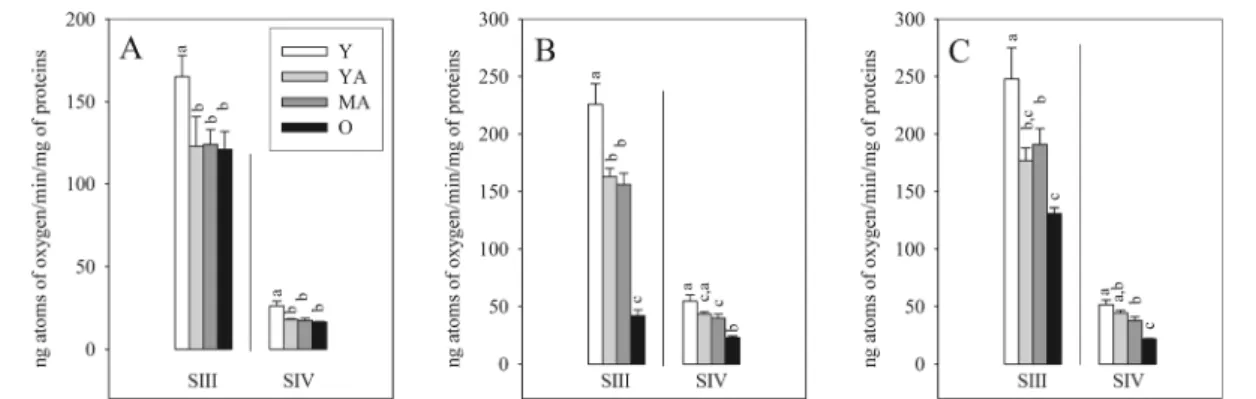
As shown in Fig. 6, whatever the substrate(s) used (glutamate/malate, succinate/rotenone, and glutamate/
Table 2 Ex vivo cardiac function
Y YA MA O
HR (beats/min) 287±20a 272±11a 253±17a 193±9b
LVDP (mmHg) 111±7a 90±4b 104±2a,b 110±8a
RPP (mmHg/min) 32.8±2.2a 23.3±1.0b 25.5±2.7b 21.9±0.5b CP before U46619 (mmHg) 68±8a 74±5a 82±3a,b 98±8b CP after U46619 (mmHg) 110±15a 123±8a,b 141±3b 139±5b The number of experiments was 7 per group. Y young, YAyoung adult, MA middle-aged, O old, HR heart rate, LVDP left ventricle developed pressure, RPP rate pressure product, CP coronary pressure. a, b: the absence of a common letter indicates a significant difference
malate/succinate) and the respiration state studied (state 3 or ADP-stimulated respiration and state 4 or
rotenone-related respiration), the respiration assayed in isolated mitochondria was reduced between 3 and 6 months (i.e., −29 % for the state 3 of glutamate/malate/succinate) and stabilized thereafter until middle age. Between 11 and 17 months, the evolution differed according to the sub-strate used. It remained stable with glutamate/malate, whereas it was further decreased with succinate/ rotenone and glutmate/malate/succinate (i.e., −73 % for the state 3 of succinate/rotenone).
Mitochondrial enzymatic activity
Aging did not alter the mitochondrial density until mid-dle age as evidenced by the activity of citrate synthase of cardiac homogenates (4.73±0.08, 4.75±0.10, and 4.57 ±0.06 mU/mg at 3, 6, and 11 months). However, it was significantly decreased in the myocardium of 17-month-old animals (3.58±0.24 mU/mg,−22 % compared with 11-month-old rats).
Regarding the mitochondrial respiratory chain com-plexes activities in isolated mitochondria, with the ex-ception of complex 2 whose activity was stable until 11 months of age and increased thereafter (+46 %), all of them had a stable activity until middle age, which de-creased at the age of 17 months (−36, −25, and −35 % for complexes 1, 3, and 4) as shown in Fig.7.
Discussion
This study was aimed at determining the effects of aging on the function of the coronary microvasculature from youth to old age by evaluating the contribution of each cellular type (ECs and SMCs) in the coronary vasodila-tation. These results were interpreted with regard to the systemic glucose tolerance, cardiac oxidative stress, and mitochondrial energy metabolism. The main results were that the EDD was progressively reduced by aging, the EID was decreased between youth and young adult-hood and stabilized thereafter, and the vasodilatation activity of the ECs was decreased between young adult-hood and old age after having been noticeably increased from youth to young adulthood.
In our study, the animals displayed a decrease in glu-cose tolerance from youth to young adulthood, which paralleled the increase in adipose fat mass during this period. Indeed, increased abdominal fat mass is known to trigger insulin resistance and glucose intolerance (Pfohl and Schatz2001). We do not know whether the cardiac
Fig. 2 Coronary reactivity in young (Y), young adult (YA), middle-aged (MA), and old (O) rats. The number of experiments was seven per group. a Endothelial-dependant vasodilatation (EDD), b endothelial cell vasodilatation activity (ECVA), c endo-thelial-independent vasodilatation (EID); SNP sodium nitroprus-side. a, b The absence of a common letter indicates a significant difference
glucose tolerance was similarly affected by aging as sys-temic glucose tolerance. However, a recent study suggests that cardiac insulin resistance precedes systemic insulin resistance (Abel2005). A diminished glucose uptake from the heart during this period together with the decrease in glutamate/malate-associated oxidative phos-phorylation and the presence of systemic oxidative stress (evidenced by the protein oxidation) could be associated with the decrease in cardiac mechanical ac-tivity and in the EDD of coronary microvasculature. The systemic oxidative stress could result from the activation of NADPH oxidase occurring at this age in the large conduit arteries (Oudot et al.2006) and/or in the adipose
tissue (Furukawa et al. 2004). Thus, the decline in cardiovascular function during aging starts early at life and is associated with events triggered by the increase in abdominal fat mass occurring during this period.
However, the abdominal fat mass was stable after 6 months of age, as well as the glutamate/malate-associated oxidative phosphorylation, suggesting the involvement of adipocytokines. This was also the case for the cardiac mechanical activity as already described in the literature (Mourmoura et al. 2011; Oudot et al. 2006). These phenomena were also accompanied by the progressive disappearance of glucose intolerance at the old age. The biochemical regulations responsible for
Fig. 3 Western blotting of the total endothelial nitric oxide synthase (eNOS), eNOS phosphorylated at serine 1177 (peNOS), and actin in cardiac homogenates. a Representative immunoblots; b quantification of peNOS; c quantification of total eNOS. The number of
experiments was seven per group. Y young animals, YA young adult animals, MA middle-aged animals, O old animals. a, b The absence of a common letter indicates a significant difference
that adaptation could involve an augmentation of the oxygen cost of energy synthesis in several organs evi-denced in the myocardium of middle-aged rats by a reduced metabolic efficiency (mechanical activity to oxygen consumption ratio) (Mourmoura et al. 2011). This would favor the wasting of excess energy substrates
such as glucose and normalize the whole body glucose tolerance. Thus, adaptation phenomena seem to occur after young adulthood allowing the normalization of glucose homeostasis reflected also at the level of oxidative phosphorylation and consequently of the cardiac mechanical function.
Fig. 4 Influence of aging on the oxidative stress. a Plasma thiol groups; b antioxidant status of the plasma (FRAP); c plasma glutathione peroxidase activity; d lactate to pyruvate ratio in the coronary effluents reflecting the cytosolic redox potential; e aconitase to fumarase ratio reflecting the mitochondrial oxidative
stress in cardiac homogenates. The number of experiments was seven per group. Y young animals, YA young adult animals, MA middle-aged animals, O old animals. a–d The absence of a com-mon letter indicates a significant difference
Fig. 5 Influence of aging on the mitochondrial H2O2production
in isolated mitochondria under basal conditions (absence of rote-none and antimycin A), after inhibition of complex I (roterote-none alone), and after inhibition of complexes I and III (rotenone in association with antimycin A) in the presence of glutamate/malate
(a), succinate (b), and glutamate/malate/succinate (c) as substrates. The number of experiments was seven per group. Y young ani-mals, YA young adult aniani-mals, MA middle-aged aniani-mals, O old animals. a–c The absence of a common letter indicates a signifi-cant difference
The modifications of the oxidative phosphorylation observed with aging were not related to the changes in respiratory chain complex activity. Indeed, activities of all the complexes were stable until middle age. There-after, they decreased for complexes 1, 3, and 4 and increased for complex 2. Those results fit perfectly with those described in the literature (see the references pre-sented in the “Introduction”). However, they do not explain the reduction of oxidative phosphorylation
reported between youth and young adulthood, the de-crease in oxygen consumption observed between mid-dle and old ages with succinate/rotenone, and the up-holding of mitochondrial oxidation noticed between middle and old ages with glutamate/malate. Taken together, these data indicate that the effects of age on the activities of the respiratory chain complexes cannot explain the aging-induced changes of oxi-dative phosphorylation.
Fig. 7 Influence of aging on the activity of the mitochondrial respiratory chain complexes (complexes 1, 2, 3, and 4 in (a)–(d), respectively) in isolated mitochondria. The number of experiments was seven per group. Y young animals, YA young adult animals, MA middle-aged animals, O old animals. a, b the Absence of a common letter indicates a significant difference
Fig. 6 Influence of aging on the state 3 (SIII) and state 4 (SIV) respiration rates of isolated mitochondria oxidizing glutamate/malate (a), succinate/rotenone (b), and glutamate/malate/succinate (c). The
number of experiments was seven per group. Y young animals, YA young adult animals, MA middle-aged animals, O old animals. a–c The absence of a common letter indicates a significant difference
Advanced aging is known to increase the mitochon-drial oxidative stress (Labunskyy and Gladyshev2014) deriving notably from the respiratory chain complex 3 due to an inhibition of this electron transfer system (Fannin et al. 1999; Lesnefsky et al. 2001; Hoppel et al. 2002). In the present study, the systemic and myocardial oxidative stress paralleled neither the glu-cose tolerance values nor the changes in energy metab-olism. Glutamate/malate, a complex 1-related substrate, increased the H2O2generation as soon as young
adult-hood and the phenomenon seemed to occur mainly at the level of complex 3. By contrast, succinate, a com-plex 2-related substrate, increased the H2O2generation
only at middle age through a production occurring by reverse electron flux at the level of complex 1. That increase was not confirmed at the old age, as the release was strongly reduced as compared with middle age, which could result from combined action of decreased complexes 1, 3, and 4 activities as well as increased complex 2 activity. A similar pattern was observed when glutamate/malate and succinate were used together.
The impact of these changes in mitochondrial oxida-tive stress was estimated ex vivo by evaluating the aconitase-to-fumarase, which was increased at 17 month of age. This was not in conflict with the results of the in vitro mitochondrial H2O2release. Indeed, glucose
was the unique energy substrate supplied to the isolated heart, and it is a complex 1-related substrate such as glutamate/malate. Aging could thus increase mitochon-drial H2O2release as soon as young adulthood. Until
middle age, the heart could have sufficient anti-oxidant defenses to scavenge the toxic molecules, which pro-gressively declined until the old age.
As reported previously, cardiac mechanical function was decreased between youth and young adulthood and stabilized thereafter. The cardiac mechanical efficiency reflecting the contractile capacities of myocardial fibers is defined as the RPP normalized to the heart dry weight. In the present study, we were not able to estimate the heart weight, as we used a part of the myocardium to prepare mitochondria. However, the heart weight is known to increase with age in the rat. This would amplify the aging-induced reduction of cardiac mechan-ical efficiency. That observation agrees well with our results of the mitochondrial oxidative phosphorylation which decreased between youth and young adulthood and stabilized thereafter. The type of perfusion chosen for our study could even contribute to attenuate the aging-related differences. Indeed, Langendorff
perfusion at low extracellular calcium concentration minimally stimulates the heart. Aging could have re-duced the cardiac mechanical work more intensively if the hearts had been maximally stimulated (working mode with high preload and perfusate calcium concen-tration). Differences could thus appear, particularly at the old age when the mitochondria display a reduced density and increased oxidative stress.
The coronary flow is an important regulator of the cardiac mechanical function. In the present study, we were not able to obtain a satisfying evaluation of the coronary flow, since we were not able to determine the heart weight. One of our recent studies (Mourmoura et al. 2011) performed in the same batch of animals revealed that the coronary flow expressed as ml/min/g of dry weight was unchanged between youth and middle age, suggesting that the coronary bed adapted in parallel with the gain of heart weight in normal aging. Further-more, the coronary flow was only slightly decreased by aging in the Langendorff hearts of 21-month-old Wistar rats (−11 %) compared with that of their 2-month-old counterparts (Oudot et al. 2006). This confirms the study of Tomanek et al. (1991), which indicates remod-eling of the coronary vessels with age. This morpholog-ical study performed in young and senescent beagles has shown that aging reduces the capillary numerical densi-ty but maintains their volume densidensi-ty through a com-pensatory increase in capillary diameter. Thus, aging could not modify the coronary flow of Langendorff-perfused hearts until the old age. We do not know if this vessel remodeling occurred in our rats, since species difference could occur. However, this allows the satis-fying estimation of the coronary reserve.
Our study showed that aging triggered a progressive decrease in EDD, which began as soon as youth. That phenomenon is well recognized now with studies in humans (Chauhan et al. 1996; Egashira et al. 1993) and laboratory animals (Toma et al.1985) agreeing well with this age-related decline. However, the contribution of each vascular cellular type is not known. Our study allows the description of that contribution. The function of the vascular SMCs of the coronary microvasculature was easily depicted in our study through the measure-ment of the EID, as the nitric oxide donor nitroprusside permits the relaxation of those cells. That parameter was high in young hearts and drastically decreased as soon as the young adulthood to values which were very close to those observed at the old age. Those results perfectly fit with the observations made by Toma et al. (1985) in
the isolated guinea pig hearts. Energy is necessary for the relaxation of muscular cells and the low oxidative phosphorylation detected in the isolated mitochondria derived from young adult, middle-aged, and old hearts (cardiac homogenates of cardiomyocytes and vascular cells) could perfectly explain the age-related decline observed for EID that contributed to the progressive age-related loss of EDD.
The function of ECs is more difficult to evaluate. In the present study, we mathematically extracted from the EDD and EID curves a parameter that depicted the vasodilatation activity of ECs. Al-though that parameter is expressed in picomoles of nitroprusside, it does not reflect the vasodilatation activity resulting only from NO production, but it also takes into account all the vasodilatation agents produced by the ECs. Interestingly, the EC vasodi-latation activity (ECVA) was strongly increased from youth to young adulthood. Thereafter, it pro-gressively decreased until the old age to reach the value measured at the young age. These changes perfectly paralleled those of the Western Blot anal-ysis of the phosphorylated eNOS, suggesting the involvement of nitric oxide. Furthermore, the amount of phosphorylated eNOS and ECVA follow-ed similar trends as the glucose intolerance. As glucose intolerance is generally associated with in-creased fat mass and body weight, the inin-creased ECVA could improve the maximal rate of coronary flow and physical activity of obese individuals.
Conclusions
The progressive changes of the functional behavior of ECs and SMCs occurring from youth to old age were studied. Our results indicate that a decline in EDD, which was due to functional modifications of both ECs and SMCs, was developed from youth to middle age. Between youth and young adulthood, the EDD decrease was due to a strong reduction of the SMCs function, but this was compensated by the enhancement of the ECs dilatation activity. Thereafter, between young adulthood and middle age, relaxation of SMCs stabi-lized, but EC dilatation activity significantly decreased. The aging-induced alterations of the SMC function could be explained by abnormalities of the energy me-tabolism, namely a reduction of the mitochondrial oxi-dative phosphorylation occurring as soon as young
adulthood. Conversely, the ECs alterations could be related to the changes in glucose tolerance on eNOS activity. Overall, our study revealed that the decline in cardiovascular and mitochondrial parameters associated with later life cardiovascular incidents is already present in young adulthood. Thus, it seems important that inter-ventions such as physical activity that aim to inverse these results and improve the cardiovascular function should begin at young age.
Acknowledgments The authors would like to thank M. Chris-tophe Cottet for carefully editing the manuscript, Cindy Tellier for animal care, Joëlle Demaison for the determination of the respira-tory chain complex activities, and Mireille Osman for the mea-surements of GPx activity, FRAP assay, and thiol groups in the plasma. This work was supported by the French National Institute of Agronomical Research (INRA), the French National Institute of Health and Medical Research (INSERM), and Joseph Fourier University, Grenoble, France.
References
Abel ED (2005) Myocardial insulin resistance and cardiac com-plications of diabetes. Curr Drug Targets 5:219–226 Bergmeyer HU (1974) Methods of enzymatic analysis. Weinheim,
Verlag Chemie
Castelluccio C, Baracca A, Fato R, Pallotti F, Maranesi M, Barzanti V, Gorini A, Villa RF, Parenti Castelli G, Marchetti M (1994) Mitochondrial activities of rat heart during ageing. Mech Ageing Dev 76:73–88
Charansonney OL (2011) Physical activity and aging: a life-long story. Discov Med 12:177–185
Chauhan A, More RS, Mullins PA, Taylor G, Petch C, Schofield PM (1996) Aging-associated endothelial dysfunction in humans is reversed byL-arginine. J Am Coll Cardiol 28: 1796–1804
Cortez MY, Torgan CE, Brozinick JT Jr, Ivy JL (1991) Insulin resistance of obese Zucker rats exercise trained at two differ-ent intensities. Am J Physiol 261:E613–E619
Cox DA, Matlib MA (1993) A role for the mitochondrial Na(+) -Ca2+exchanger in the regulation of oxidative phosphoryla-tion in isolated heart mitochondria. J Biol Chem 268:938– 947
Delaval E, Perichon M, Friguet B (2004) Age-related impairment of mitochondrial matrix aconitase and ATP-stimulated prote-ase in rat liver and heart. Eur J Biochem 271:4559–4564 Effron MB, Bhatnagar GM, Spurgeon HA, Ruaño-Arroyo G,
Lakatta EG (1987) Changes in myosin isoenzymes, ATPase activity, and contraction duration in rat cardiac muscle with aging can be modulated by thyroxine. Circ Res 60:238–245 Egashira K, Inou T, Hirooka Y, Kai H, Sugimachi M, Suzuki S, Kuga T, Urabe Y, Takeshita A (1993) Effects of age on endothelium-dependent vasodilation of resistance coronary artery by acetylcholine in humans. Circulation 88:77–81
Faloona GR, Srere PA (1969) Escherichia coli citrate synthase. Biochemistry 8:4497–4503
Fannin SW, Lesnefsky EJ, Slabe TJ, Hassan MO, Hoppel CL (1999) Aging selectively decreases oxidative capacity in rat heart interfibrillar mitochondria. Arch Biochem Biophys 372:399–407
Favier AE (1995) Analysis of free radicals in biological systems. Birkhauser Verlag, Basel
Furukawa S, Fujita T, Shimabukuro M, Iwaki M, Yamada Y, Nakajima Y, Nakayama O, Makishima M, Matsuda M, Shimomura I (2004) Increased oxidative stress in obesity and its impact on metabolic syndrome. J Clin Invest 114:1752–1761 Gardner PR, Nguyen DD, White CW (1994) Aconitase is a sensitive and critical target of oxygen poisoning in cultured mammalian cells and in rat lungs. Proc Natl Acad Sci U S A 91:12248–12252
Gobel FL, Norstrom LA, Nelson RR, Jorgensen CR, Wang Y (1978) The rate-pressure product as an index of myocardial oxygen consumption during exercise in patients with angina pectoris. Circulation 57:549–556
Günzler WA, Kremers H, Flohé L (1974) An improved coupled test procedure for glutathione peroxidase (EC 1-11-1-9-) in blood. Z Klin Chem Klin Biochem 12:444–448
Hoppel CL, Moghaddas S, Lesnefsky EJ (2002) Interfibrillar cardiac mitochondrial complex III defects in the aging rat heart. Biogerontology 3:41–44
Labunskyy VM, Gladyshev VN (2014) Role of reactive oxygen species-mediated signaling in aging. Antioxid Redox Signal (in press)
Lee HY, Oh BH (2010) Aging and arterial stiffness. Circ J 74: 2257–2262
Lesnefsky EJ, Gudz TI, Moghaddas S, Migita CT, Ikeda-Saito M, Turkaly PJ, Hoppel CL (2001) Aging decreases electron transport complex III activity i heart interfibrillar mitochon-dria by alteration of the cytochrome c binding site. J Mol Cell Cardiol 33:37–47
Liviakis L, Pogue B, Paramsothy P, Bourne A, Gill EA (2010) Carotid intima-media thickness for the practicing lipidologist. J Clin Lipidol 4:24–35
Mourmoura E, Leguen M, Dubouchaud H, Couturier K, Vitiello D, Lafond JL, Richardson M, Leverve X, Demaison L (2011) Middle age aggravates myocardial ischemia through surpris-ing upholdsurpris-ing of complex II activity, oxidative stress, and reduced coronary perfusion. Age (Dordr) 33:321–336 Mourmoura E, Vial G, Laillet B, Rigaudière JP, Hininger-Favier I,
Dubouchaud H, Morio B, Demaison L (2013) Preserved endothelium-dependent dilatation of the coronary microvas-culature at the early phase of diabetes mellitus despite the increased oxidative stress and depressed cardiac mechanical function ex vivo. Cardiovasc Diabetol 12:49
Mourmoura E, Chaté V, Couturier K, Laillet B, Vial G, Rigaudiere JP, Morio B, Malpuech-Brugère C, Azarnoush K, Demaison L (2014) Body adiposity dictates different mechanisms of increased coronary reactivity related to improved in vivo cardiac function. Cardiovasc Diabetol 13(1):54
Nuutinen EM (1984) Subcellular origin of the surface fluorescence of reduced nicotinamide nucleotides in the isolated perfused rat heart. Basic Res Cardiol 79:49–58
Oudot A, Martin C, Busseuil D, Vergely C, Demaison L, Rochette L (2006) NADPH oxidases are in part responsible for in-creased cardiovascular superoxide production during aging. Free Radic Biol Med 40:2214–2222
Pfohl M, Schatz H (2001) Strategies for the prevention of type 2 diabetes. Exp Clin Endocrinol Diabetes 109:S240–S249 Sawabe M (2010) Vascular aging: from molecular mechanism to
clinical significance. Geriatr Gerontol Int 10:S213–S220 Sawabe M, Hamamatsu A, Chida K, Mieno MN, Ozawa T (2011)
Age is a major pathobiological determinant of aortic dilata-tion: a large autopsy study of community deaths. J Atheroscler Thromb 18:157–165
Scott AD, Keegan J, Mohiaddin RH, Firmin DN (2011) Noninvasive detection of coronary artery wall thickening with age in healthy subjects using high resolution MRI with beat-to-beat respiratory motion correction. J Magn Reson Imaging 34:824–830
Skrzypiec-Spring M, Grotthus B, Szelag A, Schulz R (2007) Isolated heart perfusion according to Langendorff—still via-ble in the new millennium. J Pharmacol Toxicol Methods 55: 113–126
Takemura A, Iijima K, Ouchi Y (2010) Molecular mechanism of vascular aging: impact of vascular smooth muscle cell calci-fication via cellular senescence. Clin Calcium 20:1646–1655 Toma BS, Wangler RD, DeWitt DF, Sparks HV Jr (1985) Effect of development on coronary vasodilator reserve in the isolated guinea pig heart. Circ Res 57:538–544
Tomanek RJ, Aydelotte MR, Torry RJ (1991) Remodeling of coronary vessels during aging in purebred beagles. Circ Res 69:1068–1074
Venkataraman P, Krishnamoorthy G, Selvakumar K, Arunakaran J (2009) Oxidative stress alters creatine kinase system in serum and brain regions of polychlorinated biphenyl (Aroclor 1254)-exposed rats: protective role of melatonin. Basic Clin Pharmacol Toxicol 105:92–97
Wijnands KA, Vink H, Briedé JJ, van Faassen EE, Lamers WH, Buurman WA, Poeze M (2012) Citrulline a more suitable substrate than arginine to restore NO production and the microcirculation during endotoxemia. PLoS One 7:e37439 Wyatt AW, Steinert JR, Mann GE (2004) Modulation of theL
-arginine/nitric oxide signalling pathway in vascular endothe-lial cells. Biochem Soc Symp 71:143–156