HAL Id: inserm-01707534
https://www.hal.inserm.fr/inserm-01707534
Submitted on 12 Feb 2018
HAL is a multi-disciplinary open access archive for the deposit and dissemination of sci-entific research documents, whether they are pub-lished or not. The documents may come from teaching and research institutions in France or abroad, or from public or private research centers.
L’archive ouverte pluridisciplinaire HAL, est destinée au dépôt et à la diffusion de documents scientifiques de niveau recherche, publiés ou non, émanant des établissements d’enseignement et de recherche français ou étrangers, des laboratoires publics ou privés.
Jean-Noel Freund, et al.
To cite this version:
Camille Balbinot, Olivier Armant, Nabila Elarouci, Laetitia Marisa, Elisabeth Martin, et al.. The Cdx2 homeobox gene suppresses intestinal tumorigenesis through non-cell-autonomous mech-anisms. Journal of Experimental Medicine, Rockefeller University Press, 2018, Epub ahead of print. �10.1084/jem.20170934�. �inserm-01707534�
Ar ticle
Jour
nal of Exper
imental Medicine
INTRODUCTION
Embryonic development and tumor growth share several features. For instance, homeobox genes, which are crucial for body plan organization, can also be involved in tumori-genesis either as tumor suppressors or as oncogenes. In line with this, it is now well established that tumors represent cellular masses that are structurally organized but anatomi-cally and functionally abnormal compared with healthy or-gans (Egeblad et al., 2010). Tumor growth is driven by the intrinsic properties of the cells and by cell interactions with their environment. The role of cell interactions between tumor cells and other cell types, such as cancer-associated fi-broblasts, immune cells, or endothelial cells, has been widely described (Lujambio et al., 2013; Marusyk et al., 2014). However, much less is known about whether and how epi-thelial cells at different premalignant stages can interact and participate in tumor initiation.
Besides its role in embryonic development, the ho-meobox gene Cdx2 is an important regulator of the dy-namic homeostasis of the gut, providing tissue identity to the stem cells and coordinating cell proliferation and dif-ferentiation during the constant renewal of the epithelium (Verzi et al., 2011; Stringer et al., 2012; Simmini et al., 2014).
Its expression is frequently altered in human colorectal can-cers (CRCs) and in animal models of intestinal cancan-cers, and convergent studies in mice have established its tumor sup-pressor role in the gut (Aoki et al., 2003; Bonhomme et al., 2003; Gross et al., 2008; Hryniuk et al., 2014). Recently, a functional link between B-Raf activation and loss of Cdx2 in a subset of CRCs has demonstrated the relevance of the combination of these molecular events within tumor cells and the importance of cell differentiation dictated by Cdx2 against intestinal tumorigenesis (Sakamoto et al., 2017; Tong et al., 2017). In the present study, starting from data obtained in a collection of human CRCs, we developed an original mouse model with the goal of uncovering the importance of indirect interactions between different types of epithelial cells at premalignant stages in triggering tumorigenesis. The results highlight a novel property of Cdx2 in the gut, in that this homeobox gene exerts a non–cell-autonomous tumor suppressor activity. In addition, a new paradigm for meta-plasia emerges, in the sense that metaplastic cells, widely considered as precancerous, can induce the tumorigenic evolution of adjacent nonmetaplastic cells without them-selves becoming cancerous.
Developmental genes contribute to cancer, as reported for the homeobox gene Cdx2 playing a tumor suppressor role in the gut. In this study, we show that human colon cancers exhibiting the highest reduction in CDX2 expression belong to the serrated subtype with the worst evolution. In mice, mosaic knockout of Cdx2 in the adult intestinal epithelium in-duces the formation of imperfect gastric-type metaplastic lesions. The metaplastic knockout cells do not spontaneously become tumorigenic. However, they induce profound modifications of the microenvironment that facilitate the tumori-genic evolution of adjacent Cdx2-intact tumor-prone cells at the surface of the lesions through NF-κB activation, in-duction of inducible nitric oxide synthase, and stochastic loss of function of Apc. This study presents a novel paradigm in that metaplastic cells, generally considered as precancerous, can induce tumorigenesis from neighboring nonmetaplas-tic cells without themselves becoming cancerous. It unveils the novel property of non–cell-autonomous tumor suppressor gene for the Cdx2 gene in the gut.
The Cdx2 homeobox gene suppresses intestinal
tumorigenesis through non–cell-autonomous mechanisms
Camille Balbinot,
1Olivier Armant,
2Nabila Elarouci,
3Laetitia Marisa,
3Elisabeth Martin,
1Etienne De Clara,
1Alina Onea,
4Jacqueline Deschamps,
5Felix Beck,
6Jean-Noël Freund,
1and Isabelle Duluc
11Université de Strasbourg, Institut National de la Santé et de la Recherche Médicale, IRF AC UMR-S1113, Fédération de Médecine Translationnelle de Strasbourg,
Strasbourg, France
2Karlsruhe Institute of Technology, Institute of Toxicology and Genetics, Karlsruhe, Germany 3Cartes d’Identité des Tumeurs Program, Ligue Nationale Contre le Cancer, Paris, France 4Département de Pathologie, Centre Hospitalier Universitaire de Strasbourg, Strasbourg, France 5Developmental Biology and Stem Cell Research, Hubrecht Institute, Utrecht, Netherlands 6Barts and The London School of Medicine and Dentistry, London, England, UK
© 2018 Balbinot et al. This article is distributed under the terms of an Attribution–Noncommercial–Share Alike–No Mirror Sites license for the first six months after the publication date (see http ://www .rupress .org /terms /). After six months it is available under a Creative Commons License (Attribution–Noncommercial– Share Alike 4.0 International license, as described at https ://creativecommons .org /licenses /by -nc -sa /4 .0 /).
Correspondence to Jean-Noël Freund: jean-noel.freund@inserm.fr
O. Armant’s present address is Institut de Radioprotection et de Sûreté Nucléaire (IRSN), PRP-ENV/SER IS/LECO, Cadarache, Saint-Paul-lez-Durance, France.
on February 8, 2018
jem.rupress.org
Downloaded from
RESULTS
Human serrated-type colon cancers with a stem cell signature exhibit a strong reduction of CDX2
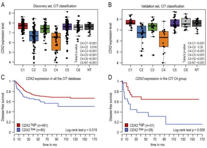
Analyzing the expression of the CDX2 homeobox gene in a cohort of 566 human CRCs (Cartes d’Identité des Tumeurs study) previously classified into six subtypes (Marisa et al., 2013) revealed a down-regulation in two subtypes: the C2 subtype, enriched with microsatellite instable and hyper-mutated tumors, and a stronger down-regulation in the C4 subtype characterized by serrated precursor neoplasia, stroma infiltration, and a stem cell–like/mesenchymal signature (Fig. 1, A and B). In the consensus classification system (Guin-ney et al., 2015), the same down-regulation was also observed in subtypes CMS1 and CMS4, including the C2 and C4 sub-types from Marisa et al. (2013) (Fig. S1). Using an unsuper-vised approach fixing the threshold at the median value of CDX2 in the C4 subtype, patients of the whole cohort below the threshold exhibited worse disease-free survival (Fig. 1 C). Within the C4 subtype, disease-free survival was even worse in patients below the threshold compared with patients above
the threshold (Fig. 1 D). Thus, the strong reduction of CDX2 correlates with poor evolution of the disease.
Loss of function of Cdx2 in the mouse intestine induces imperfect gastric-type metaplastic lesions in the cecum, which do not spontaneously undergo cancerous evolution To address the pathological relevance of the loss of expres-sion of Cdx2, mosaic gene knockout was induced in the gut
epithelium of adult AhCreERT::Cdx2f/f mice, as described
previously (Stringer et al., 2012). Mosaic knockout is man-datory for the current long-term studies because massive loss of function of Cdx2 is lethal as a result of digestive problems (Verzi et al., 2011). Within 4–6 wk after gene knockout by β-naphthoflavone and tamoxifen (βNF+Tam)
administra-tion, the AhCreERT::Cdx2f/f mice exhibited subsurface cysts
throughout the small intestine that did not evolve with time, as previously reported (Stringer et al., 2012). In addition, after 4 mo, all the mice analyzed in this study (n > 25) developed one to two polypoid lesions in the cecum, and in 20% of the mice, lesions were also found in the very distal ileum and
Figure 1. CDX2 gene expression level in 566 human colon cancers and 19 nontumoral samples of the GSE39582 dataset. (A) Boxplot of the level
of CDX2 expression in the 443 CRC samples of the discovery set organized in the six subtypes according to Marisa et al. (2013) (C1–C6). (B) Boxplot of the
CDX2 expression level in the 123 samples of the validation set organized in six subtypes. Data are given ± SD. (C) Disease-free survival comparing CDX2high
versus CDX2low CRC in the GSE39582 dataset. The cutoff for low versus high CDX2 expression is fixed at the median of the C4 group. CDX2low patients
ex-hibit a significantly reduced disease-free survival (P < 0.019). (D) Disease-free survival comparing CDX2high versus CDX2low CRC in the C4 subtype. CDX2low
patients exhibit a significantly reduced disease-free survival (P = 0.009). P-values were calculated with the log-rank test.
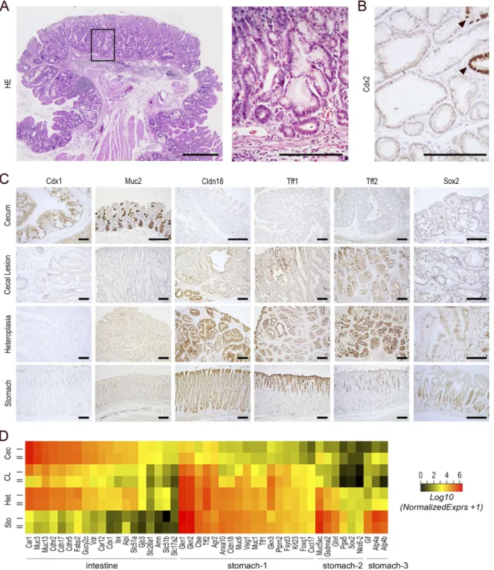
proximal colon (Fig. 2 A). These lesions did not compromise the life span of the animals (see Fig. 5 A). As shown in Fig. 2 (A and B), the lesions consisted of Cdx2-devoid glands inter-mingled with few Cdx2-intact glands, reflecting the mosaic ablation of the gene. They were covered by a single-layered polarized epithelium with local erosion in surface and exhib-ited signs of inflammation, with the presence of eosinophils and neutrophils in the stroma. Histologically, the lesions con-sisted of fundic-type glandular structures with dilated cysts together with areas of foveolar hyperplasia and antral-type glands covered by a polarized epithelium with nuclei reg-ularly distributed at the cell base. They shared histological properties with glandular cystic and hyperplastic polyps re-ported in the human stomach but differed from typical hy-perplastic polyps of the intestine. By immunohistochemistry (Fig. 2 C), the cecal lesions showed a loss of expression of intestinal proteins known to be downstream targets of Cdx2, such as the orthologous homeoprotein Cdx1 and the mucin Muc2, and conversely the onset of expression of gastric pro-teins including Claudin-18, Tff1, and Tff2. However, unlike the cecal foregut-type heteroplasia already developing during
embryogenesis in heterozygous Cdx2+/− mice (Beck et al.,
1999; Stringer et al., 2008), the lesions of AhCreERT::Cdx2f/f
mice failed to express Sox2. To further characterize them at the molecular level, we determined their transcriptomic profile and compared it with normal cecum and stomach and also with cecal foregut-type heteroplasia developing in
Cdx2+/− mice. This led to identification of a large number of
genes (5,915) altered in the lesions of AhCreERT::Cdx2f/f mice
compared with normal cecum (Table S1, sheet 1). The large number of genes is consistent with the Cdx2 protein being a major regulator of intestinal homeostasis and with its binding
to ∼14,000 chromatin sites across the genome of enterocytes
(Verzi et al., 2010). Among them, transcripts for intestinal markers were strongly reduced (i.e., Alpi, Muc3/13, Cdh17, Cdhr2/5, Fabp2, Slc51a/b, Cdx1, and Isx), whereas those for several gastric markers were turned on (i.e., Cldn18, Ctse, Gkn1/2/3, Muc1/6, Ptprn2, Tff1/2, Vsig1, Gsdma2, Krt23, Fxyd3, and Foxq1; Fig. 2 D; and Table S1, sheet 2). Yet the
le-sions of adult AhCreERT::Cdx2f/f mice exhibited a lower and
more heterogeneous expression of gastric genes compared with normal stomach and also with the foregut-type
het-eroplasia of Cdx2+/− mice; moreover, several typical markers
even failed to turn on (Fig. 2 D). Thus, we concluded that in the long term, the loss of Cdx2 produces cecal lesions char-acterized by an imperfect gastric-type metaplastic phenotype. Because metaplastic lesions are commonly considered to be precancerous in many organs, we questioned the patho-logical significance of the cecal lesions developing in
Ah-CreERT::Cdx2f/f mice. In these lesions, gene ontology terms
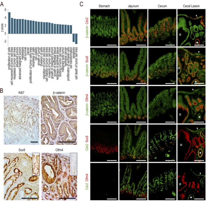
related to cancer, neoplasia, cell transformation, cell prolif-eration, cell survival, and growth of tumors were enriched, whereas those for apoptosis and cell death of tumor cells were underrepresented (Fig. 3 A). However, the pathohistological examination failed to display any dysplastic structure. Ki67
immunostaining revealed an important number of prolifer-ating cells unevenly distributed predominantly in the middle third of the lesions, which is reminiscent of the localization of proliferating cells in the isthmus of the gastric mucosa, unlike the crypts in the intestine (Fig. 3 B). The canonical Wnt pathway was not abnormally activated, as indicated by β-catenin remaining associated with the plasma membranes without cytoplasmic/nuclear translocation (Fig. 3 B). How-ever, the formation of the lesions strongly impacted the stem cell compartment, as suggested by the altered expression,
ei-ther up or down, of ∼45% (173/384) of the genes of the
intestinal stem cell signature (Muñoz et al., 2012; Table S1, sheet 3). For instance, the RNA sequencing data showed a strong stimulation of Sox9 (6×) and Olfm4 (>200×). This prompted us to analyze the tissue distribution of the corre-sponding proteins (Fig. 3, B and C). The Sox9 protein, present at the bottom of the glands in the normal stomach and at the crypt base all along the gut, was strongly expressed in both Cdx2-depleted glands of the lesions and in the few embed-ded Cdx2-intact glands, and also heterogeneously in the sur-face epithelium of the lesions. The Olfm4 protein, present in the stem/progenitor cells of the small intestine but in neither the mouse stomach nor in the cecum and colon (except for low expression in rare glands/crypts), became strongly ex-pressed in the Cdx2-depleted glands of the lesions, but it was not turned on in either the embedded Cdx2-intact glands or in the surface epithelium. Interestingly, although the Sox9
pattern was similar in the lesions of AhCreERT::Cdx2f/f mice
and in the foregut-type heteroplasia of Cdx2+/− mice, the
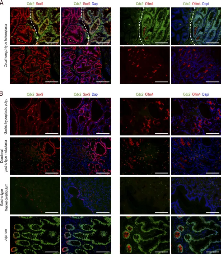
Olfm4 pattern was very different because this protein was almost absent in the heteroplasia, with the exception of few clusters of glands histologically indistinguishable from the Olfm4-negative glands (Fig. 4 A).
The patterns obtained in the cecal lesions of
AhCreERT::Cdx2f/f mice were compared with those observed
in human lesions (Fig. 4 B). In human gastric hyperplastic polyps, the expression of Sox9 and Olfm4 was strong in more than half of the glandular structures. In contrast, in gastric-type heterotopia of the duodenum, strong Olfm4 was observed in only few glands (<10%). Finally, Meckel diverticula with gastric-type differentiation showed only rare structures ex-pressing Sox9 at a low level, and no expression of Olfm4. All these human lesions failed to express Cdx2. These data are consistent with the notion that the imperfect gastric-type
metaplastic lesions arising in the cecum of AhCreERT::Cdx2f/f
mice share properties with human gastric hyperplastic polyps. Based on the high level of cell proliferation and the perturbation of the stem cell compartment in the cecal
le-sions of AhCreERT::Cdx2f/f mice, a series of five animals were
maintained alive up to 23–24 mo (20–21 mo after Cdx2 knockout) to investigate whether these lesions underwent spontaneous cancerous evolution in aged animals. Pathohisto-logical examination revealed neither dysplastic nor neoplastic
structures. At the molecular level, β-catenin remained
mem-branous without any evidence of cytoplasmic/nuclear
on February 8, 2018
jem.rupress.org
Figure 2. Cecal lesion induced by mosaic gene knockout of Cdx2 in the adult gut epithelium. (A) Histology of cecal lesions in AhCreERT::Cdx2f/f mice 4
mo after βNF+Tam administration. Bar, 500 µm. The boxed region is magnified in the right panel. Bar, 250 µm. (B) Immunodetection of the Cdx2 protein. The protein is almost absent, except in few glands with intact Cdx2 (arrowheads) entrapped in the lesions. Bar, 100 µm. In A and B, n = 20 mice from five crossings.
(C) Immunodetection of intestinal proteins (Cdx1, Muc2) and gastric proteins (Cldn18, Tff1, Tff2, Sox2) in the cecum of wild-type mice, cecal lesions of
Ah-CreERT::Cdx2f/f mice, cecal heteroplasia of Cdx2+/− mice, and the stomach of wild-type mice. n = 4 animals per genotype from two crossings. Bars, 100 µm. (D)
Heatmap comparison of transcriptomic data for intestinal and gastric genes in the cecum of wild-type mice (Cec), cecal lesions of AhCreERT::Cdx2f/f mice (CL),
cecal heteroplasia of Cdx2+/− mice (Het), and the stomach of wild-type mice (Sto). Stomach-1 represents gastric genes up-regulated in CL and Het compared
with Cec; Stomach-2 represents gastric genes up-regulated in Het but not in CL; Stomach-3 represents gastric genes up-regulated in neither CL nor in Het.
location (unpublished data). Thus, in >30 AhCreERT::Cdx2f/f
mice analyzed at various time points, none of the cecal lesions spontaneously underwent tumorigenic evolution.
Evolution of the cecal lesions in a tumor-prone context Based on these observations, the lesions were explored in a
can-cer-prone context by crossing AhCreERT::Cdx2f/f mice with
Apc+/Δ14 mice (Colnot et al., 2004). Apc+/Δ14 mice develop
adenomatous polyps predominantly in the small intestine, but also a few in the colon and rarely in the cecum (respectively 18 ± 7 and 1 ± 1 polyps in the small intestine and in the colon per mouse, and only 1 polyp every 10 mice in the cecum). As previously reported (Colnot et al., 2004), the life span of
Apc+/Δ14 animals was compromised compared with wild-type
Figure 3. Functional characterization of the cecal lesions. (A) Gene ontology enrichment analysis for terms related to cancer in the transcriptome
of the cecal lesions of AhCreERT::Cdx2f/f mice compared with the cecum of wild-type mice. (B) Immunohistochemical staining of Ki67, β-catenin, Sox9, and
Olfm4 in the cecal lesions; the insets respectively represent the Sox9 and Olfm4 patterns in wild-type cecal glands. Bars: (β-catenin) 100 µm; (Ki67, Sox9, and Olfm4) 200 µm. (C) Coimmunofluorescence detection of β-catenin and Cdx2, β-catenin and Sox9, β-catenin and Olfm4, Cdx2 and Sox9, and Cdx2 and Olfm4 in serial sections of the stomach, jejunum, and cecum of wild-type mice and in the cecal lesions of AhCreERT::Cdx2f/f mice. In the cecal lesions, open
squares show a gland depleted in Cdx2, and asterisks show a gland with intact Cdx2. The arrowheads point to the surface epithelium expressing Cdx2. Bars, 50 µm. Pictures in B and C are representative of the data obtained in n = 4 animals per genotype in two crossings.
on February 8, 2018
jem.rupress.org
Figure 4. Comparative expression patterns of the Cdx2, Sox9, and Olfm4 proteins in gastrointestinal lesions. (A) Foregut-type heteroplasia in
the cecum of Cdx2+/− mice. Top: region at the border (dotted line) between the normal cecal mucosa (right side) and the heteroplastic tissue (left side).
The normal epithelium expresses Cdx2, whereas Sox9 and Olfm4 are present at the bottom of the glands; the heteroplastic tissue, devoid of Cdx2, shows a strong expression of Sox9 but no expression of Olfm4. Bottom: rare clusters of glands in the heteroplasia expressing both Sox9 and Olfm4. Bars, 50 µm. n = 4 mice. (B) Human lesions: hyperplastic polyp in the stomach (n = 3); gastric-type metaplastic polyp in the duodenum (n = 1); and Meckel diverticulum
with gastric-type differentiation (n = 3). For the normal small intestinal mucosa (bottom), the crypt-villous axis is from left to right (n = 3). Bars, 50 µm.
mice and thus also compared with βNF+Tam-injected
AhCreERT::Cdx2f/f mice; in the cancer-prone context, the
Apc+/Δ14::AhCreERT::Cdx2f/f mice treated with βNF+Tam
showed a tendency to an even shorter life span, although this
was not significantly different from Apc+/Δ14 mice (Fig. 5 A).
The Apc+/Δ14::AhCreERT::Cdx2f/f mice examined 6–8 mo
after βNF+Tam administration exhibited polyps in the cecum
in numbers similar to the cecal lesions of AhCreERT::Cdx2f/f
mice (one to two polyps per mouse); however, polyps were nearly two times more abundant in the small intestine (32 ± 4 vs. 18 ± 7) and five times more abundant in the colon
(5 ± 2 vs. 1 ± 1) compared with Apc+/Δ14 mice (Fig. 5 B).
Within the whole population of mice carrying the ApΔ14
al-lele, we also observed rectal prolapses in higher proportion in
Apc+/Δ14::AhCreERT::Cdx2f/f mice (31.6%) than in Apc+/Δ14
mice (21.0%). The higher incidence of rectal prolapses in these animals could relate to their tendency for a reduced life span. Histologically, all the polyps (100%) present in the
cecum of the Apc+/Δ14::AhCreERT::Cdx2f/f mice showed the
same typical mixed structure characterized by the juxtapo-sition of areas resembling the imperfect gastric-type
meta-plastic lesions of AhCreERT::Cdx2f/f mice, with cell nuclei
regularly arranged at the basal side of the polarized glandular
epithelium, and areas like the adenomatous polyps of Apc+/Δ14
mice exhibiting tight dysplastic glands with an altered ar-chitecture, necrotic figures, cell polarity perturbations, and large hyperchromatic nuclei irregularly localized in the cells (Fig. 5, C and E). Thus, these polyps are referred to hereafter as mixed tumors. As in the cecum, all (100%) of the polyps
present in the colon of Apc+/Δ14::AhCreERT::Cdx2f/f mice
corresponded to mixed tumors; in the small intestine, 73% of the polyps were mixed tumors, whereas the remaining 27% corresponded to typical adenomatous polyps without gas-tric-type metaplastic structure. Evidence for invasion beyond the muscularis mucosae was observed in 15/55 mixed tumors
of Apc+/Δ14::AhCreERT::Cdx2f/f mice, which indicated
adeno-carcinomatous evolution (Fig. 5 D). Yet this is of the same
order of magnitude as observed for the polyps of Apc+/Δ14
mice (12/45). By immunohistochemistry, β-catenin showed
membranous distribution in the areas resembling the
met-aplastic lesions of AhCreERT::Cdx2f/f mice, but it shifted to
a diffuse cytoplasmic/nuclear pattern in the areas looking
like the Apc+/Δ14 adenoma (Fig. 5 D). The dysplastic pictures
and the altered distribution of β-catenin indicated that cells
in these areas have undergone tumorigenic evolution. The mixed nature of the tumors with the juxtaposition of the
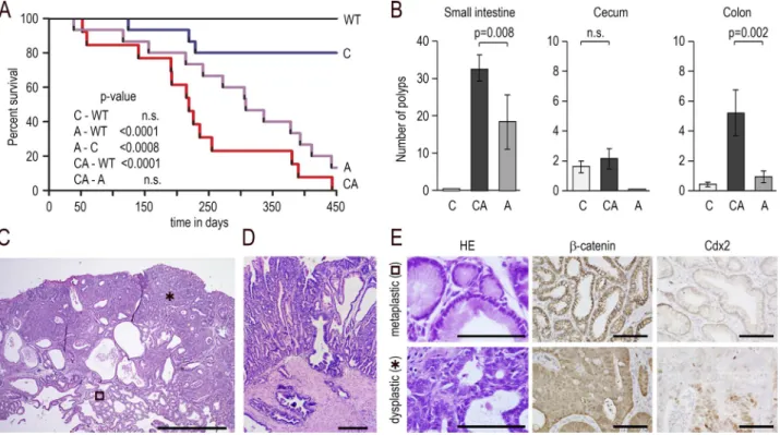
Figure 5. Mixed tumors developing by mosaic loss of Cdx2 combined with Apc heterozygosity. (A) Survival curve (Kaplan–Meier representation)
of wild-type (WT), AhCreERT::Cdx2f/f (C), Apc+/Δ14 (A), and Apc+/Δ14::AhCreERT::Cdx2f/f (CA) mice; n = 15 animals of each genotype; p-values were calculated
using the log-rank test; n.s., not significant. (B) Number of polyps in the small intestine, cecum, and colon of AhCreERT::Cdx2f/f (C), Apc+/Δ14::AhCreERT::Cdx2f/f
(CA), and Apc+/Δ14 (A) mice; n = 10 mice of each genotype; data are given ± SD; p-values were calculated using the Wilcoxon–Mann–Whitney test; n.s., not
significant. (C) Histology of a cecal mixed tumor in Apc+/Δ14::AhCreERT::Cdx2f/f mice with the juxtaposition of metaplastic-type (open square) and dysplastic
areas (asterisk). Bar, 500 µm; n = 15 mice in three crossings. (D) Invasion beyond the muscularis mucosae. Bar, 100 µm. (E) Histology and immunostaining
of β-catenin and Cdx2 in the metaplastic-type and dysplastic areas. Bars, 50 µm; n = 15 mice in three crossings. on February 8, 2018
jem.rupress.org
gastric-type metaplastic area and dysplastic areas was further confirmed by transcriptomic analyses. Indeed, Wnt pathway components were up-regulated in the cecal mixed tumors of
Apc+/Δ14::AhCreERT::Cdx2f/f mice compared with the lesions
of AhCreERT::Cdx2f/f mice (i.e., Axin2, Cldn1, Dsc3, Fosl1,
Fst, Fzd10, IL6, Lef1, Nkd1, Prox1, Tbx1, Tnfrsf19, and Wnt6), whereas gastric-type genes were turned on in the small in-testinal mixed tumors compared with small inin-testinal
dys-plastic polyps of Apc+/Δ14 mice (i.e., Atp4a, Car2, Ctse, Gif,
Gkn1/2/3, Gsdma2, Muc1/6, Pgc, Ptprn2, Tff1/2, and Vsig1; Table S2, sheets 1 and 2). Altogether, these results indicate that the loss of Cdx2 sensitizes the gut mucosa to tumorigenic progression in a cancer-prone context.
Non–cell-autonomous effect of Cdx2 on intestinal tumorigenesis
Given that, on the one hand, the knockout of Cdx2 driven by
AhCreERT is mosaic and that, on the other hand,
tumorigene-sis in Apc+/Δ14 mice results from the sporadic loss of
heterozy-gosity at the Apc locus (Colnot et al., 2004), the juxtaposition of metaplastic-type and dysplastic areas within the mixed
tu-mors of Apc+/Δ14::AhCreERT::Cdx2f/f mice may result either
from the neoplastic conversion of metaplastic Cdx2-depleted cells or from detrimental interactions between adjacent cells,
in that Cdx2-depleted cells would trigger the tumorigenic evolution of adjacent Cdx2-intact cells. Here, we addressed this issue using a lineage-tracing approach to follow the fate of the Cdx2-depleted cells. For this purpose, quadruple
trans-genic mice Apc+/Δ14::AhCreERT::Cdx2f/f::RosaCAGtdTomato
were produced, in which activation of the Cre recombinase
by βNF+Tam treatment should simultaneously disrupt the
Cdx2 gene and turn on fluorescent Tomato protein
expres-sion from the recombined RosaCAGtdTomato allele. Before
performing this experiment, two series of controls were
conducted to validate the tracing approach. First, AhCreERT::
Cdx2f/f::RosaCAGtdTomato mice were generated and used to
prove the actual correlation between cells having lost Cdx2
and cells expressing Tomato after βNF+Tam treatment (Fig.
S2B). Second, Apc+/Δ14::AhCreERT::RosaCAGtdTomato mice
were produced, treated with βNF+Tam, and analyzed 5 mo
later for fluorescence emission; the results illustrated in Fig. S2 C attest to the preservation of Tomato fluorescence in the
ad-enomatous polyps marked by cytoplasmic/nuclear β-catenin,
which conversely rules out the possibility that Tomato
ex-pression from the recombined RosaCAGtdTomato allele could
be secondarily turned off in the dysplastic context. Having
validated the tracing approach, Apc+/Δ14::AhCreERT::Cdx2f/f::
RosaCAGtdTomato mice (n = 4) were analyzed 5 mo after
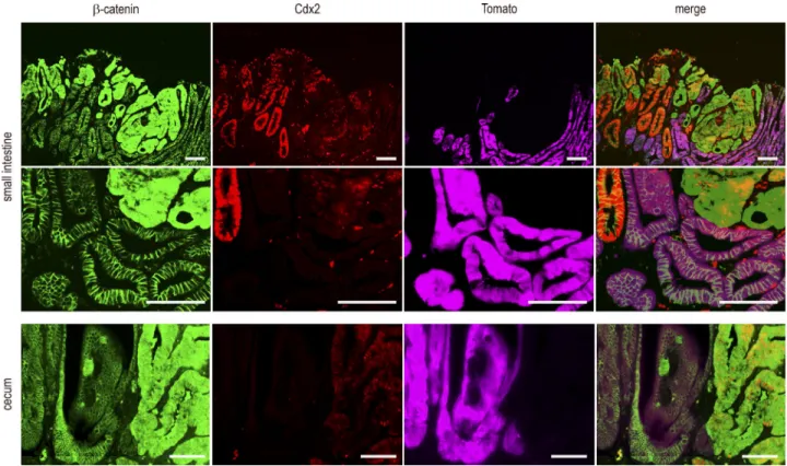
βN-Figure 6. Tracing of Cdx2-depleted cells in mixed tumors. Detection of β-catenin, Cdx2, and Tomato in small intestinal and cecal mixed tumors of Apc+/Δ14::AhCreERT::Cdx2f/f::RosaCAGtdTomato mice. The second line of pictures represents a higher magnification of the first one. Tomato was detected by
direct fluorescence emission before indirect immunodetection of β-catenin and Cdx2. The mutually exclusive patterns of Tomato and cytoplasmic/nuclear β-catenin were obtained in three mixed tumors from the cecum and three mixed tumors from the small intestine coming from four mice in two
F+Tam treatment. The results obtained in small intestinal and cecal mixed tumors (Fig. 6) led to the following con-clusions: (a) cells expressing Tomato were devoid of Cdx2
and always exhibited membranous β-catenin; (b) strong and
homogenous staining of Cdx2 was restricted to glands
ex-hibiting membranous β-catenin and absence of Tomato; (c)
Cdx2 was low and heterogeneous in areas with cytoplasmic/
nuclear β-catenin, corresponding to cells having undergone
tumorigenic evolution; and (d) dysplastic cells with
cytoplas-mic/nuclear β-catenin translocation never expressed Tomato.
These data indicated that the cells forming the dysplastic area in the mixed tumors originated from Cdx2-intact instead of Cdx2-knockout cells. Thus, metaplastic-type Cdx2-depleted areas do not themselves become tumorigenic, but they cre-ate a context that stimulcre-ates tumorigenesis from adjacent
Cdx2-intact Apc+/Δ14 tumor-prone cells. This highlights a
novel property of Cdx2 in the gut, in that this homeobox gene exerts a non–cell-autonomous tumor suppressor activity. The loss of Cdx2 modifies the stromal microenvironment To investigate how Cdx2 exerts its non–cell-autonomous tumor suppressor activity, the transcriptome of the cecal
le-sions of AhCreERT::Cdx2f/f mice was reconsidered to
iden-tify mediators of intercellular communication (Fig. 7 A and Table S1, sheets 4 and 5). Several extracellular matrix genes potentially involved in tumor growth and progression (Lu et al., 2012) were up-regulated, as well as genes for cytokines. Among them are Tnf family members, CCL and CXCL chemokines, and interleukins. For instance, CCL2 (known to shape a tumor-permissive microenvironment; Chun et al., 2015; Zhang et al., 2015), and IL6 family members (IL6, IL11, Osm, and Lif, considered potent drivers of cancer progression; Putoczki et al., 2013) were increased, whereas IL15 and IL18 (with anticancer activity; Salcedo et al., 2010; Bahri et al., 2015) were decreased. In line with this, the cellular microen-vironment was also modified, as illustrated by the widespread infiltration of macrophages and by the concentration
under-neath the surface epithelium of the lesions of CD4+ T
lym-phocytes and focally of FoxP3+ Treg lymphocytes (Fig. 7 B),
whereas CD8+ lymphocytes were barely detected.
Cytokines activate signaling pathways through
media-tors such as Erk, STATs, and NF-κB. Phospho-Erk1/2,
pres-ent in crypt base epithelial cells in the normal cecum, shifted to an irregular pattern at the surface of the cecal lesions of
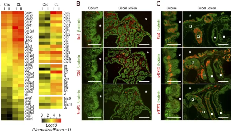
Figure 7. Modification of the microenvironment in the cecal lesions of AhCreERT::Cdx2f/f mice. (A) Heatmap comparison of transcriptomic data
for extracellular matrix genes (left) and cytokine genes (right) up-regulated in the cecal lesions of AhCreERT::Cdx2f/f mice (CL) compared with the cecum
of wild-type mice (Cec). (B) Immunodetection of Iba1+ macrophages, CD4+ T lymphocytes, and FoxP3+ Treg lymphocytes in the normal cecal mucosa of
wild-type mice and cecal lesions of AhCreERT::Cdx2f/f mice. Pictures correspond to serial sections. The asterisks indicate the lumen of the cecum. Bars: (cecum)
50 µm; (cecal lesions) 100 µm. (C) Erk1/2 and STAT3 signaling in the cecal lesions: the immunostaining illustrates the distribution of phospho-pErk1/2 and
phospho-STAT3 in the cecal lesions compared with the normal cecum. Closed and open squares respectively show Cdx2-depleted and Cdx2-intact glands. Bars: (cecum) 50 µm; (cecal lesions) 100 µm. Pictures in B and C were obtained in four mice of each genotype from two crossings.
on February 8, 2018
jem.rupress.org
AhCre ::Cdx2 mice and occasionally in subjacent glands, regardless of whether they were depleted in Cdx2 (Fig. 7 C). Phospho-STAT3 labeled infiltrating cells in the stroma of the lesions and sporadically also epithelial cells, irrespective of their Cdx2 status (Fig. 7 C). Importantly, the pattern of
NF-κB was profoundly modified in the lesions (Fig. 8).
In-deed, besides its presence in the nuclei of cells infiltrating
the stroma, NF-κB was activated and translocated into the
nucleus of epithelial cells at the surface epithelium composed of Cdx2-intact cells; this contrasts with the normal cecal
ep-ithelium, in which nuclear NF-κB is only weakly detected
with a decreasing gradient from the bottom to the top of the crypts. This observation is particularly interesting in light of recent findings reporting that gut tumorigenesis can be initi-ated from non–stem cells instead of stem cells by combining
the activation of Wnt and NF-κB signaling (Schwitalla et
al., 2013). Moreover, the activation of the NF-κB pathway
has been shown to stimulate Apc-dependent tumorigenesis in the gut, and this effect was related to the induction of
iNOS/nos2 by NF-κB (Shaked et al., 2012). Strikingly,
tran-scriptomic data revealed an approximately sixfold increase of iNOS/nos2 mRNA in cecal lesions compared with the nor-mal cecum (q < 0.0001). In addition, this correlated not only with the infiltration of the stroma by iNOS-positive cells, but also with the focal induction of iNOS in Cdx2-intact
cells also exhibiting nuclear NF-κB in the surface epithelium
of the lesions (Fig. 8).
Surface origin and iNOS-dependent tumorigenesis in the mixed tumors
These observations prompted us to investigate whether the emergence of dysplastic areas within the mixed tumors is
ac-tually associated with Apc loss of heterozygosity, and whether the process is initiated at the surface of the lesions and is iNOS dependent. PCR genotyping of individual glands
microdis-sected within cecal mixed tumors of Apc+/Δ14::AhCreERT::
Cdx2f/f mice showed that the wild-type allele of Apc was
pre-served in the metaplastic-type Cdx2-knockout glands but lost
in the dysplastic glands, as in typical Apc+/Δ14 adenomatous
polyps (Fig. 9 A). This indicates a similar molecular mecha-nism of oncogenic Wnt pathway activation through Apc loss of heterozygosity in the dysplastic area of the mixed tumors
of Apc+/Δ14::AhCreERT::Cdx2f/f mice, as in Apc+/Δ14 mice
(Colnot et al., 2004).
Next, we addressed the site of emergence of the dys-plastic area in the mixed tumors. For this purpose, the
cy-toplasmic/nuclear localization of β-catenin was used as a
marker of the loss of Apc, because Colnot et al. (2004) have
reported that β-catenin translocation already accompanies
the loss of Apc in early lesions. First, we entirely cut cecal
mixed tumors (n = 3) of Apc+/Δ14::AhCreERT::Cdx2f/f mice.
This led to identification of small invaginations in the surface epithelial layer, not connected to deeper glands as assessed by serial sections analysis, which contained cells or groups
of cells with cytoplasmic/nuclear β-catenin; these cells did
not express Olfm4 (like normal cecum in mice) but exhib-ited a reduced level of Cdx2 and an increased level of Sox9 protein compared with adjacent nontransformed cells of
the epithelial lining in which β-catenin remained
membra-nous (Fig. 9 B and Fig. S3). Second, we analyzed β-catenin–
immunostained sections coming from nine mixed tumors of
seven Apc+/Δ14::AhCreERT::Cdx2f/f mice for the position of
the dysplastic glands in the depth of the polyps. Plotting the results obtained for a total of 77 of these structures revealed
an uneven distribution: they were all connected to or located immediately underneath the surface layer and extended more or less deeply into the tumors (Fig. 9 C). Collectively, these results suggest that the loss of heterozygosity of Apc occurs in the Cdx2-intact surface epithelial layer and that the dys-plastic structures progressively cover and invade the subjacent Cdx2-depleted metaplastic tissue.
Finally, because the dysplastic areas are composed of Cdx2-intact cells and emerge from the surface of the lesion, and because the surface cells focally express iNOS, which is known to accelerate Apc loss and initiate tumorigenesis (Shaked et al., 2012), we investigated the involvement of iNOS
in the emergence of dysplasia in Apc+/Δ14::AhCreERT::Cdx2f/f
mice. For this purpose, animals were treated with βNF+Tam
and, 2 wk later, given the iNOS inhibitor aminoguanidine (AG) ad libitum in drinking water for 3 mo. Sections of the lesions developing in the cecum of these mice were analyzed
histologically and by immunostaining for Cdx2 and β-catenin
to identify and record the metaplastic-type area and dysplastic areas. As illustrated in Fig. 9 D, three of the four cecal lesions analyzed in AG-treated mice showed no dysplastic area, and the fourth lesion exhibited only a small area of this type of structure; this contrasted with the higher proportion of dys-plastic areas observed in every cecal mixed tumor of untreated mice. In parallel, we observed that the AG treatment reduced the tumor load in the small intestine (10 ± 3 vs. 28 ± 4) and
colon (2 ± 1 vs. 5 ± 2) of these Apc+/Δ14::AhCreERT::Cdx2f/f
mice compared with untreated mice, whereas it had no
sig-nificant effect in Apc+/Δ14 mice (13 ± 6 vs. 14 ± 3 tumors in
the small intestine and 1 ± 1 vs. 0 in the colon). These results suggest that iNOS actually contributes to the emergence of
dysplasia in the context of the Apc+/Δ14::AhCreERT::Cdx2f/f
mice; moreover, they are consistent with the data reported by Shaked et al. (2012) showing that the iNOS inhibitor AG
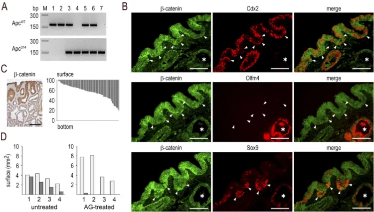
Figure 9. Surface initiation and iNOS-dependence of the dysplastic structures in mixed tumors. (A) Genotyping of microdissected glands for
the Apcwt and ApcΔ14 alleles: (1) normal cecal gland and (2) Cdx2-depleted gland in cecal lesions of AhCreERT::Cdx2f/f mice; (3) normal and (4) dysplastic
gland in Apc+/Δ14 mice; (5) normal cecal gland, (6) metaplastic-type gland, and (7) dysplastic gland in mixed tumors of Apc+/Δ14::AhCreERT::Cdx2f/f mice. PCR
results are representative of the results obtained in n = 3 mice. (B) Immunodetection of β-catenin, Cdx2, Olfm4, and Sox9 in the Cdx2-expressing surface epithelium of mixed tumors. The arrowheads point to surface epithelial cells with cytoplasmic/nuclear β-catenin; the asterisks show Cdx2-depleted glands. Pictures correspond to serial sections. They were obtained in n = 10 mice. Bars, 50 µm. (C) Left: localization of the dysplastic glands in mixed tumors by
β-catenin immunostaining, showing cytoplasmic/nuclear accumulation in glands connected to the surface epithelium. Bar, 100 µm. Right: distribution of dysplastic glands. Each bar represents the localization and extent of one dysplastic structure along the surface-to-bottom axis of mixed tumors. Results are expressed as percentage of the height of the mixed tumors. They correspond to data obtained from nine mixed tumors in seven mice. (D) Surface (square
millimeters) of the metaplastic-type areas (light gray) and dysplastic areas (dark gray) in the lesions developed in the cecum of Apc+/Δ14::AhCreERT::Cdx2f/f
mice either untreated or treated with AG. Values represent the mean surfaces in 12 sections for each sample. They were obtained from n = 4 AG-treated mice and n = 4 untreated mice.
on February 8, 2018
jem.rupress.org
knockout to address the long-term effect of this gene’s defi-ciency in the gut. Although abrogating the function of Cdx2 results in the formation of imperfect gastric-type metaplastic lesions, these lesions did not spontaneously evolve in can-cer even in aged animals. This is consistent with the fact that overexpressing a dominant-negative splicing variant of Cdx2 in the duodenum also led to the formation of lesions with gastric-type metaplastic properties without spontaneous neo-plastic evolution (Balbinot et al., 2017). Nevertheless, by devel-oping the original model that combines the mosaic knockout of Cdx2 with the stochastic loss of function of the Apc tumor suppressor gene, we uncovered that functional interactions between distinct types of noncancerous epithelial cells—met-aplastic-type and tumor-prone, respectively—can accelerate tumorigenesis. These deleterious interactions are indirect and involve modifications of the microenvironment driven by the metaplastic-type Cdx2-deficient cells that trigger chronic
ac-tivation of NF-κB and induction of iNOS in Cdx2-intact
tumor-prone cells, which subsequently undergo Apc loss of heterozygosity and generate dysplastic structures. Thus, this study highlights a novel and original property of Cdx2 in the gut, in that this homeobox gene exerts non–cell-autonomous tumor suppressor activity mediated by changes in the stromal environment. Because the cecal epithelium can be the seat of
adenomatous development in Apc+/Δ14 mice, even at a very
low rate, we believe that the environment created by the met-aplastic-type Cdx2-depleted cells would stimulate Apc loss of function in the adjacent Cdx2-intact cells rather than bring-ing out latent mutations. The non–cell-autonomous tumor suppressor activity represents a new property for this homeo-box gene, already reported to exhibit cell-autonomous tumor suppressor activity in the gut through its impact on many cel-lular and molecular functions including apoptosis, cell differ-entiation, cell proliferation, chromosome stability, and DNA repair (Aoki et al., 2003; Bonhomme et al., 2003; Renouf et al., 2012; Sakamoto et al., 2017; Tong et al., 2017). Only a few examples of non–cell-autonomous tumor suppressor activ-ity have been described so far, for instance in liver and brain tumors and also in melanoma (Andoniadou et al., 2013; Lu-jambio et al., 2013; Mescher et al., 2017), probably because it needs to develop appropriate lineage tracing approaches. This mechanism is likely underrated despite its potential not only for tumor initiation, but also in the emergence of subclonal heterogeneity (Marusyk et al., 2014).
The findings reported here are important regarding the pathological relevance of metaplastic-type lesions displayed in a wide range of epithelial organs, including, for instance, the digestive, respiratory, and urinary tracts. Indeed, metaplasia is
One possibility is related to the fact that that these cells show a strong induction of Sox9 and Olfm4, which have been re-ported to antagonize tumorigenesis (Bastide et al., 2007; Liu et al., 2016; Prévostel et al., 2016). Also remarkable is that the tumorigenic evolution of Cdx2-intact cells does not occur in glands randomly embedded in the metaplastic lesions, but at the level of Cdx2-intact cells of the surface epithelial layer. This surface is made of patches of Cdx2-intact cells, Cdx2-depleted cells, and also eroded areas. Previous studies have shown that reducing the Cdx2 level in mice increases intestinal permeabil-ity and sensitivpermeabil-ity to proinflammatory treatment (Calon et al., 2007), and that Cdx2 modulates the motility and mechanical properties of the intestinal epithelial cells (Gross et al., 2008; Platet et al., 2017). Therefore, the mosaic deletion of Cdx2 cre-ates a complex picture with abrasion and tissue repair from Cdx2-intact and Cdx2-deleted cells, which may contribute to the resulting modification of the underpinning stroma. The surface epithelial lining of the lesions is therefore at a critical position at the interface between the luminal content with the microbiota and the activated stroma, which may facilitate Apc loss of heterozygosity. The surface origin of the dysplasia rep-resents an alternative to the model of tumor initiation in the crypt stem cells (Barker et al., 2009). It is in line with previ-ous descriptions of top-down morphogenesis of colon cancers in humans (Shih et al., 2001) and with the mouse model of tumorigenesis initiated from dedifferentiated cells located in small intestinal villi by combining the activation of the Wnt
and NF-κB pathways (Schwitalla et al., 2013).
Corroborating previous studies, our data strengthen the notion that human CRCs with a strong reduction of CDX2 principally segregate within the subtype of serrated neoplasia with a stem cell signature (De Sousa E Melo et al., 2013; Bae et al., 2015; Dalerba et al., 2016) and also are characterized by the ectopic expression of gastric markers (Matsuda et al., 2010; Sentani et al., 2013; Kim et al., 2015). These cancers exhibit a worse evolution. Importantly, the lesions developing here after Cdx2 loss in mice share several stromal and im-mune properties with the serrated subtype of human CRCs (Becht et al., 2016). These include a high expression of ex-tracellular matrix molecules, myeloid chemokine Ccl2, com-plement components (C1qb, C1qc, C1ra, C1rb, C1s, C3, Cfh, and Cfi), angiogenic factors (Vegfb, Pdgfb, Pdgfc, and Pdgfd), and immunosuppressive molecules (Tgfb2, Tgfb3, Lgals1, and Lgals2). These similarities make the mice developed in this study a relevant animal model to investigate the com-plex modifications of the microenvironment leading to the neoplastic conversion of premalignant lesions. This is central in the perspective of the development of efficient preventive
on February 8, 2018
jem.rupress.org
strategies and treatments of cancer targeting the microenvi-ronment and its interaction with tumor cells.
MATERIALS AND METHODS Human CRC samples and analysis
The 566 transcriptomic profiles recorded in GSE39582l (Marisa et al., 2013) were analyzed. Comparison of the ex-pression levels of CDX2 between the C4 CRC subtype and the other subtypes was performed using independent two-group t test (function t.test, stats R package). CRC subtypes means comparison was performed using Tukey post hoc test after two-way ANO VA (function TukeyHSD, stats R pack-age). Disease-free survival was defined as the time from sur-gery to the first recurrence. Survival curves were obtained according to the method of Kaplan and Meier (function Surv, R package survival), and differences between survival distri-butions were assessed by log-rank test.
Human tissue samples were obtained at the University Hospital of Strasbourg (France) according to the recommen-dations of the French Ethical Committee and the ethical standards of the 1964 Declaration of Helsinki. Patients pro-vided written informed consent.
Mouse strains and treatments
Mice were used according to the protocol approved by the Committee on the Ethics of Animal Experiments of the University of Strasbourg (CRE MEAS, C2EA-35) under the
permit number AL/43/50/02/13. AhCreERT (Ireland et al.,
2004), AhCreERT::Cdx2f/f (Stringer et al., 2012), and Apc+/Δ14
mice (Colnot et al., 2004) have been described. Rosa-CAGtdTomato mice (strain Ai9) were provided by the Jackson
Laboratory. Strains were backcrossed at least eight times. Lit-termates were used as controls throughout this study.
Mice were genotyped by PCR amplification of tail DNA (Viagene, DirectPCR Lysis Reagent mouse tail;
Eu-romedex) using the following primers: Cdx2wt and Cdx2f
alleles, 5′-TGG GGC AAT CTT AAT GGG TA-3′ and 5′-TGT
AGC CTC GAC TTG GCT TT-3′; Apcwt allele, 5′-CTG TTC
TGC AGT ATG TTA TCA-3′ and 5′-CTA TGA GTC AAC
ACA GGA TTA-3′; ApcΔ14 allele, 5′-CTG TTC TGC AGT
ATG TTA TCA-3′ and 5′-TAT AAG GGC TAA CAG TCA
ATA-3′; AhCreERT allele, 5′-GCC TGG TCT GGA CAC
AGT CC-3′ and 5′-GGT TCA GCA TCC AAC AAG
GC-3′; RosaCAGtdTomato allele, 5′-CTG TTC CTG TAC GGC
ATGG-3′ and 5′-GGC ATT AAA GCA GCG TAT CC-3′; and
Rosawt allele, 5′-AAG GGA GCT GCA GTG GAG TA-3′ and
5′-CCG AAA ATC TGT GGG AAG TC-3′.
Mice 3 mo of age were intraperitoneally injected with
1.6 mg βNF+TAM (Sigma-Aldrich) in corn oil, once daily
for 4 d. For the treatment with the iNOS inhibitor, they
were injected with βNF+Tam, and 2 wk later they were
given 2 g/L AG (Sigma-Aldrich) in drinking water. The pathohistological evaluation of the lesions developed in the mice used in this study was performed independently by
two pathologists of the University Hospital of Strasbourg: A. Onea and M.P. Chenard.
RNA extraction and analysis by RNA sequencing
RNA was extracted from tissue fragments with Tri Reagent (Euromedex) and analyzed using nanoRNA chips on a Bio-analyser 2100 (Agilent Technologies). 1 µg of total RNA was used for the construction of the mRNA sequence libraries with Illumina’s TruSeq RNA sample kit (Illumina). Poly(A) RNA was selected by two rounds on poly-dT–coated magnetic beads, followed by fragmentation and first-strand cDNA synthesis with Superscript II (Thermo Fisher Scien-tific) using random hexamer primers. The cDNA fragments were subjected to end repair and dA tailing, ligated to in-dexed bar-coded adapters, and subjected to 12 cycles of PCR. Concentration and validation of the libraries were made with DNA1000 chips loaded on a Bioanalyser 2100. Paired-end 50-bp reads were obtained with a HiSeq1000 by multiplex-ing three libraries on one lane. Demultiplexmultiplex-ing and gener-ation of raw fastq files were performed with Casava v1.7. Mapping against the reference mouse genome GRCm38 was performed with tophat 2 (Trapnell et al., 2009) using the following options: b2-sensitive, a 5, p 5, library-type, fr-unstranded, r 180, mate-std-dev 80, and exon-exon ref-erence from Ensembl v75. Quantification of the reads was performed with HTSeq v.0.5.3p3 (Anders et al., 2015) with the following options: stranded = no, mode = union; and using the reference gene annotation from Ensembl v75. Normalization and differential expression analysis was made with DESeq2 (Love et al., 2014). Unless otherwise stated,
genes with a log2(fold change [FC]) ≥1 and adjusted p-value
(q-value) <0.01 were considered as differentially expressed. Data analysis was performed using Ingenuity Pathway Anal-ysis (Qiagen). The transcriptomic data have been deposited in the GEO database under accession number GSE89992. Histology and immunohistology
Tissue samples were fixed with 4% PFA and embedded in paraffin. Sections (5 µm) were deparaffinized and treated for antigen retrieval for 10 min in 10 mmol/L sodium citrate, pH 6, in a microwave oven for every primary antibody except an-ti-Iba1, and then blocked in 5% normal goat serum and 0.1% Triton X-100–PBS for 1 h at room temperature. Slides were incubated overnight at 4°C with primary antibodies diluted in 0.1% Triton X-100–PBS and washed in this saline buffer.
Primary antibodies were as follows: mouse anti–β-catenin
(clone 14; dilution 1:500; BD Transduction Lab), mouse an-ti-CD4 (50134-M08H; dilution 1:500; Sino Biological), goat anti-CD8b (M-20, sc-1144; dilution 1:500, Santa Cruz Bio-technology), rabbit anti-Cdx1 (Bonhomme et al., 2003; dilu-tion 1:1,000), mouse anti-Cdx2 (CDX2-88, F/MU392A-UC; dilution 1:500; Biogenex), rabbit anti-Cdx2 (EPR2764Y, ab76541; dilution 1:10,000; Thermo Fisher Scientific), rabbit anti-Cldn18 (38-8000; dilution 1:500; Invitrogen), rat anti- FoxP3 (FJK-16s, 14-5773-80; dilution 1:500; Affymetrix
on February 8, 2018
jem.rupress.org
specific; dilution 1:500; Abcam), rabbit anti-p-Erk1/2 (D11A8, mAb5683; dilution 1:500; Cell Signaling Technology), rabbit anti–p-STAT3 (ab76315; dilution 1:500; Abcam), rabbit anti-
RelA (NF-κB p65; C-20, sc-372; dilution 1:500; Santa Cruz
Biotechnology), rabbit anti-Sox2 (AB5603; dilution 1:500; Millipore), rabbit anti-Sox9 (De Santa Barbara et al., 1998; dilution 1:500), rabbit anti-Tff1 (Karam et al., 2004; dilution 1:500), rabbit anti-Tff2 (Karam et al., 2004; dilution 1:500), and rat anti-Tomato (clone 16D7; dilution 1:250; KerFast). For im-munohistochemical staining, secondary biotinylated antibodies (dilution 1:2,000; Vector Laboratories) were revealed using the Vectastain ABC kit (Vector Laboratories). For immunofluores-cence detection, secondary goat anti-mouse antibody labeled with Alexa Fluor 488 (dilution 1:1,000; Molecular Probes) and goat anti-rabbit antibody labeled with Alexa Fluor 568 (dilution 1:1,000; Molecular Probes) were used. Sections were visualized with an Axio Zoom.V16 microscope, an Axiophot microscope, or an Axio Imager Z2 microscope (Zeiss).
For the combined detection of Tomato, β-catenin, and
Cdx2 proteins in tissue sections, a two-step procedure was de-veloped. In the first step, sections were deparaffinized and cov-ered with 0.1% Triton X100–PBS buffer, and a picture of the direct fluorescence emitted by Tomato was taken. In the sec-ond step, the sections were treated for antigen retrieval for 10 min in 10 mmol/L sodium citrate, pH 6, in a microwave oven, which (a) destroys the direct fluorescence emission by Tomato
and (b) allows further detection of β-catenin and Cdx2 by
in-direct immunofluorescence labeling, as described in the above paragraph. To validate this procedure, we verified that the de-tection of Tomato by direct fluorescence emission in deparaf-finized sections was as efficient as the detection of the Tomato protein by indirect immunofluorescence in the same sections.
For this purpose, cecal sections of AhCreERT::RosaCAGtdTomato
mice treated with βNF+Tam were first processed for Tomato
detection by direct fluorescence emission, and then the To-mato protein was revealed by indirect immunofluorescence using the anti-Tomato antibody. The results illustrated in Fig. S2 A demonstrate perfect superimposable patterns.
Tissue microdissection for genomic PCR analysis
Tissue sections of 10 µm fixed in 4% PFA and embedded in paraffin were sliced on FrameSlides PET Membrane slides (Leica) and stained with Harris, and serial sections were
im-munostained for β-catenin and Cdx2 to ascertain the identity
of the microdissected glands. Areas of ∼100 cells were
micro-dissected using an LMD 6000 laser microscope (Leica Mi-crosystems), and genomic DNA was extracted using QIAamp
DNA FFPE Tissue kit (Qiagen). Identification of the Apcwt
control experiments performed for the lineage tracing ap-proach. Fig. S3 shows the immunofluorescence patterns of β-catenin and Cdx2 in serial sections of mixed tumors. Table S1 provides the list of genes differentially expressed between
the cecal lesions of AhCreERT::Cdx2f/f mice and the normal
cecum (sheet 1) and lists of selected panels of genes selected from this list (intestinal/gastric markers, stem cell markers, extracellular matrix components, and cytokines, respec-tively; sheets 2–5). Table S2 provides the list of genes
differ-entially expressed between cecal mixed tumors of Apc+/Δ14::
AhCreERT::Cdx2f/f mice and cecal metaplastic-type lesions of
AhCreERT::Cdx2f/f mice (sheet 1) and between small
intesti-nal mixed tumors of Apc+/Δ14::AhCreERT::Cdx2f/f mice and
small intestinal adenoma of Apc+/Δ14 mice (sheet 2).
ACKNOWLEDGMENTS
We thank Prof. M.P. Chenard for help in pathohistology evaluation, Prof. D.J. Winton (Cancer Research UK, Cambridge, England, UK) for the AhCreERT mice, Dr. C. Perret (Inserm
U1016, Institut Cochin, Paris) for the Apc+/Δ14 mice, Dr. C. Tomaseto (Inserm U1258, IGB MC, Illkirch) for the anti-Tff1 and -Tff2 antibodies, and Dr. A. De Reynes (Cartes d’Identité des Tumeurs Program, Ligue Contre le Cancer, Paris, France) for discussions.
This work was supported by the Ligue Contre le Cancer du Haut-Rhin (France), the Fondation ARC pour la Recherche sur le Cancer (France; PGA120140200834), and the Institut National du Cancer (INCa2014-178). C. Balbinot was funded by the Ministère de l’Enseignement Supérieur et de la Recherche (France) and the Ligue Contre le Cancer.
The authors declare no competing financial interests.
Author contributions: C. Balbinot, F. Beck, J. Deschamps, J.-N. Freund, and I. Duluc conceived and designed the study. C. Balbinot, O. Armant, N. Elarouci, L. Marisa, E. Martin, E. De Clara, A. Onea, J.-N. Freund, and I. Duluc contributed to data acquisi-tion, analysis, and interpretation. C. Balbinot, F. Beck, J.-N. Freund, and I. Duluc wrote the manuscript.
Submitted: 23 May 2017 Revised: 13 November 2017 Accepted: 18 January 2018
REFERENCES
Anders, S., P.T. Pyl, and W. Huber. 2015. HTSeq—A Python framework to work with high-throughput sequencing data. Bioinformatics. 31:166–169. https ://doi .org /10 .1093 /bioinformatics /btu638
Andoniadou, C.L., D. Matsushima, S.N. Mousavy Gharavy, M. Signore, A.I. Mackintosh, M. Schaeffer, C. Gaston-Massuet, P. Mollard, T.S. Jacques, P. Le Tissier, et al. 2013. Sox2(+) stem/progenitor cells in the adult mouse pituitary support organ homeostasis and have tumor-inducing potential.
Cell Stem Cell. 13:433–445. https ://doi .org /10 .1016 /j .stem .2013 .07 .004
Aoki, K., Y. Tamai, S. Horiike, M. Oshima, and M.M. Taketo. 2003. Colonic polyposis caused by mTOR-mediated chromosomal instability in Apc+/ Delta716 Cdx2+/- compound mutant mice. Nat. Genet. 35:323–330. https ://doi .org /10 .1038 /ng1265
Bae, J.M., T.H. Lee, N.-Y. Cho, T.-Y. Kim, and G.H. Kang. 2015. Loss of CDX2 expression is associated with poor prognosis in colorectal cancer
on February 8, 2018
jem.rupress.org
patients. World J. Gastroenterol. 21:1457–1467. https ://doi .org /10 .3748 / wjg .v21 .i5 .1457
Bahri, R., I.S. Pateras, O. D’Orlando, D.A. Goyeneche-Patino, M. Campbell, J.K. Polansky, H. Sandig, M. Papaioannou, K. Evangelou, P.G. Foukas, et al. 2015. IL-15 suppresses colitis-associated colon carcinogenesis by inducing antitumor immunity. OncoImmunology. 4:e1002721. https ://doi .org /10 .1080 /2162402X .2014 .1002721
Balbinot, C., M. Vanier, O. Armant, A. Nair, J. Penichon, C. Soret, E. Martin, T. Saandi, J.-M. Reimund, J. Deschamps, et al. 2017. Fine-tuning and autoregulation of the intestinal determinant and tumor suppressor homeobox gene CDX2 by alternative splicing. Cell Death Differ. 24:2173–2186. https ://doi .org /10 .1038 /cdd .2017 .140
Barker, N., R.A. Ridgway, J.H. van Es, M. van de Wetering, H. Begthel, M. van den Born, E. Danenberg, A.R. Clarke, O.J. Sansom, and H. Clevers. 2009. Crypt stem cells as the cells-of-origin of intestinal cancer. Nature. 457:608–611. https ://doi .org /10 .1038 /nature07602
Bastide, P., C. Darido, J. Pannequin, R. Kist, S. Robine, C. Marty-Double, F. Bibeau, G. Scherer, D. Joubert, F. Hollande, et al. 2007. Sox9 regulates cell proliferation and is required for Paneth cell differentiation in the intestinal epithelium. J. Cell Biol. 178:635–648. https ://doi .org /10 .1083 /jcb .200704152
Becht, E., A. de Reyniès, N.A. Giraldo, C. Pilati, B. Buttard, L. Lacroix, J. Selves, C. Sautès-Fridman, P. Laurent-Puig, and W.H. Fridman. 2016. Immune and stromal classification of colorectal cancer is associated with molecular subtypes and relevant for precision immunotherapy. Clin.
Cancer Res. 22:4057–4066. https ://doi .org /10 .1158 /1078 -0432 .CCR
-15 -2879
Beck, F., K. Chawengsaksophak, P. Waring, R.J. Playford, and J.B. Furness. 1999. Reprogramming of intestinal differentiation and intercalary regeneration in Cdx2 mutant mice. Proc. Natl. Acad. Sci. USA. 96:7318– 7323. https ://doi .org /10 .1073 /pnas .96 .13 .7318
Bonhomme, C., I. Duluc, E. Martin, K. Chawengsaksophak, M.-P. Chenard, M. Kedinger, F. Beck, J.-N. Freund, and C. Domon-Dell. 2003. The Cdx2 homeobox gene has a tumour suppressor function in the distal colon in addition to a homeotic role during gut development. Gut. 52:1465–1471. https ://doi .org /10 .1136 /gut .52 .10 .1465
Calon, A., I. Gross, B. Lhermitte, E. Martin, F. Beck, B. Duclos, M. Kedinger, I. Duluc, C. Domon-Dell, and J.N. Freund. 2007. Different effects of the Cdx1 and Cdx2 homeobox genes in a murine model of intestinal inflammation. Gut. 56:1688–1695. https ://doi .org /10 .1136 /gut .2007 .125542
Chun, E., S. Lavoie, M. Michaud, C.A. Gallini, J. Kim, G. Soucy, R. Odze, J.N. Glickman, and W.S. Garrett. 2015. CCL2 promotes colorectal carcinogenesis by enhancing polymorphonuclear myeloid-derived suppressor cell population and function. Cell Reports. 12:244–257. https ://doi .org /10 .1016 /j .celrep .2015 .06 .024
Colnot, S., M. Niwa-Kawakita, G. Hamard, C. Godard, S. Le Plenier, C. Houbron, B. Romagnolo, D. Berrebi, M. Giovannini, and C. Perret. 2004. Colorectal cancers in a new mouse model of familial adenomatous polyposis: Influence of genetic and environmental modifiers. Lab. Invest. 84:1619–1630. https ://doi .org /10 .1038 /labinvest .3700180
Dalerba, P., D. Sahoo, S. Paik, X. Guo, G. Yothers, N. Song, N. Wilcox-Fogel, E. Forgó, P.S. Rajendran, S.P. Miranda, et al. 2016. CDX2 as a prognostic biomarker in stage II and stage III colon cancer. N. Engl. J. Med. 374:211– 222. https ://doi .org /10 .1056 /NEJMoa1506597
De Santa Barbara, P., N. Bonneaud, B. Boizet, M. Desclozeaux, B. Moniot, P. Sudbeck, G. Scherer, F. Poulat, and P. Berta. 1998. Direct interaction of SRY-related protein SOX9 and steroidogenic factor 1 regulates transcription of the human anti-Müllerian hormone gene. Mol. Cell.
Biol. 18:6653–6665. https ://doi .org /10 .1128 /MCB .18 .11 .6653
De Sousa E Melo, F., X. Wang, M. Jansen, E. Fessler, A. Trinh, L.P. de Rooij, J.H. de Jong, O.J. de Boer, R. van Leersum, M.F. Bijlsma, et al. 2013. Poor-prognosis colon cancer is defined by a molecularly distinct subtype
and develops from serrated precursor lesions. Nat. Med. 19:614–618. https ://doi .org /10 .1038 /nm .3174
Egeblad, M., E.S. Nakasone, and Z. Werb. 2010. Tumors as organs: Complex tissues that interface with the entire organism. Dev. Cell. 18:884–901. https ://doi .org /10 .1016 /j .devcel .2010 .05 .012
Gross, I., I. Duluc, T. Benameur, A. Calon, E. Martin, T. Brabletz, M. Kedinger, C. Domon-Dell, and J.-N. Freund. 2008. The intestine-specific homeobox gene Cdx2 decreases mobility and antagonizes dissemination of colon cancer cells. Oncogene. 27:107–115. https ://doi .org /10 .1038 / sj .onc .1210601
Guinney, J., R. Dienstmann, X. Wang, A. de Reyniès, A. Schlicker, C. Soneson, L. Marisa, P. Roepman, G. Nyamundanda, P. Angelino, et al. 2015. The consensus molecular subtypes of colorectal cancer. Nat. Med. 21:1350– 1356. https ://doi .org /10 .1038 /nm .3967
Hryniuk, A., S. Grainger, J.G.A. Savory, and D. Lohnes. 2014. Cdx1 and Cdx2 function as tumor suppressors. J. Biol. Chem. 289:33343–33354. https :// doi .org /10 .1074 /jbc .M114 .583823
Ireland, H., R. Kemp, C. Houghton, L. Howard, A.R. Clarke, O.J. Sansom, and D.J. Winton. 2004. Inducible Cre-mediated control of gene expression in the murine gastrointestinal tract: effect of loss of beta-catenin. Gastroenterology. 126:1236–1246. https ://doi .org /10 .1053 /j .gastro .2004 .03 .020
Karam, S.M., C. Tomasetto, and M.-C. Rio. 2004. Trefoil factor 1 is required for the commitment programme of mouse oxyntic epithelial progenitors.
Gut. 53:1408–1415. https ://doi .org /10 .1136 /gut .2003 .031963
Kim, J.H., K.-J. Kim, Y.-Y. Rhee, J.M. Bae, N.-Y. Cho, H.S. Lee, and G.H. Kang. 2015. Gastric-type expression signature in serrated pathway-associated colorectal tumors. Hum. Pathol. 46:643–656. https ://doi .org /10 .1016 /j .humpath .2015 .01 .003
Liu, W., H. Li, S.-H. Hong, G.P. Piszczek, W. Chen, and G.P. Rodgers. 2016. Olfactomedin 4 deletion induces colon adenocarcinoma in ApcMin/+ mice. Oncogene. 35:5237–5247. https ://doi .org /10 .1038 /onc .2016 .58 Love, M.I., W. Huber, and S. Anders. 2014. Moderated estimation of fold
change and dispersion for RNA-seq data with DESeq2. Genome Biol. 15:550. https ://doi .org /10 .1186 /s13059 -014 -0550 -8
Lu, P., V.M. Weaver, and Z. Werb. 2012. The extracellular matrix: A dynamic niche in cancer progression. J. Cell Biol. 196:395–406. https ://doi .org /10 .1083 /jcb .201102147
Lujambio, A., L. Akkari, J. Simon, D. Grace, D.F. Tschaharganeh, J.E. Bolden, Z. Zhao, V. Thapar, J.A. Joyce, V. Krizhanovsky, and S.W. Lowe. 2013. Non-cell-autonomous tumor suppression by p53. Cell. 153:449–460. https ://doi .org /10 .1016 /j .cell .2013 .03 .020
Marisa, L., A. de Reyniès, A. Duval, J. Selves, M.P. Gaub, L. Vescovo, M.-C. Etienne-Grimaldi, R. Schiappa, D. Guenot, M. Ayadi, et al. 2013. Gene expression classification of colon cancer into molecular subtypes: Characterization, validation, and prognostic value. PLoS Med. 10:e1001453. https ://doi .org /10 .1371 /journal .pmed .1001453 Marusyk, A., D.P. Tabassum, P.M. Altrock, V. Almendro, F. Michor, and K.
Polyak. 2014. Non-cell-autonomous driving of tumour growth supports sub-clonal heterogeneity. Nature. 514:54–58. https ://doi .org /10 .1038 / nature13556
Matsuda, M., K. Sentani, T. Noguchi, T. Hinoi, M. Okajima, K. Matsusaki, N. Sakamoto, K. Anami, Y. Naito, N. Oue, and W. Yasui. 2010. Immunohistochemical analysis of colorectal cancer with gastric phenotype: Claudin-18 is associated with poor prognosis. Pathol. Int. 60:673–680. https ://doi .org /10 .1111 /j .1440 -1827 .2010 .02587 .x Mescher, M., P. Jeong, S.K. Knapp, M. Rübsam, M. Saynisch, M. Kranen, J.
Landsberg, M. Schlaak, C. Mauch, T. Tüting, et al. 2017. The epidermal polarity protein Par3 is a non-cell autonomous suppressor of malignant melanoma. J. Exp. Med. 214:339–358. https ://doi .org /10 .1084 /jem .20160596
on February 8, 2018
jem.rupress.org
Jo
u
r
n
a
l o
f E
x
p
e
r
ime
n
t
a
l M
e
d
ic
in
e
modifications of colon cancer cells through organization of the actin cytoskeleton. Cancer Lett. 386:57–64. https ://doi .org /10 .1016 /j .canlet .2016 .10 .040
Prévostel, C., C. Rammah-Bouazza, H. Trauchessec, L. Canterel-Thouennon, M. Busson, M. Ychou, and P. Blache. 2016. SOX9 is an atypical intestinal tumor suppressor controlling the oncogenic Wnt/ß-catenin signaling.
Oncotarget. 7:82228–82243. https ://doi .org /10 .18632 /oncotarget .10573
Putoczki, T.L., S. Thiem, A. Loving, R.A. Busuttil, N.J. Wilson, P.K. Ziegler, P.M. Nguyen, A. Preaudet, R. Farid, K.M. Edwards, et al. 2013. Interleukin-11 is the dominant IL-6 family cytokine during gastrointestinal tumorigenesis and can be targeted therapeutically. Cancer
Cell. 24:257–271. https ://doi .org /10 .1016 /j .ccr .2013 .06 .017
Renouf, B., C. Soret, T. Saandi, F. Delalande, E. Martin, M. Vanier, I. Duluc, I. Gross, J.-N. Freund, and C. Domon-Dell. 2012. Cdx2 homeoprotein inhibits non-homologous end joining in colon cancer but not in leukemia cells. Nucleic Acids Res. 40:3456–3469. https ://doi .org /10 .1093 /nar /gkr1242
Sakamoto, N., Y. Feng, C. Stolfi, Y. Kurosu, M. Green, J. Lin, M.E. Green, K. Sentani, W. Yasui, M. McMahon, et al. 2017. BRA FV600E cooperates with CDX2 inactivation to promote serrated colorectal tumorigenesis.
eLife. 6:e20331. https ://doi .org /10 .7554 /eLife .20331
Salcedo, R., A. Worschech, M. Cardone, Y. Jones, Z. Gyulai, R.-M. Dai, E. Wang, W. Ma, D. Haines, C. O’hUigin, et al. 2010. MyD88-mediated signaling prevents development of adenocarcinomas of the colon: Role of interleukin 18. J. Exp. Med. 207:1625–1636. https ://doi .org /10 .1084 /jem .20100199
Schwitalla, S., A.A. Fingerle, P. Cammareri, T. Nebelsiek, S.I. Göktuna, P.K. Ziegler, O. Canli, J. Heijmans, D.J. Huels, G. Moreaux, et al. 2013. Intestinal tumorigenesis initiated by dedifferentiation and acquisition of stem-cell-like properties. Cell. 152:25–38. https ://doi .org /10 .1016 /j .cell .2012 .12 .012
Sentani, K., N. Sakamoto, F. Shimamoto, K. Anami, N. Oue, and W. Yasui. 2013. Expression of olfactomedin 4 and claudin-18 in serrated neoplasia of the colorectum: A characteristic pattern is associated with sessile
K.W. Kinzler, and B. Vogelstein. 2001. Top-down morphogenesis of colorectal tumors. Proc. Natl. Acad. Sci. USA. 98:2640–2645. https ://doi .org /10 .1073 /pnas .051629398
Simmini, S., M. Bialecka, M. Huch, L. Kester, M. van de Wetering, T. Sato, F. Beck, A. van Oudenaarden, H. Clevers, and J. Deschamps. 2014. Transformation of intestinal stem cells into gastric stem cells on loss of transcription factor Cdx2. Nat. Commun. 5:5728. https ://doi .org /10 .1038 /ncomms6728
Stringer, E.J., C.A. Pritchard, and F. Beck. 2008. Cdx2 initiates histodifferentiation of the midgut endoderm. FEBS Lett. 582:2555– 2560. https ://doi .org /10 .1016 /j .febslet .2008 .06 .024
Stringer, E.J., I. Duluc, T. Saandi, I. Davidson, M. Bialecka, T. Sato, N. Barker, H. Clevers, C.A. Pritchard, D.J. Winton, et al. 2012. Cdx2 determines the fate of postnatal intestinal endoderm. Development. 139:465–474. https ://doi .org /10 .1242 /dev .070722
Tong, K., O. Pellón-Cárdenas, V.R. Sirihorachai, B.N. Warder, O.A. Kothari, A.O. Perekatt, E.E. Fokas, R.L. Fullem, A. Zhou, J.K. Thackray, et al. 2017. Degree of tissue differentiation dictates susceptibility to BRAF-driven colorectal cancer. Cell Reports. 21:3833–3845. https ://doi .org /10 .1016 /j .celrep .2017 .11 .104
Trapnell, C., L. Pachter, and S.L. Salzberg. 2009. TopHat: Discovering splice junctions with RNA-Seq. Bioinformatics. 25:1105–1111. https ://doi .org /10 .1093 /bioinformatics /btp120
Verzi, M.P., H. Shin, H.H. He, R. Sulahian, C.A. Meyer, R.K. Montgomery, J.C. Fleet, M. Brown, X.S. Liu, and R.A. Shivdasani. 2010. Differentiation-specific histone modifications reveal dynamic chromatin interactions and partners for the intestinal transcription factor CDX2. Dev. Cell. 19:713–726. https ://doi .org /10 .1016 /j .devcel .2010 .10 .006
Verzi, M.P., H. Shin, L.-L. Ho, X.S. Liu, and R.A. Shivdasani. 2011. Essential and redundant functions of caudal family proteins in activating adult intestinal genes. Mol. Cell. Biol. 31:2026–2039. https ://doi .org /10 .1128 /MCB .01250 -10
Zhang, Y., J. Gao, X. Wang, S. Deng, H. Ye, W. Guan, M. Wu, S. Zhu, Y. Yu, and W. Han. 2015. CXCL4 mediates tumor regrowth after chemotherapy by suppression of antitumor immunity. Cancer Biol. Ther. 16:1775–1783. https ://doi .org /10 .1080 /15384047 .2015 .1095404
on February 8, 2018
jem.rupress.org