HAL Id: hal-02079467
https://hal.archives-ouvertes.fr/hal-02079467
Submitted on 26 Mar 2019HAL is a multi-disciplinary open access
archive for the deposit and dissemination of sci-entific research documents, whether they are pub-lished or not. The documents may come from teaching and research institutions in France or abroad, or from public or private research centers.
L’archive ouverte pluridisciplinaire HAL, est destinée au dépôt et à la diffusion de documents scientifiques de niveau recherche, publiés ou non, émanant des établissements d’enseignement et de recherche français ou étrangers, des laboratoires publics ou privés.
Muscarinic M1 Receptor Modulation of Synaptic
Plasticity in Nucleus Accumbens of Wild-Type and
Fragile X Mice
Daniela Neuhofer, Olivier Lassalle, Olivier Manzoni
To cite this version:
Daniela Neuhofer, Olivier Lassalle, Olivier Manzoni. Muscarinic M1 Receptor Modulation of Synaptic Plasticity in Nucleus Accumbens of Wild-Type and Fragile X Mice. ACS Chemical Neuroscience, American Chemical Society (ACS), 2018, 9 (9), pp.2233-2240. �10.1021/acschemneuro.7b00398�. �hal-02079467�
*
S Supporting InformationABSTRACT: We investigated how metabotropic acetylcho-line receptors control excitatory synaptic plasticity in the mouse nucleus accumbens core. Pharmacological and genetic approaches revealed that M1mAChRs (muscarinic acetylcho-line receptors) trigger multiple and interacting forms of synaptic plasticity. As previously described in the dorsal striatum, moderate pharmacological activation of M1mAChR
potentiated postsynaptic NMDARs. The M1-potentiation of NMDAR masked a previously unknown coincident TRPV1-mediated long-term depression (LTD). In addition, strong pharmacological activation of M1 mAChR induced canonical
retrograde LTD, mediated by presynaptic CB1R. In the f mr1-/ y mouse model of Fragile X, we found that CB1R but not
TRPV1 M1-LTD was impaired. Finally, pharmacological blockade of the degradation of anandamide and 2-arachidonylglycerol, the two principal endocannabinoids restored f mr1-/y LTD to wild-type levels. Thesefindings shed new light on the complex influence of acetylcholine on excitatory synapses in the nucleus accumbens core and identify new substrates of the synaptic deficits of Fragile X.
KEYWORDS: Synaptic plasticity, endocannabinoid, acetylcholine, muscarinic receptors, CB1R, TRPV1R, accumbens, fragile X
■
INTRODUCTIONAcetylcholine is a major neurotransmitter and modulator in the CNS acting via ionotropic nicotinic and metabotropic muscarinic receptors. It is involved in a plethora of cognitive and executive functions.1
Five distinct muscarinic acetylcholine receptor (mAChR) subtypes (M1−M5) are expressed in the brain,
2,3
and quantitative autoradiographic studies have demonstrated that the striatum has one of the highest concentrations of muscarinic receptors,4 highlighting the importance of muscar-inic signaling in the basal ganglia. The role of dorso-striatal cholinergic transmission in the control of voluntary movement is well established.5The ventral part of the striatum, the nucleus accumbens, has been conceptualized as the“gatekeeper” of the basal ganglia, because it is ideally positioned to integrate signals originating from limbic and cortical areas and modulate reward-related motor output.6 The accumbens has been extensively studied in the context of drug abuse and addiction related behaviors.7,8 More recently, its role in rewarding social behaviors and social interactions has been highlighted.9−11 Muscarinic and nicotinic receptors in the accumbens are necessary for the acquisition of appetitive tasks,12food and drug satiety.13 How cholinergic inputs modulate glutamatergic
synaptic transmission onto medium spiny neurons (MSN) remains poorly understood.
M1mAChR activation triggers long-term depression (LTD)
in the perirhinal cortex,14 the visual cortex,15 the hippo-campus,16,17 the prefrontal cortex18 as well as axonal signal processing.19In contrast to the dorsal striatum, how mAChR modulate synaptic plasticity in the accumbens remains largely unknown.
Cholinergic dysfunction has been implicated in the pathophysiology of schizophrenia, mood disorders, as well as neurodegenerative disorders including Alzheimer’s and Parkin-son’s diseases.1,20,21 Fewer studies have addressed the implication of the cholinergic system in Fragile X syndrome (FXS), the most common monogenetic cause of inherited intellectual disability and a leading cause of autism.22−24The disease is caused by mutation of a single X-linked gene called f mr1.25The Fragile X mental retardation protein (FMRP) is a 71 kDa protein which regulates the transport and translation of
Special Issue: Synaptic Plasticity
Received: October 22, 2017
Accepted: February 27, 2018
Published: February 28, 2018
© XXXX American Chemical Society A DOI:10.1021/acschemneuro.7b00398
more than 850 mRNAs in the brain and especially in synapses.26−28In humans with FXS, the loss of FMRP results in a variety of neurological symptoms widely associated with dysfunctional synaptic plasticity in critical brain regions such as the cortex, hippocampus, and amygdala.29,30 In the f mr1-/y mice model of FXS, structural and functional deficits have been reported in multiple brain areas, most notably the hippo-campus, the cortex but also the striatum and accumbens.31−35 Although acetylcholine plays a key role in arousal and reward and FXS patients commonly show symptoms in associated behaviors,29,36 how acetylcholine-accumbens plasticity is affected in fmr1-/y mice is currently not known.
Here we used pharmacological methods to explore acetylcho-line-dependent synaptic plasticity and its underpinnings in the accumbens core region of wild-type and f mr1-/y mice. We report that two types of M1mAChR-mediated LTD and one
long-term potentiation (LTP) cohabit at excitatory synapses onto accumbens core MSN. Moderate pharmacological activation of M1 mAChR induces both a TRPV1-mediated LTD and a potentiation of NMDAR, two phenomena that occlude mutually. In response to strong activation, M1mAChRs
induce a CB1R-mediated retrograde LTD. Finally, we show that CB1R-mediated but not TRPV1-mediated M1-LTD was
affected in fmr1-/y mice and that pharmacological blockade of the degradation of anandamide and 2-arachidonylglycerol, the two principal endocannabinoids (eCBs), restored LTD in the Fragile X mouse model.
The results provide a previously unidentified link between M1 mAChR-mediated accumbal synaptic plasticity and
cognitive dysfunction in Fragile X and suggest the cholinergic system as a novel therapeutic target.
■
RESULTSDirect Activation of Muscarinic M1 Receptors Induces LTD in the Accumbens Core. Acute cholinergic stimulation induces synaptic plasticity in several cortical areas.18,37Striatal medium spiny neurons (MSN) receive cholinergic innervation from the brain stem38 and local giant cholinergic interneur-ons.39 We first tested the hypothesis that G-protein coupled muscarinic acetylcholine receptors (mAChRs) can modulate excitatory synapses in the accumbens.
Recordingfield EPSPs from MSN in the accumbens core, we first challenged slices from adult wild-type mice with brief (10 min) applications of the large spectrum muscarinic agonist Carbachol. Figure 1A shows the individual field responses in two representative experiments. Bath perfusion with 10 μM Carbachol induced a short lasting and fully reversible depression (STD), which returned to baseline levels after 20 min. In contrast, bath-perfusion of 100μM Carbachol induced a sustained LTD of synaptic efficacy in the accumbens core.
Figure 1B summarizes the averagefield responses for the three different concentrations tested. All three concentrations induced significant STD in comparison to baseline response (1μM: 79.27 ± 5.373, p = 0.006, n = 8; 10 μM: 52.89 ± 3.730, p < 0.0001, n = 9; 100μM: 42.22 ± 3.249, p < 0.0001, n = 13; one-sample t test). There was a concentration-dependent difference in the amount of STD (one-way ANOVA p < 0.0001 with Holm-Sidak’s multiple comparisons test: 1 μM vs 10 μM p = 0.0002; 1μM vs 100 μM p < 0.0001; 1 μM vs 10 μM; 10 μM vs 100μM p = 0.0216). From our results, it is clear that LTD was triggered solely in response to the highest dose of Carbachol (1μM: 96.48 ± 6.677, p = 0.6146; 10 μM: 107.3 ± 4.661, p = 0.1555; 100 μM: 76.90 ± 4.190, p < 0.0001; one-sample t test).Figure 1C shows the lack of correlation between
Figure 1.Direct pharmacological activation of M1 AChR triggers STD and LTD in the nucleus accumbens core. (A) Representativefield recording showing the effects of 10 μM and 100 μM Carbachol. The lowest dose (10 μM) induced a strong but transient depression of synaptic responses (short-term depression, STD). The highest concentration of Carbachol (100μM) induced a robust LTD. (B) Averaged fEPSPs for three different Carbachol concentrations (1μM, n = 8; 10 μM n = 9; 100 μM n = 13). All three concentrations induced STD but only 100 μM Carbachol induced LTD. (C) Pearson’s correlation showed no dependence of LTD magnitude on STD. (D) 100 μM Carbachol mediated LTD was highly sensitive to the M1 antagonist VU0225035. n = 12,*P < 0.05.
ACS Chemical Neuroscience Research Article
DOI:10.1021/acschemneuro.7b00398
ACS Chem. Neurosci. XXXX, XXX, XXX−XXX
(PFC),18,40we were surprised to observe that bath application of 10μM Carbachol was not sufficient to induce LTD. Such a discrepancy could be due to low M1R expression or poor M1
R-coupling efficiency to downstream effectors or result from multiple compensating/antagonizing M1R-mediated synaptic effects. Noteworthy, activation of M1R potentiates NMDAR
currents and offsets LTD in the dorsal striatum.5,41
To test if a similar process occurs in the accumbens core, we simply recorded NMDAR-mediated fEPSP in artificial cerebrospinal fluid (ACSF) containing 0 Mg2+and 100μM CNQX to block
ionotropic glutamate receptors fast synaptic potentials (i.e., mediated by AMPAR/KAR). fEPSP recordings were chosen to allow for the direct comparison with our LTD experiments and their robustness to pharmacological treatments. Figure 2A
NMDAR antagonism could unmask LTD in slices perfused with low Carbachol. In support of this scenario, bath perfusion of 10μM Carbachol in the presence of the NMDAR antagonist D-AP5 (50 μM) now induced a significant LTD (85.28 ± 3.365, n = 15, p = 0.0006, one-sample t test). This LTD was blocked in accumbens slices incubated with the M1 specific receptor antagonist VU0225035 (10μM) (94 ± 3.103, n = 8, p = 0.1126, one-sample t test,Figure 2B).
Together our data show that moderate activation of M1
mAChR with 10 μM Carbachol induces concomitant AMPAR LTD and NMDAR LTP. Although “low Carbachol” largely modulates AMPAR and NMDAR function, the change in synaptic transmission can only be unmasked by blocking NMDAR.
TRPV1 Receptors, not CB1R, Mediate “Low Carba-chol” LTD. Different LTD pathways allow a single neuron to engage either presynaptic CB1R or postsynaptic TRPV1 receptors.42It has been long established that M1mAChR can engage the production of eCBs to consequently modulate short and long-term synaptic plasticity.43,44In the bed nucleus stria terminalis42and also the accumbens, eCB engage presynaptic CB1R and/or postsynaptic TRPV1R depending on cell type and stimulation patterns.45,46
Hence, we explored the locus of LTD expression and the mechanism of the low Carbachol LTD. A series of experiments was performed in the presence of D-AP5 to block NMDAR and unmask LTD (Figure 3). We found that induction of low Carbachol LTD was abolished in slices incubated with the selective TRPV1 receptor antagonist AMG9810 (98.21 ± 5.984, n = 5, p = 0.7801, Student’s t test;Figure 3). However, low Carbachol LTD was unaffected by the CB1R antagonist SR141716A (83.64± 5.932, n = 10, p = 0.0220, Student’s t test;
Figure 3C). We verified the locus of LTD by simply quantifying the changes in the paired-pulse ratio from the field excitatory responses (Figure 3A, lower trace). The paired-pulse ratio quickly returned to baseline (p = 0.5818, Student’s t test), pointing toward a postsynaptic expression mechanism of LTD expression. Taken together, these data strongly suggest that low Carbachol induces a postsynaptic LTD mediated by TRPV1 receptors.
“High Carbachol” LTD Depends on Endocannabinoid Retrograde Signaling and CB1R. We next examined whether CB1R and/or TRPV1 were responsible for high Carbachol/M1mAChR LTD. The CB1R antagonist SR14176A
(5 μM) efficiently blocked high Carbachol LTD (96.44 ± 5.061, n = 7, p = 0.5082, Student’s t testFigure 4) whereas the TRPV1 receptor antagonist AMG9810 (10 μM) did not prevent the expression of LTD (89.38 ± 1.966, n = 7, p =
Figure 2.NMDAR modulates mAChR-induced synaptic plasticity. (A) Averagedfield recordings of NMDAR responses. In response to bath-application of 10μM Carbachol, transient depression was followed by a marked LTP of NMDAR-fEPSP. In response to bath-application of 100μM Carbachol, the transient depression was followed by a trend toward LTD of NMDAR-fEPSP. Inset: representativefield response before and after 10 μM Carbachol application. (B) Average field recordings of AMPAR responses. TenμM Carbachol mediated LTD was unmasked after blocking NMDAR with dAPV. This LTD was blocked with the M1 specific receptor antagonist VU0225035 (50
μM). *p < 0.05.
DOI:10.1021/acschemneuro.7b00398
ACS Chem. Neurosci. XXXX, XXX, XXX−XXX
0.0017; Student’s t test Figure 4). We conclude that high Carbachol LTD requires CB1R, not TRPV1. In the extended amygdala and accumbens, both mGluR1 and mGluR5 participate to eCB-LTD.42,46 In striking contrast, neither the mGluR5 specific antagonist MPEP nor the mGluR1 specific antagonist CPCCoEt prevented from high Carbachol LTD (p = 0.0142; n = 8, Student’s t test, data not shown).
We verified that high Carbachol LTD had a presynaptic locus of expression as typically expected if CB1R were implicated.45 Indeed, high Carbachol LTD was paralleled by a significant enhancement of the paired-pulse ratio (p = 0.0419, Student’s t test,Figure 4A, lower trace). Together these data suggest that M1mAChR LTD induced by high Carbachol is mediated by
eCB acting at presynaptic CB1R.
CB1R-Mediated but Not TRPV1-Mediated LTD Is Impaired in fmr1-/y Mice. The postsynaptic mGluR5/eCB signaling complex is impaired at accumbens synapses of f mr1-/y mice.31,32 M1 mAChR and mGluR1/5 are Gq/11-protein coupled receptors with common downstream effectors including eCB.44 Having established that activation of M1
mAChR receptors triggers eCB-mediated LTD via CB1R or TRPV1R, we next tested low and high Carbachol LTD in adult f mr1-/y mice. As forFigure 3, the experiments to characterize low Carbachol/TRPV1R- dependent LTD were performed in the presence of D-AP5 to block NMDAR and unmask LTD. The data show that low Carbachol/TRPV1R-dependent LTD was readily induced in f mr1-/y mice (84.21 ± 3.9, n = 5, p = 0.0155, Student’s t test; Figure 5A) and not different from
controls (p = 0.743, one-way ANOVA).
On the contrary, high Carbachol/CB1R-mediated LTD was not abolished (94.55± 2.084, n = 17, p = 0.0187, Student’s t test) but significantly reduced in fmr1-/y mice compared to WT littermates (Figure 5B, p = 0.0029, unpaired t test).
In the f mr1-/y mouse model, enhancing 2-AG levels by blocking its degradation with the selective monoacylglycerol lipase inhibitor JZL184, normalized synaptic and behavioral impairments.32 We attempted a similar strategy to rescue deficient high Carbachol LTD in fmr1-/y. Indeed, blocking 2-AG degradation with JZL184 restored high Carbachol LTD in
Figure 3.Postsynaptic TRPV1 mediates low carbachol LTD. (A) Averagedfield recordings of AMPAR responses showing that preincubation with the TRPV1R antagonist AMG 9810 (10μM) completely prevented the induction of LTD by 10 μM Carbachol. (B) Example traces of average field response before and after Carbachol application. (C) Summary bar graph of the pharmacological experiments characterizing low (10μM) LTD. LTD was blocked by the application of the TRPV1R antagonist AMG9810 (10μM) but not the CB1R antagonist SR141716a (5 μM). Error bars represent mean± SEM.§p < 0.05, Student’s t test. The number in each bar indicates the number of experiments.
Figure 4.Presynaptic CB1R mediates high carbachol LTD. (A) Averagedfield recordings of AMPAR responses. In slices preincubated with the CB1R antagonist SR141716A (5μM), 100 μM Carbachol induced STD but not LTD. (B) Example traces of average field response before and after Carbachol application. (C) Summary bar graph of all pharmacological experiments characterizing the effects of high-Carbachol: LTD was blocked by the application of the CB1R antagonist SR141716a but not by the TRPV1R antagonist AMG9810 (10μM). Error bars represent mean ± SEM. *p < 0.05, unpaired t test;§p < 0.05, Student’s t test. The number in each bar indicates the number of experiments.
ACS Chemical Neuroscience Research Article
DOI:10.1021/acschemneuro.7b00398
ACS Chem. Neurosci. XXXX, XXX, XXX−XXX
f mr1-/y mice (Figure 5C, 76.68 ± 4.292, n = 15, p < 0.001, Student’s t test).
The two principal signaling eCBs, anandamide and 2-arachidonoylglycerol (2-AG) are thought to have different targets. While anandamide activates CB1R and TRPV1R, 2-AG is thought to mostly engage CB1R.47−49 In f mr1-/y mice, impaired social interactions and avoidance are improved by URB-597,50,51 suggesting that elevation of anandamide levels can be beneficial to alleviate from certain Fragile X behavioral symptoms. We tested a similar strategy to relieve the synaptic deficits we had uncovered. In favor of this idea, we found that when accumbens slices from f mr1-/y mice were incubated with URB597, high Carbachol treatment induced a LTD indis-tinguishable from that of wild-type littermates (Figure 5C, 74.33 ± 4.041, n = 13, <0.001, Student’s t test). The LTD rescued by incubation with URB597 was not affected by incubation with the TRPV1 antagonist AMG9810 (59.19 ± 5.434, p = 0.0017, <0.001, Student’s t test) but by incubation with the CB1antagonist SR141716A (96.87± 8.736, Student’s t test). These results demonstrate that enhanced anandamide
are in agreement with a previous report showing that activation of TRPV1 via the endocannabinoid anandamide induces LTD in the accumbens.46 Indeed, anandamide has been demon-strated to be an endogenous TRPV1 agonist.52
It is important to note that the low Carbachol LTD was unmasked when blocking NMDAR. Several mechanisms could explain the mAChR-mediated synaptic potentiation of NMDAR in accumbens MSN. First, Calabresi et al. have demonstrated that elevation of endogenous acetylcholine increases the conductance of NMDAR.5Second, M1-dependent inhibition of SK channels boosts synaptic potentials.41 Although we cannot unequivocally determine which of these mechanisms mediates NMDAR potentiation, we clearly demonstrate that blocking this potentiation unmasks “low Carbachol” LTD.
The high Carbachol LTD required CB1R-mediated presynaptic inhibition of glutamate release. In the nucleus accumbens core, eCB-LTD implicates postsynaptic mGluR5, the production of 2-AG that retrogradely activates presynaptic CB1R.44,45 Both mGluR5 and M1 mAChR are Gq-coupled receptors, that engage similar downstream plasticity mecha-nisms.53M1mAChR regulate inhibitory and excitatory synapses via 2-AG and CB1R.54−58Thus, the current data add to the growing list of central synapses where 2-AG is the principal mediator of eCB mediated GPCR synaptic plasticity.
Whether low and high Carbachol LTD are induced simultaneously in response to strong M1mAChR stimulation
or whether they exclude each other is not completely clear: the slight decrease in LTD after application of a TRPV1 antagonist, which would demonstrate a summation of plasticity did not reach statistical significance (seeFigure 4C). The two forms of LTD could engage different signaling pathways by recruiting anandamide for postsynaptic LTD and 2-AG for presynaptic LTD. Indeed, that both eCBs are engaged in M1-LTD is supported by the present observation that LTD in f mr1-/y mice is rescued by blocking the degradation of either anandamide or 2-AG. How the activation of M1can lead to the engagement of
two different endocannabinoid signaling pathways remains to be determined.
Although the production of both endocannabinoids has been shown to depend on GPCR activation, this production can also be state dependent, e.g., depend on activation of voltage gated calcium channels59which could bias the production of one eCB over the other depending on the degree of activation. The two forms of plasticity could also be expressed separately in the two subtypes of medium spiny neurons (i.e., D1R- or D2R-expressing). Although subtype specific synaptic plasticity mechanisms have been reported with various induction
Figure 5. Altered M1R-LTD in f mr1-/y mice. (A) Summary graph showing that low Carbachol-LTD is intact in f mr1-/y mice. (B) Summary graph showing that high Carbachol-LTD is abolished in f mr1-/y mice. (C) Bar graph of pharmacological experiments (mean values of minutes 50−59). The preincubation with URB597 and JZL184 could rescue LTD. LTD rescued by incubation with URB597 could be blocked by SR41716A but not AMG9810. Error bars represent mean± SEM. *p < 0.05, unpaired t test;§p < 0.05, Student’s t test. The number in each bar indicates the number of experiments.
DOI:10.1021/acschemneuro.7b00398
ACS Chem. Neurosci. XXXX, XXX, XXX−XXX
protocols59,60 the animal models used have been ques-tioned.61−64 The unimodal distribution of LTD observed in patch clamp experiments does not support the idea that CB1R and TRPV1R are expressed in different MSN subtypes (Supplementary Figure 2).
In f mr1-/y mice, only high CB1R-mediated LTD was ablated, and TRPV1R-mediated LTD was normal. Inhibition of either 2-AG or anandamide degradation restored CB1R-LTD. Our results are compatible with recent reports showing that blocking the FAAH inhibitor with URB-597 improves perform-ance in the passive avoidperform-ance test and social impairments in f mr1-/y mice50,51
The complex regulation of synaptic plasticity in the accumbens by M1 mAChR supports the idea that the cholinergic system is a substrate of arousal and emotional deficits observed in Fragile X.
■
METHODSAnimals. Animals were treated in compliance with the European Communities Council Directive (86/609/EEC) and the United States National Institutes of Health Guide for the Care and Use of Laboratory Animals. All animals were housed with 12 h light/dark cycles and access to food and water ad libitum.
Slice Preparation. Adult male f mr1-/y mice on a C57Bl6/J genetic background aged between 60 and 95 postnatal days were used, with wild-type littermates and C57Bl6/J mice purchased from Janvier Laboratories France used as control group.32They were anesthetized with isoflurane and decapitated according to institutional regulations. The brain was sliced (300μm) in the coronal plane with a vibratome (Integraslice, Campden Instruments, Loughborough, UK) in a sucrose-based solution at 4 °C (in mM: 87 NaCl, 75 sucrose, 25 glucose, 2.5 KCl, 4 MgCl2, 0.5 CaCl2, 23 NaHCO3 and 1.25
NaH2PO4). Immediately after cutting, slices were stored for 1 h at 32
°C in a low calcium artificial cerebrospinal fluid (low Ca2+ACSF) that
contained (in mM) 130 NaCl, 11 glucose, 2.5 KCl, 2.4 MgCl2, 1.2
CaCl2, 23 NaHCO3, 1.2 NaH2PO4, and was equilibrated with 95% O2/
5% CO2. Slices were maintained at room temperature until the time of
recording.
Electrophysiology. Field potential recordings were made in coronal slices containing the accumbens core as previously described.31,45Recordings were made in the medial ventral accumbens core close to the anterior commissure.31,45
For recording, slices were placed in the recording chamber and superfused (1.5−2 mL/min) with ACSF (same as low Ca2+ACSF with
the following exception: 2.4 mM CaCl2 and 1.2 mM MgCl2). All
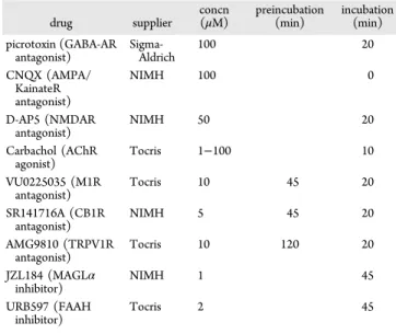
experiments were done at 25°C. Picrotoxin (100 μM) was added to the superfusion medium to block gamma-aminobutyric acid type A (GABA-A) receptors. All drugs were added at thefinal concentration to the superfusion medium (seeTable 1).
Forfield excitatory postsynaptic potential (fEPSP), the recording pipet wasfilled with ACSF and afferents were stimulated with a glass electrodefilled with ACSF and placed ∼200 μm in the dorsal-medial direction of the recording pipet. The stimulus intensity was adjusted around 80% of maximal intensity after performing an input-output curve (baseline fEPSP amplitudes ranged between 0.15 mV and 0.4 mV). Stimulation frequency was set at 0.1 Hz.
Recordings were performed with an Axopatch-200B amplifier (Axon Instrument, Molecular Devices, Sunnyvale, CA). Data were low-pass filtered at 2 kHz, digitized (10 kHz, DigiData 1440A, Axon Instrument, Molecular Devices, Sunnyvale, CA), collected using Clampex 10.2, and analyzed using Clampfit 10.2 (Axon Instrument, Molecular Devices, Sunnyvale, CA). Both fEPSPs’ area and amplitude were analyzed.
Drugs. All drugs were added at final concentration to the superfusion medium (seeTable 1for details).
Data Acquisition and Analysis. The magnitude of plasticity was calculated 40−44 min after the plasticity protocols as percentage of baseline responses. Statistical analysis of data was performed with
GraphPad Prism (GraphPad Software Inc., La Jolla, CA). All values are given as mean ± standard error. N indicates the number of experiments. At least 3−12 animals were used for each single experimental condition. The Shapiro−Wilk test confirmed the normal distribution of data sets. Therefore, depending on the experimental condition, statistical differences were assessed using t tests and one-way ANOVA post hoc tests. A confidence level of P < 0.05 was considered significant.
■
ASSOCIATED CONTENT*
S Supporting InformationThe Supporting Information is available free of charge on the
ACS Publications website at DOI: 10.1021/acschemneur-o.7b00398.
Effect of blockade of 2-AG and anandamide degradation on high carbachol LTD in slices from wild-type littermates; patch clamp recordings of AMPAR responses to 100 μM carbachol; methods for patch clamp recordings (PDF)
■
AUTHOR INFORMATIONCorresponding Author
*Mailing address: Department of Neuroscience, 173 Ashley Ave, BSB410, Medical University of South Carolina, Charleston, SC 29425. Telephone: 843-876-2246. E-mail:
neuhofer@musc.edu.
ORCID
Daniela Neuhofer:0000-0001-5726-7855
Author Contributions
D.N. and O.J.M. designed research; D.N. and O.L. performed research; D.N. and O.L. analyzed data; D.N. and O.J.M. wrote the paper.
Funding
The FRAXA Foundation (O.J.M. and D.N.), a NARSAD 2010 Independent Investigator Grant given by the Brain & Behavior Research Foundation (O.J.M.), Agence Nationale de la Recherche (ANR CortexCell and Cyfip-Aut; O.M. and O.L.) and INSERM (OJ.M.) supported this work.
Notes
The authors declare no competingfinancial interest. Table 1. Drug Suppliers, Final Concentrations, and Incubation Times drug supplier concn (μM) preincubation(min) incubation (min) picrotoxin (GABA-AR antagonist) Sigma-Aldrich 100 20 CNQX (AMPA/ KainateR antagonist) NIMH 100 0 D-AP5 (NMDAR antagonist) NIMH 50 20 Carbachol (AChR agonist) Tocris 1−100 10 VU0225035 (M1R antagonist) Tocris 10 45 20 SR141716A (CB1R antagonist) NIMH 5 45 20 AMG9810 (TRPV1R antagonist) Tocris 10 120 20 JZL184 (MAGLα inhibitor) NIMH 1 45 URB597 (FAAH inhibitor) Tocris 2 45
ACS Chemical Neuroscience Research Article
DOI:10.1021/acschemneuro.7b00398
ACS Chem. Neurosci. XXXX, XXX, XXX−XXX
line receptor proteins in brain with subtype-specific antibodies. J. Neurosci. 11, 3218−3226.
(3) Hersch, S. M., Gutekunst, C. A., Rees, H. D., Heilman, C. J., and Levey, A. L. (1994) Distribution of m1-m4 muscarinic receptor proteins in the rat striatum: light and electron microscopic immunocytochemistry using subtype-specific antibodies. J. Neurosci. 14, 3351−63.
(4) Cortes, R., and Palacios, J. (1986) Muscarinic cholinergic receptor subtypes in the rat brain. I. Quantitative autoradiographic studies. Brain Res. 362, 227−38.
(5) Calabresi, P., Centonze, D., Gubellini, P., Pisani, A., and Bernardi, G. (2000) Acetylcholine-mediated modulation of striatal function. Trends Neurosci. 23, 120−6.
(6) O’Donnell, P. (2010) In Handbook of basal ganglia structure and function (Steiner, H., and Tseng, K. Y., Eds.), pp 367−377, Elsevier Inc..
(7) Scofield, M. D., et al. (2016) The Nucleus Accumbens: Mechanisms of Addiction across Drug Classes Reflect the Importance of Glutamate Homeostasis. Pharmacol. Rev. 68, 816−871.
(8) Neuhofer, D., and Kalivas, P. (2018) Metaplasticity at the addicted tetrapartite synapse: A common denominator of drug induced adaptations and potential treatment target for addiction. Neurobiol. Learn. Mem.,DOI: 10.1016/j.nlm.2018.02.007.
(9) Dölen, G., Darvishzadeh, A., Huang, K. W., and Malenka, R. C. (2013) Social reward requires coordinated activity of nucleus accumbens oxytocin and serotonin. Nature 501, 179−184.
(10) Gunaydin, L. a, et al. (2014) Natural neural projection dynamics underlying social behavior. Cell 157, 1535−51.
(11) Manduca, A., Lassalle, O., Sepers, M., et al. (2016) Interacting Cannabinoid and Opioid Receptors in the Nucleus Accumbens Core Control Adolescent Social Play. Front. Behav. Neurosci. 10, 1−17.
(12) Crespo, J. A., Sturm, K., Saria, A., and Zernig, G. (2006) Activation of muscarinic and nicotinic acetylcholine receptors in the nucleus accumbens core is necessary for the acquisition of drug reinforcement. J. Neurosci. 26, 6004−10.
(13) Avena, N. M., and Rada, P. V. (2012) Cholinergic modulation of food and drug satiety and withdrawal. Physiol. Behav. 106, 332−6.
(14) Massey, P. V., Bhabra, G., Cho, K., Brown, M., and Bashir, Z. I. (2001) Activation of muscarinic receptors induces protein synthesis-dependent long-lasting depression in the perirhinal cortex.itle. J. Eur. Neurosci. 14, 145−52.
(15) McCoy, P. A., and McMahon, L. L. (2007) Muscarinic receptor dependent long-term depression in rat visual cortex is PKC independent but requires ERK1/2 activation and protein synthesis. J. Neurophysiol. 98, 1862−70.
(16) McCutchen, E., Scheiderer, C. L., Dobrunz, L. E., and McMahon, L. L. (2006) Coexistence of muscarinic long-term depression with electrically induced long-term potentiation and depression at CA3-CA1 synapses. J. Neurophysiol. 96, 3114−21.
(17) Dickinson, B. A., et al. (2009) A novel mechanism of hippocampal LTD involving muscarinic receptor-triggered interactions between AMPARs, GRIP and liprin-alpha. Mol. Brain 2, 18.
X syndrome. Psychopharmacology (Berl). 217, 143−51.
(24) Veeraragavan, S., et al. (2012) Genetic reduction of muscarinic M4 receptor modulates analgesic response and acoustic startle response in a mouse model of fragile X syndrome (FXS). Behav. Brain Res. 228, 1−8.
(25) Verkerk, A. J., et al. (1991) Identification of a gene (FMR-1) containing a CGG repeat coincident with a breakpoint cluster region exhibiting length variation in fragile X syndrome. Cell 65, 905−914.
(26) Ronesi, J. A., and Huber, K. M. (2008) Metabotropic glutamate receptors and fragile x mental retardation protein: partners in translational regulation at the synapse. Sci. Signaling 1, pe6.
(27) Darnell, J. C., et al. (2011) FMRP stalls ribosomal translocation on mRNAs linked to synaptic function and autism. Cell 146, 247−61. (28) Maurin, T., Zongaro, S., and Bardoni, B. (2014) Fragile X Syndrome: From molecular pathology to therapy. Neurosci. Biobehav. Rev., 242.
(29) Bear, M. F., Huber, K. M., and Warren, S. T. (2004) The mGluR theory of fragile X mental retardation. Trends Neurosci. 27, 370−377.
(30) Martin, B. S., and Huntsman, M. M. (2012) Pathological plasticity in fragile X syndrome. Neural Plast. 2012, 275630.
(31) Neuhofer, D., Henstridge, C. M., Dudok, B., et al. (2015) Functional and structural deficits at accumbens synapses in a mouse model of Fragile X. Front. Cell. Neurosci. 9, 1−15.
(32) Jung, K.-M., Sepers, M., Henstridge, C. M., et al. (2012) Uncoupling of the endocannabinoid signalling complex in a mouse model of fragile X syndrome. Nat. Commun. 3, 1080.
(33) Maccarrone, M., et al. (2010) Abnormal mGlu 5 receptor/ endocannabinoid coupling in mice lacking FMRP and BC1 RNA. Neuropsychopharmacology 35, 1500−1509.
(34) Zhang, L., and Alger, B. E. (2010) Enhanced Endocannabinoid Signaling Elevates Neuronal Excitability in Fragile X Syndrome. J. Neurosci. 30, 5724−5729.
(35) Bhakar, A. L., Dölen, G., and Bear, M. F. (2012) The pathophysiology of fragile X (and what it teaches us about synapses). Annu. Rev. Neurosci. 35, 417−43.
(36) Restivo, L., et al. (2005) Enriched environment promotes behavioral and morphological recovery in a mouse model for the fragile X syndrome. Proc. Natl. Acad. Sci. U. S. A. 102, 11557−62.
(37) Huang, C.-C., and Hsu, K.-S. (2010) Activation of muscarinic acetylcholine receptors induces a nitric oxide-dependent long-term depression in rat medial prefrontal cortex. Cereb. Cortex 20, 982−996. (38) Dautan, D., et al. (2014) A Major External Source of Cholinergic Innervation of the Striatum and Nucleus Accumbens Originates in the Brainstem. J. Neurosci. 34, 4509−4518.
(39) Lim, S. A. O., Kang, U. J., and McGehee, D. S. (2014) Striatal cholinergic interneuron regulation and circuit effects. Front. Synaptic Neurosci. 6, 1−23.
(40) Martin, H. G. S., Bernabeu, A., Lassalle, O., et al. (2015) Endocannabinoids Mediate Muscarinic Acetylcholine Receptor-Dependent Long-Term Depression in the Adult Medial Prefrontal Cortex. Front. Cell. Neurosci. 9, 1−11.
DOI:10.1021/acschemneuro.7b00398
ACS Chem. Neurosci. XXXX, XXX, XXX−XXX
(41) Giessel, A. J., and Sabatini, B. L. (2010) M1 muscarinic receptors boost synaptic potentials and calcium influx in dendritic spines by inhibiting postsynaptic SK channels. Neuron 68, 936−47.
(42) Puente, N., et al. (2011) Polymodal activation of the endocannabinoid system in the extended amygdala. Nat. Neurosci. 14, 1542−7.
(43) Kim, J., Isokawa, M., Ledent, C., and Alger, B. E. (2002) Activation of muscarinic acetylcholine receptors enhances the release of endogenous cannabinoids in the hippocampus. J. Neurosci. 22, 10182−10191.
(44) Katona, I., and Freund, T. F. (2012) Multiple functions of endocannabinoid signaling in the brain. Annu. Rev. Neurosci. 35, 529− 58.
(45) Robbe, D., Kopf, M., Remaury, A., Bockaert, J., and Manzoni, O. J. (2002) Endogenous cannabinoids mediate long-term synaptic depression in the nucleus accumbens. Proc. Natl. Acad. Sci. U. S. A. 99, 8384−8388.
(46) Grueter, B. a, Brasnjo, G., and Malenka, R. C. (2010) Postsynaptic TRPV1 triggers cell type-specific long-term depression in the nucleus accumbens. Nat. Neurosci. 13, 1519−25.
(47) Alger, B. E. (2002) Retrograde signaling in the regulation of synaptic transmission: focus on endocannabinoids. Prog. Neurobiol. 68, 247−86.
(48) Chávez, A. E., Chiu, C. Q., and Castillo, P. E. (2010) TRPV1 activation by endogenous anandamide triggers postsynaptic long-term depression in dentate gyrus. Nat. Neurosci. 13, 1511−8.
(49) Zygmunt, P. M., et al. (2013) Monoacylglycerols activate TRPV1–a link between phospholipase C and TRPV1. PLoS One 8, e81618.
(50) Qin, M., et al. (2015) Endocannabinoid-mediated improvement on a test of aversive memory in a mouse model of fragile X syndrome. Behav. Brain Res. 291, 164−171.
(51) Wei, D., et al. (2016) Enhancement of Anandamide-Mediated Endocannabinoid Signaling Corrects Autism-Related Social Impair-ment. Cannabis Cannabinoid Res. 1, 81−89.
(52) Ross, R. (2003) Anandamide and vanilloid TRPV1 receptors. Br. J. Pharmacol. 140, 790−801.
(53) Park, J.-Y., and Spruston, N. (2012) Synergistic Actions of Metabotropic Acetylcholine and Glutamate Receptors on the Excitability of Hippocampal CA1 Pyramidal Neurons. J. Neurosci. 32, 6081−6091.
(54) Uchigashima, M., et al. (2007) Subcellular arrangement of molecules for 2-arachidonoyl-glycerol-mediated retrograde signaling and its physiological contribution to synaptic modulation in the striatum. J. Neurosci. 27, 3663−76.
(55) Narushima, M., et al. (2007) Tonic enhancement of endocannabinoid-mediated retrograde suppression of inhibition by cholinergic interneuron activity in the striatum. J. Neurosci. 27, 496− 506.
(56) Zhao, Y., and Tzounopoulos, T. (2011) Physiological activation of cholinergic inputs controls associative synaptic plasticity via modulation of endocannabinoid signaling. J. Neurosci. 31, 3158−3168. (57) Rinaldo, L., and Hansel, C. (2013) Muscarinic acetylcholine receptor activation blocks long-term potentiation at cerebellar parallel fiber-Purkinje cell synapses via cannabinoid signaling. Proc. Natl. Acad. Sci. U. S. A. 110, 11181−11186.
(58) Alger, B. E., Nagode, D. A., and Tang, A.-H. (2014) Muscarinic cholinergic receptors modulate inhibitory synaptic rhythms in hippocampus and neocortex. Front. Synaptic Neurosci. 6, 1−23.
(59) Mathur, B. N., Tanahira, C., Tamamaki, N., and Lovinger, D. M. (2013) Voltage drives diverse endocannabinoid signals to mediate striatal microcircuit-specific plasticity. Nat. Neurosci. 16, 1275−83.
(60) Bateup, H. S., et al. (2010) Distinct subclasses of medium spiny neurons differentially regulate striatal motor behaviors. Proc. Natl. Acad. Sci. U. S. A. 107, 14845−50.
(61) Ade, K. K., Wan, Y., Chen, M., Gloss, B., and Calakos, N. (2011) An Improved BAC Transgenic Fluorescent Reporter Line for Sensitive and Specific Identification of Striatonigral Medium Spiny Neurons. Front. Syst. Neurosci. 5, 32.
(62) Bagetta, V., et al. (2011) Dopamine-dependent long-term depression is expressed in striatal spiny neurons of both direct and indirect pathways: implications for Parkinson’s disease. J. Neurosci. 31, 12513−12522.
(63) Kramer, P. F., et al. (2011) Dopamine D2 receptor overexpression alters behavior and physiology in Drd2-EGFP mice. J. Neurosci. 31, 126−32.
(64) Nelson, A. B., et al. (2012) A comparison of striatal-dependent behaviors in wild-type and hemizygous Drd1a and Drd2 BAC transgenic mice. J. Neurosci. 32, 9119−23.
ACS Chemical Neuroscience Research Article
DOI:10.1021/acschemneuro.7b00398
ACS Chem. Neurosci. XXXX, XXX, XXX−XXX