Development and characterization of immunogenic
genetically engineered mouse models of pancreatic
cancer
ByLaurens J. Lambert MSc, Medical Biology Radboud University, 2014
Submitted to the Department of Biology
in Partial Fulfillment of the Requirements for the Degree of Doctor of Philosophy
at the
MASSACHUSETTS INSTITUTE OF TECHNOLOGY September 2020
© 2020 Massachusetts Institute of Technology. All rights reserved.
Signature of Author………. Laurens J. Lambert Department of Biology June 16, 2020 Certified by………..………. Tyler Jacks David H. Koch Professor of Biology Investigator, Howard Hughes Medical Institute Thesis Supervisor
Accepted by………. Stephen Bell Uncas and Helen Whitaker Professor of Biology Investigator, Howard Hughes Medical Institute
Development and characterization of immunogenic
genetically engineered mouse models of pancreatic
cancer
ByLaurens J. Lambert
Submitted to the Department of Biology on June 16, 2020 in Partial Fulfillment of the Requirements for the Degree of Doctor of Philosophy in Biology
Abstract
Insights into mechanisms of immune escape have fueled the clinical success of immunotherapy in many cancers. However, pancreatic cancer has remained largely refractory to checkpoint immunotherapy. To uncover mechanisms of immune escape, we have characterized two preclinical models of immunogenic pancreatic ductal adenocarcinoma (PDAC). In order to dissect the endogenous antigen-specific T cell response in PDAC, lentivirus encoding the Cre recombinase and a tumor specific antigen (SIINFEKL, OVA257-264) was delivered to KrasLSL-G12D/+; Trp53flox/flox (KP) mice. We
demonstrate that KP tumors show distinct antigenic outcomes: a subset of PDAC tumors undergoes clearance or editing by a robust antigen-specific CD8+ T cell response, while a fraction undergo immune escape.
Subsequently, we have developed an immunogenic pancreatic tumor organoid orthotopic transplant model. In this model, immunogenic pancreatic tumors manifest divergent tumor phenotypes; 40% of tumor organoids do not form tumors (“non-progressors”), whereas 50% of organoids form aggressive tumors despite maintaining antigen expression and a demonstrable T cell response (“progressors”). Additionally, a subset (10%) of tumors show an intermediate phenotype, possibly reflective of an immune equilibrium state. We have further phenotypically and transcriptionally characterized the CD8+ T cell response to understand immune escape in this model. Our analyses reveal unexpected T cell heterogeneity, and acquisition of T cell dysfunctionality. Therapeutic combinatorial targeting of co-inhibitory receptors identified on dysfunctional antigen-specific CD8+ T cells led to dramatic regression of aggressive pancreatic tumors. Finally, we demonstrate that human CD8+ T cells isolated from pancreatic tumors co-express co-inhibitory receptors, suggesting that T cell dysfunction may be operational in human disease.
This is the first demonstration of immunoediting in an autochthonous and organoid-based model of pancreatic cancer. Further characterization of these preclinical model systems will enable rational design of novel clinical immunotherapeutic strategies for treatment of this devastating disease.
Curriculum vitae
Laurens J. Lambert
Education
2014 - MASSACHUSETTS INSTITUTE OF TECHNOLOGY (CAMBRIDGE, MA,
Present USA)
PhD, Department of Biology, The Jacks Lab
2009 - 2014 RADBOUD UNIVERSITY (NIJMEGEN, NETHERLANDS) Bachelor of Science in Biology
Master of Science in Medical Biology, Cum Laude
Professional Experience
May 2015- The David Koch Institute for Integrative Cancer Research, MIT (Tyler Present Jacks Laboratory)
PhD Student
§ Developed novel pancreatic cancer mouse models and characterized the T cell response in these models.
§ Discovered a therapeutic checkpoint combination with clinical § translatability.
§ Co-led a pancreas research team in the Jacks lab and coordinated with internal and external collaborators (Koch Institute, DFCI). § Authored a chapter in a major textbook and submitted a manuscript
to Nature (under review). Jan. 2020- MPM Capital (Cambridge, MA) Present Consultant
§ Supported key company-formation activities through the technical evaluation of scientific fields and identification of high priority development candidates.
§ Presented progress in weekly strategy meetings with the founding team at MPM Capital.
April 2019- Vida Ventures (Boston, MA) Dec. 2019 Fellow
§ Evaluated the scientific evidence, clinical paradigms, and
commercial landscape to support 15+ investment decisions for the team at the Boston office.
§ Performed technical diligence on a Vida investment (Kinnate Biopharma, Series B closed Dec. 2019).
Fall 2018 Merrimack Pharmaceuticals (Cambridge, MA)
Graduate Intern
§ Participated in the selective “Research Experience in Biopharma” course and interned at Merrimack Pharmaceuticals for the semester.
§ Assisted in target validation of the TNFRII preclinical immuno-oncology program (MM-401).
Scientific Publications
Freed-Pastor W*, Lambert LJ*, Mercer K, Pattada N, Garcia A, Ely Z, Hwang W, Lin L, Eng G, Westcott P, Yilmaz O, Jacks T (2020). Immunogenic models of pancreatic adenocarcinoma reveal the therapeutic benefit of novel checkpoint combinations. Manuscript under review (Nature). *Co-authors
Lambert LJ, Muzumdar MD, Rideout III WM, Jacks T (2017). Chapter 15: Harnessing the Mouse for Biomedical Research, in Basic Science Methods for Clinical
Researchers, ed. Jalali M, Saldanha FYL and Jalali M, Academic Press
Wefers C, Lambert LJ, Torensma R, Hato SV (2015). Cellular immunotherapy in ovarian cancer: Targeting the stem of recurrence. Gynecologic Oncology 137(2)
Lambert LJ, Walker S, Feltham J, Lee HJ, Reik W, Houseley J (2013). Etoposide Induces Nuclear Re-Localisation of AID. PLoS ONE 8(12)
Teaching & Leadership Experience
Sep. 2018- Co-Director, Industry Initiative team Present MIT Biotech Group (MBG)
Fall 2017 Teaching Assistant, Hallmarks of Cancer (7.45/7.85), MIT
Fall 2015 Teaching Assistant, Introductory Biology (7.016), MIT Awards & Grants
• Koch Institute Graduate Fellowship (2018)
• Praecis Pharmaceuticals MIT Presidential Fellowship (2014) • Radboud University Excellence Grant (2012)
Acknowledgments
I am extremely grateful for the past 6 years in the Jacks Lab. I have grown tremendously as a scientist, as a mentor, as an educator, and most of all as a person during my PhD. I firmly believe that the skills and life lessons instilled through my graduate training have prepared me well as I pursue the next chapter in my career.
In the Jacks Lab, I have had the privilege to work alongside an amazing group of smart and passionate people. I have benefited so much from the experience and scientific knowledge shared, and the friendships formed along the way. Without the tireless dedication of many people in and outside the lab, support and advice from many colleagues, friends and loved ones, the work in this thesis would not have been possible. I have many people to thank…
First and foremost, I would like to thank my thesis supervisor, Tyler, for letting me join the lab and for incredible mentorship during my graduate studies. From the early days in the lab, you have given me the freedom to pursue my interests in immunotherapy, trusted me to fail and succeed when I needed to, and through it all challenged me to think “two steps ahead”. I have also appreciated that you have always had my career at heart, and have allowed me to explore my interests in biotech while completing my PhD. Your leadership, thoughtfulness, rigor and generosity are truly inspiring to me, and are qualities I will strive for in the rest of my career.
I would also like to thank my thesis committee members, Matt and Jackie. I have benefited tremendously from your scientific insights, guidance and mentorship during my time at MIT. It was always a pleasure to share the latest updates on my science with you during my committee meetings, and I will truly miss having these stimulating conversations moving forward. The classes I took as a TA in 7.45/7.85 Cancer Biology were one of the most memorable courses I took while at MIT-- thank you making them so fun and interactive, Matt.
I would also like to thank Vijay Kuchroo for agreeing to be my external thesis defense member, and I look forward to discussing pancreatic tumor immunology with a true leader in the field.
I would like to thank Will, for being an amazing collaborator and a great friend. Your incredible drive, intellect, and dedication has pushed our shared science to greater heights. Thank you for always being willing to do the crazy experiments, for challenging the status quo, and for your unrelenting excitement when making new discoveries. I will miss our comradery, wide-ranging discussions on anything pancreatic or immunology-related, and yes maybe even 4 am scRNA-seq harvests. Those were days...
I would like to thank the Jacks Lab members who keep our lab running: Judy, Karen, Kate, Kim, and Margaret. Thank you for helping me with countless things during my time in the lab, and for your selfless efforts to make sure the lab is clean, continued to operate
smoothly and remained in stable financial waters. I really appreciate everything you have done.
Thank you to Kim for your generosity, support, and friendship. Your dedication to the mouse colony and animal experiments over the last year(s) have made much of the work in this thesis possible. I particularly want to thank you for jumping in during the COVID-19 lockdown, which has allowed me to stay focused on completing my PhD.
Thank you to two incredible technicians, Nimisha and Ana. I have really appreciated your patience, countless technical contributions, and commitment to this project. It has been rewarding to see you grow as scientists during your time in the lab, and I look forward to following your paths as you chart out on bright futures.
Thank you to Zack for jumping into the deep with this pancreas project at full pace. I am incredibly grateful for your computational analyses that have pushed the science described here forward, and I am excited to see what you will achieve during your PhD. Thank you to all current and past graduate students in the lab, in particular Sheng Rong, Rodrigo, Amy, David, Leah, Ryan, Grissel and Amanda. After “ascending” to becoming the most senior graduate student in the lab, I have come to realize that much of the experience during a PhD in the Jacks Lab is universal. I have tremendously benefited from your advice, support, comradery and expertise at many points during the past 6 years.
Thank you to all current and past postdoc students and staff in the lab, in particular Britt, Megan, Carla, Peter, Alex, Banu, Jason, Mandar, and Nik. Each one of you has helped me in a myriad of ways, be it through advice, sharing scientific insights and expertise, or even just perspectives on life—it has helped me grow as a person and allowed me to keep pushing the science forward.
Thank you to all the current and past technicians in the lab, for your dedication and hard work. Your energy and excitement for science is a big part of what makes the culture in the Jacks lab so great. I especially want to thank Grace, Sophie, Demi, Michelle, Caterina, and Da-Yae—I appreciate your willingness to always help me out and your friendship. Thank you to the amazing MIT classmates I have been fortunate enough to call friends, in particular Santi, Nikola, Chris, Danny, Spencer, and Josh—for countless dinners, movies, parties, ski and hiking trips, being part of my wedding, and numerous other things! I know our friendship is bigger than a PhD, and that is incredibly special.
To my sisters Willemijn and Nora, and my parents Hein and Jannie. Thank you for your unconditional love and support. You have always been there for when I needed it most and much of this work is inspired by the lessons you have taught me. I hope this thesis makes you proud.
Last, but most importantly, to the love of my life, Alexandra: Thank you for always believing in me, for always encouraging me to do and be my best. Thank you for your enduring patience and understanding as I spent many weekends and late nights in the lab. I could not have done this without your love and support all these years, and I am so excited to welcome our first addition to the family in August.
Laurens Lambert June 2020
Table of Contents
Abstract ... 3
Curriculum vitae ... 4
Acknowledgments ... 6
1. Introduction ... 12
1.1. History of tumor immunology ... 13
1.1.1. The early history of immunotherapy ... 13
1.1.2. Coley’s toxins and the beginning of tumor immunology ... 15
1.1.3. The dawn of tumor transplantation models ... 18
1.1.4. Tumor antigens and the immunosurveillance hypothesis ... 20
1.1.5. Cellular immunology comes of age ... 23
1.1.6. The beginning of a modern era in tumor immunology ... 25
1.2. The cancer immune response ... 28
1.2.1 Tumor antigen release leads to the initiation of immune responses ... 29
1.2.2 Dendritic cells traffic to lymph node and interact with naïve T cells ... 31
1.2.3 T cell priming triggers clonal expansion and effector cell differentiation ... 31
1.2.4 T cells use chemotactic cues to migrate to the tumor microenvironment ... 33
1.2.5 T cells infiltrate into the tumor microenvironment ... 34
1.2.6 Effector T cells recognize cancer cells through antigen presentation ... 35
1.3. Tumor-intrinsic mechanisms of immune escape ... 37
1.3.1. Antigenicity is a critical determinant in immune escape ... 37
1.3.2. Immunoediting can mediate tumor immune escape ... 39
1.3.3. Oncogenic pathways drive T cell exclusion ... 41
1.4.2. Cancer-associated fibroblasts (CAFs)... 45
1.4.3. Myeloid-derived suppressor cells (MDSCs) and tumor-associated macrophages (TAMs) .. 47
1.4.4. Tumor-associated macrophages (TAMs) ... 48
1.4.5. T cell-intrinsic dysfunction ... 49
1.4.6. CD4+ T cells ... 56
1.4.7. Tumor microenvironments can exist in distinct inflammatory states ... 58
1.5. Pancreatic cancer ... 59
1.5.1. The disease progression and the genetic basis of pancreatic adenocarcinoma ... 61
1.5.2. The pancreatic tumor microenvironment ... 64
1.5.3. Immunotherapy in pancreatic cancer ... 70
1.6. Synopsis ... 74
1.7. References ... 77
2. TIGIT-based therapy induces potent anti-tumor responses in pancreatic cancer ... 103
2.1 Abstract ... 104
2.2 Main results ... 105
2.2.1 Preclinical modeling of immunogenic pancreatic cancer ... 105
2.2.2 Immune evasive pancreatic tumors retain antigen expression and presentation ... 109
2.2.3 Multiple classes of antigen-specific CD8+ TILs within immune evasive pancreatic tumors 114 2.2.4 TIGIT-directed combination immunotherapy as a novel therapeutic approach in PDAC ... 119
2.3 Discussion ... 122
2.4 Acknowledgements ... 123
2.5 Methods ... 124
3. Discussion ... 161
3.1. Genetically engineered mouse models offer unique insights into tumor immunoediting 161 3.2. Outlook – the potential of autochthonous models and outstanding questions ... 167
3.3. Organoid tumor immune escape may be mediated by tumor-extrinsic mechanisms of immune evasion ... 169
3.4. Transcriptional profiling of the antigen-specific T cell compartment revealed heterogenous effector states ... 173
3.5. Outlook – The future of immunotherapy in pancreatic ductal adenocarcinoma through the lens of genetically engineered mouse models ... 175
3.6. References ... 177 Appendix I ... 180 Abstract ... 181 Main text ... 181 Discussion ... 183 Methods ... 184 Acknowledgments ... 188 References ... 189
1. Introduction
It is now widely appreciated that the immune system plays a critical role in tumorigenesis, and is considered a “hallmark of cancer”1. Recent clinical successes with
therapeutic agents that modulate the immune system, rather than the tumor itself, have truly revolutionized cancer treatment in recent years.
Given these clinical successes, it may come as a surprise that the tumor immunology field is over a century old, and for most of its recent scientific history, has been regarded with much skepticism. Prevailing dogma asserted that because cancers arise from the body’s own tissues, the immune system is unable to detect these cells and remains ignorant of the growing tumor. Beginning in the 1990s, this concept was slowly overturned by revisions to concept of cancer immunosurveillance2, the identification of
human tumor antigens3, and the demonstration of immune checkpoint blockade efficacy
in tumor transplantation models4. These findings have sparked an enthusiasm for the
development of immunotherapeutic anti-cancer agents, which has led to regulatory approval of the first generation of immune checkpoint inhibitors (ICIs). The durable clinical remissions achieved with these therapies are arguably the most convincing evidence that the immune system can be mobilized to elicit anti-tumor responses5.
While immunotherapies have made tremendous clinical impact on many different types of cancer, pancreatic adenocarcinoma (PDAC) has remained largely treatment-refractory. The overall five-year survival rate of metastatic PDAC, which constitutes the majority of diagnosed pancreatic cancer cases, has not improved over the last decade. Therefore, novel therapies to combat this devastating disease are urgently needed. Genomic profiling has revealed that (a subset of) PDAC harbors high affinity
neoantigens6, suggesting that this cancer is not intrinsically resistant to anti-tumor T cell
responses. Therefore, a deeper understanding of the tumor-immune microenvironment is necessary to dissect the mechanisms may limit the efficacy of immunotherapeutic approaches.
This introductory chapter includes a historical overview of the field of tumor immunology, and reviews our current understanding of the anti-tumor immune response. Following this overview, the tumor-intrinsic and tumor-extrinsic mechanisms of immunoevasion are described. This chapter concludes with a summary of our current understanding of the genetic basis of pancreatic cancer, the role of the tumor microenvironment in PDAC progression, and the ongoing clinical efforts focused on harnessing immunotherapy for the treatment of pancreatic cancer.
1.1.
History of tumor immunology
1.1.1. The early history of immunotherapy
The concept of utilizing the immune system to treat human disease, broadly referred to as ‘immunotherapy’, dates back millennia. The Greek philosopher and historian, Thucydides, first described a link between the survival of a disease, “the plague of Athens”, and the acquisition of immunity in 430 BC7. However, it would take many
centuries for these observations to become actionable. During the 10th century, Chinese
doctors noticed that “ripe pus” from smallpox patients could be transferred to other individuals on dried cotton tips, and that this sometimes conferred protection against the disease8. A Chinese text from 1597 suggested the use of “powdered cow ice” for the
The practice of these ‘variolation’ techniques (or inoculation) appears to have arisen independently in China, India, and Africa before slowly spreading to Europe and America. Lady Mary Wortley Montagu, wife of the British ambassador of the Ottoman Empire has been widely credited for introducing this idea into Western civilizations. Having witnessed the practice firsthand in Turkey, she had her son secretly variolated by the Scottish physician Charles Maitland in 17168. When Maitland subsequently performed
a successful variolation on Montagu’s second child under the observation of prominent Royal Society physicians, he was granted a medical license to perform a “clinical trial”. For his variolation experiment in 1721, Maitland chose 6 condemned prisoners whom he infected with smallpox pus. Much to his surprise, all individuals recovered from their symptoms, and even had subsequent immunity when exposed to smallpox patients8.
That same year, the city of Boston (U.S.) was plagued with a smallpox epidemic; nearly half of its 12,000 citizens developed the disease. Desperate to fight the spread of smallpox, the physician Zabdiel Boylston, and Reverend Cotton Mather, variolated nearly 300 Bostonians, and compared the disease outcomes of these treated individuals against the naturally infected patients9. In what is arguably one of the first examples of a rigorous
clinical statistical analysis, they were able to show that smallpox variolation dramatically reduced subsequent disease mortality. However, the practice was vehemently opposed by townsmen; at some point ‘anti-variolators’ went as far as bombing Mather’s house. While certainly a progressive medical thinker in his time, Mather’s legacy is nowadays mired in controversy due his written justifications of the Salem witch trials.
The success from this large-scale variolation campaign did not go unnoticed at the time, and the practice became gradually commonplace across European nations and in
the U.S. throughout the 18th century. However, smallpox variolation was not without
complications, as these “vaccines” were frequently contaminated and could cause syphilis. Moreover, (excessive) transfer of smallpox virus to nonimmunized individuals would sometimes result in active infection, as opposed to the protective immune response that the treatment was supposed to confer.
It would take until the turn of the 18th century before prophylactic vaccination
against smallpox became safer. Growing up in the county of Berkeley (England), the physician Edward Jenner had heard stories about dairy milkmaids that were protected against smallpox after suffering from the milder cowpox disease. He decided to investigate these observations further, and in 1796 transferred cowpox from an infected maid to an 8-year-old boy10. When he subsequently inoculated the boy with smallpox, no
disease developed. Buoyed by this finding, Jenner submitted a treatise to the Royal Society, which was abruptly rejected. After adding a few more cases, Jenner published a small booklet, in which he coined the practice of ‘vaccination’, derived from the cowpox-causing Vaccinia virus10. This proved enough to convince a number of leading British
physicians, and as a result the procedure spread quickly throughout the country; it is estimated that over 100,000 people were vaccinated by 18018. In 1840 the British
government officially banned the practice of variolation. However, it would take another 176 years before global incidence of smallpox was completely reduced to zero.
1.1.2. Coley’s toxins and the beginning of tumor immunology
The concept that human diseases could be prevented through prophylactic vaccination undoubtedly laid the foundation for tumor immunology. Throughout the history
of medicine, individual cases of spontaneously regressing tumors have been reported, often accompanied by seemingly unrelated infections11. During the late 19th century,
German physicians Wilhelm Busch and Friedrich Fehleisen independently noted a connection between an opportunistic bacterial skin infection (erysipelas) and tumor regression. Fehleisen identified the causative pathogen as the Gram-positive
Streptococcus pyogenes bacterium. These observations led both physicians to
experiment with the intentional induction of erysipelas in cancer patients, which reportedly led to tumor shrinkage12. The practice, however, was not widely adopted by other
physicians at the time.
Frustrated by an inoperable case of head and neck sarcoma, a young American surgeon, William Coley, took note of these cases in 1891 and decided to perform his own experimentation in his cancer ward at the Memorial Hospital in New York. In a series of treatments on 6 inoperable sarcoma and 4 carcinoma patients, Coley administered S.
pyogenes inoculations that he had received from Robert Koch’s laboratory in Germany.
The results were mixed: in 4 patients he was able to induce full erysipelas and observed robust tumor regressions. However, the condition of 4 other patients only temporarily improved, but they managed to achieve a fully developed infection. Worse yet, 2 patients did develop erysipelas, and actually succumbed to a pathogenic attack instead13,14. This
led Coley to try combining heat-killed S. pyogenes with toxins from the Gram-negative bacterium Serratia marcescens, thereby creating the first ever mixed bacterial vaccine (MBV)13. Unbeknownst at the time, this combination of Gram-positive and Gram-negative
bacteria creates a potent immunostimulatory cocktail, and is now thought to induce the release of multiple inflammatory cytokines, including interleukin-12 (IL-12)15.
Coley and colleagues at the Memorial Hospital in New York would go on to treat nearly 1200 patients with these “Coley’s toxins” over the next 40 years; 270 patients reportedly achieved long-term remissions with MBV, sometimes even lasting decades13,16. Many of the best responding tumors included soft tissue sarcomas, although
responses in other tumor types were achieved as well16. These cases were carefully
documented by Helen Coley Nauts (William Coley’s daughter). In honor of her late father, Nauts would establish the Cancer Research Institute (CRI) for advancement of tumor immunology in 1953.
However, the widespread adoption of MBV outside of New York was hampered by poor documentation of Coley’s practices and the limited potency of commercial preparations13. Coley frequently changed how he produced MBV and would inject the
vaccines using various routes; many of these attempts were later shown to be ineffective16. Furthermore, the scientific field of tumor immunology was still in its infancy
at the turn of the 20th century, and a mechanistic basis for the efficacy of Coley’s toxins was lacking. As radiation therapy and chemotherapy, which promised similar remission rates as MBV, became more popular for cancer treatment, Coley’s toxins fell out of favor. Ironically, James Ewing, one of Coley’s staunchest opponents and his director at Memorial Hospital, discovered a bone sarcoma (Ewing’s sarcoma) that could be effectively treated Coley’s toxins17. While MBV is no longer used in the clinic today,
William Coley’s efforts undeniably set the practice of cancer immunotherapy in motion. He is now recognized as one of the founding fathers of the field.
1.1.3. The dawn of tumor transplantation models
It was the German physician Paul Ehrlich who was the first to formulate the concept that the human immune system is capable of recognizing and fighting off cancerous cells18. In his work published in 1909, Ehrlich suggested that “aberrant cells arising during
fetal and post-fetal development” could remain latent due to the body’s “positive mechanisms” 18. However, the scientific underpinnings of immunology at the time were
not advanced enough to experimentally validate his hypothesis, nor was a connection made between Ehrlich’s theory and Coley’s toxins.
Instead, the nascent field of tumor immunology became fixated on understanding immune responses through tumor transplantation studies. Experiments performed around the turn of the 20th century by Loeb and Jensen showed that thyroid tumors
and spontaneous alveolar carcinomas could sometimes be transplanted within the species they originated from, and that the growth of these transplants resulted directly from the transferred cells19. These results argued against one of the influential theories
at that time, which stated that cancer was caused by an (unknown) etiologic agent and was essentially a “transmissible disease”19. Loeb and a number of his contemporaries
then showed that tumor origin (“race”) was an important factor in determining the outcome of tumor transplantation20. Over ten years later, Clarence Little and Ernest Tyzzer at
Harvard would revisit these studies by carefully examining the tumor transmissibility of inbred strains. Their work led to the discovery of a genetic basis for tumor transplantation, and importantly raised the question whether tumor transplant rejections were simply the result of transferring between “genetically impure” mouse strains20–22.
This issue would continue to frustrate tumor immunologists throughout the 1920s and 1930s22. In an expansive literature review, William Woglom gloomily concluded
that “it would be as difficult to reject the right ear and leave the left ear intact as it is to immunize against cancer”23,24, a dogmatic belief that was shared by many geneticists
around the time.
It would take until 1936 before further progress on the genetic basis of tumor transplantation was made. Through the ingenious use of human and rabbit antisera, Peter Gorer discovered the presence of distinct blood groups in mice of different inbred strains25. When Gorer transferred tumors originating from mice containing “blood group
II” were transferred to mice with “blood group I”, these recipients rapidly rejected the tumor. This suggested to Gorer that the blood antigens were involved in mediating the resistance to tumor rejection. George Snell, working at the Jackson Laboratory, had similarly set out to pursue mapping of the genetic basis of tumor transplantation. As a classically trained geneticist, Snell approached the problem by generating a series of genetically identical mouse strains only differing in a single locus, so-called ‘congenic’ strains. Harnessing the antisera against “blood group II” on different congenic lines, Snell and Gorer were able to map the genetic site of tumor transplantation to the histocompatibility locus in 194825,26. In recognition of the “blood group II” serum that led
to its discovery, this locus became known as the histocompatibility locus II (H-2). As the
H-2 locus was shown to be the most critical determinant mediating tumor rejection, while
in in addition to being highly polymorphic in nature, it is referred to as the major
with Baruj Benacerraf for the discovery of the H-2 locus; unfortunately, Peter Gorer had passed away 19 years prior.
A final insight into tumor transplantation came from the work of the British scientist, Peter Medawar, working in the separate field of transplantation immunology. Medawar focused his efforts on elucidating the basis of allogeneic skin graft rejection (‘allografts’). In seminal papers published in 1953 and 1956, Medawar and Bellingham described the concept of ‘acquired tolerance’ by demonstrating that the immune system plays a fundamental role in the rejection of allogeneic transplants27–29. Medawar’s work confirmed
the earlier hypothesis formulated by Macfarlane Burnet in 1948, that the immune system (in Burnet’s theory “antibodies”) could acquire the ability to discriminate ‘self’ from ‘non-self’ during development30. Both scientists would be awarded the Nobel Prize in 1960.
1.1.4. Tumor antigens and the immunosurveillance hypothesis
While the study of tumor transplantation models shaped much of the first 50 years of tumor immunology, leading to the discovery of the MHC locus and immunological tolerance, it did little to illuminate how the immune system could respond to tumors arising in its own host. To study this question, immunologists increasingly turned to the chemical carcinogen methylcholanthrene (MCA) to induce sporadic tumors in mice. In 1943 Polish scientist Ludwig Gross was the first to demonstrate that an MCA-induced sarcoma line from the inbred C3H strain was rejected by genetically identical C3H recipients31,
suggesting that the sarcoma contained antigens that could be recognized by the host’s immune system. However, his work left open the possibility that the sarcomas had acquired mutations during repeated transplantation. Edward Foley confirmed Gross’
observations in 1953 by demonstrating that methylcholanthrene-induced tumors were antigenic upon direct transplantation in isogeneic animals32. Raymond Prehn and Joan
Main would further extend these results through the characterization of several fibrosarcoma tumor lines that were rejected upon transplantation into isogenic strains33.
However, these studies were viewed with skepticism, as they could still be explained by a lack of a genetically pure background between strains (‘residual heterozygosity’). George Klein and colleagues would finally settle this debate by demonstrating that autochthonous tumors (in addition to syngeneic tumors) could be rejected upon pre-immunization of the hosts34.
Collectively, these studies raised the idea that the immune system is actively involved in the elimination of cancerous cells arising in the body. This led Macfarlane Burnet and Lewis Thomas to formulate their influential cancer immunosurveillance hypotheses in the late 1950s, echoing Paul Ehrlich’s theory half a century earlier. Inspired by the concept of immune tolerance, Burnet focused on his hypothesis on the accumulation of “antigenic potentialities” (neoantigens) by cancer cells, which would be sufficiently different from the own body to trigger immune responses that restrained tumors from becoming clinically apparent35,36. Thomas’ early theory was more
evolutionary in nature, suggesting that multicellular organisms have developed mechanisms to protect against transformed cells in order to maintain tissue homeostasis, similar to the protection against foreign tissues (during allograft rejection)37,38.
Experimental validation for these hypotheses soon followed with demonstrations that carcinogen-induced and oncoviral experimental models contained unique tumor
antigens39. However, evidence for the existence of human antigens was absent, and
whether these models accurately reflected human cancer remained unclear.
The field of tumor immunology would soon face a setback that would again cause skepticism about the perceived ability of the immune system to ward off cancer cells. A logical extension of the cancer immunosurveillance hypothesis was an absence of immune function (‘immunodepression’) would result in higher incidence of tumor formation. The development of athymic (Nu/Nu) mice in the late 1960s made it possible to experimentally test this idea40,41, as these mice largely lacked mature T lymphocytes.
Working with these ‘nude’ mice at Memorial Sloan Kettering, Osias Stutman demonstrated in the 1970s that MCA induction at birth did not lead to a statistically higher number of sarcomas in Nu/Nu mice compared to their normal, heterozygous counterparts (Nu/+)42,43. He concluded that his results did not support the immunosurveillance
hypothesis. Additionally, no evidence of increased susceptibility to spontaneous tumors was found when 27 murine lines were systemically investigated by Harold Hewitt and colleagues44. Indeed, even Foley, Prehn, and Main, in their landmark papers during the
1950s, had noted the differences between carcinogen-induced and spontaneous tumor models32,33, raising doubts that the MCA-induced tumors were just an anomaly.
While these results certainly dampened the excitement around cancer immunotherapies over the next decade, we now know that Stutman’s studies were flawed by several technical factors unknown to him. While athymic mice have severely compromised immune responses to infectious agents, some basal T cell functionality is maintained45–47. Additionally, natural killer (NK) cells, developing normally in these mice,
with MCA is highly efficient in the CBA/N strain (the only Nu/Nu model available to Stutman), raising the possibility that early tumor induction simply overwhelmed an immature immune system51. Indeed, later studies using mouse strains lacking T, B, and
NK cells (Rag2-/-; gc-/- mice), the interferon-g receptor (Ifngr1-/-) or the
cytotoxicity-mediating protein perforin (Prf1-/-) have shown elevated susceptibility to MCA-induced tumorigenesis52–55. Thus, Stutman’s rejection of tumor immunity ultimately proved
premature; rather, genetic murine models as well as epidemiological observations of elevated cancer incidence in immunocompromised humans have firmly cemented the immune system’s role in cancer surveillance.
1.1.5. Cellular immunology comes of age
While tumor immunology remained focused on experimental models of cancer, discoveries made in different immunological fields during the 1960s and 1970s would lay the foundation for our modern understanding of the cellular components of the anti-tumor response. Drawing upon Medawar’s concepts of transplant tolerance, James Gowans and colleagues showed that thoracic duct lymphocytes (TDLs) were involved in the initiation of alloreactive immune responses, although they could not distinguish cellular from humoral responses56. Jacques Miller soon followed these results by demonstrating
that neonatal thymectomized mice were unable to reject their skin grafts, which suggested that the thymus was the source of these reactive immune cells57,58. His results stood in
stark contrast to the widely held belief that the thymus was simply a vestigial organ where lymphocyte would “go to die”. Additionally, Miller found that adult thymectomized mice were unable to regenerate their lymphocytes upon total body irradiation or initiate immune
responses upon antigenic challenges with sheep erythrocytes, further solidifying the role of the thymus in immunocompetence59–61. In a series of experiments using irradiated CBA
mice grafted with syngeneic thymi, TDLs, and allogeneic bone marrow, Mitchell and Miller then unequivocally proved that thymus-derived lymphocytes (‘T cells’) were immunologically distinct from bone marrow-derived lymphocytes (‘B cells’)62–65.
Subsequent use of different antisera helped to further distinguish these lymphocytic cell types by their respective cell surface markers66,67, and led to the
subdivision of T cells into CD8+ (originally Ly-2, Ly-3) and CD4+ (Ly-4) cells68,69. The
development of monoclonal antibodies in 1975 by Kohler and Milstein70 would usher in a
new age where lymphocytes could be studied in increasingly greater detail, particularly with the advent of flow cytometric methods71.
In an effort to dissect immune responses in vitro, Mishell and Dutton developed an assay during the 1960s that mixed antigens with splenic lymphocytes, which led to the realization that antibody formation required an additional cell type present in this mixture72. Working with these cultures in 1973, Steinman and Cohn first described the
dendritic cell (DC) as the third cell type73,74. Steinman and colleagues would go on to
decipher much of the biology of DCs over the next decades75–78, and establish a critical
role for this antigen-presenting cell (APC) in the initiation of adaptive immune responses. The structure of antibodies was uncovered in an influential set of studies during the 1950s and 1960s79,80, setting the stage for the discovery of immunoglobulin (Ig)
rearrangement by Hozumi and Tonegawa81. By early 1980s it was well accepted that the
B cells used Ig molecules to recognize soluble antigen, however the nature of the T cell receptor (TCR) and how T cells recognized antigens remained topics of debate82,83. The
latter was clarified by Zinkernagel and Doherty, who established that T cell recognition was governed by the MHC molecule of the syngeneic host, a concept that became known as ‘MHC restriction’84–86. Importantly, their results argued that a single TCR could
recognize both antigen and MHC—a model proven correct when the first MHC peptide epitopes were uncovered87,88. The nature of the TCR was uncovered when Davis and
Mak isolated the first murine and human TCR chains89,90, before rapidly cloning of the
remaining TCR chains91–93. Collectively, these studies demonstrated that primary antigen
sensing receptors (the TCRa,b chains) were part of a larger transmembrane protein complex that associated with g, d, and the two e, z polypeptide chains94,95.
Additional discoveries in cellular immunology made in the 1970s and 1980s included with the discovery of interleukin-2 (IL-2)96, which allowed the sustained in vitro
culture of cytotoxic lymphocytes, and the generation of transgenic TCR mouse strains97.
Together, these advancements would form the basis for novel immunotherapeutic strategies pioneered over the following decade, as the field of tumor immunology regained its former momentum.
1.1.6. The beginning of a modern era in tumor immunology
While tumor immunologists still grappled with the implications of Stutman’s results throughout the 1980s, Thierry Boon’s work offered a glimmer of hope for the field. In 1982, Boon and van der Pel showed that vaccination with mutagenized leukemia clones could induce the immune rejection of spontaneous tumors of the same origin98, which
suggested that these tumors, rather than lacking antigens, failed to stimulate strong immune responses. These findings called the results obtained with experimental models
in the decades before into question, and raised the possibility that human tumors could be antigenic as well. The Boon group would continue their focus on tumor antigens, which culminated with the identification of the first human T cell antigen in 1991, encoded by the
MAGE-1 gene3. MAGE proteins are part of a diverse group of cancer-testis antigens
(CTAs), which are normally restricted to immunoprivileged reproductive organs, but become aberrantly expressed in many tumor types99. In the immediate years following
the initial identification of MAGE-1, a wealth of other murine and human antigens were characterized, including MAGE, BAGE, GAGE, RAGE (all CTAs), Tyrosine, gp100/pmel 17 (differentiation antigens), E7 (a viral antigen), HER2/Neu (an overexpression antigen),
CDK4, and b-catenin (neoantigens)100.
Further mechanistic insights into the relationship between the immune response and cancer antigenicity were generated by Bob Schreiber and Lloyd Old. In a landmark study published in 2001, Schreiber, Old and Shankaran carefully compared the re-transplantation kinetics of MCA-induced sarcomas derived in the absence of an immune-selective environment (Rag2-/- mice), or derived in an immunocompetent environment (wild-type 129/SvEv mice)54. They were able to demonstrate that tumor immune escape
was intimately linked to the prior elimination of immunogenic cancer clones by T cells. These experiments, together with data from genetic knockout mice that showed increased susceptibility to cancer formation, and the experimental demonstration of an intermediate state of “stasis” between the actions of immune cells and tumor emergence101, have led
to the definition of the ‘cancer immunoediting’ process. Cancer immunoediting is thought to occur in three distinct phases: immune elimination, immune equilibrium, and finally immune escape2,38.
The 1990s would also set the stage for much of the clinical excitement around immune checkpoint inhibitors nearly two decades later. In a landmark study published in 1996, James Allison and Max Krummel demonstrated that the cytotoxic T-lymphocyte-associated protein 4 (CTLA-4) was a negative regulator of T cell responses in murine transplant models4. Just two years later, Tasuku Honjo and Hiroyuki Nishimura used a
genetic Pdcd-1 knockout mouse to elucidate the function of PD-1 in the maintenance of peripheral tolerance102. The discovery of these two ‘immune checkpoints’ has had
enormous impact on the field. Research into the mechanisms of immune tumor detection, tumor immune evasion and efficacy of novel immunotherapies is now truly flourishing. Indeed, much of the biology described in the following sections of this introduction has only been fairly recently elucidated; a testament to incredible pace of discovery in this field.
1.2.
The cancer immune response
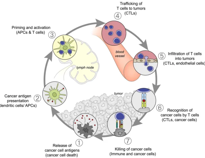
The immune response to a growing tumor can be conceptualized as a multistep iterative process, termed the “cancer-immunity cycle” by Chen and Mellman103. As will be
reviewed in detail below, this immunological sequence begins with the release of tumor antigens, which trigger immune recognition and the mobilization of a T cell response. This culminates in destruction of antigen-expressing cancer cells by effector T cells, which in turn liberates additional antigens and thus amplifies the existing response (see Figure
1).
Figure 1: The various stages of the anti-tumor immune response visualized as a cycle. Source: Daniel S. Chen & Ira Mellman, Immunity (2013)
1.2.1 Tumor antigen release leads to the initiation of immune responses
The oncogenic transformation of normal cells is characterized by a number of altered cellular processes, including uncontrolled proliferation, altered metabolism, resistance to cell death and the acquisition of invasive motility1. Cancer cells often
experience genetic instability over the course of neoplastic growth, leading to the acquisition of many single nucleotide variations (SNVs) and can even gain or lose whole chromosomes104. While this instability may seem detrimental to individual cells, these
genomic alterations are thought to be beneficial to the bulk tumor by increasing clonal heterogeneity, which enables rapid selection of the “fittest” clones. However, as a consequence of mutagenic processes, cancer cells can also produce altered proteins that contain neoepitopes with strong binding affinity to MHC molecules105. As these
‘neoantigens’ essentially represent foreign peptides when presented to the human immune system, they are not subjected to immunological tolerance and can thus elicit a strong T cell attack. Indeed, accumulating evidence over the past decade has implicated neoantigens as a critical source for tumor-directed immune responses, both during endogenous immune recognition106,107 as well as upon therapeutic modulation108–110.
Many neoantigens identified through the use of high-throughput screening and T cell reactivity assays are contained in mutated proteins that are unlikely to fulfill oncogenic roles. However, on rare occasion, driver mutations have been found to generate neoepitopes capable of strongly binding HLA alleles (e.g. KRASG12D on HLA-C*08:02111
and TP53R175H on HLA-A*0201112). Targeting these driver neoantigens with
immunotherapy is attractive, as these mutations are often highly clonal and T cell cytotoxicity can be precisely directed to cancer cells while sparing normal tissues.
Viral antigens constitute a different type of neoantigens and are foreign to the immune system as well. For example, the E6/7 oncoproteins that drive cervical and oropharyngeal carcinomas can mediate protective immunological responses in therapeutic settings113,114.
Finally, aside from neoantigens, several other classes of tumor antigens are present in many cancer types. These antigens are the result of aberrant re-expression of early lineage proteins (differentiation antigens), endogenous DNA viruses (retrovirus antigens), or the misexpression of proteins from immunoprivileged sites (cancer-testis antigens)115. These antigens derive their immunogenicity from a partial or complete lack
of tolerance116.
It is clear that antigen release by tumor cells initiates the cancer-immunity cycle
(step 1, see Figure 1). Cellular stress (ER or oxidative stress) or therapeutic treatment (chemotherapy or radiotherapy) can trigger a type of ‘immunogenic’ cell death (ICD) that is distinct from apoptosis117,118. ICD results in the release of damage-associated
molecular patterns (DAMPs, e.g. calreticulin, ATP, and HMGB1) into the microenvironment, which in turn induces phagocytotic activity and intracellular Toll-like receptor signaling in responding innate immune cells119. This allows antigen-presenting
cells, such as dendritic cells (DCs), to capture and process tumor antigens, and undergo maturation to upregulate co-stimulatory surface ligands (CD80, CD86, MHC class II)120.
Dendritic maturation is critical for productive T cell engagement, as the absence of co-stimulation can lead to a T cell anergy and clonal deletion121. The inflammatory cues
innate cells receive are thus critically important in determining the outcome of this early phase of cancer immunity.
1.2.2 Dendritic cells traffic to lymph node and interact with naïve T cells
DC maturation allows an APC to migrate to nearby lymph nodes, where they encounter naïve T cells cycling through lymph nodes via the circulation (step 2, see
Figure 1). In the lymph node, the APC and T cells congregate in a discrete anatomical
location (the ‘T cell zone’) supported by a network of reticular cells122. Each T cell carries
a uniquely rearranged TCR capable of recognizing its ‘cognate’ antigen. It has been estimated that the odds of a “match” between an APC and T cell are on the order of 1 in 105-106, based on the precursor frequency of naïve antigen-specific CD8+ T cells123.
Therefore, during this initial phase of antigen presentation, multiple T cells transiently engage with the APC; their motility resembling an unguided, random walks124. Rapid T
cell scanning of the surface of APC increases the likelihood of successful T cell antigen encounter. Once a T cell has found its cognate antigen, the search pattern changes to more a directed movement124, and the process of T cell priming is initiated.
1.2.3 T cell priming triggers clonal expansion and effector cell differentiation
T cell recognition of cognate antigen leads to the formation of a stable immunological synapse with the APC, known as ‘signal 1’ in T cell priming (step 3, see
Figure 1)125. The CD8 co-receptor is required to form a stable interaction with the MHC
class I for antigen binding, while the CD4 co-receptor forms a complex with MHC class II. The immune synapse recruits the transmembrane phosphatase CD45 to the TCR protein complex, setting an intracellular signal transduction pathway in motion. CD45 activity at the synapse allows for the dephosphorylation of the basally active kinase Lck125, which in
turn phosphorylates the immunoreceptor tyrosine-based activation motif (ITAM) residues on the TCR. The ITAMs are then are bound by ZAP70 kinase to initiate proximal TCR signaling and activation of the MAPK, PI3K, and NFAT pathways125,126.
While the interaction between TCR and peptide-bound MHC molecules (pMHC) is relatively low affinity (in the millimolar range), T cells are nonetheless exquisitely sensitive to cognate antigen stimulation; even 1 pMHC molecule can trigger cytokine secretion127.
Although several models have been put forward to reconcile these seemingly paradoxical findings, the mode by which the TCR transduces signals remains a topic of debate125,128,129.
A second positive signal results from the interaction between co-stimulatory receptors on the T cell surface and the ligands expressed on the APC (CD80, and CD86). This ‘signal 2’ is required to achieve full activation of naïve T cells. The intracellular pathways engaged upon co-stimulatory receptor ligation largely overlap with TCR signaling, thereby reinforcing T cell activation, differentiation and acquisition of effector function130. Co-stimulatory receptor expression is regulated in a complex spatiotemporal
manner; while certain receptors are found on naïve T cells (e.g. CD28, CD27), other receptors are induced upon activation and impact differentiation and effector functionality (e.g. OX40, HVEM)130.
To balance the activity of co-stimulatory receptors, co-inhibitory receptors also become upregulated upon T cell activation131. These receptors provide critical feedback
inhibition by negatively impacting TCR signaling pathways. Co-inhibitory receptors are thus thought to act as ‘immune checkpoints’ to restrain excessive T cell proliferation, ensuring that T cell responses are inherently self-limited. Robust prolonged engagement
of T cell co-inhibition can render T cells dysfunctional, a state also termed ‘T cell exhaustion’. This dysfunctional state will be further examined later in this chapter.
Following extended T cell priming and the engagement of proliferative, survival, and cellular differentiation pathways, T cells undergo a period of rapid cell divisions leading to clonal expansion. During clonal expansion, CD8+ T cells receive significant cytokine support from CD4+ T cells through the secretion of interleukin 2 (IL-2), as well as ‘licensed’ APCs. The majority of activated T cells ultimately acquire a terminal effector cell state, characterized by an ability to produce cytotoxic molecules (granzymes, perforin, FasL) to kill a pMHC-expressing target cell.
1.2.4 T cells use chemotactic cues to migrate to the tumor microenvironment
T cell migration through the body is not a passive process, but instead finely tuned through the surface expression of cell-adhesion molecules and chemokine receptors. For example, expression of CCR7 and CD62L endows naïve T cells with the ability to home to lymphoid tissues132,133, where they can receive antigen stimulation. During the clonal
expansion phase of the CD8+ T cell response, a different homing profile is imprinted upon terminally differentiated cytotoxic T lymphocytes (CTLs). CTLs downregulate CCR7 and instead upregulate the chemokine receptor CXCR3, which allows re-entry into the circulation and migration to the tumor site (step 4, in the cancer-immunity cycle, see
Figure 1)132. The canonical CXCR3 ligands CXCL9 and CXCL10 have been shown to
correlate with increased T cell infiltration in several tumor types134–136, indicating that
these chemokines also play a crucial role in guiding T cells to the tumor microenvironment (TME). Although other chemokine axes (e.g. CCR5-CCL5 and CCR2-CCL2) have also
been implicated in T cell migration, genetic evidence suggests that CXCR3 is absolutely required137.
1.2.5 T cells infiltrate into the tumor microenvironment
As T cells access tumors through the circulation, the endothelial cells (ECs) lining blood vessels are critically important mediators of entry to the tumor microenvironment
(step 5, see Figure 1). The movement across endothelial cells (‘diapedesis’) is a
coordinated, multistep process, where T cells first initiate rolling movements across the EC barrier, before arresting and adhering to ECs138,139. These cell-cell adhesions are
guided by the interactions of lymphocyte integrin receptors (LFA-1, Mac-1, and VLA-4) with their respective endothelial ligands (ICAM-1, ICAM-2, and VCAM-1)138. Both T cells
and endothelial cells subsequently undergo extensive intracellular cytoskeletal remodeling, which allows the extravasation of T cells through endothelial cell junctions (‘paracellular migration’) or even directly through individual endothelial cells (‘transcellular migration’)140,141.
Given the importance of chemotactic cues for proper immune cell homing to tumor sites, it is perhaps not surprising that cancer cells frequently deregulate chemokine expression132. By attracting immunosuppressive cell types such as myeloid-derived
suppressor cells (MDSCs) and T regulatory cells (Tregs) and avoiding anti-tumorigenic effector T cells, cancer cells can shape the immune composition of the tumor microenvironment. These “immunoevasive” strategies will be described in more detail later in this chapter.
1.2.6 Effector T cells recognize cancer cells through antigen presentation
Upon successfully crossing the endothelial barrier, the positioning of T cells in the tumor microenvironment depends on the presence of chemoattractive molecules (‘chemokines’), stromal cells, and crucially, the extracellular matrix (ECM) (step 6, see
Figure 1).
The ECM has well-established roles in sustaining tumor proliferation, invasive motility, maintenance of cancer stem cell niches, and drug resistance142. Although the
composition and “stiffness” of the ECM can vary considerably between different tumors, components of the ECM include collagen, proteoglycans, laminin, and fibronectin143,144.
Both cancer cells, as well as cancer-associated fibroblasts (CAFs), are active contributors to the ECM, which can make up 60 percent of the tumor mass by some estimates145. It
was shown that ECM density of lung tumors plays an important role in the T cell localization: tumor islets that contained abundant ECM components, largely excluded T cells146. The initial movement of CTLs through the TME is believed to be largely
random124,147. However, when effector T cells come in contact with tumor cells expressing
their cognate antigen, migration halts and long-lasting connections between the two cells are formed147,148.
When cytotoxic T lymphocytes detect cognate antigens on a tumor, this triggers a series of cell-surface and intracellular cytoskeletal changes in the effector cell (step 7,
see Figure 1). The first step involves the formation of an ‘immunological synapse’
between the CTL and the tumor cell, and tight clustering of TCR receptors (also referred to as the ‘central supramolecular activation cluster’; cSMAC) with pMHC molecules on cancer cells149. The synapse is reinforced by a peripheral ring of adhesion receptors
(LFA-1 and talin proteins; the ‘pSMAC’)149. The intracellular cytoskeletal network of the T
cell also undergoes dynamic rearrangement, leading to the positioning of the microtubule organizing center (MTOC) near the cortex of the cSMAC150. This allows for directed
delivery of a “lethal hit” by the T cell, while preventing potential exposure of nearby cells to cytotoxic molecules. CTLs appear to be capable of cooperating in the killing of target cells and in some instances, can serially kill several target cells151.
Cytotoxic T cells employ two distinct mechanisms to kill their target cell. The Fas and TNF-related apoptosis-inducing ligand (TRAIL) pathways mediate cytotoxicity through the binding of complementary ‘death receptor’ ligands on the surface of target cells152. In contrast, the granule exocytosis pathway relies on disruption of the target
plasma membrane by perforin molecules for intracellular delivery of toxic granzyme molecules153,154. After intracellular release in the target cell, granzymes are capable of
triggering apoptotic cell death directly through proteolytic cleavage of the effector caspases 3 and 7, and indirectly through the mitochondrial intrinsic pathway and apoptosome formation153,154. The Fas and TRAIL pathway also converge on caspases 3
and 7 activation, but instead mediate signaling through the Fas-associated death domain (FADD) adaptor and procaspase 8 proteins152,153.
This final step in the cancer immunity cycle thus involves the killing of cancer cells. In the course of dying, cancer cells may release new tumor antigens in the TME. These tumor antigens are required to initiate the next cancer immunity cycle, and may also redirect T cell responses to different antigens, a process known as ‘epitope spreading’.
While the immune cycle described in detail in the previous sections is an idealized example of an effective anti-tumor response, it nonetheless forms the basis of what most
cancer immunotherapies aim to achieve. However, cancer immunosurveillance appears defective in most, if not all, cancer types. This dysfunction can be driven by both tumor-intrinsic and tumor-extrinsic (microenvironmental) mechanisms, leading to tumor escape from immune pressure, and ultimately, the clinical manifestation of a tumor. In the sections below, several mechanisms that can suppress a productive anti-tumor response will be reviewed.
1.3.
Tumor-intrinsic mechanisms of immune escape
Robust evidence for extensive crosstalk between the immune system and human tumors has only recently accumulated. Many aspects of tumorigenesis appear to be shaped by selective immune pressure; and conversely, several tumor cell oncogenic pathways impact and disrupt immune function. These ‘tumor-intrinsic’ (cell-autonomous) mechanisms allow tumors to evade and escape detection of immune cells.
1.3.1. Antigenicity is a critical determinant in immune escape
Lack of tumor antigenicity was one of the earliest recognized mechanisms of immune escape155. As initiation of an effector T cell response fundamentally requires the
presence of immunogenic peptides that are presented by APCs, a paucity or complete lack of antigens would naturally not lead to a T cell response. Several pathways have shown to be involved in driving a loss of tumor antigenicity, including loss of antigens, and deregulation of the antigen presentation machinery (APM). Tumor antigenicity can also be shaped by the immune response, which can lead to the escape of non-antigenic ‘immunoedited’ tumors.
As described in the previous section, many human antigens are simply the product of ‘neutral’ mutations (leading to neoantigens), deregulated transcription (cancer-testis antigens, differentiation antigens, endogenous retroviruses), or oncogenic viral etiology (viral antigens). Therefore, cancer cells can often repress or genetically delete these antigenic sequences without overtly affecting their cellular fitness. Indeed, many experimental cancer models have documented the emergence of ‘antigen-loss variants’ that evaded immune control106,156–159. More recently, antigen loss has also been
documented in clinical settings; both treatment with TILs160, as well as chimeric antigen
receptor (CAR)-T cell therapy161–163 have led to patient relapses harboring
antigen-negative tumors.
The surface presentation of antigens on tumors, and indeed on all nucleated cells, is a complex biological pathway involving peptide import into the ER by the TAP1/2 transporters, peptide loading on MHC-b2-microglobulin complexes, and the transport of pMHCs to the plasma membrane164. At steady state, a cell will constantly present many
peptides resulting from protein turn-over in the cell; the vast majority of these will not provoke an immune response. Exposure of the cell to IFN-g can lead to upregulation of several components of the antigen presentation machinery (APM). Tumors have been shown to frequently deregulate components of their APM, including downregulation of HLA alleles through genomic hypermethylation165,166, acquisition of inactivating B2M
mutations167–169 and subsequent loss of heterozygosity of the β2m region on chromosome
15q21170, and genetic loss of TAP transporter proteins164,171,172. Similarly, mutations in JAK/STAT transducers of the IFN-g pathway can dampen a cancer cell’s ability to present antigens through HLA molecules169. Further evidence of the impact of selective,
endogenous immune pressure comes from longitudinal studies documenting B2M loss in metastatic melanomas samples in the absence of immunotherapy173,174, and
computational analyses showing frequent subclonal and focal HLA loss of heterozygosity in metastatic lung cancer sites175. Thus, frequent loss of antigen and wide-spread
deregulation of antigen presentation across many cancers demonstrates that lack of tumor antigenicity is a major component in driving immune escape.
1.3.2. Immunoediting can mediate tumor immune escape
It is now well appreciated that structural alterations in antigenicity and the presentation machinery of cancer cells can arise as a result of immune cell pressure during the early phases of tumor growth. To explain how the immune system may target and “sculpt” tumor cell antigenicity, the process of ‘immunoediting’ was defined by Schreiber, Old, and Smyth. Immunoediting occurs through three distinct phases: elimination, equilibrium, and escape (the three “E’s”)2,38.
During the elimination phase, innate and adaptive immune cells cooperate to detect and kill antigenic tumors. One of the initiating factors of these anti-tumor responses is the immunogenic cell death of tumor cells, leading to the release of antigens and DAMPs119. Other factors, such as expression of stress-induced ligands that activate NK
cells, may play a role in recruiting additional immune cells, and promote a tumor microenvironment that favors immune tumoricidal activity2,176. Direct evidence for the
existence of an elimination phase in human cancer has been difficult to obtain, and instead has been drawn from different lines of correlative studies. First, the presence of tumor infiltrating lymphocytes (TILs) is associated with favorable disease prognoses in
many cancer types, including breast cancer177,178, melanoma179–181, and colorectal
cancer182–184 (reviewed in refs. 185,186). Second, several retrospective studies have
demonstrated correlations between increased cancer susceptibility and the use of immunosuppressive drugs in patients with organ transplants187–190 or HIV/AIDS191–193.
These associations suggest that the immune system at baseline is involved in elimination of (pre-)malignant clones before these cancer cells can manifest as a clinical disease. Finally, immune elimination has been inferred from the greater frequency of chemical-induced tumors or spontaneous tumor penetrance in mice lacking key components of the immune system (e.g. Rag2-/-, Prf1-/- (perforin), IFNGR1-/- (IFN-g receptor) and others, as summarized in ref. 194).
The equilibrium phase is characterized by the continuous emergence of new tumor clones and subsequent immune cell removal of particularly antigenic clones, thereby gradually “sculpting” the immunogenicity of the tumor. Although the existence of a clinically non-apparent, dormant tumor state has not been observed in human disease, evidence from a number of experimental models suggests that the equilibrium phase can persist for extended periods. For example, in a model of low-dose MCA-treatment, only disruption of the immune system (through CD8+ T cell or IFN-g depletion) led to rapid sarcoma formation, suggesting that cytotoxic T cells previously had controlled tumorigenesis101. Although it is still largely unknown which factors dictate the equilibrium
state, the cytokine milieu in the TME195, and the balance between (anti-tumorigenic)
CTLs, NK cells and γδ T cells and (pro-tumorigenic) monocytic MDSCs are likely to be important196.