HAL Id: hal-01609882
https://hal.sorbonne-universite.fr/hal-01609882
Submitted on 4 Oct 2017HAL is a multi-disciplinary open access archive for the deposit and dissemination of sci-entific research documents, whether they are pub-lished or not. The documents may come from teaching and research institutions in France or abroad, or from public or private research centers.
L’archive ouverte pluridisciplinaire HAL, est destinée au dépôt et à la diffusion de documents scientifiques de niveau recherche, publiés ou non, émanant des établissements d’enseignement et de recherche français ou étrangers, des laboratoires publics ou privés.
Distributed under a Creative Commons Attribution - NonCommercial| 4.0 International
comparative study
Pierre-Antoine Juge, Laure Berard, Salma Kotti, Levon Doursounian, Alain
Sautet, Tabassome Simon, Francis Berenbaum, Geoffroy Nourissat, Jérémie
Sellam
To cite this version:
Pierre-Antoine Juge, Laure Berard, Salma Kotti, Levon Doursounian, Alain Sautet, et al.. Car-diometabolic risk factors in primary centred and rotator cuff-related shoulder osteoarthritis: a com-parative study. RMD Open : Rheumatic & Musculoskeletal Diseases, EULAR ; BMJ, 2017, 3 (1), pp.e000429. �10.1136/rmdopen-2016-000429�. �hal-01609882�
AbstrAct
Background Risk factors for shoulder osteoarthritis
(SOA) have been poorly studied. SOA has two anatomical subtypes: primary centred SOA (centred SOA) and rotator cuff-related OA (non-centred SOA). We examined whether cardiometabolic risk factors are preferentially associated with centred than mechanical-induced non-centred SOA.
Methods This 2004–2012 retrospective multicentric study
included patients with SOA. Data on clinical characteristics, especially cardiometabolic risk factors, were collected. We compared patients with radiographic-centred and non-centred SOA and tested the association between cardiometabolic risk factors and subtypes of SOA.
Results We included 147 patients (101 women (68.7%);
mean age 75.8±10 years); 99 had centred SOA. As compared with patients with non-centred SOA, those with centred SOA were older (77.5±9 vs 72.4±11 years; p=0.004) with no difference in cardiometabolic disturbances or their accumulation. Multivariable analyses indicated that older age was independently associated with centred SOA (OR 1.06;95% CI 1.02 to 1.1; p=0.004), and cardiovascular diseases were less associated with this subtype (OR 0.27; 95% CI 0.089 to 0.824; p=0.02) than with the non-centred one.
Conclusion Cardiometabolic risk factors were not more
prevalent with primary centred than rotator cuff-related SOA. They may participate in the pathophysiology of both SOA subtypes through cartilage and tendon disruption.
IntroductIon
The current view of osteoarthritis (OA) now distinguishes patients by risk factors (ie, metabolic, ageing and injury), because such an approach may delineate several OA phenotypes characterised by a specific pathophysiology, preferential localisation and tailored therapeutic management.1 The
phenotype metabolic OA has been delineated and is supported by the association between cardiometabolic risk factors (ie, including obesity, type 2 diabetes mellitus, dyslipidemia
and hypertension) and OA.2 3 In this setting,
low-grade inflammation and adipose-tissue products, namely adipokines, may participate in cartilage disruption leading to OA and may also explain, along with sedentary behaviour, the association between cardiovascular diseases or atherosclerosis and OA.4
The association between cardiometabolic risk factors, cardiovascular diseases or athero-sclerosis and OA has been deeply studied in knee and hand joints5 6 but never in the
shoulder, a common location of OA. Shoulder OA (SOA) is divided into two anatomical subtypes: 1) primary SOA (ie, centred SOA), in which tendons are generally preserved and the humerus head is centred with the glenoid cavity and 2) rotator cuff-related SOA (ie, non-centred SOA), in which chronic lesions of the rotator-cuff lead to its rupture, which is responsible for humerus head ascension.
The new phenotypic approach of OA may help in better understanding the pathogenic
ORIgInAl ARTICle
Cardiometabolic risk factors in
primary centred and rotator
cuff-related shoulder osteoarthritis: a
comparative study
Pierre-Antoine Juge,1 Laure Berard,2 Salma Kotti,3 Levon Doursounian,4
Alain Sautet,4 Tabassome Simon,3 Francis Berenbaum,1 Geoffroy Nourissat,4,5
Jérémie Sellam,1 Rhumatologique français de l'Epaule (GREP)
To cite: Juge P-A, Berard l, Kotti S, et al. Cardiometabolic risk factors in primary centred and rotator cuff-related shoulder osteoarthritis: a comparative study. RMD Open 2017;3:e000429. doi:10.1136/ rmdopen-2016-000429 ►Prepublication history for this paper is available online. To view these files, please visit the journal online (http:// dx. doi. org/ 10. 1136/ rmdopen- 2016- 000429). Received 27 December 2016 Revised 4 April 2017 Accepted 24 April 2017 1Rheumatology Department, Saint-Antoine hospital, Assistance Publique – Hôpitaux de Paris (AP-HP), Univ Paris 06, Inserm UMRS_938, DHU i2B, Paris, France
2Rheumatology Department, le Havre Hospital, le Havre, France 3Unité de Recherche Clinique de l’est Parisien, AP-HP, Saint-Antoine hospital, Paris, France 4Orthopedic surgery department, AP-HP Saint-Antoine hospital, Univ Paris 06, Paris, France 5Clinique des Maussins-nollet, Paris, France
Correspondence to
Professor Francis Berenbaum; francis. berenbaum@ aphp. fr
Key messages
What is already known about this subject?
► OA is now divided in different phenotypes such as metabolic OA but shoulder OA has been porrly studied regarding this pehnotypic approach. What does this study add?
► Metabolic cardiovascular risk factors and diseases were not preferentially associated with primary centred or rotator-cuff related shoulder OA. How might this impact on clinical practice?
► Metabolic cardiovascular risk factors and diseases may be related to both subtype of shoulder OA because metabolic-related stress may affect both cartilage and tendons.
mechanisms of these SOA subtypes: rotator cuff-related OA may be considered a mechanical disease because of anatomic destabilisation of the shoulder joint, whereas primary SOA could preferentially involve metabolic low-grade inflammation (ie, ‘meta-inflammation’)7 and
thus could be preferentially associated with cardiomet-abolic risk factors, atherosclerosis and cardiovascular diseases as compared with mechanical-induced rotator cuff-related SOA.3
We investigated the association of cardiovascular risk factors and diseases with shoulder OA subtype.
PAtIents And methods study population
In this multicentric retrospective study, we included patients who were hospitalised between 2004 and 2012 for SOA in three different settings: one rheuma-tology department (Saint-Antoine University Hospital (centre #1) and two orthopaedic surgery departments (Saint-Antoine University Hospital (centre #2) and private Maussins-Nollet Hospital (centre #3). All patients from centres #2 and #3 were seen at the time of shoulder arthroplasty. All patients were screened by the International Classification of Diseases 10 code by using the keywords ‘primary SOA’, ‘secondary SOA’, ‘rapidly destructive SOA’, ‘shoulder arthroplasty’ and ‘chronic rotator-cuff tear’. Centred and non-centred SOA was diagnosed by standard radiography, which was analysed by at least one of the authors of this study. Exclusion criteria were other causes of SOA (post-traumatic OA, OA due to chronic inflammatory rheumatism, avascular necrosis of the humeral head). Patients aged <30 years were more likely to suffer from a secondary SOA such as dysplasia or chronic inflammatory rheumatism-related SOA and were thus excluded.
The following data were collected from the patient’s medical and anaesthetic record: age, sex, weight and height for body mass index (BMI) calculation, smoking status, hyperuricemia, dyslipidemia (defined as known status and/or use of lipid-lowering agents, and/or abnormal available dosage of triglycerides or cholesterol levels), type 2 diabetes (defined as known status and/or use of antidiabetic drugs), hypertension (defined as known status and/or use of antihyperten-sive agents), history of cardiovascular diseases (heart attack, coronary insufficiency, cardiac deficiency, stroke, thromboembolic event) and hypothyroidism. In case of missing data, patients were called by phone in order to get them.
All participants gave their written consent to partici-pate in the study. The study was approved by the French institutional review board (Comité de Protection des Personnes, Paris Ile de France 5) and the Commission Nationale de l'Informatique et des Libertés (reference number 1796934), the French data protection authority.
statistical analyses
Qualitative variables were described with number and percentage. Quantitative variables were described with mean±SD. We compared all characteristics and especially cardiovascular risk factors between the two SOA subpop-ulations by Fisher's exact test or χ2 test for qualitative variables and Student's t-test for quantitative variables.
Variables with p<0.20 significance on these univariate analyses and with a logical explanation for the association with SOA subtypes were entered in multivariate models to determine factors independently associated with centred versus non-centred SOA.
Logistic regression analysis was used to test the associa-tion between cardiovascular factors and subtypes of SOA. Metabolic syndrome was tested using different defi-nitions in order to search for an additive effect of the different cardiometabolic factors. Patients were defined by combining 1, 2, 3 or 4 of the cardiometabolic risk factors among obesity, dyslipidemia, diabetes mellitus and hypertension. Results of this analysis are presented with adjusted OR and 95% CIs; p<0.05 was considered statistically significant. SAS V.9.3 (SAS Institute, Cary, North Carolina, USA) was used for analysis.
results
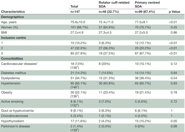
We screened 219 patients from whom 72 patients were excluded due to secondary SOA. Thus, 147 patients were included and analysed in our study. Among the 147 patients (101 woman (68.7%); mean age 75.8±10 years; mean BMI 27.2±4.89 (range 17.3–45.7)), most were recruited from orthopaedic surgery departments (89.8%) at the time of shoulder arthroplasty (table 1). Overall, 18 patients (13%) had cardiovascular events, 17 (11.6%) has hypothyroidism, 5 (3.4%) had chondrocal-cinosis and 2 (1.4%) had Parkinson's disease. In total, 99 patients (67.4%) had primary SOA and 48 (32.7%) had rotator cuff-related SOA.
Many patients (67.7%) with primary SOA were from centre #3 (table 1). The patients with primary SOA were significantly older than those with non-centred shoulder OA (mean age 77.5±9.1 vs 72.4±11.0 years, p=0.004) and more frequently had hypothyroidism (n=15, 15%; vs n=2, 4%; p=0.05). The two groups did not differ in frequency of cardiometabolic risk factors such as BMI, type 2 diabetes mellitus and hypertension, but cardio-vascular diseases were numerically more frequent in the rotator cuff-related SOA (n=10/99, 10% vs n=8/48, 20%; p=0.12).
Multivariable model with a logical clinical expla-nation included age, cardiovascular diseases and hypothyroidism. Age was independently associated with primary SOA (adjusted OR 1.06; 95% CI 1.02 to 1.1; p=0.004). Conversely, cardiovascular diseases were asso-ciated with rotator cuff-related SOA (adjusted OR 0.27; 95% CI 0.09 to 0.82; p=0.02). Hypothyroidism was asso-ciated but not significantly with primary SOA (OR 3.47; 95% CI 0.67 to 18.9; p=0.14).
Table 1 Characteristics of all patients with shoulder osteoarthritis (SOA) and those with primary centred and rotator cuff-related SOA
Characteristics
Total Rotator cuff-related SOA Primary centredSOA
p Value n=147 n=48 (32.7%) n=99 (67.4%) Demographics Age, years 75.8±10.0 72.4±11.0 77.5±9.1 <0.01 Women (%) 101 (68.7%) 31 (64.6%) 70 (70.7%) 0.45 BMI 27.2±4.9 27.3±4.5 27.2±5.0 0.86 Inclusion centre 1 15 (10.2%) 3 (6.3%) 12 (12.1%) <0.01 2 47 (32.0%) 27 (56.3%) 20 (20.2%) <0.01 3 85 (57.8%) 18 (37.5%) 67 (67.7%) <0.01 Comorbidities Cardiovascular diseases* 18 (13%) (139†) 8 (20%) 10 (10.1%) 0.12 Diabetes mellitus 21 (14.3%) 7 (14.6%) 14 (14.1%) 0.94 Dyslipidemia 51 (34.7%) 15 (31.3%) 36 (36.4%) 0.54 Hypertension 95 (65.1%) (146†) 30 (63.8%) 65 (65.7%) 0.83 Obesity 30 (22.1%) (136†) 11 (23.4%) 19 (21.4%) 0.78 Active smoking 8 (6.1%) (132†) 3 (7.0%) 5 (5.6%) 0.72 Gout or hyperuricemia 9 (6.1%) 3 (6.3%) 6 (6.1%) 1 Chondrocalcinosis 5 (3.4%) 1 (2.1%) 4 (4.0%) 1 Hypothyroidism 17 (11.6%) 2 (4.2%) 15 (15.2%) 0.05 Parkinson’s disease 2 (1.4%) (139†) 2 (5.0%) 0 (0%) 0.08
*Cardiovascular diseases includes: heart attack, coronary insufficiency, cardiac deficiency, stroke, thromboembolic event. †Total number of analysed patients when missing data.
Data are no. (%) or mean±SD.
Table 2 Accumulation of cardiometabolic factors (ie, obesity, dyslipidemia, hypertension, diabetes mellitus) by shoulder osteoarthritis (SOA) subtype
No. of criteria Rotator cuff-related SOA Primary centred SOA p Value
Two or more criteria
Total population=138 18 (38.3%) 47 (52.7%) 0.14 Having three or more criteria Total population=144 9 (19.2%) 10 (10.3%) 0.14
Having four criteria
Total population=145
3 (6.5%) 0 (0%) 0.03
Data are no. (%). Total population for each analysis differs because of missing data.
To determine whether accumulation of cardiomet-abolic risk factors was associated with an SOA subtype, we compared the number of patients with ≥2, or ≥3 or four criteria among obesity, dyslipidemia, diabetes and
hypertension (table 2). Unexpectedly, there were signifi-cantly more patients combining all the four criteria in the non-centred SOA than in the primary centred SOA (6.5% vs 0%; p=0.03).
dIscussIon
Considering that the recently defined individualised metabolic component of OA could be preferentially involved in primary SOA than rotator cuff-related SOA, which may be the mechanical consequence of rotator cuff rupture, we compared these two SOA subtypes in terms of cardiometabolic risk factors and found no differences between the subtypes. Conversely, ageing was independently associated with primary SOA, and surprisingly cardiovascular diseases were associated with rotator cuff-related SOA. Moreover, a combination of the four metabolic cardiovascular risk factors seemed to be more frequent with rotator cuff-related than primary SOA, but only three patients had a combination of the four criteria limiting the interpretation of this result.
SOA is a common localisation of OA, but its pathophys-iology has not been well studied in the light of the new approach to OA pathophysiology by subtype. A recent study showed increased adipokine levels in SOA joints and that BMI was correlated with synovial fluid and serum adipokine levels (ie, leptin, adiponectin and resistin), which suggests that BMI may affect non-weight-bearing joints such as the shoulder via a systemic component responsible for a metabolic stress.8 However, this
hypoth-esis was not analysed by anatomical type of SOA.
The two subtypes of SOA, primary and rotator cuff-re-lated, may involve different pathogenic mechanisms. We initially hypothesised that primary SOA may be due to metabolic stress on joint tissues such as cartilage, subchondral bone and synovium, notably through an adipokine effect. Conversely, rotator cuff-related SOA is due to chronic lesions of the rotator cuff tendons leading to their tearing and to elevated humeral head.9 However,
along with this mechanical process, chronic lesions of the rotator cuff tendons could also be induced or aggra-vated by metabolic disturbances. Indeed, in patients with obesity, chronic low-grade inflammation can damage tendons.10 Moreover, animal models showed that obesity
and type 2 diabetes compromised tendon homeostasis and tendon repair.11 Likewise, metabolic stress may act
on cartilage and tendons, which may explain our results. Assessment of adipokine levels in synovial fluid by SOA subtypes would help strengthen this hypothesis, but syno-vial fluid was not available for this retrospective study. So, metabolic syndrome could lead to both primary and rotator cuff-related SOA, which could explain why our study did not show any association between metabolic syndrome and one or the other SOA type.
Besides the metabolic OA phenotype, the age-related OA phenotype may involve specific pathophysiological pathways such as advanced glycation end-products or chondrocyte senescence.1 In our study, patients with
primary SOA were older than those with rotator cuff-re-lated SOA. Such a finding suggests that primary SOA may preferentially belong to this age-related OA phenotype, with a greater direct effect of ageing on cartilage than on tendons. However, this suggestion does not exclude the occurrence of rotator cuff-related SOA with age, but possibly indirectly because of repeated trauma and meta-bolic stress throughout life.
Despite the older age of the patients with centred than non-centred SOA, the latter group more frequently had cardiovascular diseases and all four of the cardiometa-bolic risk factors (ie, obesity, dyslipidemia, diabetes and hypertension). Likewise, we may hypothesise that the meta-inflammation occurring in cardiovascular diseases may also produce rotator cuff rather than cartilage lesions as suggested by recent studies.10 12 13 However, we cannot
exclude selection bias, because most of the patients with centred SOA were from a private clinic, whereas patients with non-centred SOA were from university hospi-tals, which usually care for patients with more severe comorbidities. The number of patients with Parkinson’s
disease was also different between the two groups (two patients in non-centred SOA vs 0 patient in centred SOA, p=0.08). Parkinson’s disease has been previously associ-ated with cardiovascular diseases so it could have caused a cofounder but only two patients were affected by this disease in our study.14 15
Our work has some limitations. First, because its design was retrospective, we could not collect all data such as steroid consumption or markers of atherosclerosis. However, we extensively reviewed all medical records, including the anaesthetic record, which contain cardio-vascular risk factors and diseases because of the planned shoulder surgery. Moreover, patients were contacted by phone if data were lacking. In addition, we used several ways to define type 2 diabetes and hypertension, including declarative data, results of biological inves-tigations if necessary and drug prescriptions. Second, our study may lack power because of the low number of included patients. Along this line, analysis on meta-bolic syndrome should be cautiously interpreted because of the small number of patients (19 patients having 3 or more criteria and only 3 patients having 4 criteria, table 2). However, this was an exploratory study including almost 150 patients from both surgical and medical departments and from both public and private institutes. Thus, our population may reflect the great heterogeneity of patients with shoulder OA. Third, we did not have a control group of patients without SOA matched on age to compare the prevalence of cardiovas-cular risk factors with the general population. However, the purpose of our study was not to compare metabolic cardiovascular risk factors with those in the general population, but to determine whether these factors are more frequent in primary versus rotator cuff-related SOA.
In conclusion, metabolic cardiovascular risk factors and diseases were not preferentially associated with one of the two subtypes of SOA. This finding could be explained by a deleterious impact of the metabolic stress on the tendon tissues in addition to cartilage. Longitu-dinal and larger as well as translational studies will be helpful to determine how metabolic stress may partici-pate in SOA pathophysiology.
Acknowledgements We would like to thank all participants of this study.
Contributors JS, lB, gn, FB and P-AJ contributed to study conception, data acquisition, data analysis and interpretation. SK and TS contributed to statistical analysis. FB, lD and AS contributed to data analysis and interpretation. All the authors reviewed the manuscript.
Competing interests none declared.
Patient consent Obtained.
Ethics approval Comité de Protection des Personnes + Commission nationale de l'Informatique et des libertés.
Provenance and peer review not commissioned; externally peer reviewed.
Open Access This is an Open Access article distributed in accordance with the Creative Commons Attribution non Commercial (CC BY-nC 4.0) license, which permits others to distribute, remix, adapt, build upon this work non-commercially, and license their derivative works on different terms, provided the original work is properly cited and the use is non-commercial. See: http:// creativecommons. org/ licenses/ by- nc/ 4.0/
© Article author(s) (or their employer(s) unless otherwise stated in the text of the article) 2017. All rights reserved. no commercial use is permitted unless otherwise expressly granted.
RefeRences
1. Bijlsma JW, Berenbaum F, Lafeber FP. Osteoarthritis: an update with relevance for clinical practice. Lancet 2011;377:2115–26.
2. Sellam J, Berenbaum F. Is osteoarthritis a metabolic disease? Joint Bone Spine 2013;80:568–73.
3. Courties A, Gualillo O, Berenbaum F, et al. Metabolic stress-induced joint inflammation and osteoarthritis. Osteoarthritis Cartilage
2015;23:1955–65.
4. Nüesch E, Dieppe P, Reichenbach S, et al. All cause and disease specific mortality in patients with knee or hip osteoarthritis: population based cohort study. BMJ 2011;342:d1165.
5. Hoeven TA, Kavousi M, Ikram MA, et al. Markers of atherosclerosis in relation to presence and progression of knee osteoarthritis: a population-based cohort study. Rheumatology 2015;54:1692–8. 6. Jonsson H, Helgadottir GP, Aspelund T, et al. Hand osteoarthritis
in older women is associated with carotid and coronary atherosclerosis: the AGES Reykjavik study. Ann Rheum Dis
2009;68:1696–700.
7. Hotamisligil GS, Shargill NS, Spiegelman BM. Adipose expression of tumor necrosis factor-alpha: direct role in obesity-linked insulin resistance. Science 1993;259:87–91.
8. Gandhi R, Takahashi M, Rizek R, et al. Obesity-related adipokines and shoulder osteoarthritis. J Rheumatol 2012;39:2046–8. 9. Huegel J, Williams AA, Soslowsky LJ. Rotator cuff biology and
biomechanics: a review of normal and pathological conditions. Curr Rheumatol Rep 2015;17:476.
10. Abate M. How obesity modifies tendons (implications for athletic activities). Muscles Ligaments Tendons J 2014;4:298–302.
11. David MA, Jones KH, Inzana JA, et al. Tendon repair is compromised in a high fat diet-induced mouse model of obesity and type 2 diabetes. PLoS One 2014;9:e91234.
12. Scott A, Zwerver J, Grewal N, et al. Lipids, adiposity and
tendinopathy: is there a mechanistic link? critical review. Br J Sports Med 2015;49:984–8.
13. Abboud JA, Kim JS. The effect of hypercholesterolemia on rotator cuff disease. Clin Orthop Relat Res 2010;468:1493–7.
14. Liang HW, Huang YP, Pan SL. Parkinson disease and risk of acute myocardial infarction: a population-based, propensity score-matched, longitudinal follow-up study. Am Heart J 2015;169:508–14. 15. Günaydın ZY, Özer FF, Karagöz A, et al. Evaluation of cardiovascular risk in patients with Parkinson disease under levodopa treatment. J Geriatr Cardiol 2016;13:75–80.
osteoarthritis: a comparative study
centred and rotator cuff-related shoulder
Cardiometabolic risk factors in primary
and Jérémie Sellam
Nourissat Alain Sautet, Tabassome Simon, Francis Berenbaum, Geoffroy
Pierre-Antoine Juge, Laure Berard, Salma Kotti, Levon Doursounian,
doi: 10.1136/rmdopen-2016-000429
2017 3:RMD Open
http://rmdopen.bmj.com/content/3/1/e000429 Updated information and services can be found at:
These include:
References
#BIBL
http://rmdopen.bmj.com/content/3/1/e000429
This article cites 15 articles, 5 of which you can access for free at:
Open Access
http://creativecommons.org/licenses/by-nc/4.0/
non-commercial. See:
provided the original work is properly cited and the use is
non-commercially, and license their derivative works on different terms, permits others to distribute, remix, adapt, build upon this work
Commons Attribution Non Commercial (CC BY-NC 4.0) license, which This is an Open Access article distributed in accordance with the Creative
service
Email alerting
box at the top right corner of the online article.
Receive free email alerts when new articles cite this article. Sign up in the
Notes
http://group.bmj.com/group/rights-licensing/permissions To request permissions go to:
http://journals.bmj.com/cgi/reprintform To order reprints go to:
http://group.bmj.com/subscribe/ To subscribe to BMJ go to: