Abstract. We investigated mitochondrial toxicity of four
lipophilic stains (cerivastatin, fluvastatin, atorvastatin, simvastatin) and one hydrophilic statin (pravastatin). In L6 cells (rat skeletal muscle cell line), the four lipophilic statins (100 µmol/l) induced death in 27–49% of the cells. Pravastatin was not toxic up to 1 mmol/l. Cerivastatin, fluvastatin and atorvastatin (100 µmol/l) decreased the mitochondrial membrane potential by 49–65%, whereas simvastatin and pravastatin were less toxic. In isolated rat skeletal muscle mitochondria, all statins, except
pravas-tatin, decreased glutamate-driven state 3 respiration and respiratory control ratio. Beta-oxidation was decreased by 88–96% in the presence of 100 µmol/l of the lipophilic statins, but only at higher concentrations by pravastatin. Mitochondrial swelling, cytochrome c release and DNA fragmentation was induced in L6 cells by the four lipo-philic statins, but not by pravastatin. Lipolipo-philic statins impair the function of skeletal muscle mitochondria, whereas the hydrophilic pravastatin is significantly less toxic.
Keywords. Rhabdomyolysis, statins, apoptosis, mitochondria, β-oxidation, respiratory chain
Introduction
Statins (3-hydroxy-3-methyl-glutaryl coenzyme A reduc-tase inhibitors, HMG-Co A reducreduc-tase inhibitors) impair hepatocellular cholesterol production by inhibiting the synthesis of mevalonate, a critical intermediary product in the cholesterol pathway. They are generally well toler-ated, but can produce a variety of skeletal muscle-asso-ciated, dose-dependent adverse reactions, ranging from muscle pain to frank rhabdomyolysis. Rhabdomyolysis is a serious adverse reaction of these drugs for both patients and the pharmaceutical industry, as evidenced by numer-ous case reports and case series [1, 2] and the withdrawal from the market of cerivastatin in August 2001 [3]. The frequency of rhabdomyolysis is low, with a reported incidence of approximately 1 : 10 000 patient years [4] and a death rate of 0.15 per million prescriptions [3, 5]. The fact that the frequency of myotoxicity observed for cerivastatin was higher than for the other statins raised the
question of whether there are differences in the myotoxic potential of the statins and whether such differences are related to their physicochemical properties. As shown in Table 1, differences in the physicochemical properties of statins can result in a variable kinetic behavior, including bioavailability, tissue distribution and metabolism, which may affect their toxic potential on skeletal muscle [6]. For example, inhibition of cytochrome P450 (CYP) isozymes can lead to increased bioavailability of lipophilic statins [7], thereby elevating the potential for myotoxicity. How-ever, since myotoxicity has been reported with all statins on the market, the variability in their pharmacokinetic properties does not adequately explain the susceptibility to develop statin-induced myotoxicity.
Little is known regarding the mechanisms by which statins produce skeletal muscle injury. HMG-CoA reduc-tase catalyses the formation of mevalonate from HMG-CoA. Mevalonate is an important precursor not only of cholesterol but also of ubiquinone, dolichols and other isoprenoids [8]. All of these compounds are involved in various essential cell functions. A deficit in them may
Research Article
Toxicity of statins on rat skeletal muscle mitochondria
P. Kaufmann, M. Török, A. Zahno, K. M. Waldhauser, K. Brecht and S. Krähenbühl*
Division of Clinical Pharmacology and Toxicology and Department of Research, University Hospital, 4031 Basel (Switzerland), Fax: +41 61 265 4560, e-mail: kraehenbuehl@uhbs.ch
Received 21 May 2006; received after revision 28 July 2006; accepted 25 August 2006 Online First 29 September 2006
* Corresponding author. © Birkhäuser Verlag, Basel, 2006
therefore adversely affect myocytes, rendering them vul-nerable to myotoxic events [8, 9]. This hypothesis was strengthened by the observation that the myotoxicity of statins on myocytes in vitro could be decreased by the addition of mevalonate [10, 11]. Ubiquinone, whose bio-synthesis is reduced in the presence of statins [8, 9], is utilized by mitochondria for the transport of electrons be-tween enzyme complexes of the electron transport chain. Reduced levels of ubiquinone are present in specific forms of mitochondrial myopathies and are considered to result in impaired mitochondrial electron transport chain function and decreased adenosine triphosphate (ATP) synthesis [12, 13]. Regarding statins and mitochondria, light microscopic changes observed in muscle biopsies of patients with statin-associated myopathy were similar to the findings in patients with mitochondrial myopathies [14–17]. In a patient with rhabdomyolysis associated with simvastatin, MELAS syndrome manifested in the recov-ery phase, suggesting that so far unnoticed mitochondrial diseases may represent risk factors for statin-associated myopathy [18]. Furthermore, in a recent study, Vladutiu et al. [19] described biochemical and/or genetic abnor-malities of proteins or genes involved in skeletal muscle energy metabolism in more than 50% of patients with statin-associated myopathy. Recent in vitro investigations indicate that simvastatin interferes with mitochondrial calcium homeostasis and inhibits complex I of the elec-tron transport chain [20].
On the basis of these reports, we hypothesized that statins could act as mitochondrial toxins and that the patients with an underlying mitochondrial disease could react preferentially with myopathy. Since the data about toxic-ity of statins on mitochondria are still rare, we decided to investigate the effects of several statins on isolated rat skeletal muscle mitochondria and on L6 cells, a rat skel-etal muscle cell line.
Materials and methods Materials
Fluvastatin was a gift from Novartis Pharma (Basel, Swit-zerland), simvastatin from Merck Sharp & Dohme
(Ra-haway, NJ, USA), cerivastatin from Bayer (Zürich, Swit-zerland) and pravastatin from Bristol-Myers Squibb (San-kyo, Japan). Atorvastatin was provided by Prof. J. Drewe (University Hospital Basel, Switzerland). Simvastatin lactone was converted to the corresponding acid as de-scribed previously [21]. JC-1 was obtained from Alexis Biochemicals (Lausen, Switzerland) and [1-14C]palmitic
acid from Amersham (Dübendorf, Switzerland). FAS li-gand was prepared as described previously [22].
Fetal calf serum, all supplements and the culture me-dium were from Gibco (Paisley, UK). The 96-well plates were purchased from Becton Dickinson (Franklin Lakes, NJ, USA) and the 8-chamber slides from Nalge Nunc (Rochester, NY, USA). The VybrantTM Apoptosis Assay
Kit #2 was purchased from Molecular probes (Eugene, OR, USA). All other chemicals used were of best quality available and purchased from Sigma–Aldrich (Schnell-dorf, Germany).
Animals
Male Sprague Dawley rats (Charles River, Les Onins, France) were used for all experiments. They were fed ad libitum, held on a 12-h dark and light cycle and weighed before their use. The study protocol had been accepted by the local Animal Ethics Committee.
Cells
L6 cell lines (rat skeletal muscle myoblasts) were ob-tained from LGC Promochem (Wesel, Germany). The cell line was cultured in Dulbecco’s modified Eagle’s medium (Gibco 61965026; with 4 mmol/l GlutaMAX®,
4.5 g/l glucose and sodium bicarbonate) supplemented with 10% heat-inactivated fetal calf serum, 1 mmol/l so-dium pyruvate and 5 µl/ml penicillin-streptomycin. Cul-ture conditions were 5% CO2 and 95% air atmosphere at
37 °C.
Isolation of rat skeletal muscle mitochondria
At the time of killing, the rat weight averaged 389 g. The animals were first treated with carbon dioxide and then Table 1. Physiochemical properties, pharmakokinetic parameters and metabolism of the statins studied [5, 9].
Statin Lipophilicity Hepatic extraction [%] Bioavailability [%] Protein binding [%] Volume of distribution (L) Metabolism Renal/fecal elimination [%]
Cerivastatin high < 40 60 99 21 CYP3A4/2C8 a 30/70
Fluvastatin high > 68 6 98 30 CYP2C9/3A4 6/90
Atorvastatin high > 70 12 80 381 CYP3A4 2/70
Simvastatin high ∼ 80 5 95 nk b CYP3A4 13/58
Pravastatin low 44–66 18 50 35 Conjugation 20/71
a CYP: cytochrome P450. b nk: not known.
killed by decapitation. The skeletal muscle of the hind legs (mean 19.0 g) was removed, freed from fat and con-nective tissue, minced with scissors and homogenized according to Kerner and Hoppel [23]. From this homoge-nate, skeletal muscle mitochondria were isolated accord-ing to Palmer et al. [24].
The mitochondrial protein content was determined using the biuret method with bovine serum albumin (BSA) as a standard [25].
In vitro cytotoxicity assays
Cell injury was assessed by the determination of the activity of lactate dehydrogenase (LDH) in the super-natant of statin-treated as compared with LDH activity in the supernatant of lysed cells (0.8% Triton X-100) [22]. LDH activity was analyzed as described by Vas-sault [26]. Different concentrations of the compounds investigated and 100 mmol/l mevalonate (only to se-lected incubations, see Results) were added to the cell cultures in a 96-well-plate for 24 h before the superna-tants were harvested and analyzed. Control incubations were treated with the vehicle used to dissolve the sub-stances investigated.
Mitochondrial membrane potential
Cells were detached from the cell culture flasks by add-ing 10 mmol/l ethylenediamine-tetraacetic acid (EDTA) in phosphate-buffered saline pH 7.4 (PBS). After filtra-tion, cells were adjusted to a density of 0.5 × 10⁶ cells/
ml and incubated in complete medium in the dark. Be-fore incubation, test substances, JC-1 (4 µg/ml) and 100 µmol/l mevalonate (only to selected incubations, see Results) were added. Flow cytometry was performed after an incubation time of 10 min using a FACSCali-bur flow cytometer (Becton Dickinson, San José, CA, USA). Changes in mitochondrial membrane potential (∆Ym) were monitored by measuring the JC-1
fluores-cence using FL-1 and FL-2. FL denotes the measured fluorescence intensity in the respective channel (FL-1 = 530 ± 15 nm, FL-2 = 585 ± 21 nm). Dinitrophenol (an uncoupler) and benzbromarone (depolarizes the ∆Ym
[22]) served as controls.
Oxygen consumption
Oxygen consumption was monitored polarographically using a 1-mL chamber equipped with a Clark-type oxygen electrode (Yellow Springs Instruments, Yellow Springs, OH, USA) at 30 °C as described previously [27]. The fi-nal concentration of l-glutamate was 20 mmol/l.
Oxygen consumption by intact mitochondria. The
re-spiratory control ratio (RCR) was calculated according to
Estabrook [28]. The RCR represents the ratio between the rate of oxygen consumption in the presence of a substrate and ADP (state 3) and the rate after complete conversion of ADP to ATP (state 4).
The test compounds were added to the mitochondrial in-cubations before the addition of the respective substrate. Control experiments were carried out in the presence of the solvent (1% DMSO) containing no inhibitor.
Oxygen consumption of L6 muscle cells. Cells (1 × 10⁶)
were treated with oligomycin (final concentration 5 µg/ ml) to inhibit F1F0-ATPase. After 2 min, test compounds
were added to the incubation chamber and the oxygen consumption was determined. Control experiments were carried out with solvent (1% DMSO).
Activity of NADH-oxidase
The activity of NADH-oxidase was determined at 30 °C using freeze-thawed mitochondria as described originally by Blair et al. [29] with the modifications described ear-lier [30].
In vitro mitochondrial β-oxidation and carinitine palmitoyltransferase activity
The β-oxidation of [1-14C]palmitic acid by skeletal
mus-cle mitochondria was assessed according to Sherratt et al. [31]. The incubation vials, which contained 500 µg mitochondrial protein in 900 µl incubation solution, were closed with a rubber stopper and incubated for 15 min at 30 °C. A scoop containing a filter paper soaked with 90 µl 0.1 mol/l KOH was fixed at the rubber stopper and was used to trap the volatile 14CO
2.
Carinitine palmitoyltransferase (CPT) activity was mea-sured by the formation of palmitoyl-[3H]carnitine from
palmitoyl-CoA and l-[3H]carnitine [32], a reaction mainly
reflecting activity of CPT1. Palmitoylcarnitine was ex-tracted with 1.4 ml water-saturated 1-butanol, which was washed with 600 µl butanol-saturated water [33] and quantified by liquid scintillation counting.
Activities of mitochondrial β-oxidation enzymes
All enzyme activities were determined using spectro-photometric assays at 37 °C. Freeze-thawed mitochon-dria were treated 1 : 1 with 5% cholic acid to disrupt the mitochondrial membranes. The solution was then diluted one hundred times with 50 mmol/l potassium phosphate buffer (pH 7.4). Acyl-CoA dehydrogenase was determined according to Hoppel et al. [27], using palmitoyl-CoA as a substrate. β-Hydroxy-acyl-CoA de-hydrogenase was determined in the reverse direction ac-cording to Brdiczka et al. [34] using acetoacetyl-CoA as substrate. β-Ketothiolase was determined using
ace-toacetyl-CoA as a substrate according to Hoppel et al. [27].
Mitochondrial swelling
Mitochondrial swelling was monitored by measuring the decrease in light scattering at 540 nm using a Spectra-MAX 250 plate reader (Paul Bucher Analytik und Bio-technologie, Basel, Switzerland). The decrease in light scattering has been shown to correlate closely with the percentage of the mitochondrial population undergoing permeability transition [35]. Freshly isolated mitochon-dria were suspended in isotonic swelling buffer (pH 7.3; 150 mmol/l KCl, 20 mmol/l MOPS, 10 mmol/l Tris, 2 mmol/l nitrilotriacetic acid and 2 µmol/l calcium iono-phore A23187) and exposed to test compounds at room temperature. Swelling was calculated from the slope be-tween 60 and 2000 s of exposure.
Cytochrome c immunocytochemistry
For immunocytochemistry, cells were grown in an eight-chamber-slide for 24 h at 37 °C and then treated with the test compounds for 24 h. Cytochrome c was visualized by immunocytochemistry as described previously [22].
Cellular ATP content
Rat myoblasts (500 000 L6 cells/well) were transferred into a 12-well plate and treated for 24 h with test com-pounds. Following treatment, cells were collected and the ATP content measured as described previously [22]. ATP concentrations were calculated using an ATP standard curve.
Determination of apoptosis
Both assays were performed using L6 muscle cells cul-tured on poly-d-lysine coated (0.1 mg/ml, 30 min) cell
culture dishes.
Hoechst 33342 nuclear staining. A confluent cell layer
was treated for 24 h with test compounds, then incubated for 30 min at room temperature with Hoechst 33342 dye (50 µmol/l in PBS) and visualized by fluorescence mi-croscopy (Olympus IX 50, Hamburg, Germany).
Annexin V and propidium iodide staining. An in situ
apoptosis detection kit was used for Annexin V bind-ing and propidium iodide (PI) stainbind-ing (VybrantTM
Apo-ptosis Assay Kit #2). After a 24-h incubation with the test compounds, cells were stained with 25 µl Annexin V-Alexa Fluor® 488 and 2 µl PI (final concentration:
1.5 µg/l). After 15 min of incubation at room tempera-ture, samples were analyzed by flow cytometry, using
a FACSCalibur flow cytometer (Becton Dickinson, San José, USA).
Statistical analysis
Data are presented as mean ± standard error of the mean (SEM). For statistical comparisons, data of groups were compared by analysis of variance (ANOVA). The level of significance was p ≤ 0.05. If ANOVA revealed signifi-cant differences, comparisons between the control and the other incubations were performed by Dunnett’s post test procedure. A t-test (unpaired, two-tailed) was performed if only two groups were analyzed.
Results
In vitro cytotoxicity
Cytotoxicity was investigated by treating L6 cells with various concentrations of the different statins for 24 h. As shown in Figure 1, 100 µmol/l cerivastatin, fluvastatin, atorvastatin or simvastatin showed significant toxicity. In contrast, pravastatin did not cause any cell damage up to 1 mmol/l. Statin-associated cytotoxicity could not be prevented by the addition of 100 µmol/l mevalonate to the incubations (data not shown). Since lipophilic statins were toxic to muscle cells, further experiments were per-formed to find out the mechanisms for cytotoxicity.
Mitochondrial membrane potential
Since statin-induced myopathy has been shown to be as-sociated with mitochondrial dysfunction [14, 15], we first
Figure 1. Cytotoxicity of the test compounds. Cerivastatin, fluvas-tatin, atorvastatin and simvastatin caused a concentration-depen-dent release of LDH from L6 cells into the cell culture media. In contrast, the cells were not destroyed by pravastatin up to 1 mmol/l. Data are expressed as the percentage of total LDH activity released into the cytoplasm and are presented as mean ± SEM of at least three individual experiments. *p < 0.05 vs. control, **p < 0.01 vs. control.
focused on mitochondria. In a first step, we measured the
∆Ym, since this potential is critical for myocyte survival
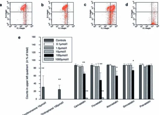
[36] and its dissipation can be associated with induction of apoptosis [37]. As shown in Figure 2, at a
concen-tration of 100 µmol/l, 35% of the cells showed a dissi-pated mitochondrial membrane potential in the presence of cerivastatin, and 51%, 20% or 26% in the presence of fluvastatin, atorvastatin or simvastatin, respectively. Figure 2. Assessment of the mitochondrial membrane potential. After labelling the cells with JC-1, mitochondrial depolarization could be visualized by a shift of the fluorescence emission from green to red. In the upper left quadrant, cells with polarized mitochondria are located (a, 1% DMSO), whereas cells with dissipated potential are found in the upper or lower right panels (b–d, fluvastatin 10 µmol/l, 100 µmol/l or 1000 µmol/l, respectively). In (e), the counts retrieved in the upper left quadrant are given as the percentage of total counts (defined as 100%). Data are given as mean ± SEM of at least three individual experiments. *p < 0.05 vs. control, **p < 0.01 vs. control.
Figure 3. Uncoupling effect of statins. Uncoupling was determined by assessing the effect of statins on state 4 respiration in the presence of l-glutamate and oligomycin using L6 cells. In coupled mitochondria, blocking of the F1F0-ATPase by oligomycin results in a restricted electron transport and oxygen consumption similar to state 4. If a test compound works as an uncoupler, oxygen consumption increases, in spite of inhibited phosphorylation (a). Of the tested statins, only cerivatstatin worked as an uncoupler, whereas the other statins did not in-crease oxygen consumption. Dinitrophenol and benzbromarone served as positive controls. *p < 0.05 vs. control, **p < 0.01 vs. control.
The dissipation of the mitochondrial membrane poten-tial could not be prevented by the addition of 100 µmol/l mevalonate to the incubations (data not shown).
Mitochondrial respiration
To further specify the mitochondrial defects, we assessed the toxicity of the statins on oxidative metabolism of iso-lated rat skeletal muscle mitochondria. In the presence of
l-glutamate as a substrate, cerivastatin, fluvastatin,
ator-vastatin and simator-vastatin induced a progressive depression of the RCR, which reflects the activity of the electron transport chain and the tightness of the coupling of oxida-tive phosphorylation (see Table 2). The concentrations as-sociated with a 50% decrease in the RCR were: 57 µmol/l for cerivastatin, 72 µmol/l for fluvastatin, 113 µmol/l for
atorvastatin and 78 µmol/l for simvastatin. In contrast, pravastatin did not significantly affect the RCR up to 400 µmol/l.
While the depression of the RCR by fluvastatin, atorvas-tatin and simvasatorvas-tatin was mainly due to inhibition of state 3 respiration (inhibition of the electron transport chain), for cerivastatin, the decrease was mostly due to accelera-tion of state 4, suggesting uncoupling. We therefore de-termined oxygen consumption by L6 cells in the presence of oligomycin, an inhibitor of F1F0ATPase (Fig. 3). Under
these conditions, only cerivastatin showed an increase in state 4 respiration, demonstrating that cerivastatin uncou-ples oxidative phosphorylation.
To exclude the possibility that the observed lack of toxic-ity of pravastatin is due to its hydrophilictoxic-ity, which could impair membrane permeation, the activity of the mito-chondrial NADH oxidase (reflecting activities of complex I, III and IV) was measured using broken mitochondria. Also in the absence of membrane barriers, pravastatin up to 1 mmol/l did not impair the activity of the electron transport chain, whereas the lipophilic statins revealed significant inhibitory effects at ≥100 µmol/l (results not shown).
Fatty acid metabolism
For the assessment of β-oxidation by skeletal muscle mi-tochondria, both the formation of acid soluble products and of CO2 were determined. [31] As shown in Figure 4a,
cerivastatin (100 µmol/l), fluvastatin, atorvastatin and simvastatin (each 200 µmol/l) inhibited mitochondrial
β-oxidation between 82% and 96%. For pravastatin, a significant inhibition of β-oxidation was found at (300 µmol/l). The corresponding IC50 were: 14 µmol/l
for cerivastatin, 9.0 µmol/l for fluvastatin, 29 µmol/l for atorvastatin, 75 µmol/l for simvastatin and 300 µmol/l for pravastatin.
To localize the inhibitory effect of β-oxidation in more de-tail, three enzymes of the β-oxidation were investigated. Acyl-CoA dehydrogenase was inhibited by 30–40% in the presence of 100 µmol/l cerivastatin, 200 µmol/l fluvastatin, atorvastatin or simvastatin, or 400 µmol/l pravastatin, and β-hydroxy-acyl-CoA dehydrogenase by 10–20% in the presence of 100 µmol/l cerivastatin, 200 µmol/l fluvastatin, atorvastatin or simvastatin, or 400 µmol/l pravastatin. In contrast, β-ketothiolase was not significantly inhibited by the statins used at the same concentrations as above.
Since there was some discrepancy between the inhibition of the β-oxidation pathway (using intact mitochondria) and individual enzymes (using disrupted mitochondria), we also determined the activity of CPT, which can be rate limiting for fatty acid oxidation [38]. As shown in Figure 4b, fluvastatin and cerivastatin inhibited CPT1 activity, whereas the other statins revealed no signifi-Table 2. Effects of cerivastatin, fluvastatin, atorvastatin,
simvas-tatin and pravassimvas-tatin on oxidative metabolism of l-glutamate by isolated rat skeletal muscle mitochondria. See method section for experimental details. Mean ± SEM of at least three experiments using different mitochondrial preparations.
Control (no inhibitor) State 3 State 4 RCR 233 ± 21 66 ± 14 3.7 ± 0.8 Cerivastatin (µmol/l) 2 256 ± 23 69 ± 10 4.0 ± 0.6 5 240 ± 31 69 ± 6 3.5 ± 0.4 25 289 ± 35 88 ± 10 3.4 ± 0.5 50 274 ± 35 137 ± 15* 2.0 ± 0.0 100 187 ± 19 187 ± 19** 1.0 ± 0.0** Fluvastatin (µmol/l) 5 241 ± 15 63 ± 7 3.9 ± 0.2 25 224 ± 8 42 ± 21 3.5 ± 0.3 50 266 ± 24 65 ± 6 2.3 ± 0.4 100 192 ± 41 126 ± 28* 1.7 ± 0.5* 200 116 ± 11** 116 ± 11* 1.0 ± 0.0** Atorvastatin (µmol/l) 5 171 ± 28 49 ± 3 3.4 ± 0.3 25 173 ± 20 56 ± 1 3.1 ± 0.3 50 161 ± 45 45 ± 9 3.5 ± 0.6 100 149 ± 6* 71 ± 11 2.2 ± 0.3* 200 45 ± 5** 45 ± 5 1.0 ± 0.0** Simvastatin (µmol/l) 5 200 ± 32 56 ± 6 3.0 ± 0.3 25 213 ± 56 74 ± 16 2.8 ± 0.4 50 157 ± 41 86 ± 18 1.9 ± 0.5* 100 123 ± 15* 80 ± 13 1.7 ± 0.5* 200 83 ± 9* 83 ± 9 1.0 ± 0.0** Pravastatin (µmol/l) 50 272 ± 25 92 ± 10 3.0 ± 0.4 100 338 ± 41 95 ± 28 2.9 ± 0.5 200 271 ± 36 82 ± 5 3.4 ± 0.5 300 247 ± 41 90 ± 12 2.7 ± 0.1 400 278 ± 73 88 ± 10 3.1 ± 0.5 *p < 0.05 vs. control; **p < 0.01 vs. control.
cant inhibitory effect up to 200 µmol/l (atorvastatin) or 1 mmol/l (simvastatin, pravastatin).
Mitochondrial swelling and release of cytochrome c
As already demonstrated, statins did cause a loss of the mitochondrial membrane potential. We therefore hypoth-esized that statins might induce mitochondrial permeabil-ity transition, which can result in mitochondrial swelling and cytochrome c release [39]. As shown in Figure 5, cerivastatin, fluvastatin, atorvastatin and simvastatin in-duced mitochondrial swelling in a concentration-depen-dent manner, whereas pravastatin did not cause an open-ing of the mitochondrial permeability transition pore. As a consequence of the mitochondrial permeability transition pore opening, cytochrome c release from the mitochondrial intermembrane space into the cytoplasm can occur, which can trigger the mitochondrial pathway of apoptosis [40]. To assess this possibility, cytochrome c release into cytoplasm was determined. As shown in Figure 6, only pravastatin did not induce a release of cyto-chrome c from the mitochondria, whereas in the presence of the lipophilic statins, cytochrome c was spilled into the cytoplasm.
Mechanisms of cell death
Cytoplasmic cytochrome c can activate caspases and induce apoptosis [41]. DNA fragmentation occurring
Figure 4. Effect of statins on β-oxidation and CPT1 activity. Fatty acid oxidation (a) was determined using isolated rat skeletal muscle mitochondria and was impaired by all lipophilic statins in a concentration-dependent manner (IC50 9.9–76 µmol/l). In contrast, pravastatin inhibited fatty acid oxidation with an IC50 of 300 µmol/l. Data are given relative to control values (100% activity corresponds to 3.90 nmol/ mg mitochondrial protein/min). In (b), the effect of statins on CPT1 activity using isolated mitochondria was determined. Cerivastatin and fluvastatin are inhibitors of CPT1, whereas the other three statins investigated do not affect CPT1 activity. Data are given relative to control values (100% activity corresponds to 6.45 nmol/mg mitochondrial protein/min). Data are given as mean ± SEM of at least three individual experiments. *p < 0.05 vs. control, **p < 0.01 vs. control.
Figure 5. Induction of mitochondrial permeability transition. Mi-tochondrial permeability transition was monitored as a decrease in absorbance at 540 nm. Upon pore opening, osmotically driven influx of water into the mitochondrial matrix leads to mitochon-drial swelling, causing a change in the light scattering properties of mitochondria. Ca2+ was used as positive control. Among the tested statins, only pravastatin did not induce mitochondrial permeability transition, whereas all lipophilic statins caused a concentration-dependent increase in mitochondrial size. *p < 0.05 vs. control; **p < 0.01 vs. control.
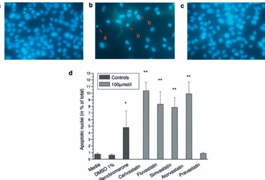
during apoptosis can be visualized by fluorescence mi-croscopy using dyes intercalating with DNA. As shown in Figure 7, untreated cells showed an apoptosis rate of 1.1%. At a concentration of 100 µmol/l, the proportion of apoptotic cells was 13% for cerivastatin, 11% for
flu-vastatin, 6% for atorflu-vastatin, 6% for simvastatin and 1% for pravastatin.
Another hallmark of the early stages of apoptosis is the translocation of phosphatidylserine from the inner to the outer leaflet of the plasma membrane, which can be de-tected by Annexin V. Cells were co-stained with PI as a marker of cell membrane permeability, which increases during the later stages of apoptosis and also in necro-sis. Since PI only enters cells with already disintegrated membranes, early apoptotic cells can be distinguished from late apoptotic and necrotic cells. Similar to the stainings with Hoechst 33342, flow cytometric analysis of the cells revealed a progressive increase of the An-nexin V signal (early apoptotic cells) with increasing concentrations of lipophilic statins. At 100 µmol/l of these compounds, between 9% and 17% of cells under-went early apoptotic changes. In contrast, only 3% of cells treated with 100 µmol/l pravastatin were Annexin V positive (data not shown). Fas ligand, used as a positive control for apoptosis induction, induced early apoptosis in 12% of cells. Similarly, only the lipophilic statins, but not pravastatin, caused late apoptosis/necrosis (data not shown).
Since ATP is essential for executing apoptosis [39, 42], the cellular ATP content was assessed. The ATP content in control incubations was 1.26 ± 0.08 µmol/106
myo-cytes and remained stable in presence of 100 µmol/l flu-vastatin, atorflu-vastatin, simvastatin or pravastatin. In the presence of 100 µmol/l cerivastatin, it dropped to 0.16 Figure 6. Mitochondrial release of cytochrome c. L6 cells were
incubated with test compounds (each 100 µmol/l) for 24 h and cytochrome c was detected by immunocytochemistry. In the pres-ence of 1% DMSO (a, negative control) or pravastatin (g), no leak-age into the cytoplasm occurred (the nuclei are clearly visible and cytochrome c is distributed in the cytoplasm with a granular pat-tern). After treatment with benzbromarone (positive control, b), cerivastatin (c), fluvastatin (d ), atorvastatin (e) or simvastatin ( f ), cytochrome c was released from the mitochondrial intermembrane space into the cytoplasm (most nuclei covered by cytochrome c and the cytoplasm stained diffusely).
Figure 7. Detection of apoptosis by staining with Hoechst 33342. L6 cells were treated with test compounds at a concentration of 100 µmol/ l for 24 h and subsequently stained with Hoechst 33342. The cell nuclei were visualized using fluorescence microscopy. (a) Control, (b) atorvastatin, (c) pravastatin. Apoptotic cells were characterized by DNA condensation (b, arrow a) and/or DNA fragmentation (b, arrow b). (d ) Summary of the percentage of cells undergoing apoptosis. Pravastatin was the only statin not inducing apoptosis. *p < 0.05 vs. control; **p < 0.01 vs. control.
±0.09 µmol/106 myocytes, suggesting that also necrosis
occurred.
Discussion
Our investigations demonstrate that lipophilic statins are mitochondrial toxins affecting the electron transport chain, coupling of oxidative phosphorylation and/or mito-chondrial β-oxidation. These alterations in mitochondrial function are associated with dissipation of the electric po-tential across the inner mitochondrial membrane. The mi-tochondrial membrane potential is mainly maintained by a proton gradient generated by the proton pump activity of complexes I, III and IV of the electron transport chain. Any perturbation of these enzyme complexes and/or un-coupling of oxidative phosphorylation can be expected to dissipate the mitochondrial membrane potential, which can therefore be regarded as a marker of mitochondrial function and integrity. A decrease in the mitochondrial membrane potential can lead to mitochondrial membrane permeabilization, which is considered to be a point-of-no-return for initiating apoptotic cell death [41, 43]. Mi-tochondrial membrane permeabilization can culminate in rupture and therefore loss of barrier function of the outer mitochondrial membrane, with consequent release of apoptosis-inducing proteins (e.g. cytochrome c) from the mitochondrial intermembrane space.
Our study shows that lipophilic statins act along this en-tire sequence: they inhibit the function of the electron transport chain, impair mitochondrial β-oxidation (lim-ited availability of NADH for the function of complex I of the electron transport chain), uncouple oxidative phos-phorylation (cerivastatin), decrease the mitochondrial membrane potential, and induce mitochondrial swelling and apoptosis. The induction of apoptosis by statins may not only be initiated by the mitochondrial pathway, how-ever. Due to inhibition of the conversion of HMG-CoA to mevalonate, not only the synthesis of cholesterol, but also of intermediates between HMG-Co A and cholesterol and products of these intermediates (e.g. ubiquinone and dolichols) is reduced [8]. As a consequence, cells may become poor in prenylated (farnesylated or geranylated) small GTP proteins, such as Rho, Ras or Rac, which pro-mote cell maintenance and cell survival [3, 44, 45]. Since mevalonate could not prevent statin-associated mito-chondrial damage and cytotoxicity, mitomito-chondrial toxic-ity and not impaired prenylation of GTP proteins appears to initiate apoptosis of L6 cells. Patients treated with statins may have an increased basal rate of apoptotic cell death, which is compensated by cell proliferation under normal conditions. In the case an additional insult, e.g. a large increase in the statin plasma and tissue concentra-tions due to a drug-drug interaction and/or an underlying metabolic disease [19], there may be a massive increase
in apoptotic cell death, possibly leading to organ damage such as rhabdomyolysis.
Our findings support the results of Kubato et al. [46], who showed that lipophilic statins can cause apoptosis of hepatocytes. Also in these experiments, pravastatin nei-ther reduced cell viability nor induced apoptosis. Since a reduction in cholesterol synthesis could also be shown for pravastatin, the hydrophilic pravastatin also had to have entered the hepatocytes to execute its pharmacological action. At least in hepatocytes, inhibition of cholesterol synthesis is therefore not sufficient to induce apoptosis. Although all lipophilic statins tested induced apoptosis of skeletal muscle cells, their effects on mitochondrial function differed among each other. For instance, only cerivastatin uncoupled oxidative phosphorylation (see Fig. 3). The uncoupling capacity of cerivastatin is obvi-ously not related to its pharmacological action, but may offer an explanation for its higher myotoxicity compared with the other statins [2, 5, 6, 47]. When mitochondrial uncoupling occurs, protons are transported into the mi-tochondrial matrix by bypassing the F1F0-ATPase. As a
consequence, the mitochondrial membrane potential is dissipated and mitochondrial ATP synthesis is decreased, possibly leading to cellular ATP depletion and cytotoxic-ity [39]. Furthermore, uncouplers may be transported into and accumulate within the mitochondrial matrix, which could enhance their toxicity. The higher myotoxicity of cerivastatin compared with other statins may therefore be explained by its higher bioavailability (see Table 1) and by its capability to uncouple oxidative phosphorylation of skeletal muscle mitochondria.
Having investigated muscle toxicity of statins in vitro, the question arises, to what extent our findings are relevant for the in vivo situation. For atorvastatin, pravastatin and atorvastatin, typical plasma concentrations after oral doses of 20–40 mg are in the range of 0.1 µmol/l [5, 48], which is considerably lower than the toxic concentrations determined in our investigations. Since data of statin concentrations in skeletal muscle of humans are so far lacking, possible tissue concentrations reached have to be estimated. The volume of distribution of lipophilic statins (Table 1) is high for atorvastatin, low for the statins with high protein binding (cerivastatin and fluvastatin) and un-known for simvastatin (intermediate protein binding). At least for atorvastatin (and possibly also for simvastatin), accumulation in peripheral tissues can be assumed, pos-sibly also in skeletal muscle. Furthermore, most cases of rhabdomyolysis with statins have been described in patients having a drug-drug interaction [1], leading to plasma (and possibly also tissue) concentrations which are higher (in some cases tenfold or even more [49]) than normal concentrations. Since it is known that even in the presence of a drug-drug interaction, only a minority of the patients develop rhabdomyolysis [50], patients affected by muscular problems may have an underlying (possibly
mitochondrial) disease, rendering them more sensitive to lower statin concentrations [19]. Taken together, our find-ings may be relevant for the in vivo situation in sensitive humans, although the statin concentrations associated with mitochondrial toxicity found in our study appear to be high.
Similar to the study of Kubota et al. [46], our investi-gations do not offer an explanation for in vivo skeletal muscle toxicity of pravastatin, which appears to have an equal frequency as for atorvastatin and simvastatin [4]. In our assays, pravastatin revealed only a slight inhibition of mitochondrial β-oxidation at high concentrations, but did not induce apoptosis or necrosis of L6 cells. A lack of ac-cess to cell organelles can be excluded by our own studies (NADH oxidase) and also by the studies of Kubota et al. [46]. The possibilities remain that a metabolite of pravas-tatin is the culprit or that L6 cells are not a suitable cell system to show toxicity for this particular statin.
In conclusion, the lipophilic statins impair several func-tions of skeletal muscle mitochondria, whereas as the hy-drophilic pravastatin does not reveal a relevant mitochon-drial toxicity in vitro. Although the toxic concentrations of the statins on isolated mitochondria and L6 cells are considerably higher than their plasma concentrations in humans, mitochondrial toxicity may trigger rhabdomy-olysis in sensitive patients. Since the hydrophilic pravas-tatin is also associated with rhabdomyolysis, additional mechanisms may exist.
Acknowledgements. The study was supported by a grant of the
Swiss National Science Foundation to SK (3100-59812-03/1). 1 Omar, M. A. and Wilson, J. P. (2002) FDA adverse event
re-ports on statin-associated rhabdomyolysis. Ann. Pharmacother. 36, 288–295.
2 Staffa, J. A., Chang, J. and Green, L. (2002) Cerivastatin and reports of fatal rhabdomyolysis. N Engl J Med. 346, 539–540. 3 Thompson, P. D., Clarkson, P. and Karas, R. H. (2003)
Statin-associated myopathy. JAMA 289, 1681–1690.
4 Graham, D. J., Staffa, J. A., Shatin, D., Andrade, S. E., Schech, S. D., La Grenade, L., Gurwitz, J. H., Chan, K. A., Goodman, M. J. and Platt, R. (2004) Incidence of hospitalized rhabdomy-olysis in patients treated with lipid-lowering drugs. JAMA 292, 2585–2590.
5 Ballantyne, C. M., Corsini, A., Davidson, M. H., Holdaas, H., Jacobson, T. A., Leitersdorf, E., Marz, W., Reckless, J. P. and Stein, E. A. (2003) Risk for myopathy with statin therapy in high-risk patients. Arch. Intern. Med. 163, 553–564.
6 Rosenson, R. S. (2004) Current overview of statin-induced my-opathy. Am. J. Med. 116, 408–416.
7 Williams, D. and Feely, J. (2002) Pharmacokinetic-pharmaco-dynamic drug interactions with HMG-CoA reductase inhibi-tors. Clin. Pharmacokinet. 41, 343–370.
8 Liao, J. K. (2002) Isoprenoids as mediators of the biological effects of statins. J. Clin. Invest. 110, 285–288.
9 Evans, M. and Rees, A. (2002) Effects of HMG-CoA reductase inhibitors on skeletal muscle: are all statins the same? Drug Saf. 25, 649–663.
10 Flint, O. P., Masters, B. A., Gregg, R. E. and Durham, S. K. (1997) Inhibition of cholesterol synthesis by squalene synthase inhibitors does not induce myotoxicity in vitro. Toxicol. Appl. Pharmacol. 145, 91–98.
11 Guijarro, C., Blanco-Colio, L. M., Massy, Z. A., O’Donnell, M. P., Kasiske, B. L., Keane, W. F. and Egido, J. (1999) Li-pophilic statins induce apoptosis of human vascular smooth muscle cells. Kidney Int. Suppl. 71, S88–91.
12 Di Giovanni, S., Mirabella, M., Spinazzola, A., Crociani, P., Silvestri, G., Broccolini, A., Tonali, P., Di Mauro, S. and Servi-dei, S. (2001) Coenzyme Q10 reverses pathological phenotype and reduces apoptosis in familial CoQ10 deficiency. Neurology 57, 515–518.
13 Ogasahara, S., Engel, A. G., Frens, D. and Mack, D. (1989) Muscle coenzyme Q deficiency in familial mitochondrial en-cephalomyopathy. Proc. Natl. Acad. Sci. USA 86, 2379–2382. 14 Giordano, N., Senesi, M., Mattii, G., Battisti, E., Villanova, M.
and Gennari, C. (1997) Polymyositis associated with simvas-tatin. Lancet 349, 1600–1601.
15 Schalke, B. B., Schmidt, B., Toyka, K. and Hartung, H. P. (1992) Pravastatin-associated inflammatory myopathy. N. Engl. J. Med. 327, 649–650.
16 Phillips, P. S., Haas, R. H., Bannykh, S., Hathaway, S., Gray, N. L., Kimura, B. J., Vladutiu, G. D. and England, J. D. (2002) Statin-associated myopathy with normal creatine kinase levels. Ann. Intern. Med. 137, 581–585.
17 England, J. D., Walsh, J. C., Stewart, P., Boyd, I., Rohan, A. and Halmagyi, G. M. (1995) Mitochondrial myopathy developing on treatment with the HMG CoA reductase inhibitors – simv-astatin and pravsimv-astatin. Aust. N. Z. J. Med. 25, 374–375. 18 Chariot, P., Abadia, R., Agnus, D., Danan, C., Charpentier, C.
and Gherardi, R. K. (1993) Simvastatin-induced rhabdomyoly-sis followed by a MELAS syndrome. Am J Med. 94, 109–110. 19 Vladutiu, G. D., Simmons, Z., Isackson, P. J., Tarnopolsky, M.,
Peltier, W. L., Barboi, A. C., Sripathi, N., Wortmann, R. L. and Phillips, P. S. (2006) Genetic risk factors associated with lipid-lowering drug-induced myopathies. Muscle Nerve 71, 1324– 1330.
20 Sirvent, P., Bordenave, S., Vermaelen, M., Roels, B., Vassort, G., Mercier, J., Raynaud, E. and Lacampagne, A. (2005) Simv-astatin induces impairment in skeletal muscle while heart is pro-tected. Biochem. Biophys. Res. Commun. 338, 1426–1434. 21 Bogman, K., Peyer, A. K., Torok, M., Kusters, E. and Drewe,
J. (2001) HMG-CoA reductase inhibitors and P-glycoprotein modulation. Br. J. Pharmacol. 132, 1183–1192.
22 Kaufmann, P., Torok, M., Hanni, A., Roberts, P., Gasser, R. and Krahenbuhl, S. (2005) Mechanisms of benzarone and benzbro-marone-induced hepatic toxicity. Hepatology 41, 925–935. 23 Kerner, J. and Hoppel, C. L. (2002) Radiochemical
malonyl-CoA decarboxylase assay: activity and subcellular distribution in heart and skeletal muscle. Anal. Biochem. 306, 283–289. 24 Palmer, J. W., Tandler, B. and Hoppel, C. L. (1977) Biochemical
properties of subsarcolemmal and interfibrillar mitochondria iso-lated from rat cardiac muscle. J. Biol. Chem. 252, 8731–8739. 25 Gornall, A. G., Bardawill, G. J. and David, M. (1949)
Deter-mination of serum proteins by means of the biuret reaction. J. Biol. Chem. 177, 751–766.
26 Vassault, A. (1983) Lactate dehydrogenase. In: Methods of En-zymatic Analysis. vol. III, pp. 118–125, Bergmeyer, H. U. (ed.), VCH, Weinheim.
27 Hoppel, C., DiMarco, J. P. and Tandler, B. (1979) Riboflavin and rat hepatic cell structure and function. Mitochondrial oxidative metabolism in deficiency states. J. Biol. Chem. 254, 4164–4170.
28 Eastabrook, R. (1967) Mitochondrial respiratory control and polarographic measurement of ADP:O ratios. Methods Enzy-mol. 10, 41–47.
29 Blair, P. V., Oda, T. and Green, D. E. (1963) Studies on the Electron Transfer System. LIV. Isolation of the Unit of Electron Transfer. Biochemistry 128, 756–764.
30 Krahenbuhl, S., Chang, M., Brass, E. P. and Hoppel, C. L. (1991) Decreased activities of ubiquinol:ferricytochrome c oxidoreductase (complex III) and ferrocytochrome c:oxygen
oxidoreductase (complex IV) in liver mitochondria from rats with hydroxycobalamin[c-lactam]-induced methylmalonic ac-iduria. J. Biol. Chem. 266, 20998–21003.
31 Sherratt, H. S., Watmough, N. J., Johnson, M. A. and Turnbull, D. M. (1988) Methods for study of normal and abnormal skeletal muscle mitochondria. Methods Biochem. Anal. 33, 243–335. 32 McGarry, J. D. and Brown, N. F. (1997) The mitochondrial
car-nitine palmitoyltransferase system. From concept to molecular analysis. Eur. J. Biochem. 244, 1–14.
33 Kiorpes, T. C., Hoerr, D., Ho, W., Weaner, L. E., Inman, M. G. and Tutwiler, G. F. (1984) Identification of 2-tetradecylglycidyl coenzyme A as the active form of methyl 2-tetradecylglycidate (methyl palmoxirate) and its characterization as an irreversible, active site-directed inhibitor of carnitine palmitoyltransferase A in isolated rat liver mitochondria. J. Biol. Chem. 259, 9750– 9755.
34 Brdiczka, D., Pette, D., Brunner, G. and Miller, F. (1968) Com-partmental dispersion of enzymes in rat liver mitochondria. Eur. J. Biochem. 5, 294–304.
35 Haworth, R. A. and Hunter, D. R. (1979) The Ca2+-induced membrane transition in mitochondria. II. Nature of the Ca2+ trigger site. Arch. Biochem. Biophys. 195, 460–467.
36 Jones, S. P., Teshima, Y., Akao, M. and Marban, E. (2003) Sim-vastatin attenuates oxidant-induced mitochondrial dysfunction in cardiac myocytes. Circ. Res. 93, 697–699.
37 Ding, W. X. and Nam Ong, C. (2003) Role of oxidative stress and mitochondrial changes in cyanobacteria-induced apoptosis and hepatotoxicity. FEMS Microbiol. Lett. 220, 1–7.
38 Drynan, L., Quant, P. A. and Zammit, V. A. (1996) Flux control exerted by mitochondrial outer membrane carnitine palmitoyl-transferase over beta-oxidation, ketogenesis and tricarboxylic acid cycle activity in hepatocytes isolated from rats in different metabolic states. Biochem. J. 317, 791–795.
39 Lemasters, J. J., Qian, T., Bradham, C. A., Brenner, D. A., Cas-cio, W. E., Trost, L. C., Nishimura, Y., Nieminen, A. L. and Her-man, B. (1999) Mitochondrial dysfunction in the pathogenesis of necrotic and apoptotic cell death. J. Bioenerg. Biomembr. 31, 305–319.
40 Newmeyer, D. D. and Ferguson-Miller, S. (2003) Mitochon-dria: releasing power for life and unleashing the machineries of death. Cell 112, 481–490.
41 Zamzami, N. and Kroemer, G. (2003) Apoptosis: mitochon-drial membrane permeabilization – The (w)hole story? Curr. Biol. 13, R71–R73.
42 Nicotera, P., Leist, M. and Ferrando-May, E. (1998) Intracel-lular ATP, a switch in the decision between apoptosis and ne-crosis. Toxicol. Lett. 102–103, 139–142.
43 Waterhouse, N. J., Ricci, J. E. and Green, D. R. (2002) And all of a sudden it’s over: mitochondrial outer-membrane permeabi-lization in apoptosis. Biochimie 84, 113–121.
44 Nishida, M., Nagao, T. and Kurose, H. (1999) Activation of Rac1 increases c-Jun NH(2)-terminal kinase activity and DNA fragmentation in a calcium-dependent manner in rat myoblast cell line H9c2. Biochem. Biophys. Res. Commun. 262, 350– 354.
45 Kaneta, S., Satoh, K., Kano, S., Kanda, M. and Ichihara, K. (2003) All hydrophobic HMG-CoA reductase inhibitors induce apoptotic death in rat pulmonary vein endothelial cells. Athero-sclerosis 170, 237–243.
46 Kubota, T., Fujisaki, K., Itoh, Y., Yano, T., Sendo, T. and Oishi, R. (2004) Apoptotic injury in cultured human hepatocytes in-duced by HMG-CoA reductase inhibitors. Biochem. Pharma-col. 67, 2175–2186.
47 Farmer, J. A. (2001) Learning from the cerivastatin experience. Lancet 358, 1383–1385.
48 Lennernas, H. (2003) Clinical pharmacokinetics of atorvas-tatin. Clin. Pharmacokinet. 42, 1141–1160.
49 Neuvonen, P. J., Kantola, T. and Kivisto, K. T. (1998) Simvas-tatin but not pravasSimvas-tatin is very susceptible to interaction with the CYP3A4 inhibitor itraconazole. Clin. Pharmacol. Ther. 63, 332–341.
50 Ratz Bravo, A. E., Tchambaz, L., Krahenbuhl-Melcher, A., Hess, L., Schlienger, R. G. and Krahenbuhl, S. (2005) Preva-lence of potentially severe drug-drug interactions in ambula-tory patients with dyslipidaemia receiving HMG-CoA reduc-tase inhibitor therapy. Drug Saf. 28, 263–275.
![Table 1. Physiochemical properties, pharmakokinetic parameters and metabolism of the statins studied [5, 9].](https://thumb-eu.123doks.com/thumbv2/123doknet/14837305.623081/2.892.120.820.149.267/table-physiochemical-properties-pharmakokinetic-parameters-metabolism-statins-studied.webp)