HAL Id: tel-02064973
https://tel.archives-ouvertes.fr/tel-02064973
Submitted on 12 Mar 2019
HAL is a multi-disciplinary open access archive for the deposit and dissemination of sci-entific research documents, whether they are pub-lished or not. The documents may come from teaching and research institutions in France or abroad, or from public or private research centers.
L’archive ouverte pluridisciplinaire HAL, est destinée au dépôt et à la diffusion de documents scientifiques de niveau recherche, publiés ou non, émanant des établissements d’enseignement et de recherche français ou étrangers, des laboratoires publics ou privés.
AstA signaling functions as an evolutionary conserved
mechanism timing metamorphosis and growth in
Drosophila Melanogaster
Derya Deveci
To cite this version:
Derya Deveci. AstA signaling functions as an evolutionary conserved mechanism timing metamorpho-sis and growth in Drosophila Melanogaster. Cellular Biology. Université Côte d’Azur, 2018. English. �NNT : 2018AZUR4090�. �tel-02064973�
1
La voie de signalisation AstA, qui est conservée au
cours de l’évolution, contrôle le déclenchement de la
métamorphose et régule la croissance chez Drosophila
melanogaster
Derya DEVECI
Equipe LEOPOLD- Institute Biology Valrose
Présentée en vue de l’obtention du grade de docteur en Interactions
moléculaires et cellulaires
d’Université Côte d’Azur Dirigée par : Pierre Leopold Soutenue le : le 31 octobre 2018
Devant le jury, composé de :
Pierre Leopold, PhD, Institute Biology Valrose Florence Besse, PhD, Institute Biology Valrose
Kim Furbo Rewitz, Associate Prof, University of Copenhagen María Domínguez Castellano, Prof, Instituto de
Neurociencias de Alicante
Cedric Maurange, PhD, Institut de Biologie du Développement de Marseille
2
3
UNIVERSITE CÔTE D’AZUR
SVS Sciences
Thèse
Présentée pour obtenir le titre de
DOCTEUR DE L’UNIVERSITE CÔTE D’AZUR
Discipline : Interactions moléculaires et cellulaires Présentée et soutenue par
Derya DEVECI
CNRS UMR 7277 / INSERM U1091Institut Biologie Valrose
La voie de signalisation AstA, qui est conservée au cours de l’évolution, contrôle le déclenchement de la métamorphose et régule la croissance chez Drosophila melanogaster
Thèse dirigée par Pierre Leopold Soutenue le 31 octobre 2018 devant le jury composé de :
Dr. Pierre Leopold Directeur Institute Biologie Valrose Dr. Kim Furbo Rewitz Rapporteur University of Copenhagen
Dr. María Domínguez Castellano Rapporteur Instituto de Neurociencias de Alicante
Dr. Cedric Maurange Examinateur Institut de Biologie du Développement Marseille Dr. Florence Besse Président Institute Biologie Valrose
4
UNIVERSITY CÔTE D’AZUR
Doctoral School Life Sciences
PhD Thesis
To obtain the title ofPhD of Science
of the University Côte d’Azur
Speciality: Molecular and Cellular interactions
Defended by:
Derya DEVECI
CNRS UMR 7277 / INSERM U1091Institute of Biology Valrose iBV
AstA signaling functions as an evolutionary conserved
mechanism timing metamorphosis and growth in
Drosophila Melanogaster
Thesis advisor: Pierre Leopold Defence on 31st of October 2018
Jury:
Dr. Pierre Leopold Director Institute Biologie Valrose Dr. Kim Furbo Rewitz Reporter University of Copenhagen
Dr. María Domínguez Castellano Reporter Instituto de Neurociencias de Alicante
Dr. Cedric Maurange Examiner Institut de Biologie du Développement Marseille Dr. Florence Besse President Institute Biologie Valrose
5
Titre
La voie de signalisation de l’allatostatine A, conservée au cours de l’évolution, contrôle le déclenchement de la métamorphose et régule la croissance chez Drosophila melanogaster.
Resume
La maturation sexuelle se fait en réponse à l'intégration de divers signaux internes homéostatiques et externes. Jusqu'à présent, on ignore en grande partie quels mécanismes sensoriels internes sont impliqués dans le couplage de ces signaux. Chez les mammifères, le début de la puberté est associé à des pulsations élevées de GnRH conduisant à un pic d'hormone stéroïdienne. Le système ligand/récepteur, KISS/KISSR, est un régulateur en amont des neurones producteurs de GnRH. Chez Drosophila melanogaster, un pic d'hormone prothoracicotrope (PTTH) produite par deux paires de neurones (PTTHn) conduit à la production de l'ecdysone, la principale hormone stéroïdienne chez les insectes. PTTH est l'un des premiers signaux à activer la cascade d'événements menant à la maturation. Si la production de PTTH est bloquée, un retard dans le début de la transition du stade juvénile au stade adulte est observé, tandis qu'une maturation précoce est observée lors de la surexpression de PTTH. Ceci indique donc un rôle important des PTTHn dans l'intégration des signaux. Afin de découvrir les signaux intégrés par les PTTHn, nous avons criblé une collection d’ARN interférents dans les PTTHn. Nous avons ainsi identifié le récepteur à l’allatostatine A (AstA-R1) comme un régulateur positif des PTTHn. La perte de fonction de AstA-R1 retarde la maturation avec une augmentation de la taille finale de l’organisme. Une réduction de la quantité du ligand allatostatine A (AstA) a également une incidence sur le déclenchement de la maturation. AstA est produite dans le cerveau par une paire de neurones bilatéraux qui étendent leurs axones vers les dendrites des PTTHn. De plus, les neurones AstA projettent également
6
leurs axones vers les cellules productrices d’insuline (IPCs), connues pour réguler le taux de croissance larvaire. L’inactivation d’AstA-R1 dans les IPCs donne des organismes plus petits. Nos résultats impliquent que les neurones AstA sont capables de réguler le rythme de croissance ainsi que le déclenchement de la maturation en jouant sur deux circuits différents ciblant les PTTHn et les IPCs. De façon intéressante, AstA/AstAR1 est homologue à KISS/KISSR (GPR54), un facteur d’entrée dans de la puberté humaine. Ceci suggère donc qu’un circuit neuronal est conservé au cours de l’évolution pour l'intégration des signaux qui contrôlent le déclenchement de la maturation sexuelle.
7
Title
AstA signaling functions as an evolutionary conserved mechanism timing metamorphosis and growth in Drosophila Melanogaster
Abstract
The onset of puberty occurs in response to the integration of various internal homeostatic and external signals. Up until now, it remains largely unknown which internal sensory mechanisms are involved in the coupling of those signals. In mammals, the onset of puberty is associated with elevated GnRH pulsations leading to a peak of steroid hormones. The KISS/KISSR system is a pivotal upstream regulator of GnRH producing neurons. In Drosophila melanogaster a peak of prothoracicotropic hormone (PTTH) produced by two pairs of neurons (PTTHn) leads to the production of the insect steroid hormone ecdysone. PTTH is one of the first signals to activate the cascade of events leading to maturation. Once PTTH production is blocked, a delay is observed in the onset of the transition from juvenile to adult stage, whereas precocious maturation is observed upon PTTH over-expression, denoting an important role for PTTHn in the integration of cues. In order to uncover signals integrated by PTTHn we have conducted a biased RNAi screen in PTTHn. After two rounds of screening we identified the GPCR Allatostatin A receptor 1 (AstA-R1) as a positive regulator of PTTHn. AstA-R1 knock down delays maturation with a subsequent increase in final pupal size. Down regulation of its ligand, Allatostatin-A (AstA) on the brain is also affecting the timing of maturation. We found that AstA is produced in the central brain by a bilateral pair of neurons that extend their axons towards the PTTHn dendrites. In addition, AstA neurons also project their axons towards the Drosophila insulin producing cells (IPCs) that are known to regulate larval growth. Knockdown of AstA-R1 on the IPCs leads to smaller pupae. These findings imply that the AstA neurons are able to regulate growth and maturation timing by interacting with 2 different circuits: the
8
PTTHn and IPCs. Unexpectedly, AstA-R1 and AstA genes share a common evolutionary origin with KISSR and KISS, respectively, suggesting a common mechanism between insects and mammals for the integration of signals that control the onset of puberty.
9
Sealed by a KISS..
‘The KISS1 gene was discovered and named to the location of where it was discovered
(Hershey, Pennsylvania, home of Hershey's Kisses)’
10
Acknowledgements
And now for the part that is most- read in this thesis and the part that is the most fun to write! My thesis would have never been completed without the help, love and support from a big group of colleagues, friends and family. The journey took a lot of hard work and dedication with a whole bunch of ups and downs (but mostly ups). Firstly, I want to thank you, Pierre, for allowing me to join your lab. I truly have enjoyed my time in the lab and for sure this was the best decision I could have taken to do my PhD in your lab. I was promised a pregnancy and/or a baby by the end of my PhD but it seems that I managed to escape the curse! I want to wish you all the best in Paris and I hope you will not forget the sunshine and people in Nice. Don’t forget to invite me to the next insect hormone meeting in Crete! Then for the one person who has kept me from losing my sanity and who has guided and supported me through all these years, Nuria. If it weren’t for you, Nuria, I would’ve never understood anything about fly genetics and I would’ve accidentally set the lab on fire. I cannot explain how much I have learnt from you, not only scientifically, but also important life lessons that I will use later on in life. I will miss your positivity, our extremely loud laughs and gossips and I wish you all the best with your future steps in science (which I know will be bright). And now my other awesome boss, Paco. During my rotation in the lab, after just one day, you managed to convince me to work with flies. I knew at that time that I didn’t want to do a PhD anywhere else than in this lab and I have never regretted it for a moment, so a huge thank you, Boss. I wish you all the best in your own lab and with everything else in life! If it weren’t for Gisele I would’ve probably ended up on the streets without a Carte Vitale. I cannot thank you enough for helping me and for being my French mum. Ditte, I have really enjoyed spending time with you and it was really nice to have someone to chat about everything and anything. Thank you for the laughs and conversations we have had and for your wise advices. Congrats again on getting the grant and I wish you and Julien all the best in Denmark. Just don’t forget to pack an umbrella! And now you, Julien, thank you for the fun times in the lab and for being my drinking partner at the many parties we have had! Go teach the Danish how to have some fun! Laura, thank you for the nice chats and the nice time in the lab. I admire how you managed to combine a pregnancy and baby with your hard work in the lab. Best of luck in the future with your beautiful family. Marc, it was a pleasure having you around in the lab and being able to always chat with you. Enjoy your retirement because a lot of people would like11
to be where you are right now! Nath, the always smiling and always helpful one, we had a bunch of fun together and I hope you’ll be surrounded with many nice cheerful people in Marseille! Marianne, my other cheerful drinking buddy from the North. You have always brought life to the lab (and to parties) and I have truly enjoyed my time with you and I wish you the best of luck with everything! Eleonora & Caterina, my fellow PhD victim in the lab, it was so great to have each other around to laugh and cry during our time in the lab. Good luck with your defense this year and remember than the end is almost there! J May the force be with you! Renald, it was an absolute pleasure to speak and laugh with you during our coffee breaks. Thanks for all of the scientific input and nice time in the lab and I wish you all the best with your new project. Sadly, no more Pokémon Go without you. Tomas, thank you for all your help with my annoying experiments and for the fun conversations we have had together. I wish you all the happiness and health with your son. Alessandra, it was really great having you around in the lab and I have to thank you for all your help with my experiments. You are definitely not forgotten! I wish you and your beautiful family all the best! Lolo, I would be nowhere without all the help you have given me. I have always admired your hard work and energy to help others in need and I wish you all the best with every project you’re involved in (in & outside the lab). Cecile & Tanvi, I enjoyed our times together at the 5th floor and I hope to join you in the Post-PhD-world soon! Then, the entire 5th floor for all the support, coffee breaks, apero times, celebration times and any other occasions! We had a great time together at our floor and I will truly miss the good times we have had so thank you to the entire floor! It’s a pity Paula and Parisa joined the lab so late but nevertheless I wanted to thank you both for your cheerful spirits in the last few months of our lab. Good luck in Paris and I hope to see you both soon! Thanks to Ramona, George, Ranya, Gaia, Tomas, Johan, Maria and Christina I managed to maintain my sanity during the past few years in Nice. I am so thankful to have met you all and I hope we will cross paths again one day (or more days!). I wish you all the best with your future plans and I hope we will all end up in exotic places with exotic jobs J. Thank you mom and dad for all the love you have given me and for supporting me. I know it was difficult to grasp what I was doing all these years but nevertheless you have been very sweet and proud of my accomplishments. Thank you dad for always believing in me and for supporting me with all the difficult decisions I had to take over the years. Sizi cok seviyorum ve ozliyorum annecim ve babacim ♥ . Even though there was a distance with my family, I
12 received support and love from them and I look forward to seeing them again once I am back in the Netherlands. Thank you Peter and Michele for all the support and kindness you have given me over the past year. You have taken such good care of me even though we barely knew each other at that time (damn kidney infection). I am truly happy to have such nice, caring and sweet parents in law! Then the one person who brought so much happiness and love in my life, who made my final year bearable and who has always been supportive and caring is my dear fiancé Robert-Jan. I wouldn’t have survived the toughest final year of my PhD without all the love, attention and support from you. I cannot thank you enough for keeping up with my crazy schedules, the weird flies I took with me, and all the PhD stress I was going through. Your presence (and voice over the phone) was the only thing that would keep me from losing my sanity during this crazy journey. My words cannot express how thankful I am to have you in my life and for all the support & love you have given me. I cannot wait to start our new chapter in our lives together! Ik hou van jou! ♥
13
Table of Contents
Acknowledgements ... 10 Abbreviations ... 15 Abstract ... Error! Bookmark not defined.Introduction
1. General introduction ... 151.1 Puberty is induced by steroid production ... 17
1.3 The effect of the time of pubertal onset on adult size ... 18
1.2 GnRH regulates pubertal onset ... 20
1.3 Kiss/GPR54 signaling times puberty ... 24
1.4 Kiss/GPR54 signaling regulates GnRH pulsations during pubertal stages ... 26
1.5 Metabolic signals modulate the HPG- axis ... 31
1.6 Steroidal feedback is important in HPG- axis ... 32
1.7 There is possibly not one ‘trigger’ for puberty ... 35
2. Drosophila melanogaster as a model to study the regulation of steroid production ... 37
2.1 Drosophila has a life cycle of two weeks ... 37
2.2 Drosophila offers a wide variety of genetic tools ... 39
2.3 Steroid production induces sexual maturation in Drosophila ... 40
2.4 Insulin signaling controls systemic growth and determines final size ... 43
2.5. Fat body is a nutritional relay for systemic growth control ... 45
2.5 Steroid signaling controls growth ... 48
2.6 Ecdysone production in the PG is tightly regulated ... 50
2.6 Sexual maturation is coordinated with systemic growth ... 53
3. PTTH as a gatekeeper of sexual maturation in Drosophila ... 56
3.1 PTTH times onset of metamorphosis ... 56
3.2 PTTH does not have a homology in vertebrates ... 58
14
3.4 PTTH regulates final body and organ size ... 62
3.5 PTTH acts as a modulator of environmental adaptive plasticity ... 66
3.6 PTTH mediates light avoidance behavior ... 67
3.7 PTTH acts as a signal integrator of Dilp8 and critical weight ... 70
3.8 PTTH is differentially regulated in other insects ... 74
Results
Aim of the thesis ... 76 Manuscript ... 77 Abstract ... 78 Introduction ... 79 Results ... 81 Discussion ... 86 References ... 89 Figures ... 94Materials and Methods ... 100
Supplementary figures ... 105
Complementary unpublished experiments ... 111
1. Regulation of AstA-R2 in controlling larval growth ... 111
2. Regulation of AstA-R2 in timing metamorphosis ... 113
3. AstA mutant larvae exhibit both timing and growth defects ... 114
4. Pulsatile activity of PTTHn in PG ... 115
General conclusion ... 116
Discussion
There is an evolutionary conserved mechanism in the brain controlling puberty ... 117Interplay of steroid production on maturation and growth ... 121
What is upstream of AstA? ... 122
15
Abbreviations
20-E 20-hydroxyecdysone ACPR AKH/CRZ-related peptide AKH adipokinetic hormone
AKHR adipokinetic hormone receptor ARC hypothalamic arcuate nucleus AstA allatostatin- A
AstA-R1 allatostatin- A receptor 1
AVPV anteroventral periventricular nucleus
CA corpora allata
CC corpora cardiaca
CPP central precocious puberty CRZR corazonin receptor
DHR4 Drosophila hormone receptor 4
Dilp Drosophila insulin-like peptides
EcR Ecdysone receptor
EEDCs estrogen-like endocrine disrupting chemicals ERα estrogen receptor α
ERK extracellular signal-regulated kinase FSH follicle stimulating hormone
GCL growth coordinating Lgr3 (GCL) neurons GFP green fluorescent protein
GH growth hormone
GnRH gonadotropin-releasing hormone
HH idiopathic hypogonadotropic hypogonadism
IIS insulin/insulin-like growth factor (IGF)-like signaling InR insulin receptor
IPCs insulin- producing cells
JH juvenile hormone
Kiss kisspeptin
LH luteinizing hormone
PG prothoracic gland
PGS pubertal growth spurt
PIP2 phosphatidylinositol bisphosphate
PLC phospholipase C
PTTH prothoracicotropic hormone UAS Upstream Activation Sequence
16
1.
General introduction
An emerging trend in human physiology in the past decade is a decline in the average age of puberty onset. Whereas the average age in the USA used to be 14.2 in 1900, it dropped to 12.5 in the year 2000 (Bellis, Downing, and Ashton 2006). When children undergo puberty earlier, especially when compared to their peers, they could be more prone to psychopathologies such as depression that can persist into adulthood. Another possible health risk that is linked to precocious pubertal onset is the increased risk of breast cancer development in woman (Bodicoat et al. 2014). General improvements in health care and child nutrition could act as contributors to accelerate puberty. However, other factors such as obesity, increased animal protein, high dairy consumption and exposure to estrogen-like endocrine disrupting chemicals (EEDCs) have been reported to have a positive correlation with accelerated puberty onset (Berkey et al. 2000; Günther et al. 2010; W. Li et al. 2017; Roy, Chakraborty, and Chakraborty 2009). Due to the health risks associated with aberrant timing of puberty, it is important that puberty occurs at the right time. Therefore it is of fundamental interest to identify the underlying signals that interplay in pubertal onset to understand how puberty is triggered. Indeed, in the last decades, several permissive factors like leptin and estrogen have been identified that allow puberty to occur or to progress. Puberty onset is a complex process wherein multiple signals are integrated. Thus, rather than the existence of having one trigger signal, puberty seems to be the consequence of a large number of integrative factors. The identification of novel signals could therefore allow us to understand the emerging trend of early puberty.
17
1.1 Puberty is induced by steroid production
Puberty is the developmental transition of juveniles into sexually mature adults. Sexual maturation is indispensable for reproduction and is therefore tightly controlled by a sophisticated system that stems from an interplay of a complex network of signals integrated in the brain. The so called hypothalamic-pituitary-gonadal axis (HPG-axis) is the regulatory mechanism controlling the onset of sexual maturation relying on the intercommunication of these organs (Fig. 1). At the very top of this axis is gonadotropin-releasing hormone (GnRH) produced by GnRH producing neurons within the hypothalamus. GnRH is released in the hypophyseal portal bloodstream in the brain in a pulsatile manner and binds to its receptor GnRHR in the anterior pituitary. This subsequently stimulates the gonadotropes to release two gonadotropins: luteinizing hormone (LH) and follicle stimulating hormone (FSH). The pulsatile release of LH and FSH subsequently activates the gonads (testis and ovaries) to release the sex steroids estrogen, progesterone and testosterone to mature the gonads, inducing puberty. At the same time, estrogen and testosterone signal back to the hypothalamus to generate positive and negative regulatory feedback signals for the HPG- axis. Further details about these feedback regulatory mechanisms will be addressed in Chapter 1.7.
18
1.3 The effect of the time of pubertal onset on adult size
Humans are one of the few mammals that undergo a major pubertal growth spurt (PGS) during adolescence. After puberty, adolescents can still grow in height for up until an average of 2 years during this PGS phase. Statural growth, or growth in height is the result of elongation of long bones due to cartilage formation at the epiphyseal plates (growth plate). During puberty onset, growth velocity increased and decreases at late puberty when the growth plates fuse, halting bone growth (Shim 2015). The exact molecular mechanism underlying the fusion of Figure 1. Schematic of the hypothalamic-pituitary-gonadal axis. During pubertal onset, gonadotropin-releasing hormone (GnRH) is produced from GnRH neurons in the hypothalamus and secreted in a pulsatile manner to stimulate the production and pulsatile release of LH and FSH from the pituitary gland. LH and FSH then activate the gonads (testis and ovaries) to produce sex steroid hormones, testosterone, estrogen and progesterone to mature the sexual organs and to provide feedback regulatory loops back to the hypothalamus. Adapted from (Bodicoat et al. 2014)
19
growth plates remains elusive since it is likely to involve interplay of multiple neuroendocrine factors. For instance, the sex steroid estrogen induces growth hormone (GH), that in turn stimulates the production of insulin-like growth factor-I (IGF-I). Both GH and IGF-1 function in bone growth. It is suggested from mice studies, however, that low doses of estrogen during early puberty is important for the growth spurt mediated by GH and IGF-1, whereas the high levels of estrogen signal to the growth plate cartilage to induce growth plate fusion at late puberty (Börjesson et al. 2013).
It has therefore been proposed that precocious puberty could have a negative correlation with total height gain due to early fusion of the growth plate. Conversely, adolescents that undergo delayed puberty would likely have increased height gain. Clinical reports, however, claimed the opposite effect, suggesting that adolescents undergoing puberty early undergo their PGS earlier, that in turn also lasts longer. This would result in an increase in total height gain. On the contrary, adolescents that undergo puberty at a late age would delay their PGS that would also last shorter, leading to lesser height gain. Nevertheless, these same studies have remarked that similar adult height is reached. Interestingly, a more recent mathematical approach revealed no correlation between final height and age of puberty onset from a linear regression analysis based on an observational retrospective study (Limony, Kozieł, and Friger 2015). However, when the authors took into account the initial height of PGS onset, there was a significantly high correlation between final height and PGS onset (Fig. 2) . These findings suggests that when adolescents with the same height at PGS, the absolute loss in the final height gain is greater in adolescents that undergo maturity earlier versus adolescents that undergo maturity later, both in girls and boys. These reports together highlight the controversy in the field as well as the complexity of the exact mechanism behind the time of pubertal onset, statural growth and final adult height.
20
1.2 GnRH regulates pubertal onset
The discovery of gonadotropin- release hormone (GnRH) in neuroendocrinology as a gatekeeper of puberty was recognized by a Nobel Prize of Medicine in 1977. Mutations in either GnRH or in GnRHR have been implication in causing a large number of serious congenital defects, including hypogonadotropic hypogonadism (HH), as reviewed in (Maggi et al. 2016). This condition is defined by disrupted gonadal function due to an absence or impaired secretion of GnRH, that can result in absent, or delayed puberty.
GnRH is synthesized from a large precursor consisting of 92 amino acids that is enzymatically cleaved into multiple transcript variants. GnRH is a small 10 amino acid long neuropeptide with a short half life of a few minutes due to peptidase cleavage (Wetsel and Fig. 2 Mathematic regression analysis of the relationship of final height and average age of pubertal growth spurt in girls. The difference of final height corresponding to the average of pubertal onset is depicted as height- standard deviation score (SDS). It is assumed that the SDS value at pubertal onset is zero. When girls undergo pubertal growth spurt (PGS) at a young age, the final height obtained as an adult will be lower compared to girls that undergo PGS later. Adapted from (Limony, Kozieł, and Friger 2015)
21
Srinivasan 2002). GnRH is produced by GnRH neurons located in the arcuate nucleus of the medial basal hypothalamus. GnRH neuronal activity is already detectable at late gestation, or around the time of birth, when GnRH secretion is very high, before it drastically drops during juvenile stages (T M Plant 1982). It is only until the pre- pubertal stages when GnRH neurons become reactivated again, secreting GnRH in a pulsatile manner after which a large surge is observed during puberty onset, which is the hallmark of pubertal onset.
GnRH is secreted and binds to its receptor GnRHR, which is a rhodopsin-like G protein-coupled receptor located in the pituitary gonadotropin cells (Chi et al. 1993). Once activated, GnRHR undergoes a conformational change, activating phospholipase C (PLC) that in turn transmits its signal to diacylglycerol (DAG) and inositol 1,4,5-trisphosphate (IP3) (Fig. 3) (Kraus, Naor, and Seger 2001). Next, IP3 stimulates the release of Ca2+, that in turn binds and
activates Calmodulin (CaM) and then Ca2+/calmodulin-dependent protein kinase (CaMK) to
induce gonadotropin gene expression and secretion. In parallel, DAG activates the intracellular protein kinase C (PKC) pathway, including the mitogen-activated protein kinases (MAPKs), including extracellular-signal-regulated kinase (ERK) and p38 MAPK that ultimately translocate to the nucleus to induce transcription factors to activate gene expression of gonadotropins (LH/FSH) in the pituitary gonadotrope cells.
GnRH pulsatile activity is important for subsequent pulsatile release of FSH and LH. High frequency GnRH release is important for LH pulses, whereas low frequency GnRH pulsations stimulate FSH release. Prolonged high- dose secretion of GnRH results in loss of GnRHR response caused by rapid uncoupling of GnRHR from intracellular signaling molecules and subsequent reduction in GnRHR mRNA levels (Olefsky and Webster 2010). Therefore, GnRH pulsations are under tight regulation by multiple positive and negative cues . Two major neurotransmitters modulate GnRH neuronal excitation and synchronous GnRH release: Gamma-amino butyric acid neurons (GABA) and glutamate. Both inputs of GABA and
22
glutamate trigger Ca2+ oscillations by acting through their receptors, GABA
AR/GABABR and
AMPA/kainate glutamatergic receptors, respectively (Moore, Shang, and Wray 2002). Other important regulators of GnRH are estrogen and progesterone originating from the gonads, to provide both negative and positive feedback signals. These signals, however, do not directly signal to GnRH since their receptors are not expressed in GnRH. Instead, these steroids signal to neighboring intermediary neurons to exerts their modulatory effects in GnRH secretion (such as glutamate-, GABA-, or Kiss-neurons). GnRH itself is also able to regulate its own secretion through autocrine signaling via its receptor GnRHR (Krsmanovic et al. 1999). The main synchronizing regulator of GnRH pulsations, however, is agreeably the small neuropeptide kisspeptin that is produced by Kiss neurons. It is proposed that Kiss neurons are responsible not only for the activation of GnRH neurons to induce pulsatile GnRH release, but also to induce the large GnRH surge during puberty.
23
Fig 3. GnRHR signal transduction pathway. GnRH binds to GnRHR which is a seven transmembrane Gαq/11-coupled receptor. Once activated, it activates phospholipase Cβ (PLCβ), converting converting phosphatidylinositol bisphosphate (PIP2) into inositol 1,4,5-trisphosphate (IP3), causing Ca2+ mobilization. PIP2 also induces diacylglycerol (DAG) that activates the intracellular protein kinase C (PKC) pathway, leading to the phosphorylation of Raf-1, protein tyrosine kinase SRC and MAPK. This ultimately leads to the phosphorylation of Elk-1, c-Fos and c-Jun, inducing transcription of the gonadotropins follicle-stimulating hormone (FSH) and Luteinizing hormone (LH) as well as PLCβ. Adapted from (Aguilar-Rojas, Pérez-Solis, and Maya-Núñez 2016)
24
1.3 Kiss/GPR54 signaling times puberty
Kisspeptins were originally discovered in 1996, owing its name to its discovery in Hershey, Pennsylvania, home to the famous Hershey’s chocolate brand. The gene encoding for kisspeptins, KISS1, is an important regulator of GnRH secretion, both in the pulsatile behavior during pre- pubertal stages as well as the preovulatory gonadotropin surge during puberty onset. In mice models, inactivating mutations in KISS1 or in its receptor GPR54 results in failure of gonadal maturation that is associated with low FSH and infertility (Lapatto, Pallais, Zhang, Chan, Mahan, Cerrato, Wei, et al. 2007). In humans, inactivating mutations in KISS1 or GPR54 can cause idiopathic hypogonadotropic hypogonadism (HH), which is a disorder characterized by delayed puberty, or a complete absence of puberty (Seminara et al. 2003; Topaloglu et al. 2012). On the contrary, central precocious puberty (CPP) has been observed with a GPR54 activating mutation (Arg386Pro), due to prolonged activation of intracellular signaling pathways revealed by in vitro studies (Trarbach et al. 2008).
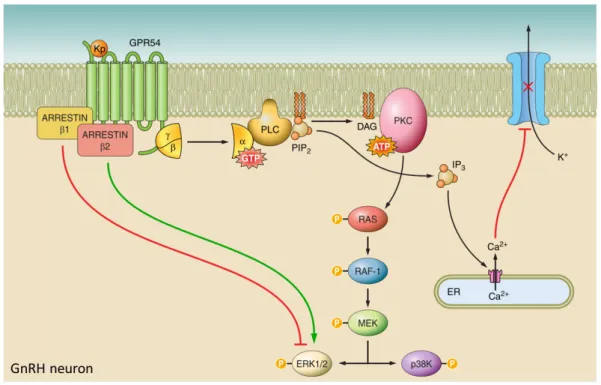
The KISS1 gene encodes for a precursor peptide of 145 amino acids long that is subsequently cleaved and processed to give rise to four kisspeptin: Kp-54, Kp-14, Kp-13 and Kp-10. Kisspeptins are produced by kisspeptin neurons (Kiss1 neurons) located in the hypothalamic brain nuclei (Brock and Bakker 2013; Clarkson and Herbison 2006; Smith et al. 2005). All four kisspeptin peptides possess an Arg–Phe–NH2 motif on the C-terminal, that
allows them to bind to their receptor, GPR54/KISS1R that is expressed in the hypothalamus and in a number of other organs. GPR54 is a member of rhodopsin family and is a seven transmembrane Gq/11-coupled receptor. Once bound to kisspeptin, phospholipase C (PLC) is
activated, subsequently converting phosphatidylinositol bisphosphate (PIP2) into inositol
1,4,5-trisphosphate (IP3) (Fig. 4) (Castaño et al. 2009). This results in the immobilization of Ca2+
from the endoplasmic reticulum (ER), causing changes in ion channel permeability, inducing depolarization. In parallel, the rise of PIP2 also leads to diacylglycerol (DAG) formation,
25
inducing protein kinase C (PKC) which phosphorylates mitogen-activated protein kinases (MAPKs), Ras, Raf and MEK, to induce phosphorylation of ERK1/2 and p38. Since pharmacological blockade of ERK1/2 and p38 kinase reduces kisspeptin- induced GnRH secretion, it is proposed that not only Ca2+ mediated depolarization is required for GnRH
secretion but also the recruitment of ERK1/2 and p38 through the MAPK pathway. (Castellano et al. 2006). In addition, kisspeptin and GPR54 modulate proliferation and migration in a tumoral setting since GPR54 signaling also plays role in metastasis suppression (Cho et al. 2012).
Fig 4. Kiss/GPR54 signaling in GnRH neurons. Kisspeptin (Kp) binds to the seven transmembrane Gq/11-coupled receptor GPR54. Upon Kp binding, phospholipase C (PLC) becomes activated, converting phosphatidylinositol bisphosphate (PIP2) into inositol 1,4,5-trisphosphate (IP3), causing Ca2+ mobilization from the endoplasmic reticulum (ER). PIP2 also induces diacylglycerol (DAG), activating intracellular protein kinase C (PKC) leading to the phosphorylation of Ras, Raf-1 and MEK. This ultimately leads to the phosphorylation of ERK1/2 and p38 kinase (P38K). In parallel, GPR54 activation induces arrestin-β1 and -β2, which provide negative and positive feedback on receptor regulation, respectively. Adapted from (Pinilla et al. 2012)
26
Even though KISS1 and GPR54 are obvious candidates in stimulating sexual maturation as described in a large number of studies conducted in rodents (Colledge 2008), a small number studies reported variable hypogonadism phenotypes or a complete absence of phenotype in
KISS1- and GPR54- knockout mice (Lapatto, Pallais, Zhang, Chan, Mahan, Cerrato, Le, et al.
2007; Mayer and Boehm 2011). This, however, could possibly be explained by residual activity in the HPG- axis, or compensatory mechanisms since acute disruption of Kiss neurons does alter fertility as described in the same study. These pathologies uncover the pivotal role of kisspeptin/GPR54 signaling for pubertal onset as well as for timing.
1.4 Kiss/GPR54 signaling regulates GnRH pulsations during pubertal stages
Kisspeptin plays a crucial role in controlling puberty through its regulatory function of GnRH pulsatile secretion (S.-K. Han 2005; K et al. 1999). Pulsatile secretion of kisspeptin induces the pulsatile behavior of GnRH secretion, that in turn regulates the pulsatile release of LH/FSH necessary for maturation of the sexual organs. It is known that disruptions in GnRH pulse frequencies are often associated with delayed, or precocious puberty (Balasubramanian et al. 2010). Similar to GnRH, kisspeptin secretion has a pulsatory behavior of interpulse interval of approximately 60 minutes that is correlated with subsequent LH pulses (Keen et al. 2008).
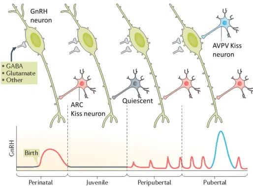
Kisspeptins are produced by kisspeptin neurons (Kiss neurons) that are subdivided into two populations, depending on the location in the rodent hypothalamic brain nuclei: hypothalamic arcuate nucleus (ARC) and anteroventral periventricular nucleus (AVPV) (Brock and Bakker 2013; Clarkson and Herbison 2006; Smith et al. 2005). It is proposed that the Kiss population in the ARC is more important in the robust pulsatile secretion of GnRH during pre-
27
pubertal stages, whereas the population in AVPV are more involved in the large preovulatory gonadotropin surge during puberty (Fig. 5).
The AVPV Kiss neuronal population increases towards early pubertal stages, which is proposed to increase kisspeptin- induced GnRH secretion. Rodent studies have revealed that the KISS1 mRNA levels as well as the number of Kiss neurons in the AVPV increases towards late- juvenile stages (Bentsen et al. 2010; Clarkson and Herbison 2006). Moreover, a previous study observed significantly more Kiss neuronal fibers towards the GnRH neurons located in the preoptic area (POA), going in line with the previous findings that suggest Kiss neurons maturate towards pre- pubertal stages in order to maximize kisspeptin- stimulated GnRH secretion (Clarkson and Herbison 2006).
Fig 5 Postnatal development of GnRH- and Kiss neuronal network. GnRH secretion is high during birth, before drastically dropping during juvenile stages. GnRH neurons (green) receive inputs from the ARC Kiss neurons (red), as well as from other neurons (GABA, Glutamate). It is suggested that the ARC Kiss neurons become inactivated during early juvenile stages (in grey) before being re-activated again in pre- pubertal stages. During pre- pubertal stages, GnRH is released in a pulsatile manner. At the same time, the number of dendritic spines in the GnRH neurons increases. The GnRH surge occurs in the pubertal period, suggested to be enabled by the AVPV Kiss neuronal population (blue). Adapted from (Herbison 2016b)
28
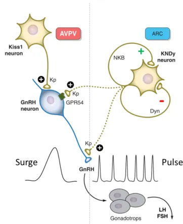
The other Kiss neuronal population is located in the ARC and are also referred to as KNDy neurons, owing its name to the co-expression neurokinin-B/tachykinin-3 (NKB/TAC3) and dynorphin (Dyn) in these Kiss neurons (Goodman et al. 2007). NKB acts as a stimulator of kisspeptin release and subsequent GnRH secretion, acting through its receptor neurokinin-3 receptor/tachykinin receptor-3 (NK3R) that are also expressed on the ARC neurons (Amstalden et al. 2010). Similar to defects in KISS1/GPR54, inactivating mutations of human NK3R have been reported to cause delayed or absent puberty (Topaloglu et al. 2009). Dyn, on the contrary, acts as an inhibitory signal for kisspeptin secretion which might be coordinating the complex pulsatile release of kisspeptin, and subsequent pulsatile release of GnRH. Whether or not the Dyn receptor is expressed in the ARC Kiss neurons is yet to be confirmed. Since kisspeptin is released in a pulsatile manner from the ARC Kiss neural population due to the integration of positive and negative signals, it is proposed that these neurons have a more prominent function in synchronizing the GnRH pulsatile release from the GnRH neurons (Fig. 6). In vitro rodent coronal brain slices containing GnRH neurons reveal that once kisspeptin is administered in a pulsatile manner, GnRH pulsatile expression became robustly synchronized accompanied with pulsatile secretion of GnRH, revealing that pulsatile kisspeptin stimulation is important for synchronizing the pulsations of GnRH release (Choe et al. 2013).
Both ARC and AVPV Kiss neuronal populations express receptors for the sex steroids estrogen and testosterone, nevertheless, the response of KISS1 acts in an opposite manner. Estrogen and testosterone signaling downregulates KISS1 in the ARC, whereas they induce the upregulation of KISS1 in the AVPV. The difference in response could be mediated by differential recruitment of transcriptional coactivators or corepressors during estrogen signaling in the different Kiss populations. The steroid- induced feedback mechanism in the HPG- axis will be further discussed in Chapter 1.6.
29
Even though it is suggested the two Kiss neuronal populations could have different functions in stimulating GnRH, the direct effects of these Kiss neuronal populations on GnRH pulsations remain unknown since most studies conducted so far are done in rodents and by administration of kisspeptin in vivo and have mainly used the pulsatile LH levels in the blood as an indirect measure to study GnRH. Further research is required to elucidate the regulatory effects of ARC- and AVPV- Kiss neurons on GnRH secretion.
Fig. 6 A proposed mode of action of two Kiss neural populations in regulating GnRH secretion. A hypothetical model of the differential regulation of the GnRH surge during puberty and the preceding pulsations in pre- pubertal stages. The AVPV Kiss neural population increases in size towards puberty onset, and mainly provides positive regulation to GnRH neurons. The ARC Kiss population mediates both inhibitory (Dyn) and positive (NKB) signals to generate a pulsatile release of kisspeptin and therefore a pulsatile secretion of GnRH from GnRH neurons. This in turn signals to the gonadotrops to secrete LH and FSH. Based on data obtained from rodent students. Adapted from (Pinilla et al. 2012)
30
Concomitant with drastic remodeling of the AVPV Kiss neuronal populations to enhance GnRH secretion, its receptor GPR54 is also subjected to changes to enhance kisspeptin/GPR54 signaling. Studies conducted in monkeys revealed that GPR54 mRNA levels in the hypothalamus during juvenile stages increases by approximately three fold in mid-pubertal stages (Shahab et al. 2005). Further studies have revealed that kisspeptin- induced LH secretion is more persistent during pubertal stages, compared to adulthood. Chronic infusion of kisspeptin has a remarkable stimulatory effect on LH secretion, even after seven days of administration in pubertal mice (J. Roa et al. 2008). This effect might be mediated by reduced desensitization of the GPR54 receptor during pubertal stages, in order to achieve maximum kisspeptin signaling. Similarly, a study conducted in early postnatal stages of rats showed that kisspeptin administration already has a potent stimulatory function on GnRH and LH, whereas LH responsiveness to low doses of kisspeptin was significantly enhanced during pubertal stages (Castellano et al. 2006). These findings suggest that kisspeptin and GPR54 undergo dramatic changes during early- juvenile to pre-pubertal stages in order to achieve maximum signaling of GnRH.
31
1.5 Metabolic signals modulate the HPG- axis
The HPG- axis induces not only puberty, but also controls the reproductive ability since a surge in LH in adult females triggers ovulation. Especially in females, extreme underfeeding can decrease LH levels, postponing the reproductive axis until conditions improve. It is proposed that the HPG- axis that controls puberty can also be regulated by nutritional and internal metabolic cues that regulate the reproductive axis. It is proposed that metabolic signals originating from peripheral tissues can signal to GnRH, mostly indirectly through other neurons. Kiss neurons are the most widely studied neurons to integrate these hormones since Kiss expresses receptors for leptin and ghrelin (Elias 2011; Forbes et al. 2009a).
Leptin, or the ‘starvation hormone’, is an important hormone produced by the adipose tissues in response to fat stores to regulate energy balance by signaling to the brain to suppress appetite (Fig. 7). Leptin plays a crucial role in sexual maturation as it was demonstrated that leptin deficient mice do not sexually mature and show lower levels of KISS1 expression (Chehab, Lim, and Lu 1996). Leptin administration in leptin deficient mice restored sexual maturation, but did not rescue KISS1 expression. Furthermore, further research revealed that sexual maturation is not altered when leptin receptor is specifically removed from Kiss neurons (Donato Jr. et al. 2011). Re- introduction of leptin receptor specifically in Kiss neurons in leptin receptor knock- out mice also did not rescue the absent sexual maturation phenotype (Cravo et al. 2013). These findings together demonstrate that leptin signaling in Kiss neurons is not essential nor necessary for puberty, and likely acts through intermediate neurons.
Another important hormone that controls energy balance and food intake is ghrelin, that opposes the actions of leptin, acting as the ‘hunger hormone’ that is produced from the gut in response to negative energy balance to promote food intake. Ghrelin is suggested to be important in suppression of the HPG-axis in woman who over- exercise or that suffer from anorexia. In vitro experiments revealed that ghrelin suppresses the pulsatile release of GnRH
32
and LH (Cravo et al. 2013). This effect might be mediated through ghrelin- induced suppression of KISS1 mRNA levels in the AVPV Kiss neurons but not in the ARC neurons that are suggested to be crucial for the GnRH surge during puberty onset (Forbes et al. 2009b). This suggests that ghrelin might be more important in suppressing starvation- induced ovulation, more than affecting puberty onset itself.
Insulin has also been suggested to be involved in regulating GnRH since in vitro studies on hypothalamic neurons revealed a stimulatory effect on GnRH secretion as well as expression (Forbes et al. 2009b). However, silencing insulin receptor specifically in the GnRH neurons, as well as in other neurons projecting to GnRH, did not alter puberty (Forbes et al. 2009b).
1.6 Steroidal feedback is important in HPG- axis
Sex steroids are produced by the gonads in response to GnRH- induced LH/FSH secretion. These steroids play a pivotal role in providing feedback regulatory loops onto the GnRH secretory behavior by signaling through the Kiss neurons (Fig. 7). Kiss neurons express several sex steroid hormone receptors, such as neuronal estrogen receptor α (ERα) and ERβ, androgen receptor, and progesterone receptor. Of these receptors, ERα is suggested to be important in timing puberty since specific ablation of ERα in Kiss neurons in female mice accelerated puberty onset due to a loss of GnRH inhibition from the ARC Kiss neurons (Mayer et al. 2010). In addition to this E2/Estrogen inhibitory signal in the ARC Kiss neurons, studies in rodents have demonstrated the existence of a positive feedback where E2 stimulates KISS1 mRNA and kisspeptin levels in the AVPV (Forbes et al. 2009b). It is proposed that the Kiss neural network acts as an E2 dependent amplifier of the HPG- axis during puberty.
Disruption of the E2 feedback regulatory loops can have drastic changes in timing the onset of puberty as stated above. Estrogen-like endocrine disrupting chemicals (EEDCs) are substances that disrupt the endocrine system by interfering with the endogenous synthesis,
33
metabolism or receptor signaling of E2. These chemicals are present in pesticides, plastic products, flame retardants, pharmaceuticals and more products that are used on a daily basis. The EEDCs that act as E2 mimicker or blocker both have been proposed to cause precocious puberty onset, genital abnormalities as reviewed in Roy, Chakraborty, and Chakraborty 2009. The effect of human exposure to EEDCS present in the environment needs to be further elucidated. However, the EEDCs effect on pubertal onset currently lacks a good model since rodent KISS1 mRNA levels are only affected at very high doses (Roy, Chakraborty, and Chakraborty 2009). However, whether E2 truly functions as a permissive or driving signal still remains to be elucidated, especially since specie dependent differences of E2 sensitivity exist. To summarize, E2 from the gonads plays an important function in providing a regulatory feedback mechanism to the HPG- axis. It acts in two distinct manners, depending on the stage of pubertal onset. During pre- pubertal stages, E2 acts on the ARC Kiss neuronal population by providing negative feedback in order to generate kisspeptin pulsations to induce puberty. Once puberty is induced, E2 acts as an amplifier of the HPG- axis by providing positive feedback signals to the AVPV Kiss neurons.
34
Fig. 7 A proposed mode of action of two Kiss neural populations in regulating GnRH secretion. A hypothetical model of the differential regulation of the GnRH surge during puberty and the preceding pulsations in pre- pubertal stages. The AVPV Kiss neural population increases in size towards puberty onset, and mainly provides positive regulation to GnRH neurons. The ARC Kiss population mediates both inhibitory (Dyn) and positive (NKB) signals to generate a pulsatile release of kisspeptin and therefore a pulsatile secretion of GnRH from GnRH neurons. This in turn signals to the gonadotrops to secrete LH and FSH. Based on data obtained from rodent students. Adapted from (Pinilla et al. 2012)
35
1.7 There is possibly not one ‘trigger’ for puberty
Since the reproductive maturity of an organism is a mechanism with no point of return, its temporal regulation is of fundamental importance. Given the hierarchical function of kisspeptin and GnRH act as potent activator of the HPG- axis, it is important to understand in response to which signals they may act to activate the cascade of events leading to the onset of puberty. All the regulatory signals of the HPG- axis summarized above highlight the multifaceted regulatory network involved in the pubertal onset. These signals seem to be acting more in a permissive manner rather than as a driving force to directly induce the onset of puberty. These signals act more in a permissive manner rather than as a driving force suggesting that puberty is under the control of a dynamic interplay between endogenous and environmental cues that are integrated in the HPG- axis to allow pubertal plasticity. This would ensure the complex energy demanding process of puberty to occur under optimal conditions. The nature of this precise and multifaceted mechanism timing puberty has only begun to be elucidated where several signals and feedbacks currently remain unknown.
Other than the known metabolic, nutritional and E2 signals as described before it could be plausible for the HPG-axis to also receive intrinsic signals originating from other organs. For instance, there could be a coupling between growing organs and the HPG- axis to ensure they have grown to an appropriate size before puberty starts and growth is fixed by closure of the bone plates.
In addition, in 1969 it was suggested that pheromones may signal into the HPG-axis since prepubertal female mice would undergo puberty earlier when they were in the presence of adult males (Vandenbergh 1969). Further studies in C. elegans and in mice showed similar results when females were kept in a place that was previously exposed to males (Aprison and Ruvinsky 2016; Flanagan, Webb, and Stowers 2011). However, the exact pheromone and pheromone receptor in both cases remains unknown and requires further research. Nevertheless,
36
these findings suggest that vertebrates and invertebrates could potentially sense similar signals and integrate them to modulate sexual maturity. Due to the conservation of steroids timing the onset of sexual maturation, the large number of genetic tools and the ease of conducting genetic screens, we propose to use Drosophila melanogaster as a model organism to dissect the timing mechanism of puberty.
37
2. Drosophila melanogaster as a model to study the regulation of
steroid production
2.1 Drosophila has a life cycle of two weeks
Drosophila melanogaster is extensively used as a model organism in biology due to its
ease in use for genetic modifications that enabled the identification of novel genes in human pathologies. Its rapid development, short life cycle and easy upkeeping brings major advantages for fundamental research. A single fertile pair of flies can give rise to fertile offspring in only 10 to 12 days when kept at 25°C. The duration of the life cycle can be shortened or accelerated by temperature of nutrient availability. The life cycle of Drosophila can be classified in four distinct phases: the embryonic developmental phase, juvenile growth phase, sexual maturation (metamorphosis) and adult stage (Fig. 8).
Fig. 8 Developmental stages of Drosophila melanogaster. The developmental progression of
Drosophila is under the control of the steroid hormone ecdysone. Ecdysone titers are high during
embryonic stages and during larval molts. Growth occurs exclusively during larval stages and halts at the end of the 3rd instar larval stage that is induced by a large titer of ecdysone, causing the larvae to pupariate and undergo metamorphosis. Sexual maturation takes place during maturation stages, giving rise to mature adult flies. The entire life cycle of Drosophila takes approximately 9 days. Adapted from King-Jones: http://www.biology.ualberta.ca/kingjones_lab/KKJ_lab/steroids.html
38
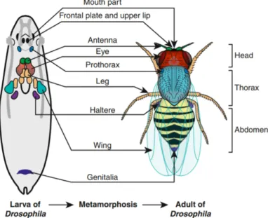
Upon egg deposition, Drosophila embryonic development takes place in a time window of around 22 hours. Once eclosed (hatched), larvae feed and grow extensively, undergoing approximately 200 fold increase in body mass (Robertson 1963). Larvae contain larval tissues and organs as well as small sac- like epithelial structures that are named imaginal discs that act as precursors of the adult fly organs (Fig. 9).
During the juvenile growth phase larvae undergo three instar developmental transitions that are marked by visible molts. These molts are induced by production of the insect steroid hormone ecdysone. Larvae spend around 24 hours in first larval instar stage, another day in second larval instar stage and the 3rd and final larval instar stage takes two to three days, before
undergoing pupariation. Sexual maturation in Drosophila and other holometabolous insects occurs during pupariation stages through a process named metamorphosis. Upon pupariation, larval tissues are broken down or undergo autophagy whereas the imaginal discs undergo differentiation to give rise to adult organs. During this time sexual organs also undergo maturation after which adult flies emerge after approximately 4 days.
Fig. 9 The larvae imaginal discs in the 3rd larvae instar stage and the corresponding adult
structures. The schematic representation and localization of the imaginal disc structures in the last instar larvae stage. During metamorphosis, a large number of larval tissues degenerate whereas the imaginal discs give rise to the corresponding adult structures. There are 19 discs in total, of which 9 bilateral pairs that give rise to the adult epidermis of the head, thorax and limbs, whereas the genitalia is derived from one medial imaginal disc. Adapted from (Aldaz and Escudero 2010)
39
2.2 Drosophila offers a wide variety of genetic tools
The whole genome of Drosophila was sequenced in 2000, and subsequently compared to the human sequence one year later (Adams et al. 2000). Drosophila only has four chromosomes: Chromosome I containing one pair sex- chromosomes (two X- chromosomes for females, one X and one Y for males), and three pairs of autosomes (II-IV), of which IV is the smallest, containing only 2% of the fly genome. The entire Drosophila genome encodes for roughly ~13.600 genes, versus ~22.000 genes in humans. It has been proposed that approximately 60-75% of all genes involved in human pathologies could have functional orthologous in Drosophila (Pandey and Nichols 2011).
Drosophila is a commonly used model to study gene expression and function due to the
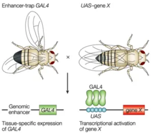
binary Gal4- UAS system that was developed by Brand and Perrimon in 1993. This system can allows tissue specific manipulation (Fig. 10). The system is based on the interplay of two parts: Gal4, encoded by the yeast transcriptional activator protein, and UAS (Upstream Activation Sequence). Gal4 containing flies have an activator protein but do not have the UAS- target gene to activate. The UAS- target gene fly lines, on the other hand, have a silent target gene since the activator gene is absent. Only when the UAS- and Gal-4 containing fly lines are crossed with each other, they will give rise to progeny containing the activated target gene. Gal4 is placed under a specific gene promoter or driver gene to be expressed in a specific subset of cells or tissues, referred to as the reporter or driver line. UAS is placed next to the desired gene, such as a gene RNA (for gene knock- down), a gene of interest (to induce overexpression) or GFP (to visualize tissues), for example.
Additionally, a second binary system, named the LexA- LexOP system can be used simultaneously with the Gal4-UAS system to allow multiplexing.
40
2.3 Steroid production induces sexual maturation in Drosophila
Similar to vertebrates, sexual maturation of Drosophila is initiated by a surge of steroid hormone. It takes place during metamorphosis, marking the transition of a growing juvenile larvae into a non- feeding pupae (Fig. 11). The production of the insect steroid hormone ecdysone takes place in the insect endocrine tissue prothoracic gland (PG), and controls the time of the two larval molts and metamorphosis (Warren et al. 2006).
Upon activation of the PG, a large number of enzymatic steps are required for the biosynthesis of 20E (active ecdysone). These steps are mediated by a group of ecdysone biosynthetic enzymes encoded by the Halloween genes that are members of the cytochrome P450 enzyme superfamily as reviewed extensively by Niwa and Niwa in 2014. First, cholesterol
Fig. 10 The Drosophila Gal4-UAS system. UAS (Upstream Activation Sequence) acts as an enhancer for Gal4 (yeast transcriptional activation protein) to induce gene transcription. Gal4 is generally placed under a specific gene promoter or driver gene to be expressed in a specific subset of cells or tissues (enhancer-trap). UAS is placed next to the desired gene, such as a gene RNAi (for gene knock-down), GFP (to visualize cells/tissues), or different other tools. Once these lines are crossed with each other, it induces tissue specific manipulations in the progeny. Adapted from https://media.nature.com/full/nature-assets/nrg/journal/v3/n3/images/nrg751-i1.gif
41
is biosynthesized into 7- dehydrocholesterol by an enzyme named Neverland (nvd) and then becomes synthesized into ketodiol by a yet unknown mechanisms, but are known to involve the enzymes Shroud (sro), Spook (spo) and Spookier (spok). Next, Phantom (phm) synthesizes ketotriol and then is converted into 2- deoxyecdysone via Disembodied (dib). Finally, Shadow (sad) converts it into ecdysone (E) before being released from the PG into the hemolymph, the fly equivalent of blood. E becomes hydroxylated by peripheral tissues such as the fat body, via Shade (shd) into the bioactive form 20-hydroxyecdysone, 20E, or also referred to as ecdysone in the rest of the thesis.
Similar to vertebrates, steroid signaling is mediated through activation of the nuclear receptor superfamily that are ligand- regulated transcription factors. Bioactive ecdysone binds to its receptor EcR, that heterodimerizes with a second nuclear receptor, Ultraspiracle (USP)(L M Riddiford, Cherbas, and Truman 2000). Once activated, ecdysone signaling induces a larval stage- specific response by transcriptionally regulating a subset of nuclear receptors as well as regulating the expression of its receptor, EcR. Other downstream targets are Drosophila
hormone receptor 3 (DHR3), DHR4, E75B (primary response gene), E78, and ftz transcription
factor 1 (βFTZ-F1), that in turn also regulate each other, as reviewed in King-Jones and Thummel 2005.
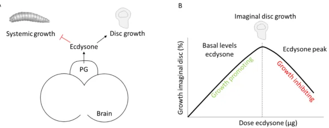
Other than its function in initiating metamorphosis due to a large increase at the late larval instar stages, ecdysone is also released at continuous low levels during larval development. These basal levels of ecdysone have been suggested to function in negatively finetuning larval growth rate and positively in promoting imaginal disc growth during larval development. Further studies need to be conducted to better understand the mechanisms underlaying these functions. A more detailed description will be addressed in Chapter 2.7.
42
Fig. 11 The biosynthesis of ecdysone. The conversion of cholesterol into the bioactive form of ecdysone is the result of multiple steps involving biosynthetic enzymes that are encoded by a group of genes that are referred to as ‘Halloween genes’. The first biosynthetic step is mediated by Neverland to convert cholesterol into 7-dehydrocholesterol. Next, 5β-Ketodiol is produced by a yet an unknown mechanism, that is referred to as the ‘Black Box’. These steps are mediated by the biosynthetic enzymes Shroud, Spook, Spookier. 5β-Ketodiol is then converted into 5β-Ketrotriol via the enzyme Phantom, before being converted into 2-Deoxyecdysone by Disembodied. The last step involves Shadow to produce ecdysone (E), that in turn is released in the hemolymph where it is hydroxylated into the bioactive form 20-Hydroxyecdysone (20E) via Shade. Adapted from (Niwa and Niwa 2014)
43
2.4 Insulin signaling controls systemic growth and determines final size
The onset of sexual maturation coincides with the end of the larval growth period. Therefore, it is important to have a coordination between metamorphosis and growth mechanisms to ensure properly sized adult flies. In almost all organisms insulin/insulin-like growth factor (IGF)-like signaling (IIS) relays nutritional information to organ and tissue growth and plays a fundamental role in physiology, governing growth, metabolism, longevity and development. As described above, insulin signaling in flies plays a pivotal role in controlling larval growth and subsequently final adult size. IIS signaling in Drosophila is under the control of Drosophila insulin-like peptides (Dilps). To date, eight Dilps (Dilp1-8) have been identified and characterized and with the exception of the newly identified Dilp8, all signal through the fly canonical IIS pathway to promote growth.Dilp1-7 bind to one common tyrosine kinase receptor, the Drosophila insulin receptor (InR), that is the homologue of the mammalian insulin- like growth factor type 1 receptor (IGF-1R). Overexpression of any of these Dilps leads to an increase in larval size, of which the most dramatic phenotype was seen in Dilp2 overexpression, giving rise to adults that are 51% heavier (Ikeya et al. 2002)
Dilps exhibit specific temporal and spatial expression patterns and can be differentially regulated, either at transcriptional level or at the level of secretion. In fact, only four Dilps,
Dilp1, Dilp2, Dilp3 and Dilp5 are expressed from the insulin- producing cells (IPCs)(Fig. 12).
IPCs are two clusters of seven neurosecretory cells located in each side of the brain hemisphere with axonal projections towards the corpora cardiaca (CC), and aorta. The IPCs are the functional homologous of the mammalian islet β- cells, the insulin producing cells within the pancreas. Interestingly, it was described that not only the neuroblast progenitors of the IPCs are homologous to the islet β- cells, but also a second pair of neuroblast progenitors were found of the neurosecretory cells of the CC to share homology with the islet α- cells that secrete glucagon which counterbalances the actions of insulin (Wang et al. 2007). The identification of these
44
parallels provides evidence that the endocrine axis of both flies and mammals might have evolved from a common ancestor. Genetic ablation of the IPCs results in growth retardation and extended life span, similar to the phenotype observed in mice when IGF-1R receptor is silenced (Holzenberger et al. 2002; Rulifson, Kim, and Nusse 2002). Out of all Dilps expressed from the IPCs, Dilp2 is the most prominently expressed and is considered to be the most potent growth promoter. Dilp2 overexpression alone is sufficient to rescue the growth perturbation phenotype and reduction in adult size caused by IPC ablation (Rulifson, Kim, and Nusse 2002).
Out of the Dilps that are discovered, Dilp2, Dilp3, Dilp5 and Dilp6 are under the control of nutritional availability. Whereas Dilp3 and Dilp5 transcript levels decrease upon starvation,
Dilp2 expression is not affected (Ikeya et al. 2002). It was only until later when it was found
that Dilp2 is regulated at the level of secretion since amino acid starvation caused Dilp2 to be retained in the IPCs (Rulifson, Le, and Ge 2009). Dilp6 is expressed in the fat body at high levels during the non- feeding wandering stage of 3rd instar larvae as well as the pupal stage
(Slaidina et al. 2009). Animals carrying a deletion of the Dilp6 gene are smaller in size and are Fig. 12 Schematic representation of the IPCs in the Drosophila larval brain. The insulin producing cells (IPCs) consist of two pairs of seven median neurosecretory cells in the brain. The IPCs project towards the corpora cardiaca (CC) in the ring gland, and aorta. In response to nutrition, Drosophila insulin-like peptides (Dilps) are secreted, inducing larval growth. IPCs express Dilp1,2,3 and 5.
45
even smaller upon starvation when compared to wild-type animals under the same conditions. Therefore, Dilp6 expression is induced upon nutrient deprivation and functions to induce growth during the nonfeeding stages of Drosophila. This highlights the importance of the fat body in controlling growth in response to nutritional inputs.
2.5. Fat body is a nutritional relay for systemic growth control
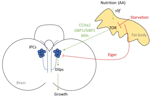
The Drosophila fat body is the functional equivalent to the mammalian liver and adipocytes. Old studies showed that when the fat body was co- cultured with larval imaginal discs or quiescent imaginal neuroblasts, nonautonomous tissue growth and cellular proliferation were induced, respectively (Britton and Edgar 1998; Davis and Shearn 1977). This suggested at that time that the fat body possesses growth promoting signals. In the recent years, the fat body has become an extensively studied organ in Drosophila when it was revealed that the fat body acts as a relay of nutritional availability to modulate growth. It was discovered that when the amino acid transporter slimfast (slif) was downregulated in the fat body, it would cause a perturbation in systemic growth that is mediated by TOR1 signaling in the fat body (Colombani et al. 2003). The underlying molecular mechanism that couples growth with slif and TOR1 signaling was only revealed later when it was shown that when slif or TOR1 were perturbed in the fat body, Dilp2 was accumulated in the IPCs, inhibiting systemic growth (Rulifson, Le, and Ge 2009). This suggests that the fat body signals nutritional information to the IPCs to modulate systemic growth by acting on Dilp secretion, possibly through releasing an x- factor in the hemolymph that is able to signal to the IPCs (Fig. 13). Studies over the recent years focused on identifying these x- factors and this lead to the identification of several humoral signals controlling growth that will be summarized below.
46
In 2015, Drosophila CCHamide-2 (CCHa2) was identified as a fat body derived signal that relays protein availability to control insulin signaling in the IPCs. Through its receptor CCHa2-R, CCHa2 regulates Dilp2 and Dilp5 secretion in the IPCs and therefore controls systemic growth (Sano et al. 2015). A year later, another two fat body derived peptides were discovered to promote systemic growth, Growth-Blocking Peptide (GBP1) and CG11395 (GBP2) . In response to amino acids and TOR signaling GPB1 and GBP2 stimulate Dilp2 and Dilp5 secretion from the IPCs to induce systemic growth (Koyama and Mirth 2016). That same year, a genetic screen in the IPCs led to the identification of a secretin-incretin receptor subfamily member Methuselah (Mth) to couple nutrition with growth (Renald Delanoue et al. 2016). Its ligand is identified as Stunted (Sun) that is a fat body secreted factor required for the IPCs to release Dilp2 and induce growth. Sun is released in the hemolymph under fed conditions and not in starved larvae, suggesting that Sun acts as a nutritional relay for the IPCs to induce growth.
Fig. 13 The fat body acts as a nutritional relay to control growth through the IPCs in the
Drosophila larval brain. In response to nutrition, the amino acid transporter slimfast (slif) in the
fat body activates TOR signaling that controls systemic growth by acting on the insulin producing cells (IPCs). The fat body does so by secreting Drosophila CCHamide-2 (CCHa2), Growth-Blocking Peptide 1 and 2 (GBP1/GBP2) and Methuselah (Mth) that positively regulate Dilp secretion from the IPCs under fed conditions. In response to starvation, the fat body secretes Eiger that negatively affects the Dilps from the IPCs. The green arrows represent positive regulation, whereas the red arrows represent negative regulation.