Université de Montréal
Endophytes of commercial Cranberry cultivars that control
fungal pathogens
Par
Karima Essid Elazreg
Département de Biochimie et Médecine Moléculaire, Faculté de Médecine
Mémoire présenté à la Faculté de Médecine en vue de l’obtention du grade de Maîtrise en Biochimie, option Génomique Humaine
November 2020 © Karima Elazreg, 2020
ii
Université de Montréal
Département de Biochimie et Médecine Moléculaire, Faculté de Médecine
Ce mémoire intitulé(e)
Endophytes of commercial Cranberry cultivars that control fungal pathogens
Présenté par
Karima Essid Elazreg
A été évalué(e) par un jury composé des personnes suivantes
Gerardo Ferbeyre Président-rapporteur B. Franz Lang Directeur de recherche Jesse Shapiro Membre du jury
i
Résumé
Les endophytes sont des microorganismes (généralement des bactéries et des champignons) qui vivent dans les tissus végétaux mais n'activent pas le système immunitaire/défense des plantes, contrairement aux pathogènes végétaux qui activent généralement les réponses immunitaires des plantes. Des recherches récentes ont montré que pratiquement toutes les plantes cultivées en plein champ contiennent un certain nombre d'endophytes, et que certains endophytes stimulent la croissance des plantes et renforcent la résistance contre les agents pathogènes. Les endophytes sécrètent des composés chimiques (métabolites secondaires) qui suppriment la croissance des agents pathogènes, un processus connu sous le nom de biocontrôle. En raison de ces propriétés de biocontrôle, les endophytes sont une alternative potentielle aux pesticides chimiques pour lutter contre les maladies des plantes. En conséquence, le biocontrôle est devenu un domaine de recherche important.
Mon projet de recherche comportait les objectifs spécifiques suivants : (i) isoler les endophytes des plants de canneberges acquis auprès de deux producteurs commerciaux de canneberges de la variété Stevens situés au Québec, Canada (Bieler Cranberries Inc, et Gillivert Inc.) ; (ii) tester l'activité de biocontrôle des endophytes contre une collection de champignons pathogènes et ensuite inoculer les endophytes les plus actifs dans des plants de canneberges obtenus par germination de la variété Stevens (Bieler Cranberries Inc. ) et Scarlet Knight (Daniele Landreville) ; et (iii) identifier des groupes de gènes de métabolites secondaires en séquençant, assemblant et annotant le génome d'un endophyte qui présentait de fortes caractéristiques de biocontrôle. Dans le cadre de ce projet de recherche, des tests antagonistes in vitro ont été réalisés avec des endophytes de la canneberge et un champignon pathogène, qui ont montré que Pseudomonas sp. CSWB3, Pseudomonas sp. CLWB12 et la souche fongique Lachnum sp. EFK28 étaient les plus actifs et ces souches ont donc été sélectionnées pour des études plus approfondies. Des expériences de germination de semis in vitro et d'inoculation d'endophytes ont montré que les souches bactériennes Pseudomonas sp. CSWB3 et Pseudomonas sp. CLWB12 amélioraient la croissance des semis de canneberges de la variété Stevens.
Comme les Pseudomonas sp. CSWB3 et Pseudomonas sp. CLWB12 ont tous deux un effet antagoniste élevé sur les champignons pathogènes, un seul (Pseudomonas sp. CSWB3) a été
ii
soumis à une analyse du génome. Le séquençage, l'assemblage, l'annotation et l'analyse du génome de Pseudomonas sp. CSWB3 a révélé que cette souche possède cinq groupes de gènes biosynthétiques de métabolites secondaires qui codent pour les protéines responsables de la biosynthèse des composés antifongiques/antimicrobiens : pyrrolnitrine, pyoluteorine, putisolvine, 2,4-diacétylephloroglucinol, bicornutine A1 et bicornutine A2.
Sur la base des résultats de ces travaux, nous concluons que certains endophytes de la canneberge qui possèdent des groupes de gènes codant pour des métabolites secondaires antifongiques peuvent supprimer les pathogènes fongiques et améliorer la croissance des plantes.
Mots-clés: endophyte, bactéries, champignons, pathogène, biocontrôle, activité antifongique,
métabolites secondaires, canneberge, Pseudomonas sp., analyse du génome, groupes de gènes, génomique comparative.
iii
Abstract
Endophytes are microorganisms (typically bacteria and fungi) that live within plant tissue but do not activate the plant defense/immune system, unlike plant pathogens that typically do activate plant immune responses. Recent research has shown that virtually all plants grown under field conditions contain a number of endophytes, and that certain endophytes stimulate plant growth and enhance resistance against pathogens. Endophytes secrete chemical compounds (secondary metabolites) that suppress pathogen growth, a process known as biocontrol. Because of these biocontrol properties, endophytes are a potential alternative to chemical pesticides for combatting plant disease. Accordingly, biocontrol has become an important field of research.
My research project was comprised of the following specific aims: (i) isolate endophytes from cranberry plants that were acquired from two commercial producers of cranberries of the Stevens variety located in Quebec, Canada (Bieler Cranberries Inc, and Gillivert Inc.); (ii) test the biocontrol activity of endophytes against a collection of fungal pathogens and then inoculate the most active endophytes into cranberry seedlings that were obtained by germinating Stevens (Bieler Cranberries Inc.) and Scarlet Knight (Daniele Landreville) seeds; and (iii) identify secondary metabolite gene clusters by sequencing, assembling, and annotating the genome of one endophyte that exhibited strong biocontrol characteristics.
As part of this research project, in vitro antagonistic tests were conducted with cranberry endophytes and fungal pathogen, which showed that Pseudomonas sp. CSWB3, Pseudomonas sp. CLWB12, and the fungal strain Lachnum sp. EFK28 were the most active and therefore these strains were selected for further studies. In vitro seedling germination and endophyte inoculation experiments showed that the bacterial strains Pseudomonas sp. CSWB3 and Pseudomonas sp. CLWB12 enhanced the growth of cranberry seedlings of the Stevens variety.
Since Pseudomonas sp. CSWB3 and Pseudomonas sp. CLWB12 both had a high antagonistic effect on fungal pathogens, only one (Pseudomonas sp. CSWB3) was subjected to genome analysis. Sequencing, assembly, annotation, and analysis of the Pseudomonas sp. CSWB3 genome revealed that this strain possesses five secondary metabolite biosynthetic gene clusters that encode proteins responsible for the biosynthesis of the antifungal/antimicrobial compounds pyrrolnitrin, pyoluteorin, putisolvin, 2,4-diacetylephloroglucinol, bicornutin A1, and bicornutin A2.
iv
Based on the results of this work, we conclude that certain cranberry endophytes that possess gene clusters encoding antifungal secondary metabolites can suppress fungal pathogens and enhance plant growth.
Keywords: endophyte, bacteria, fungi, pathogen, biocontrol, antifungal activity, secondary
v
Table of contents
Résumé ... i
Abstract ... iii
List of Tables ... viii
List of Figures ... ix
List of Symbols ... x
List of Abbreviations ... xi
Acknowledgments ... xiii
CHAPTER.1 – LITERATURE REVIEW ... 1
1. Literature Review ... 1 1.1. Endophytes ... 1 1.2. Diversity of Endophytes ... 1 1.3. Symbiotic Interactions ... 3 1.4. Plant Pathogens... 3 1.4.1. Overview of Pathogenicity ... 3
1.4.2. Plant Defense against Pathogens ... 4
1.5. Mechanisms of Enhancing Plant Growth ... 7
1.5.1. Endophytes and Plant Nutrition ... 7
1.5.2. Phytohormone production and regulation of ethylene levels ... 7
1.5.3. Biocontrol of pathogens ... 8
1.6. Biocontrol Mechanisms ... 9
1.7. Cranberry Plants and Endophytes ... 12
vi
1.7.2. Cranberry Pathogens ... 13
1.8. Hypothesis ... 15
1.9. Objectives ... 15
CHAPTER 2 – MATERIALS AND METHODS ... 16
2. Materials and Methods ... 16
2.1. Endophytes and growth medium ... 16
2.2. Sample collection and handling ... 16
2.2. Surface sterilization ... 16
2.3. Isolation of endophytes ... 17
2.4. Biocontrol of fungal pathogens by endophytes ... 17
2.6. Compatibility test ... 17
2.7. Molecular characterization of endophytes ... 18
2.8. Seed germination ... 19
2.9. Endophyte inoculation ... 20
2.10. Seedling measurement ... 21
2.11. Isolation of genomic DNA from Pseudomonas sp. CSWB3 ... 21
2.12. Genome sequencing using Illumina MiSeq ... 22
2.13. Genome assembly and annotation of Pseudomonas sp. CSWB3 ... 22
2.14. Genome analysis of Pseudomonas sp. CSWB3 and comparative genomics ... 23
2.15. Statistical analysis ... 24
CHAPTER 3 – RESULTS ... 26
3. Results ... 26
3.1. Previous results ... 26
vii
3.2.1. Endophytes isolated from cranberry plants ... 27
3.2.2. Biocontrol activity of cranberry endophytes on pathogens ... 28
3.2.3. Compatibility between endophytes ... 30
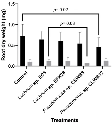
3.2.4. Effect of Pseudomonas sp. CSWB3, Pseudomonas sp. CLWB12 and Lachnum sp. EFK28 on the growth of Scarlet Knight and Stevens cranberry seedlings ... 32
3.2.5. Genome analysis of Pseudomonas sp. CSWB3 ... 36
3.2.6. Comparative analysis of Pseudomonas sp. CSWB3 with four biocontrol bacterial strains ... 41
CHAPTER 4 – DISCUSSION, CONCLUSIONS, AND PROSPECTIVE FUTURE WORK ... 46
4.1. Discussion ... 46
4.1.1. Isolated endophytes ... 46
4.1.2. Biocontrol activity ... 46
4.1.3. Seedling inoculation with endophytes ... 47
4.1.4. Pseudomonas sp. CSWB3: secondary metabolites and their mechanism of action ... 47
4.1.5. Comparison of the Pseudomonas sp. CSWB3 genome with the genomes of Pseudomonas sp. EB42, Pseudomonas protegens Pf-5, Bacillus velezensis EB37, and Bacillus velezensis EBFV ... 48
4.1.6. Limitations of this study ... 49
4.2. Conclusions and prospective future work ... 50
viii
List of Tables
Table 1.- NRPS and PKS gene clusters and bioactive secondary metabolites produced by
B. velezensis RC218. ... 12
Table 2.- Total number of endophytes isolated from cranberry plants. ... 27
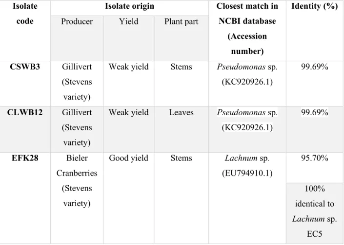
Table 3.- Origin and the closest match of CSWB3, CLWB12, and EFK28 isolates based on BLAST analysis of 16S and ITS sequences ... 28
Table 4.- Inhibition of fungal pathogens by the most active endophytes (Pseudomonas sp. CSWB3, Pseudomonas sp. CLWB12, and Lachnum sp. EFK28). ... 29
Table 5.- The average root:stem dry weight ratio ± standard error of cranberry seedlings of two varieties (Stevens and Scarlet Knight) ... 36
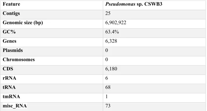
Table 6.- General features of the Pseudomonas sp. CSWB3 genome ... 37
Table 7.- Taxonomy of Pseudomonas sp. CSWB3 ... 39
ix
List of Figures
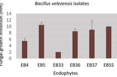
Figure 1. – Inhibition of Colletotrichum gloeosporioides by six Bacillus velezensis isolates..26
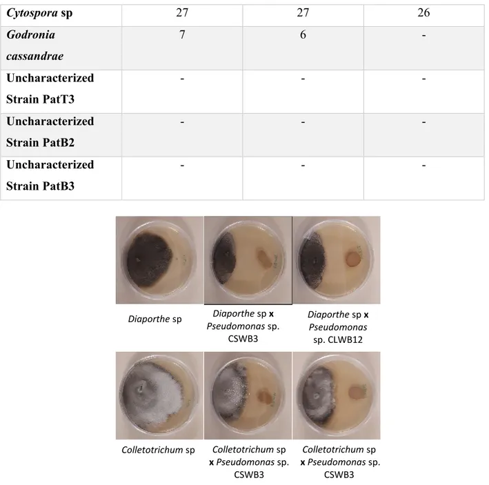
Figure 2. – Growth inhibition of the fungal pathogens Diaporthe sp. and Colletotrichum sp. by cranberry endophytic bacteria Pseudomonas sp. CSWB3 and Pseudomonas sp. CLWB12 at day 30………..30
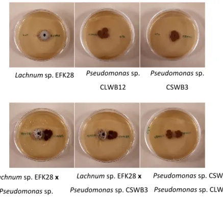
Figure 3. – Compatibility tests between Pseudomonas sp. CSWB3, Pseudomonas sp. CLWB12, and Lachnum sp. EFK28………..31
Figure 4. – The effect of endophyte inoculation on the stem length (cm) of cranberry seedlings obtained from two varieties (Stevens and Scarlet Knight)………..32
Figure 5. – The effect of endophytes on cranberry seedling stem and leaf dry biomass (mg). Cranberry seedlings of two cranberry varieties (Stevens and Scarlet Knight)……….34
Figure 6. – The average dry root biomass (mg) of cranberry seedlings of two varieties (Stevens and Scarlet Knight)………35
Figure 7. – Circular distribution display of the annotated genes of Pseudomonas sp. CSWB3………38
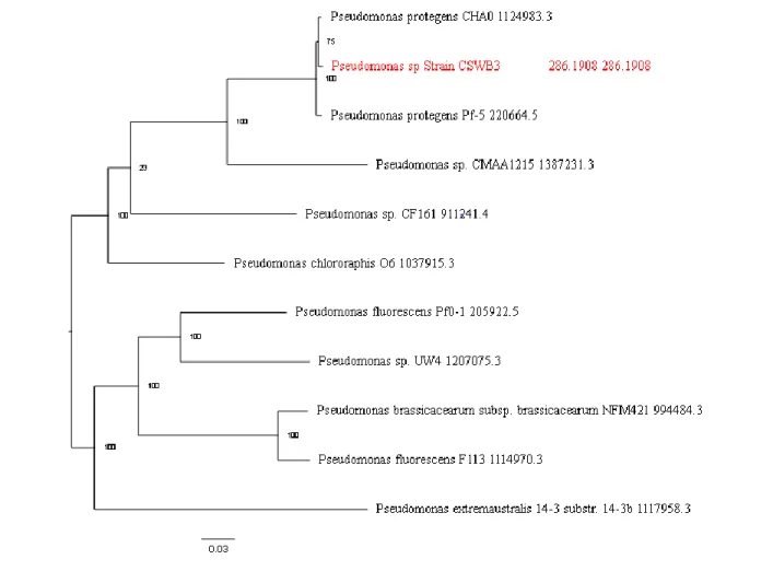
Figure 8. – Phylogenetic tree of Pseudomonas sp. CSWB3……….40
Figure 9. – Comparison of OrthoANI values between the genomes of Pseudomonas sp. CSWB3, Pseudomonas sp. EB42, Pseudomonas protegens Pf-5, Bacillus velezensis EB37, and Bacillus velezensis EBFV……….42
Figure 10. – Venn diagram generated by Orthovenn2 showing orthologous gene clusters for Pseudomonas sp. CSWB3, Pseudomonas sp. EB42, Pseudomonas protegens Pf-5, Bacillus velezensis EB37, and Bacillus velezensis EBFV………..44
x
List of Symbols
µl: microliter °C: degree Celsius min: minute s: second nm: nanometer Kb: kilobase g: gram mg: milligram L: Liter ml: milliliter %: percentage µg : microgram SE : Standard Errorxi
List of Abbreviations
NADH: Nicotinamide adenine dinucleotide ABA: Abscisic Acid
ACC: S-adenosyl-L-methionine and cyclic non-protein amino acid AMF: Arbuscular Mycorrhizal Fungi
ANOVA: Analyzed of Variance BCA: Biocontrol Agent
BGCs: Biosynthetic Gene Clusters
BLAST: Basic Local Alignment Search Tool bp: base pair
CDS: Coding Sequence
C-endophytes: Clavicipitaceous endophytes DAPG: 2, 4-diacetylphloroglucinol
DNA: Deoxyribonucleic acid
DNTPs: Deoxyribonucleotide Triphosphate EDTA: Ethylenediaminetetraacetic acid ET: Ethylene
ETI: Effector-Triggered Immunity faa: protein FASTA file
GAs: Gibberellins
gbk format: gene bank format IAA: Indole-3-Acetic Acid
xii
ITS: Internal Transcribed Spacer
MAMPs: Microbial-Associate Molecular Patterns miscRNA: miscellaneous Ribonucleic Acid MTI: MAMPs Triggered Immunity
NC-endophytes: Non-Clavicipitaceous endophytes NRPS: Nonribosomal peptide Synthetase
OD: Optical Density
PATRIC: Pathosystems Resource Integration Center PCR: Polymerase Chain Reaction
PDA: Potato Dextrose Agar
PGPR: Plant Growth–Promoting Rhizobacteria pH: potential hydrogen
PKS: Polyketides Synthetase
PRRs: Pathogen-Recognition Receptors rDNA: Ribosomal Deoxyribonucleic acid RN: Root Nodule
rRNA: ribosomal Ribonucleic Acid
SDS-PAGE: Sodium Dodecyl Sulfate–Polyacrylamide Gel Electrophoresis SPAdes: St. Petersburg genome assembler
tmRNA: transfer-messenger Ribonucleic Acid tRNA: transfer Ribonucleic Acid
xiii
Acknowledgments
This work would not have been possible without the financial support of the Ministry of Higher Education, Libya.
I am especially indebted to Prof. B. Franz Lang, my research supervisor, who has been supportive of my career goals and who actively worked to provide me with the required academic time that allowed me to pursue my research goals.
I would like to acknowledge Bieler Cranberries Inc., Gillivert Inc., and Groupe Landreville Nadeau Inc. for providing us with the cranberry plants and seeds that were used in our experiments. Nobody has been more important to me in the pursuit of this project than the members of my family. I would like to send special greetings and prayers to my Father’s spirit in his grave for his previous support and encouragement. I would like to thank my entire family, whose love and guidance are with me in whatever I pursue. Most importantly, I wish to thank my loving and supportive husband, Ahmed, and my two wonderful children, Sohaib and Mossab, who provide unending inspiration.
I am grateful to all of those with whom I have had the pleasure to work with during this and other related projects. Each of the members of my Thesis Committee has provided extensive personal and professional guidance and taught me a great deal about both scientific research and life in general.
Finally, I would like to acknowledge the laboratory researcher Lise Forget and my colleagues Lila Salhi and Peniel Bustamante who supported me immensely and were always willing to help.
1
CHAPTER.1 – LITERATURE REVIEW
1. Literature Review
1.1. Endophytes
Plant endophytes are microorganisms that live within plant tissue, at least for part of their life cycle. Endophytes were first identified in 1809 by the German botanist Heinrich Friedrich (Dey, Datta, Saha, Parida, & Panda1, 2019), who defined endophytes as a specific group of parasitic fungi that live in plants. Later, in 1991, the description of endophytes became more factual and practical, defined by Orlando Petrini as “all organisms inhabiting plant organs that at some time in their life cycle can colonize internal plant tissues without causing apparent harm to their host” (Hardoim et al., 2015).
In an agronomical sense (i.e., practical, non-scientific sense), endophytes may have beneficial effects by stimulating plant growth as well as by inhibiting plant pathogens (Gupta et al., 2016). However, the qualifications ‘beneficial’ or ‘without causing apparent harm to their host’ are problematic and need to be defined for a specific, given context, as some endophytes are pathogenic to certain plants but enhance the growth of other plants. This may be caused by many factors, including the environment, the precise genotype of the endophyte isolate, and other similar types of factors. We therefore use the term ‘endophyte’ in a more general sense, as ‘microbes (bacteria or fungi) that colonize internal plant tissues’, without referring to pathogenicity or agronomic benefits.
1.2. Diversity of Endophytes
Endophytes encompass a wide variety of microorganisms, in particular bacteria and fungi. More than 200 plant-associated species of bacteria have been identified, the majority of which belong to the phyla Actinobacteria, Proteobacteria, and Firmicutes (Golinska et al., 2015). The phylum Actinobacteria is comprised of Gram-positive bacteria that includes Streptomyces, whereas the phylum Proteobacteria is comprised of Gram-negative bacteria, including Pseudomonas, Escherichia, and Salmonella. The majority of the phylum Firmicutes is comprised of Gram-positive bacteria, such as Bacillus and Clostridium (Golinska et al., 2015; Hui, Yan, Qing, Renyuan, & Yongqiang, 2013). Many of these bacterial endophytes secrete secondary metabolites
2
that possess antibiotic, antifungal, and antitumor activity; secondary metabolites produced by Streptomyces species have been investigated in great detail (Berdy, 2012).
Fungal endophytes are found within the tissue of a wide variety of plants. The relationship between fungal endophytes and plants may be mutualistic and asymptomatic (i.e., no signs of disease throughout the fungus life cycle), in which plants provide food and energy sources to endophytes while in return endophytes enhance the resistance of plants against pathogens (Saikkonen, Faeth, Helander, & Sullivan, 1998). However, as noted above, fungal endophytes may also exhibit pathogenic traits. Fungal endophytes have been widely studied because of their relationship with plants and their association with plants, which started approximately 400 million years ago (Krings et al., 2007).
Based on taxonomy, host and tissue specificity, and function, endophytic fungi are classified into two main groups (Bamisile, Dash, Akutse, Keppanan, & Wang, 2018), which are clavicipitaceous endophytes (C-endophytes) and non-clavicipitaceous endophytes (NC-endophytes; for instance the wide-spread zygomycete AMF, see below). C-endophytes are associated with different grasses and produce bioactive metabolites that enhance the resistance of plants to insects, nematodes, and fungal pathogens. C-endophytes also produce alkaloids that are toxic to humans and animals but are tolerated by the plant host. C-endophytes include Hypocreales and other Ascomycota. NC-endophytes contribute to several functions in the host plant, including avoidance of abiotic stress, inducing synthesis of plant hormones, and protecting the plant from pathogenic fungi (Rodriguez, White, Arnold, & Redman, 2009).
Endophytic bacteria and fungi have been isolated from the Vaccinium genus within the family Ericaceae, which includes cranberries, blueberries, and strawberries. Ericoid mycorrhizal fungal endophytes have been isolated in China from blueberries; these fungal endophytes colonize the roots of blueberry plants and help increase the supply of nutrients to the plant under harsh environmental conditions. The ericoid mycorrhizal fungi isolated from blueberry belong to the genera Clavaria, Oidiodendron, Lachnum, Acephala, and Phialocephala (Yang et al., 2018). Additionally, a diverse range of endophytic bacteria has been found to be associated with blueberry plants. These endophytic bacteria produce indole acetic acid and other bioactive compounds involved in biological control activities that stimulate plant growth. In a study conducted by Ortiz-Galeana et al., the most common bacterial endophytes found to be associated with blueberry plants
3
belong to the genera Pantoea, Pseudomonas, Burkholderia, and Bacillus. The majority of the species belonging to these genera perform activities that are advantageous to the blueberry plant (Ortiz-Galeana et al., 2018).
1.3. Symbiotic Interactions
Plants and endophytes communicate through biological mechanisms that lead to changes at the genetic, signaling, and metabolic levels. Two types of symbiotic communication between plants and either fungi or bacteria have been vigorously studied. The first and most common type are arbuscular mycorrhizal fungi (AMF) (Kawaguchi & Minamisawa, 2010) and zygomycete fungi (a paraphyletic taxon) that interact with a large variety of plants. Interactions between these fungi and plants initiate with the entry of fungal hyphae into the root epidermis, followed by an expansion of hyphae to reach the inner cortex where arbuscules are formed. These branched hyphae enhance absorbance of phosphate and other nutrients by the fungus from the soil (Lee, Eo, Ka, & Eom, 2013). The second type of bacterial symbiotic interaction is called root nodule (RN), which consists of a nitrogen-fixing symbiotic interaction that is formed between rhizobacteria and the roots of leguminous plants. With RN, chemicals are produced that mediate highly specific signaling and communication between the host plant and the rhizobacteria (Clua, Roda, Zanetti, & Blanco, 2018). The process of nodulation initiates by the secretion of flavonoids by the plant root, which subsequently stimulate the expression of genes responsible for nodulation. This leads to production of lipochito-oligosaccharide, which is a nodulation factor that stimulates the growth of cells in the root cortex that ultimately leads to the formation of root nodules (Q. Wang, Liu, & Zhu, 2018). Symbiotic interactions between roots and nodules may also be a mechanism that allows bacterial pathogens to enter plant tissue, including certain non-nitrogen-fixing species that harbor antibiotic resistance genes specific to cefoxitin, ampicillin, and cefuroxime-axetil (Muresu, Maddau, Delogu, Cappuccinelli, & Squartini, 2010).
1.4. Plant Pathogens
1.4.1. Overview of Pathogenicity
Plant pathogenicity is defined as the potential of microorganisms to cause damage to crops. The majority of plant pathogens are fungi. Fungal pathogens are classified according to their pattern of nutrition into necrotrophic, hemibiotrophic, and biotrophic. Necrotrophics include bacteria and fungi that secrete lytic enzymes (e.g., cell wall-degrading enzymes) that weaken plant defenses,
4
which ultimately can lead to destruction of plant tissue (necrosis). The fungus Botrytis cinerea that causes grey mold disease is an example of a necrotrophic pathogen. Botrytis cinerea forms an infectious structure on the outer surface of the leaf that mediates penetration of plant leaves, which is followed by secretion of lytic enzymes that dissolve cutin (Laluk & Mengiste, 2010).
Unlike necrotrophic pathogens, biotrophic pathogens establish a mutualistic relationship with host plants. Biotrophic pathogens avoid host recognition and detection by secreting lytic enzymes that suppress the immunity of the host plant, which allows these pathogens to live within the plant tissue and benefit from plant-derived nutrients that gradually weakens and damages the host plant (Laluk & Mengiste, 2010). Biotrophic pathogens can also penetrate plant tissue and produce protein-based inhibitors of β-1,3-glucanases, which are enzymes produced by plants that are capable of dissolving fungal cell walls (Gebrie, 2016). An example of a biotrophic pathogen is Hyaloperonospora arabidosidis, which causes downy mildew disease in Arabidopsis. Sequencing of the H. arabidosidis genome revealed 134 RXLR genes that encode for RXLR effectors. The function of most of these RXLR effectors is not known, and only 13 of the RXLR effectors were shown to have a small effect on the host immune response, which suggests that inhibition of the host plant immune response by H. arabidosidis pathogen is possibly caused by the combined action of all 134 RXLR effectors (Coates & Beynon, 2010; Pel et al., 2014).
Nutrient acquisition by hemibiotrophic pathogens requires a living host plant. Hemibiotrophic pathogens go through a biotrophic phase during the primary stages of pathogenicity then switch to the necrotrophic phase. An example of a hemibiotrophic pathogen is the fungus Zymoseptoria tritici that is the causal agent of septoria leaf blotch, which affects the leaves of wheat. Zymoseptoria tritici infections start with the growth of hyphae on the outer surface of leaves, followed by pathogen penetration. The fungus grows slowly inside plant tissue. The infected plant does not display any pathogenic symptoms for 8–11 days, and then the fungus switches to its necrotic phase, which results in plant death. Following one month of infection, the pathogen starts to form sexual structures (Garcia-Sanchez, Bernales, & Cristobal, 2015).
1.4.2. Plant Defense against Pathogens
Several morphological, biochemical, and molecular mechanisms are involved in the response of plants to pathogenic infections. Following infection, a plant attempts to kill or weaken the
5
pathogen. Disease symptoms manifest after the plant’s defense mechanisms are sufficiently weakened by a pathogen. There are two types of plant defense mechanisms, described below.
1) Constitutive defense mechanisms
Structural defense mechanisms: This type of plant defense mechanism is considered to be the first line of defense against invading pathogens. Structural defense mechanisms include the wax layer and cuticle found on the surface of the plant, which give support and rigidity to plant tissue. Other plant structures act as physical barriers to prevent pathogen penetration, including the epidermal layer, actin cytoskeleton, guard cells, and trichomes (Doughari, 2015).
Biochemical defense: Various chemicals are produced by plants in response to invading pathogens. Toxic or lytic reactions mediated by these chemicals may directly affect the pathogen or may indirectly affect the pathogen by stimulating microflora on the plant surface to produce toxic chemicals. These types of biochemical compounds that are secreted by plant tissues include antimicrobial organic substances such as saponins and alkaloids that act as inhibitors of microorganism growth. Another biochemical defense mechanism is mediated by toxic inhibitors, which are metabolites found in plant tissue that degrade toxins secreted by pathogens, which contributes to the plant’s resistance against the invading pathogen (Doughari, 2015).
2) Induced defense mechanisms
As previously described, pathogens have the ability to suppress the immune system of host plants and produce inhibitors of cell wall-degrading enzymes produced by the host plant. However, if a pathogen overcomes the plant’s pre-existing defenses, additional structural, cellular, or biochemical response mechanisms may be induced in order to kill the pathogen (van Baarlen, van Belkum, Summerbell, Crous, & Thomma, 2007).
Structural defense: Structural defenses include lignification of the cell wall that increases its rigidity, which prevents penetration of fungal pathogen hyphae (Jones & Dangl, 2006). Another structural defense is suberization that includes conversion of the cell wall into cork tissue by suberin formation, which helps to isolate infected cells away from healthy tissue, which ultimately helps to minimize the spread of the infection (van Baarlen et al., 2007). Another important structural defense mechanism is gum deposition within infected cells (Doughari, 2015), and formation of a secondary wall and papillae. Papillae are polysaccharide polymers that are secreted
6
in response to an infection, especially in cereals, to increase their resistance (Anderson et al., 2010; Freeman & Beattie, 2008).
Biochemical defenses: Biochemical defenses are the last line of host defense against penetrating pathogens (Jan, Azam, Ali, & Haq, 2011). Biochemical immunity consists of two main layers: microbial-associated molecular pattern (MAMP)-triggered immunity (MTI) and effector-triggered immunity (ETI). Protection against an invading pathogen initiates with pathogen recognition, which stimulates a defensive response (termed a hypersensitive response) that induces infected cell death, which helps to protect neighboring uninfected cells. A group of proteins called pathogen-recognition receptors (PRRs) are localized to the plant cell membrane and the cytoplasm and recognize MAMPs that subsequently activates MTI. Pathogens secrete effectors that bind PRRs, which inhibits plant MTI (Swarupa, Ravishankar, & Rekha, 2014).
ETI is stimulated by a plant’s intracellular resistance gene in response to the detection of pathogenic type III secreted effectors (T3SEs); ETI is linked to the programmed cell death of infected cells (Doughari, 2015). Plants produce various other biochemical molecules in response to infection, including toxic substances such as phenolic compounds and phytoalexins. Additionally, plants produce proteins such as lectins, ricin, protease inhibitors, and hydrolytic enzymes such as chitinases and glucanases that play important roles in inhibiting pathogens (Doughari, 2015).
The innate immune system of plants can discern between endophytes and pathogens, which allows endophytes to enter the host plant without triggering the immune system. This lack of immune response helps to maximize the benefits received from this interaction, which ultimately boosts the immune reaction of plant-endophytes against invading pathogens (Khare, Mishra, & Arora, 2018). As mentioned previously, the first line of plant defense is the recognition of MAMPs by PRRs located on the surface of plant tissue (Swarupa et al., 2014). Fungal endophytes can avoid host immune recognition by producing chitin deacetylases that deacetylate chitosan oligomers that are part of the plant cell wall. Certain endophytic bacteria possess MAMPs that inhibit recognition by the PRRs found on the surface of the plant cell wall (Khare et al., 2018).
In conclusion, the entry of endophytes into host plants is facilitated by the ability of endophytes to avoid the plant immune system. Additionally, endophytes help plants to defend themselves against pathogens by secreting biochemical substances that suppress pathogen growth.
7
1.5. Mechanisms of Enhancing Plant Growth 1.5.1. Endophytes and Plant Nutrition
Bacterial and fungal endophytes in the soil (termed the rhizosphere) can enhance plant growth by increasing the availability and uptake of nutrients by plants; hence, they are often referred to as biofertilizers. The number of plant-growth-promoting rhizobacteria (PGPR) in the soil depends on the environmental conditions and the soil type. PGPR are found near the roots of a plant and induce plant growth via direct and indirect mechanisms. Direct mechanisms include bacterial secretions, nitrogen fixation, and solubilization of phosphorus (Olanrewaju, Glick, & Babalola, 2017). Nitrogen and phosphorus are essential nutrients for plant growth. However, these nutrients are often only available in limited quantities in the soil, due to the loss of nitrogen by leaching and the low bioavailability of phosphorus (due to it predominant insoluble form as aluminum and iron phosphates). Collectively, PGPR are important for plant growth because of their nitrogen fixation and phosphorous solubilization functions (Martinez-Viveros, Jorquera, Crowley, Gajardo, & Mora, 2010). In addition, PGPR can increase the absorbent surface of plant roots by stimulating root growth and branching as well as stimulating other non-pathogenic symbionts (endophytes) of the host (Saia et al., 2015; Vessey, 2003). Certain PGPR known as endophytic plant growth-promoting bacteria employ unknown mechanisms to enter plant tissue. The various functions performed by endophytic plant growth-promoting bacteria are similar to those performed by non-endophytic PGPR, including facilitating the acquisition of nutrients such as nitrogen, phosphorus, and iron from the rhizosphere and the direct transfer of these nutrients into plant roots (Santoyo, Moreno-Hagelsieb, Orozco-Mosqueda Mdel, & Glick, 2016). With fungal endophytes, nutrients can be transferred via mycelia that extend from mycorrhizal fungi living in plant root tissue (White et al., 2019). Endophytes such as Bacillus spp. that colonize plant roots have high affinity for organic acid-metal complexes and facilitate the transfer of these complexes from the soil into plants (White et al., 2019).
1.5.2. Phytohormone production and regulation of ethylene levels
Phytohormones are bioactive molecules that act as messengers in signaling pathways, which contributes to plant growth and developmental regulation. The biological, morphological, and physiological processes of a plant can be affected by very low concentrations of phytohormones
8
(Martinez-Viveros et al., 2010). Several endophyte species (both bacteria and fungi) can produce phytohormones (Tsavkelova, Klimova, Cherdyntseva, & Netrusov, 2006), including gibberellins (GAs), indole acetic acid (IAA), abscisic acid (ABA), and jasmonic acid (JA). JA and ABA play crucial roles in the regulatory processes that control the heat-stress response, while GAs and IAA enhance plant growth and development (Waqas et al., 2015). Interactions between plants and microorganisms such as endophytes, rhizobacteria, free-living bacteria, and some pathogens results in conversion of tryptophan to IAA, which is one of the auxins produced from tryptophan. IAA may also be produced via tryptophan-independent pathways. The main function of IAA is to stimulate the division, expansion, and differentiation of plant cells, which enhances plant growth (Martinez-Viveros et al., 2010). (Swain, Naskar, & Ray, 2007) showed that the length of the stem and root of the white yam plant (Dioscorea rotundata) increased following inoculation of IAA-producing Bacillus subtilis as compared to non-inoculated plants.
GA is another phytohormone that regulates plant growth and plays an important role in seed germination, leaf elongation, and flowering, and works with auxins to increase root length. GA levels are regulated by various factors, such as light, temperature, and auxin concentrations (Stamm & Kumar, 2010). A recent study by (Hamayun et al., 2017) showed that treatment of soybean plants with GA-producing Porostereum spadiceum (an endophytic fungus) resulted in enhanced growth as compared to untreated plants.
Ethylene (ET) is a gaseous phytohormone produced from the precursor methionine via S-adenosyl-L-methionine and cyclic non-protein amino acid (ACC). ET is involved in many stages of plant growth, including the ripening of fruit (Wani, Kumar, Shriram, & Sah, 2016). High concentrations of ET may inhibit plant growth by affecting cellular processes and stimulating defoliation. Rhizobacteria have the capacity to regulate ET levels in the soil by producing ACC deaminase, which degrades ACC into alpha-ketobutyrate and ammonium (Martinez-Viveros et al., 2010).
1.5.3. Biocontrol of pathogens
Microbial pathogens can cause various diseases in plants that ultimately leads to decreased crop yields. Chemical pesticides have been widely used to reduce or eliminate pathogenic infections, but these compounds negatively impact human health and the environment. In addition, with prolonged use, pathogens may become resistant to chemical pesticides, and consequently new pesticides must be developed. Based on previous research, biological methods may be suitable
9
alternatives to chemical pesticides, which would reduce the harmful impact of chemicals. These biological methods are based on using soil microorganisms and plant-associated endophytes as bio-pesticides, since they exhibit biocontrol activity against pathogens. Biocontrol is the term used to describe the secretion of bioactive compounds (e.g., antibiotics, hydrogen cyanide, and many other secondary metabolites) that suppress pathogen growth and consequently promote plant growth (Martinez-Viveros et al., 2010). The biocontrol properties of endophytes are the core subject of this research and are discussed in more detail in the following section.
1.6. Biocontrol Mechanisms
Biocontrol, an abbreviation for “biological control”, describes the use of microorganisms to suppress the growth of pathogens that ultimately reduces or eliminates disease symptoms. An antagonistic microbe with bioactive properties that suppress the growth of a pathogen is known as a biocontrol agent (BCA). BCAs produce compounds that influence the plant host as well as the pathogen. These compounds are secreted in response to specific and non-specific interactions between the plant, endophytes, and pathogens. BCAs have been widely used in the field of plant pathology (Pal & Gardener, 2006).
It is well known that various microorganisms, including bacteria and fungi, secrete bioactive compounds, some of which have been used as therapeutic drugs. Penicillin, obtained from Penicillium glaucoma in 1896, was the first such fungal bioactive substance identified, and has been widely used as an antibiotic for treating a wide range of bacterial infections (Suryanarayanana et al., 2009).
Various mechanisms of biocontrol have been described that enhance plant growth by suppressing or killing pathogens, including induction of host resistance (which was explained in the section on plant defense) and secretion of antibiotics and lytic enzymes.
1. Antibiotics
The production and secretion of antibiotics is one of the most powerful biocontrol mechanisms. Our research is focused on secondary metabolites secreted by endophytes that function as biocontrol agents, which include antibiotics. Antibiotics are low molecular weight organic substances that are secreted by various microbes. Previous studies showed that the antibiotics produced by PGPR have an impact on the growth of pathogens; i.e., production of sufficient levels
10
of an antibiotic by endophytes suppresses pathogens in close proximity. A single antibiotic can control one or more pathogens (Pal & Gardener, 2006).
Recent studies have shown that certain bacteria, such as Bacillus spp. and Pseudomonas spp., function as biocontrol agents by producing bioactive compounds with antimicrobial activity that suppress the growth of fungal pathogens. Certain Bacillus spp. and Pseudomonas spp. harbor gene clusters that are responsible for the secretion of secondary metabolites such as non-ribosomal peptide synthetases (NRPSs) and polyketide synthases (PKSs) (Palazzini, Dunlap, Bowman, & Chulze, 2016; Strano, Bella, Licciardello, Caruso, & Catara, 2017).
NRPSs and PKSs are large enzymes or enzyme complexes involved in the biosynthesis of various natural bioactive compounds known as non-ribosomal peptides and polyketides, which are compounds that possess remarkable bioactivity, including antifungal and antimicrobial activity. Non-ribosomal peptides are synthesized by sequential condensation of amino acids, whereas polyketides are synthesized by repetitive insertion of two carbon ketide units obtained from thioester of acetate (Ansari, Yadav, Gokhale, & Mohanty, 2004). The biosynthetic gene clusters (BGCs) that encode NRPSs and PKSs are commonly found in the phyla Proteobacteria, Actinobacteria, Firmicutes, and Cyanobacteria (H. Wang, Fewer, Holm, Rouhiainen, & Sivonen, 2014). NRPS often have three catalytic domains: the adenylation (A) domain that recognizes and activates amino acids, the peptidyl-carrier (thiolation) domain that transfers the activated amino acids, and the condensation (C) domain that is responsible for peptide bond formation and peptide chain elongation. Certain NRPS contain additional domains, such as the epimerization domain (E) that convert L-amino acids into D-amino acids, the dual/epimerization domains (E/C) that are responsible for epimerization and condensation, cyclization domains (Cy) that can functionally replace C domains, the oxidation domain (Ox) that is responsible for the oxidation of the thiazoline ring that leads to the formation of an aromatic thiazol, and thioesterase domains (TE) that are responsible for cyclization or hydrolysis of the final peptide in the NRPS module that releases it (Amoutzias, Chaliotis, & Mossialos, 2016). The resulting NRPS molecules may be further modified by subsequent biochemical reactions, leading to an even broader spectrum of bioactive substances.
11
Type I PKSs typically have three main domains: the acyl-transferase (AT) domain that incorporates malonyl or methylmalonyl-CoA, the KS domain that is responsible for C-C bond formation, and the acyl-carrier (thiolation) domain, which is similar to the peptidyl-carrier domain of NRPSs. PKSs have additional domains, such as the ketoreduction (KR) domain, the dehydration (DH) domain, and the enoylreductase (ER) domain (Amoutzias et al., 2016).
NRPSs and PKSs are structurally and functionally similar and therefore often form hybrid gene clusters (hybrid NRPS/PKS) (Muller et al., 2015). BGCs that encode both an NRPS and a PKS can produce complex NRPS/PKS-derived hybrid bioactive compounds called peptide-polyketide secondary metabolites, which can be produced directly by the NRPS/PKS hybrid enzyme or indirectly as non-ribosomal peptides and polyketides that bind together (Amoutzias et al., 2016).
Pseudomonas spp. harbor gene clusters that encode various NRPs. NRP compounds include pyoverdines, pyochelin, pseudomonine, paerucumarin, pseudoverdine, lipopeptides, safracin, tabtoxin, phaseolotoxin, pyrrolnitrin, and indole-3-acetic acid (IAA) (Gross & Loper, 2009). Bacillus velezensis produces NRPs such as fengycin, surfactin, bacilysin, iturin and corynebactin (Palazzini et al., 2016).
PKSs produce several different natural products that possess antibiotic, chemotherapeutic, and phytotoxic activities. Polyketides secreted by Pseudomonas spp. include mupirocin (pseudomonic acid A), 2,4-diacetylphloroglucinol (DAPG), and 2,5-dialkylresorcinols (Gross & Loper, 2009), while polyketides secreted by Bacillus velezensis include bacillaene, difficidin and macrolactin (Palazzini et al., 2016).
A previous study found that the biocontrol isolate Bacillus velezensis RC 218 produces non-ribosomal peptides and polyketides that can suppress the fungal pathogen Fusarium, which can be used in agricultural applications. This same study also analyzed the genome of B. velezensis RC218 and identified the BGCs responsible for the biocontrol activity of this strain (Table 1) (Palazzini et al., 2016).
12
Table 1.- NRPS and PKS gene clusters and bioactive secondary metabolites produced by
B. velezensis RC218. Adapted from (Palazzini et al., 2016)
Compound Synthetase type
Genes Size
(kb)
Bioactivity Fengycin NRPS fenA, B, C, D, E 37.7 Antifungal
Surfactin NRPS srfAA, AB, AC, AD 26.2 Surfactant, antibiotic
Bacilysin (Chlorotetaine)
NRPS bacA, B, C, D, E, F, ywfH 6.7 Antibacterial
Iturin NRPS ituD, A, B, C 37.2 Antifungal
Bacillaene PKS baeB, C, D, E, acpK, baeG, H, I, J, L, M, N, R, S 72.5 Antibacterial Difficidin PKS dfnA, Y, X, B, C, D, E, F, G, H, I, J, K, M, L 69.5 Antibacterial Macrolactin PKS mlnA, B, C, D, E, F, G, H, I 53.2 Antibacterial 2. Lytic enzymes
Endophytes can directly suppress the activity and growth of pathogens by secreting lytic enzymes, which prevent the growth of plant pathogens by destroying their cell walls. Lytic enzymes include β-1,3 glucanases, chitinases, and cellulases. β-1,3 glucanases are involved in the biocontrol activity of Lysobacter enzymogenes strain C3 and have been found to suppress fungal pathogens of plants (Pal & Gardener, 2006). In fact, mutations in the β-1,3 glucanase genes of L. enzymogenes strain C3 lead to a reduction in the biocontrol effects against Pythium and Bipolaris, which causes damping-off of sugar beets and leaf spot in fescue, respectively (Gao, Dai, & Liu, 2010).
1.7. Cranberry Plants and Endophytes
American cranberries (Vaccinium macrocarpon) are a member of the evergreen species of Ericaceae. American cranberries are prostrate and relatively short plants and grow mainly in North
13
America (i.e., Canada and USA) (Hisano, Bruschini, Nicodemo, & Srougi, 2012; Polashock et al., 2014). North America is the main producer of cranberries worldwide (Diaz-Garcia et al., 2019).
1.7.1. Cranberry Endophytes
A large number (several hundreds) of endophytes were previously isolated from cranberry plant tissue in B. F. Lang’s laboratory over a period of approximately five years. In this research more endophytes were isolated to increase the chance of isolating strains with stronger biocontrol, suppression of the largest possible spectrum of fungal pathogens, and potentially, the future identification of novel antifungal antibiotics of medical interest.
The most common endophytes that showed biocontrol properties when tested against various cranberry fungal pathogens include the following:
Bacillus velezensis: B. velezensis is a Gram-positive bacterium that forms endospores. B. velezensis produces enzymes, antibiotics, insecticides, and other bioactive compounds that inhibit pathogens and promote plant growth (Ruiz-Garcia, Bejar, Martinez-Checa, Llamas, & Quesada, 2005). The B. velezensis genome contains several gene clusters that encode secondary metabolites that function as fungicides; thus, B. velezensis may potentially be a powerful biocontrol agent that inhibits pathogens and enhances plant growth (Liu et al., 2017).
Lachnum sp.: Lachnum is a genus of fungi in the family Hyaloscyphaceae that produces abundant amounts of bioactive compounds such as antimicrobial substances that have been used for medicinal and agricultural purposes (Rukachaisirikul, Chantaruk, Pongcharoen, Isaka, & Lapanun, 2006). An L. abnorme strain was previously isolated from stems of the Ardisia cornudentata Mez plant; additionally, it was shown that this fungus and three other Lachnum species can produce at least 35 secondary metabolites, such as benzenoids and coumarins. Subsequent investigation of these metabolites revealed that they possess biocontrol activity (Chang et al., 2016).
1.7.2. Cranberry Pathogens
A variety of fungal pathogens are capable of infecting cranberry plants and can cause losses in total fruit production of up to 33% if fungicides are not used (Conti, Cinget, Vivancos, Oudemans, & Belanger, 2019). Fruit rot is the most common disease that affects cranberry plants, which is caused by approximately 12 different fungal pathogens. Identification of the fungus responsible for fruit rot can be accomplished by culturing rotten cranberry fruit on culture medium followed by analysis
14
of growth characteristics. Different fungal pathogens cause different symptoms in cranberry plants (Wells & McManus, 2013).
Coleophoma empetri: This fungal species causes Ripe Rot disease. During growth on culture medium, the mycelia initially have a white color then turn dark brown or black.
Colletotrichum acutatum: This fast-growing fungal species causes a disease called Bitter Rot. During growth on culture medium, the color of this fungus is initially white then turns pinkish-orange because of the red pigments produced by this fungus.
Colletotrichum gloeosporioides: Similar to C. acutatum, C. gloeosporioides is fast-growing and causes Bitter Rot. C. gloeosporioides is white in color during the early stages of growth on culture medium, then becomes dark gray as the colonies mature.
Phomopsis vaccinii: This fungal species causes Viscid Rot/Upright Dieback disease. During the early stages of growth on culture medium, this fungus is white in color then turns dark gray. Phyllosticta vaccinii: This fungal pathogen causes Early Rot disease and has a very slow growth rate as compared to other cranberry fungal pathogens. It produces mycelia that are olive to black in color with irregular edges during growth on culture medium.
Phyllosticta elongata: This fungal pathogen causes Berry Speckle/Botryosphaeria fruit rot. The mycelia of this fungus are greenish-gray in color and has a very similar appearance to P. vaccinii during the early stages of growth on culture medium, then becomes recognizable by its morphology and fast growth rate.
Physalospora vaccinii: This fungal pathogen causes Blotch Rot. Two different strains were previously isolated from rotten fruit: a dark strain that produces brown-gray mycelia and a light strain that produces white mycelia.
15
1.8. Hypothesis
Bacterial and fungal endophytes have the potential to enhance cranberry growth and yield because of their ability to inhibit the growth of fungal pathogens via secretion of bio-pesticides (secondary metabolites).
1.9. Objectives
The objectives of this research are to:
1- Isolate endophytes from cranberry plants obtained from two commercial sources and then classify the isolated endophytes into three groups based on fruit yield: very good yield, good yield, and weak yield.
2- Identify which isolates are the most potent in terms of suppressing cranberry pathogens.
3- Use young plants obtained by seed germination to evaluate the biocontrol activity of the most potent isolates identified in Aim 2.
4- Classify the most active biocontrol isolates by ribotyping, and then sequence, assemble, and annotate the genome of one isolate, followed by the identification of the genes potentially involved in biocontrol.
16
CHAPTER 2 – MATERIALS AND METHODS
2. Materials and Methods
2.1. Endophytes and growth medium
Endophytes were isolated from cranberry plants obtained from two commercial sources: Gillivert Inc. and Bieler Cranberries Inc. Endophytes were cultured on potato dextrose agar (PDA), which is a commonly used general purpose medium for culturing fungi. PDA was prepared by adding 12 grams of potato dextrose broth and 7.5 grams of potato agar to 500 ml of distilled water in a one-liter glass bottle. The bottle was placed on a stirrer to mix and dissolve the contents. The pH of the PDA was adjusted to 7.0 by the gradual addition of NaOH. The PDA medium was then sterilized at 121°C in an autoclave, and once sufficiently cooled, the medium was poured into Petri plates.
2.2. Sample collection and handling
Professor Lang’s laboratory team collected cranberry plants from two cranberry fields of the Stevens variety: one field owned by Bieler Cranberries Inc. and the other owned by Gillivert Inc., both located in Quebec. Plants were classified into three groups based on fruit yield: very good yield, good yield, and weak yield. Similarly, plants from the Gillivert field were classified into two groups: good yield and weak yield. Plants were labelled and placed in a cold room for long-term preservation.
2.2. Surface sterilization
Cranberry plants were selected randomly and washed for 15 minutes with tap water. Roots were placed in tap water overnight to eliminate soil particles. The surface disinfection procedure was performed under laminar air flow as follows: cranberry plants from very good yield, good yield, and weak yield of Bieler Cranberries and Gillivert fields were cut into three parts: roots, stems, and leaves. Each group was separately sterilized. Plant parts were immersed for 2 minutes in a 2% mild liquid detergent solution (Neutrad, Decon Lab Inc,) and were then immersed for 3 minutes in a solution of 0.79% sodium hypochlorite (bleach) and 0.1% Tween 80. Plant parts were then dipped into 70% ethanol for 30 seconds and were then rinsed three times with sterilized distilled water. Disinfected plant parts were placed in sterilized petri plates to dry.
17
2.3. Isolation of endophytes
Surface-sterilized plant parts (leaves, stems, and roots) were cut into small segments using a sterilized scalpel blade that were then transferred onto fresh PDA medium petri plates. Inoculated plates were incubated at 25°C. After 4-7 days of incubation, endophyte growth was visually noticeable. Each endophyte was transferred from a small section of growth to a fresh PDA plate (purification step) that was then incubated at 25°C. Several rounds of endophytes re-isolation were performed to obtain pure isolates.
2.4. Biocontrol of fungal pathogens by endophytes
The antagonistic activity of isolated endophytes were tested on 19 fungal pathogens. For this, four endophytes were grown at the edge of a fresh PDA plate for 4 days, and then one of the 19 fungal plant pathogens was placed in the center of the PDA plate. Control PDA plates contained pathogens only in the center. Three replicates of each plate were prepared. The plates were incubated at 25°C. Endophyte and pathogen growth was evaluated every 3 days and measurements of pathogen growth were taken after 3, 6, 15 and 30 days of growth.
The 19 fungal pathogens that were tested included Rhizopus sp., Trichoderma sp., Cadophora luteo-olivacea, Botrytis cinerea, Alternaria alternata, Physalospora vaccinii, Fusarium graminearum, Verticilium dahlia, Diaporthe vaccinii, Cytospora sp., Godronia cassandrae, Peniophora sp., Diaporthe sp., Penicillium sp., Colletotrichum sp., Physalospora sp., and three uncharacterized fungal pathogens.
2.6. Compatibility test
Confrontation tests were performed to determine which combinations of endophytes have the potential to enhance the biocontrol effect when inoculated together in plant tissue. A combination consisting of two endophytes was tested by placing the combination on a fresh PDA plate at short distance 5-10 mm. Controls containing only the tested endophytes were done. The plates were incubated at 25°C and were observed each day. Incompatible endophytes were identified via inhibition zone formation. Experiments were repeated three times to generate statistically relevant results.
18
2.7. Molecular characterization of endophytes
An SDS-based laboratory protocol was used to extract genomic DNA from endophytic fungus. A small piece of the fungus was placed in an Eppendorf tube containing glass beads and then 50 µl of TE (2 ml Tris 100mM + 5 µl EDTA 5mM) were added. The fungal piece was crushed and mixed vigorously with a sterile plastic pestle to lyse the cells. Next, 150 µl of TE, 4 µl of 20% SDS, and 4 µl of proteinase K were added, followed by incubation at 37 ͦC for 30 min. Next, the tube was centrifuged for 20 min at 11,000 rpm (maximum speed). The supernatant was transferred to a new Eppendorf tube and then a ¼ volume of 5 M NaCl was added. The tube was vortexed, placed on ice for 1 hour, then centrifuged for 10 min. Using a pipette, the supernatant was transferred to a new Eppendorf tube and then EtOH/AMC (95% ethanol/0.5 M ammonium acetate) (2.5-times the volume) mixed with 2.5 of the volume of, the tube was placed on ice for 20 min and centrifuged for 15 min at maximum speed. The supernatant was carefully discarded making sure DNA pellet is still in the tube, 175 µl of 70% ethanol were added, the sample was mixed gently and was then centrifuged for 5 min at 11,000 rpm (maximum speed). Next, the ethanol was carefully discarded using a pipette and then 21 µl of TE were added, followed by gentle mixing. Extracted DNA was stored at 4 ͦC or -20 ͦC.
The fungus isolate was identified by ribotyping in which the ITS (internal transcribed spacer) regions of the rDNA were PCR amplified using primers BMB-CR-fwd: 5'-GTACACACCGCCCGTCG-3’ (forward primer) and ITS4 5'-TTCCTCCGCTTATTGATATGC-3' (reverse primer) (Ihrmark et al., 2012).
Endophytic bacteria were identified by PCR amplifying 16S rDNA using the universal primers 27F 5'-AGAGTTTGATCCTGGCTCAG-3' (forward primer) and LPW58 5′-AGGCCCGGGAACGTATTCAC-3 (reverse primer) (Sabat et al., 2017). Rapid PCR on a single colony was performed.
The final volume of each PCR was 50 µl and contained 5 µl of 10x Taq polymerase buffer, 1 µl of 10 mM dNTPs, 0.8 µl of (50ng/ml) RNase, 5 µl of 20 mM MgSO4, 2 µl of 10 pM forward primer, 2 µl of 10 pM reverse primer, 33.8 µl of PCR-grade water, and a bacterial colony. Fungal ITS amplification reactions contained 5 µl of 10x Taq polymerase buffer, 1 µl of 10 mM dNTPs, 0.8 µl of (50ng/ml) RNase, 5 µl of 20 mM MgSO4, 2 µl 10 pM of forward primer, 2 µl of 10 pM reverse primer, 28.8 µl of PCR-grade water, and 5 µl of fungal genomic DNA. PCR was performed
19
on a Bio-Rad thermal cycler using the following cycling parameters: for bacteria, 96°C for 5 min, followed by 31 cycles of denaturation at 95°C for 15 s, primer annealing at 56°C for 15 s, and primer extension at 72°C for 50 s. For fungi, 96°C for 5 min, followed by 31 cycles of denaturation at 95°C for 15 s, primer annealing at 58°C for 15 s, and primer extension at 72°C for 1 min. Agarose gel was prepared by adding 1 g of agarose to 100 ml of TAE (40 mM Tris-acetate, 1mM EDTA) buffer. The solution was heated until agarose was completely dissolved. The 1% agarose gel was placed on the bench to cool down to about 50°C. 30 ml of 1% agarose gel was poured into a plastic beaker, and 2.5 µl of Ethidium bromide was added to the agarose gel and mixed well. Agarose gel with Ethidium bromide was poured into an agarose tray with a comb in the appropriate place to create the wells and then placed on the benchtop to cool down. The gel tray was then placed into the electrophoresis apparatus with TAE (40 mM Tris-acetate, 1mM EDTA) buffer. Gel electrophoresis was used to visualize the PCR products. A 2 µl aliquot of each sample was mixed with 1 µl of loading dye and 2 µl of nano-pure water. A 1µl aliquot of the 1 kb ladder was loaded into a well as a size reference and 5 µl of each sample mixture were added to each respective well. Agarose gel electrophoresis was conducted at 100 V for 50 minutes.
DNA fragments in the agarose gel were visualized using a UV trans-illuminator with an attached camera. The size of the amplified DNA fragments was estimated based on the 1 kb ladder. To identify the species corresponding to the most active isolates, PCR products were extracted from the gel and were sent to IRIC | Université de Montréal for Sanger sequencing. Sequencing results were analyzed by BLAST searches of nucleotide data collections on the NCBI website, and species were identified based on similarity matches.
2.8. Seed germination
Mature seeds were collected from cranberry fruits obtained from two varieties, the Stevens variety from Bieler Cranberries Inc and the Scarlet Knight variety from Groupe Landreville Nadeau. Seeds were sterilized using the surface sterilization protocol described above. Seeds were dried under laminar air flow on sterilized petri plates.
Minimal medium was used to germinate and grow cranberry seeds. Minimal medium per liter was prepared by combining 800 ml of Nanopure distilled water, 1 g of sucrose, 4 g Gel Gro (Gelzan, 0.4% agar), 10 ml macroelement solution (100x), 10 ml Ca(NO3)2∙4H2O solution (100x), 5 ml of
20
NaFe EDTA (200x), 1 ml microelements solution (1000x), and 1 ml of potassium iodide (KI) (1000x) in a sterilized 1L glass bottle . The bottle was placed on a heated stirrer to mix and dissolve the contents and then Nanopure distilled water was added to bring the volume to 1 L. The pH of the solution was adjusted to 5.5 with KOH. The solution was then autoclaved at 121°C. After cooling, 1 ml of vitamin solution (1000x) was added under sterile conditions under laminar air flow. Minimal medium was poured into sterilized plastic boxes (with lids) that were used for seed germination.
Ten cranberry seeds were placed into each box (25 boxes per field) that were then incubated in the dark at 25 ͦC for one month. Boxes were checked every week and contaminated boxes were eliminated. The study contained three treatments (Pseudomonas sp. CSWB3, Pseudomonas sp. CLWB12, Lachnum sp. EFK28) and two controls (negative and positive). The positive control was Lachnum sp. EC5; a biocontrol fungus that was previously isolated in the laboratory by Lila Salhi; this strain was obtained from the Stevens variety cranberries of a Pierre Fortier field (Notre-Dame-de-Lourdes, Quebec). Each treatment was repeated four times (separate boxes).
2.9. Endophyte inoculation
One month after seed germination, the small cranberry plants were inoculated with four endophytes: (i) Lachnum sp. EC5; (ii) Lachnum sp. EFK28 (Bieler Cranberries); and (iii) and (iv) two Pseudomonas spp. (CSWB3 and CLWB12) (Gillivert). Fresh bacterial liquid cultures were prepared in potato dextrose broth one day before inoculation and were incubated at 25 ͦC. The next day, the optical density (OD) of the bacterial culture was measured at 600 nm using a spectrophotometer. The OD of the bacterial culture was adjusted to 0.1 prior to inoculation of plants.
For Lachnum sp. EC5, 10-15 days prior inoculation of the plants, fresh potato dextrose broth containing a small piece of the fungus was incubated at 25 ͦC. Two days prior to inoculation, the fungus was removed from the liquid culture and ground in a sterilized blender to break up the septate hyphae. A 20 µl aliquot of the fungal suspension was grown on a PDA plate in order to estimate fungal viability and to exclude potential bacterial contamination that would interfere with the plant growth assay described below.
Twenty boxes containing small seedlings (Steven’s cultivar) were inoculated as follows: four boxes were inoculated with 20 µl (in each hole) of sterilized potato dextrose broth as negative controls,
21
four boxes with inoculated with 20 µl of Lachnum sp. EC5 as positive controls, four boxes were inoculated with 20 µl of Lachnum sp. EFK28, four boxes were inoculated with 20 µl of Pseudomonas sp. CSWB3, and four boxes were inoculated with 20 µl of Pseudomonas sp. CLWB12.
The same procedure was repeated using another 20 boxes with small seedlings obtained from Scarlet Knight seeds. The inoculated boxes were incubated at 25 ͦC for two months and observations were made one a week.
2.10. Seedling measurement
Two months following endophyte inoculation, the seedlings were harvested, washed carefully with tap water to remove remaining agar and endophytes, and measured and weighed. Next, stem lengths were measured, and each small plant was separated into two stems and roots that were transferred into petri plates and dried at 55°C for 24 hours. Dry weights of stems and roots were measured and recorded using a micro balance.
2.11. Isolation of genomic DNA from Pseudomonas sp. CSWB3
The bacterial isolate Pseudomonas sp. CSWB3 was selected for whole genome sequencing, because it had the strongest biocontrol effect on fungal pathogens. An overnight culture (10 mL) of Pseudomonas sp. CSWB3 was grown in potato dextrose broth and the optical density was measured at 600 nm using a spectrophotometer. The ideal OD for bacterial DNA extraction is 0.3 at 600 nm (Genomic-tip 20/G manual from QIAGEN, preparation negative and some Gram-positive bacterial samples). A 1 mL aliquot of the overnight culture was subjected to genomic DNA purification. Sample preparation and processing followed the protocol supplied with the QIAGEN DNA purification columns (Genomic-tip 20/G manual from QIAGEN, preparation of buffers B1, B2, QBT, QC and QF). The 1 ml aliquot of the bacterial culture was centrifuged at 5000 x ɡ for 10 min to pellet the bacterial cells, from which the supernatant was removed and discarded. The bacterial pellet was resuspended in 1 ml of buffer B1 (containing 2 µl of RNase A solution) by vortexing at top speed. Next, 20 µl of lysozyme solution (100 mg/ml) and 45 µl of QIAGEN proteinase K stock solution were added, and the tube was incubated at 37 ͦC for 30 min. Following incubation, 0.35 ml of buffer B2 were added and the tube was inverted several times to mix, followed by incubation at 50 ͦC for 30 min. A Genomic-tip 20/G was equilibrated with 1ml of buffer QBT and was allowed to empty by gravity flow. The DNA sample was then transferred to the
22
equilibrated QIAGEN genomic-tip and entered the resin by gravity flow. The column was then washed with 3 x 1 ml of buffer QC. The genomic DNA was eluted with 2 x 1 ml of buffer QF, precipitated with ethanol, and centrifuged at 5000 x g for 15 min at 4 ͦC. The DNA pellet was washed with 1 ml of cold 70% ethanol, air-dried, resuspended in 50 µl of TE buffer, and dissolved at 55 ͦC for 2 hours. The quantity and quality of the extracted genomic DNA was estimated using gel electrophoresis and NanoDrop spectrometry.
2.12. Genome sequencing using Illumina MiSeq
The Pseudomonas sp. CSWB3 DNA was sequenced by Le Centre d'Expertise et de Services Génome Québec (Montréal, Québec, Canada), using an Illumina MiSeq instrument and the NEBNext Ultra II DNA Library Preparation Kit for Illumina (New England Biolabs). Sequencing was conducted as paired-end reads with a maximum read length of ~300 bp. The resulting FASTQ read files were downloaded from the genome center’s website for further processing.
2.13. Genome assembly and annotation of Pseudomonas sp. CSWB3 2.13.1. Genome assembly
Paired-end FASTQ files encompassing both forward and reverse reads were cleaned and corrected using Trimmomatic and Rcorrector (using the clean-MISEQ script developed by our research group). Trimmomatic performs quality control of the paired-end reads (i.e., suppression of reads below a given read call probability and removal of primer sequences). Read errors are corrected by Rcorrector, which uses an approach based on k-mer counts.
Corrected reads were assembled using SPAdes (St. Petersburg genome assembler), a de novo genome assembler based on Paths P and Q algorithms that can assemble paired and unpaired reads in either FASTA or FASTQ formats (Bankevich et al., 2012). SPAdes assembles genomes based on four principal steps: (i) construction of a de Bruijn assembly graph based on k-mers of a given length followed by graph simplification, (ii) k-bimer adjustment, (iii) construction of a paired assembly graph, and (iv) inference of large contigs (Bankevich et al., 2012).
2.13.2. Genome annotation
Genome annotation was performed using Prokka (Seemann, 2014), which is a tool used for rapid bacterial genome annotation. The assembled contigs in FASTA format served as the input data for Prokka. The following external prediction tools are used by Prokka: Prodigal (for prediction of