ORIGINAL ARTICLE
A procedure for tissue freezing and processing applicable
to both intra-operative frozen section diagnosis
and tissue banking in surgical pathology
Susanne Steu&Maya Baucamp&Gabriela von Dach&Marion Bawohl&Susanne Dettwiler&Martina Storz&
Holger Moch&Peter Schraml
Received: 21 September 2007 / Revised: 10 January 2008 / Accepted: 15 January 2008 / Published online: 6 February 2008 # Springer-Verlag 2008
Abstract Different methods for snap freezing surgical human tissue specimens exist. At pathology institutes with higher work loads, solid carbon dioxide, freezing sprays, and cryostat freezing are commonly used as coolants for diagnosing frozen tissue sections, whereas for tissue banking, liquid nitrogen or isopentane cooled with liquid nitrogen is preferred. Freezing tissues for diagnostic and research purposes are therefore often time consuming, laborious, even hazardous, and not user friendly. In tissue banks, frozen tissue samples are stored in cryovials, capsules, cryomolds, or cryocassettes. Tissues are addition-ally embedded using freezing media or wrapped in plastic bags or aluminum foils to prevent desiccation. The latter method aggravates enormously further tissue handling and processing. Here, we describe an isopentane-based work-flow which concurrently facilitates tissue freezing and processing for both routine intra-operative frozen section and tissue banking and satisfies the qualitative demands of pathologists, cancer researchers, laboratory technicians, and tissue bankers.
Keywords Rapid frozen section . Tissue banking . Tissue freezing procedures . OCT
Introduction
The use of modern technologies in genomics and proteomics permits the identification of disease-associated mechanisms at molecular levels. Research data are mostly derived from cell lines or animal models. To translate this knowledge into clinical applications, it is necessary to examine numerous, well-documented human tissues of high quality. Tissue collections appropriate for such investigations exist, partic-ularly in pathology institutes at universities.
For diagnostic purposes, tissue samples are generally treated in two ways. Almost all specimens are formalin fixed and paraffin embedded (FFPE). This procedure is routinely used for hematoxylin staining and immunohisto-chemical analysis of tissue sections cut from paraffin blocks. For rapid intra-operative sections, tissue samples are generally directly placed onto a metal chuck provided with optimal cutting temperature (OCT) compound and frozen either using solid carbon dioxide or the cooling chamber of a cryostat [8]. Slow freezing of tissues produces artifacts due to aggregation of water molecules into ice crystals, which is significantly reduced with liquid nitrogen [8, 11]. Because of the very low temperature of liquid nitrogen, extremely soft tissues, such as brain, spleen, or lymph node, become brittle and rigid and therefore are difficult to cut. In addition, liquid nitrogen evaporates rapidly particularly in small containers, which consequently have to be refilled several times a day.
As FFPE tissue is of limited quality for molecular analyses [12], additional tissue pieces are also kept frozen for future diagnostic and research purposes in many pathology institutes at universities. However, as with rapid sectioning, the protocols designed for freezing tissues for
S. Steu
:
M. Baucamp:
G. von Dach:
M. Bawohl:
S. Dettwiler:
M. Storz
:
H. Moch:
P. Schraml (*)Department of Pathology, Institute of Surgical Pathology, Zurich University Hospital,
Schmelzbergstrasse 12, Zurich, Switzerland
tissue banks commonly differ and are not always standard-ized. For tissue banking, “superfluous” tissue pieces not needed for diagnostic purposes are commonly frozen in liquid nitrogen or in cooled isopentane. Different strategies are applied to preserve tissue specimens for tissue banks. Cold-resistant cryovials or cryomolds are commonly used because they can be labeled easily and do not require much space [1, 2, 6]. Freezing medium is also added to protect tissue pieces from desiccation. Alternatively, tissue samples can also be wrapped in plastic bags or aluminum foils [1,3]. Sometimes tissues were brought into shape depending on the size of the containers/tissue cassettes. All these methods are disadvantageous as tissues are difficult to handle for further processing. Snap-frozen, non-embedded tissues pieces are also transferred directly into plastic bags, which are placed in a box containing numerous other plastic bags with different tissue samples. The storage of a growing number of plastic bags in a cryocontainer may hamper determining the location of the tissues of interest and thus becomes, as the tissue collection may grow rapidly, more and more unmanageable. An additional important and critical issue of tissue banking is that in most of the cases the quality of the morphology of stored frozen tissue is unknown. Consequently, there is no guarantee for which purposes/methods the collected tissues may ideally be used for research projects.
Here, we describe a protocol that is applicable to both rapid intra-operative section and tumor tissue banking. The effects of solid carbon dioxide, isopentane, and liquid nitro-gen on tissue morphology and molecular structures (RNA, DNA, and protein) of tumor tissues embedded in OCT are also discussed.
Materials and methods
Tissue samples Eleven tumors derived from patients who underwent surgery (between May and June 2006) at Zurich University Hospital were randomly selected for this study. There were one renal cell carcinoma, two breast carcino-mas, three lung carcinocarcino-mas, one endometrial carcinoma, one stomach carcinoma, one seminoma, and two ovarian carcinomas. Three different protocols were applied to test and compare the effects of different freezing media on the morphological structure of each of the tumor samples. (a) For rapid sectioning, macro-dissected tumor material was frozen using the carbon dioxide quick-freeze method (carbon dioxide gas), where the coolant flows directly over the tissue [8]. This procedure has routinely been applied at our institute for the last two decades. (b) In parallel, one tissue piece of each of the 11 tumors not further needed for diagnostic purposes was wrapped in a plastic bag, put into a cryocassette, and snap frozen in liquid nitrogen for 10 s. This method has been used for collecting frozen tissues for
our tissue bank since 1992. (c) An additional piece of tissue from each of the tumors was transferred to a cryocassette provided with a plastic mold. The tissue in the plastic mold was covered with OCT compound and snap frozen by immersing the closed cryocassette in pre-cooled isopentane at −80°C for 30 s using the mechanic freezer SnapFrost® (Alphelys, France). HE sections from all frozen tissue specimens were reviewed by a pathologist (H.M.). Immunohistochemistry Frozen sections (4 μm) were treated with acetone (10 min) and then fixed in buffered formalin for 30 min. Following antigen retrieval for 10 min at 96°C in 0.1 M citrate buffer, tissue sections were incubated using MIB-1 (1:20 dilution; DAKO, Glostrup, Denmark). Standard im-munohistochemical detection of bound antibody was per-formed using goat anti-rabbit IgG secondary antibodies followed by the ABC (Vectastain) and peroxidase substrate kits (1:200 dilution; Vector Laboratories, Burlingame, CA, USA).
DNA and RNA extraction Ten 20-μm sections were cut from each of the tumor samples either frozen in liquid nitrogen at −196°C or in isopentane at −80°C. DNA (five sections from each tumor) and RNA (five sections from each tumor) were extracted using the Biorobot EZ1 workstation (Qiagen) in combination with the recommended kit (EZ1 DNA tissue and EZ1 RNA Universal Tissue, respectively) according to the instructions of the manufacturer. DNA and RNA yield and purity was analyzed using a NanoDrop 1000 spectrophotometer. RNA integrity was assessed using the 2100 bioanalyzer (Agilent).
PCR mRNA expression of β-actin was determined in all isopentane and liquid nitrogen frozen tumors using con-ventional reverse transcriptase polymerase chain reaction (RT-PCR). Briefly, 1μg total RNA was reverse transcribed using 50 ng of random hexamers, 100 U of M-MLV reverse transcriptase, and 20 U of RNAse Out (all reagents and enzymes from Invitrogen) for 10 min at 25°C and 60 min at 37°C. cDNA (2μl each) was serially diluted (1:50, 1:250, and 1:500). A 485-bp fragment of β-actin was amplified using 2 µl GeneAmp® 10× PCR buffer (Applied Bio-systems), 2 mM MgCl2, 0.5μM primers (forward primer:
agc ctc gcc ttt gcc ga; reverse primer: gag gcg tac agg gat agc ac), and 1 U of AmpliTaq Gold® Taq polymerase (Applied Biosystems). PCR was performed in a total volume of 20 µl for 10 min at 94°C and 35 cycles with 30 s at 94°C, 30 s at 55°C, and 1 min at 72°C. Fifty nanograms of genomic DNA of each of the 11 tumors was used to amplify sequences of exon 1 of the von Hippel Lindau gene (267 and 503 bp) [7] and the PAX-2 gene (927 bp; forward primer: ggg tac aag acg ccc agt agt agt; reverse primer: ctt cct tcc tct ctt tct ggt cct).
Immunoblot Protein was extracted from 20-μm tissue sections using RIPA buffer (Sigma). Total protein concen-tration was measured using the bicinchoninic acid protein assay (Pierce) and NanoDrop 1000 spectrophotometer. Twenty micrograms of protein of each sample was separated on NuPage™ 4–12% Bis–Tris gels (Invitrogen) and transferred to nitrocellulose membrane following standard protocols. The membrane was incubated for 1 h at room temperature with mouse-monoclonal antibody againstβ-actin (1:5,000, MAB1501, Chemikon) in 1% fat-free dry milk in 1× TBST. HRP-conjugated rabbit anti-mouse secondary antibody (1:5,000, ab6728, Abcam) was used to detect β-actin. For signal detection, the ECL™ Western Blotting analysis system in combination with Hyperfilm™ (Amersham Pharmacia) was used.
Statistics Paired Student’s t tests were applied to assess significant variances between the DNA, RNA, and protein values obtained from differentially treated tumors.
Results
Tissue pieces from 11 different tumor tissue specimens were snap frozen either with carbon dioxide snow, liquid nitrogen, or isopentane, which was pre-cooled at −80°C using the mechanical freezer SnapFrost®. The procedure for the latter method is outlined in Fig.1a–c. HE sections were reviewed to study the effects of the three freezing media on tumor tissue morphology. As demonstrated in Fig. 2, the carbon dioxide quick-freeze method damaged the tissue structure due to ice crystal formation. In contrast, no artifacts were seen in tissues that were frozen in isopentane or liquid nitrogen. The morphological evalua-tion and the amounts of RNA, DNA, and protein obtained from each of the differently treated tumors are listed in Table1. Statistical comparison of RNA, DNA, and protein concentrations showed no significant differences between tumors frozen in isopentane or liquid nitrogen (Table2).
Positive results of immunohistochemical, Western blot, RNA and DNA analyses were obtained from all tumors that were frozen in liquid nitrogen or isopentane. Examples of protein, DNA, and RNA data of one seminoma and one endometrial carcinoma embedded in OCT and frozen in isopentane are illustrated in Fig.3.
Discussion
In view of the different procedures used for rapid intra-operative section and tissue banking in many pathology
institutes, our study aimed at establishing a reliable method which is applicable to both fields of activity. The use of a mechanical freezer and isopentane not only improves significantly the quality of the diagnosis of rapid sections, but also preserves the cellular and molecular structures of tumor tissues, which is a requisite for high-quality tissue banking.
Compared to tissues treated with carbon dioxide gas, the morphology of the tissues frozen with isopentane and liquid nitrogen was significantly more homogeneous and repre-sentative. It also seems obvious that the morphology of tumor types displaying more complex and irregular patterns, such as carcinomas of the lung, endometrium, and kidney, was best preserved with isopentane (Table 1, Fig.2b). In contrast to isopentane, which directly transmits the temperature to the specimen, liquid nitrogen creates a layer of nitrogen gas around the tissue, which delays the freezing rate. This may explain our observation that certain tissues frozen in isopentane retain a slightly better mor-phology. Accordingly, this method is superior to quick-freeze procedures commonly used for routine frozen section examination and clearly facilitates evaluating routine rapid sections.
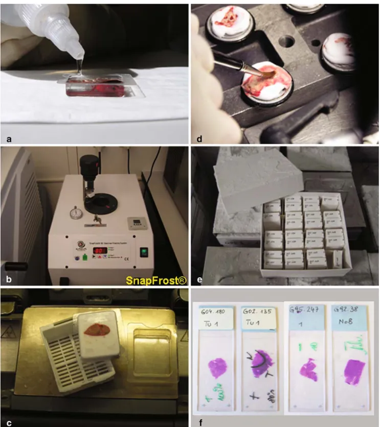
Embedding tissues in a frozen tissue matrix, such as OCT compound (Tissue Tek®), is required for reviewing properly HE sections from tumors. We used plastic molds in which tissue pieces of a maximal volume of 20×15×5 mm can be placed and embedded in OCT and which fit exactly in cryocassettes. Freezing tissues in plastic molds results in regular-shaped tissue blocks (Fig.1c). To generate frozen sections, the OCT tissue block can easily be fixed on a chuck in a cryostat by gently pressing against the base of the plastic mold. This procedure has been used for both rapid section and tissue banking. For the latter method, it is important to prevent the cut tissue from desiccation. As OCT protects tissues from the freeze-burn effect of liquid nitrogen, we re-sealed transparently the tissue surface with OCT diluted 1:1 with aqua bidest using a brush (Fig. 1d). The tissue block was then transferred to a mechanically labeled cryocassette and stored in a cryobox at −80°C for arranging concisely archived frozen tissue until reuse (Fig. 1e).
Knowing the histological composition and the quality of stored tissue is a basic prerequisite for tumor tissue banking (Fig.1f). Morphological features, such as tumor area, non-neoplastic area, and percentage of tumor cells within the tumor area, are directly documented on the glass slide with the HE section. This allows tracking precisely those tumor tissue specimens suited for the molecular method required for future cancer research projects. For analyzing reliably gene mutations, gene or protein expression patterns in tumors, tissue samples used for extractions should prefer-ably not or only be low contaminated with non-neoplastic
cells. It is of note, however, that RNA extracted from tumors with more than 50% tumor cells is apparently still sufficient for yielding a gene expression signature as predictor of survival in breast cancer [10]. Very
heteroge-neous tumors, in which the cell population of interest constitutes only a small fraction, can still be used for in situ analyses, immunostainings, laser micro-dissection, or PCR-based microbial DNA/RNA detection.
Fig. 1 Tissue processing for rapid section (a–c) and tumor tissue
banking (a–f). Tumor tissue in plastic mold embedded with OCT (a).
Tissue holder with cryocassette containing tissue prepared for freezing
at −80°C in isopentane using the SnapFrost® (b). Resulting OCT
tissue block (c). Resealing of the cut tissue surface with diluted OCT (d). Tissue storage (e). Quality control of tumor tissue for biobanking (f). +/− Good/bad morphology, T tumor, N normal, Necr necrosis, % tumor cells
According to the recommendations of the European Human Frozen Tumour Tissue Bank (TuBaFrost) [5] and the guidelines for the collection, handling, and storage of specimens from the Breast International Group (BIG) and National Cancer Institute (NCI) Cooperative Group breast cancer clinical trials (http://ctep.cancer.gov/forms/guidelines_ fresh_tissue.pdf), isopentane is the medium of choice for snap freezing tissues as it is a very efficient cryoconductor and in comparison to liquid nitrogen, causes fewer cryo-artifacts. In addition, OCT is an option for tumor biobanking as it gives
high-quality results for histology study, preserves morpholo-gy by protecting tissue from the lyophilization effect of liquid nitrogen, and minimizes biological cross contamination. However, there is hardly any literature data that described the possible chemical reactions of isopentane and OCT with cellular structures and its impact on molecular assays. Using a modified protocol for immunostaining frozen tissue sections and Western blot, we observed Ki-67 andβ-actin expression patterns that were detectable in all tumor types and virtually identical between the differently treated tissue pairs. By
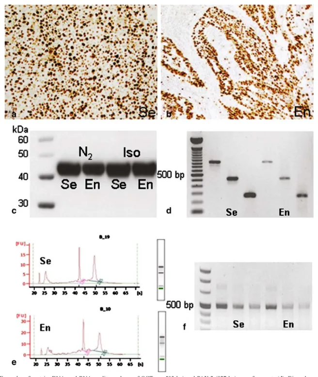
Fig. 2 Examples of HE stained sections of a seminoma (a–c) and an endometrial carcinoma (d–f) (magnifications ×10 and ×40). Tissue frozen
comparing the results obtained with liquid nitrogen, we conclude that IHC and Western results were not influenced negatively, neither by isopentane nor by OCT.
Previous studies suggested that the presence of OCT in DNA extracts can inhibit amplification by polymerase chain reactions [9]. Using a column-based system, which
highly purifies DNA from any residual components, we were able to amplify DNA fragments ranging from 267 to 927 bp in all tumors. This strongly suggests that a successful amplification of larger PCR products mainly depends on the protocol used for extracting DNA from OCT embedded tumors. It is also of note that there were no
Table 1 The impact of freezing medium on tumor morphology, RNA, DNA, and protein quality
Tumor type Medium Morphologya RNA
(μg/μl)b
260/ 280 nm
rRNA ratio RIN DNA
(μg/μl)b
Protein
(μg/μl)b
Renal cell carcinoma Isopentane +++ 1.43 2.1 1.5 8.4 0.71 4.32
Liquid nitrogen ++ 1.01 2.08 1.7 8.5 0.56 4.0
CO2 + nd nd nd nd nd nd
Breast carcinoma Isopentane ++ 0.97 2.07 1.2 8.1 0.34 5.4
Liquid nitrogen ++ 8.2 2.06 1.3 8.3 0.42 4.2
CO2 + nd nd nd nd nd nd
Lung adenocarcinoma Isopentane ++ 1.09 2.05 1.2 7.9 0.67 3.8
Liquid nitrogen ++ 1.31 2.08 1.1 7.3 0.59 4.5
CO2 + nd nd nd nd nd nd
Endometrial carcinoma Isopentane +++ 2.46 2.11 1.6 7.4 1.56 5.6
Liquid nitrogen ++ 2.18 2.12 1.7 7.6 1.72 6.8
CO2 + nd nd nd nd nd nd
Stomach carcinoma Isopentane ++ 1.55 2.09 1.4 7.9 0.89 3.9
Liquid nitrogen ++ 2.48 2.1 1.7 7.5 1.47 5.8
CO2 + nd nd nd nd nd nd
Lung adenocarcinoma Isopentane ++ 1.28 2.1 1.4 7.5 0.36 4.1
Liquid nitrogen ++ 0.84 2.07 1.1 7.9 0.29 3.5
CO2 + nd nd nd nd nd nd
Seminoma Isopentane ++ 0.78 2.01 1.3 6.4 0.14 6.0
Liquid nitrogen ++ 0.91 2.04 1.3 6.3 0.16 6.4
CO2 + nd nd nd nd nd nd
Ovarian carcinoma Isopentane ++ 0.81 2.04 1.8 8.3 0.32 3.8
Liquid nitrogen ++ 0.77 2.08 1.6 8.2 0.4 3.6
CO2 + nd nd nd nd nd nd
Ovarian carcinoma Isopentane ++ 1.37 2.09 1.0 7.2 0.96 4.9
Liquid nitrogen ++ 1.35 2.11 1.0 7.3 0.78 4.5
CO2 + nd nd nd nd nd nd
Breast carcinoma Isopentane ++ 0.88 2.11 1.4 8.8 0.56 2.2
Liquid nitrogen ++ 1.23 2.08 1.3 8.5 0.72 3.0
CO2 + nd nd nd nd nd nd
Lung adenocarcinoma Isopentane +++ 0.92 2.07 1.3 7.9 0.7 4.8
Liquid nitrogen ++ 0.57 2.02 1.4 7.9 0.46 4.2
CO2 + nd nd nd nd nd nd
nd Not done a
Evaluation of morphology: + problematic; ++, +++ well evaluable; +++ morphology better than ++ b
Total volume: 50μl
Table 2 Mean values ± standard deviations of RNA, DNA, and protein extracted from tissues frozen in isopentane and liquid nitrogen
Medium RNA (μg/μl) 260/280 nm rRNA ratio RIN DNA (μg/μl) Protein (μg/μl)
Isopentane 1.18+0.54 2.08+0.03 1.41+0.22 7.89+0.47 0.66+0.37 4.44+1.01
Liquid nitrogen 1.15+0.65 2.08+0.03 1.38+0.25 7.85+0.45 0.69+0.46 4.59+1.17
p value ns ns ns ns ns ns
significant differences between the freezing media, tumor types, and DNA yields (Table1).
Although RNA seems to be stable in non-fixed surgical specimens for up to 16 h [4], it is widely accepted that comprehensive gene expression profiling studies rely on RNA of high quality. To test the integrity of the RNA of our tissue samples, snap freezing was done as
recommen-ded [5] within 30 min after surgery. As shown in Table1, RNA integrity numbers (RIN) were comparable between the differently treated tumor pairs being highest for one breast carcinoma (8.8) and lowest for one seminoma (6.3). RIN, ribosomal RNA ratios, optical density, RNA yields, and RT-PCR results showed that intact RNA was obtained from all tumor types (Table1, Fig.3e,f) embedded in OCT.
Fig. 3 Examples of protein, DNA, and RNA quality analyses of OCT embedded tumors frozen in isopentane. Ki-67 stained tissue sections (a,
b). Western blot showingβ-actin expression in matched tumors frozen in
liquid nitrogen or isopentane (c). DNA amplification of VHL (267 and
503 bp) and PAX-2 (927 bp) gene fragments (d). Bioanalyzer scans of
extracted total RNA (e). RT-PCR of β-actin mRNA (485 bp) with
diluted cDNA (1:50, 1:250, and 1:500; f). Se Seminoma, En endometrial
Long-term storage of tissue samples is one of the most challenging issues for tissue banking. As our protocol was first established in January 2006 at our institute, we currently do not know whether the integrity of cellular and molecular structures of our tissues becomes compro-mised after storage for a longer period of time. By following strictly the guidelines of TuBaFrost [5], BIG, and NCI (http://ctep.cancer.gov/forms/guidelines_fresh_ tissue.pdf) for tissue banking, the procedure described here should ensure the quality of tissue samples. In fact, re-analysis of the tumor tissues after 1 year of storage at−80° C demonstrated clearly that the high quality of morphology, DNA, and RNA remained unchanged (data not shown).
Safety reasons also strongly argue for using a system like the SnapFrost®. Carbon dioxide originating from liquid carbonic acid stored in a cylinder is often used for rapid section. Depending on the type of disease, treating tissues using this method may cause formation of harmful aerosol, which may be infectious when inhaled. For tissue banking, liquid nitrogen is commonly filled in containers that have to be opened many times for freezing tissues, which causes strong evaporation. In contrast, isopentane is a non-degass-ing fluid which is kept in a closable freeznon-degass-ing chamber. This considerably lowers the risks for cold burns and infections.
We developed an isopentane-based protocol that may help to standardize snap-freezing tissues for both routine rapid section and tumor biobanking in pathology institutes. This procedure is time saving, simplifies significantly tissue handling and processing, and preserves excellently the integrity of cellular and molecular structures in different tumor types, thus satisfying the demands of pathologists, scientists, technicians, and tissue bankers.
Acknowledgments We thank Dr. Kirsten Struckmann and Silvia
Behnke for their excellent technical support. This study was supported by UBS AG (made possible by an anonymous donor), Zurich Cancer League, and the Foundation Biobank-Suisse. We declare that the experiments comply with the current laws of the Kanton Zürich.
Conflict of interest statement We declare that we have no conflict
of interest.
References
1. Grizzle WE, Aamodt R, Clausen K, LiVolsi V, Pretlow TG, Qualman S (1998) Providing human tissues for research: how to
establish a program. Arch Pathol Lab Med 122:1065–1076
2. Loken SD, Demetrick DJ (2005) A novel method for freezing and
storing research tissue bank specimens. Hum Pathol 36:977–980
3. McElroy HH, Shih MS, Parfitt AM (1993) Producing frozen sections of calcified bone. Biotech Histochem 68:50–55 4. Micke P, Ohshima M, Tahmasebpoor S, Ren ZP, Ostman A, Ponten
F, Botling J (2006) Biobanking of fresh frozen tissue: RNA is stable in nonfixed surgical specimens. Lab Invest 86:202–211
5. Morente MM, Mager R, Alonso S, Pezzella F, Spatz A, Knox K, Kerr D, Dinjens WN, Oosterhuis JW, Lam KH, Oomen MH, van Damme B, van de Vijver M, van Boven H, Kerjaschki D, Pammer J, Lopez-Guerrero JA, Llombart Bosch A, Carbone A, Gloghini A, Teodorovic I, Isabelle M, Passioukov A, Lejeune S, Therasse P, van Veen EB, Ratcliffe C, Riegman PH (2006) TuBaFrost 2: standardising tissue collection and quality control procedures for a European virtual frozen tissue bank network. Eur J Cancer
42:2684–2691
6. Naber SP (1996) Continuing role of a frozen-tissue bank in
molecular pathology. Diagn Mol Pathol 5:253–259
7. Schraml P, Struckmann K, Hatz F, Sonnet S, Kully C, Gasser T, Sauter G, Mihatsch MJ, Moch H (2002) VHL mutations and their correlation with tumour cell proliferation, microvessel density, and patient prognosis in clear cell renal cell carcinoma. J Pathol 196:186–193
8. Silva EG, Kraemer BB (1987) Intraoperative pathologic diagnosis. Frozen section and other techniques. Williams & Wilkins, Baltimore
9. Turbett GR, Sellner LN (1997) The use of optimal cutting temperature compound can inhibit amplification by polymerase
chain reaction. Diagn Mol Pathol 6:298–303
10. van de Vijver MJ, He YD, van’t Veer LJ, Dai H, Hart AA, Voskuil
DW, Schreiber GJ, Peterse JL, Roberts C, Marton MJ, Parrish M, Atsma D, Witteveen A, Glas A, Delahaye L, van der Velde T, Bartelink H, Rodenhuis S, Rutgers ET, Friend SH, Bernards R (2002) A gene-expression signature as a predictor of survival in
breast cancer. N Engl J Med 347:1999–2009
11. Vonsattel JP, Aizawa H, Ge P, DiFiglia M, McKee AC, MacDonald M, Gusella JF, Landwehrmeyer GB, Bird ED, Richardson EP Jr. et al (1995) An improved approach to prepare human brains for research. J Neuropathol Exp Neurol 54:42–56
12. Went PT, Dirnhofer S, Bundi M, Mirlacher M, Schraml P, Mangialaio S, Dimitrijevic S, Kononen J, Lugli A, Simon R, Sauter G (2004) Prevalence of KIT expression in human tumors. J