1
Supplemental data
In vitro antitubercular activity of extract and constituents from the stem bark of Disthemonanthus benthamianus Baill. (Ceasalpinaceae)
Jean Noel Evina1, Dominique Serge Ngono Bikobo1,5*, Auguste Abouem A Zintchem1,2, Norbert Mbabi Nyemeck II1,3, Esther Del Florence Moni Ndedi4, Patrick Hervé Betote Diboué4, Maximilienne Ascension Nyegue4, Alex de Théodore Atchadé1, Dieudonné Emmanuel Pegnyemb1 Ulrich Koert3 and Christian Bochet5
1
Department of Organic Chemistry, Faculty of Science, University of Yaoundé I, P.O Box 812, Yaoundé, Cameroon;
2
Department of Chemistry, Higher Training College, University of Yaoundé I, P.O Box 47, Yaoundé, Cameroon;
3 Philipps-Universität Marburg, Faculty of Chemistry, Hans-Meerwein-Strasse, D-35032
Marburg, Germany;
4 Department of Microbiology Faculty of Science, University of Yaoundé I, P.O Box 812,
Yaoundé, Cameroon;
5
Department Chemie, Universität Fribourg, CH du Musée 9, 1700 Fribourg, Switzerland;
*Corresponding author: Tel: +237 675 05 54 68; Fax: +237 242 22 18 73 E-mail:
[email protected] (D. S. Ngono Bikobo).
Abstract
A new C-glycosylflavone, apigenin 7-methyl ether 6-C-[ β-xylopyranosyl-(1→3)-β-glucopyranoside] named distemonanthoside (1) was isolated from the stem bark of
Distemonanthus benthamianus, along with six known compounds, sitosterol 3-O-
β-D-glucopyranoside (2), 4-methoxygallic acid (3), syringic acid (4), quercetin (5), 6 ″-O-acetylvitexin (6), quercetin 3-O-β-D-glucopyranoside (7). The structures of those compounds and others were determined through spectral analyses. Compounds 1, 2, 3 and 5 were tested against a clinical isolate strain of Mycobacterium tuberculosis AC 45; they exhibited good to moderate antitubercular activities with MIC values ranged from 31.25 to 125 µg/ml.
Keywords: Ceasalpinaceae; Distemonanthus benthamianus; flavonoids; distemonanthoside; antitubercular activity; phenolic acid
2 Fig.S1. UV spectrum of compound 1
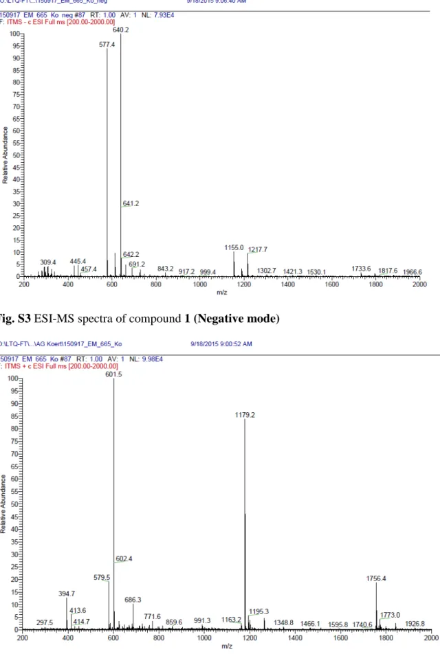
3 Fig. S3 ESI-MS spectra of compound 1 (Negative mode)
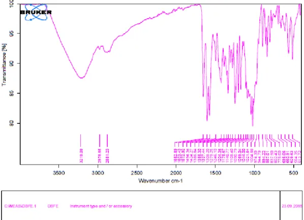
4 Fig. S5 IR spectrum of compound 1
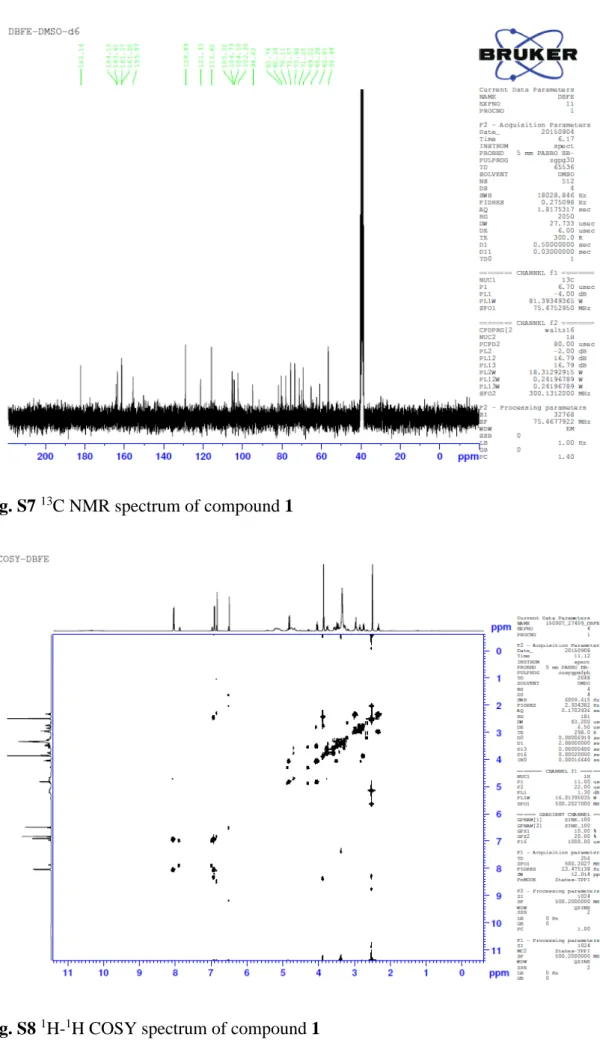
5 Fig. S7 13C NMR spectrum of compound 1
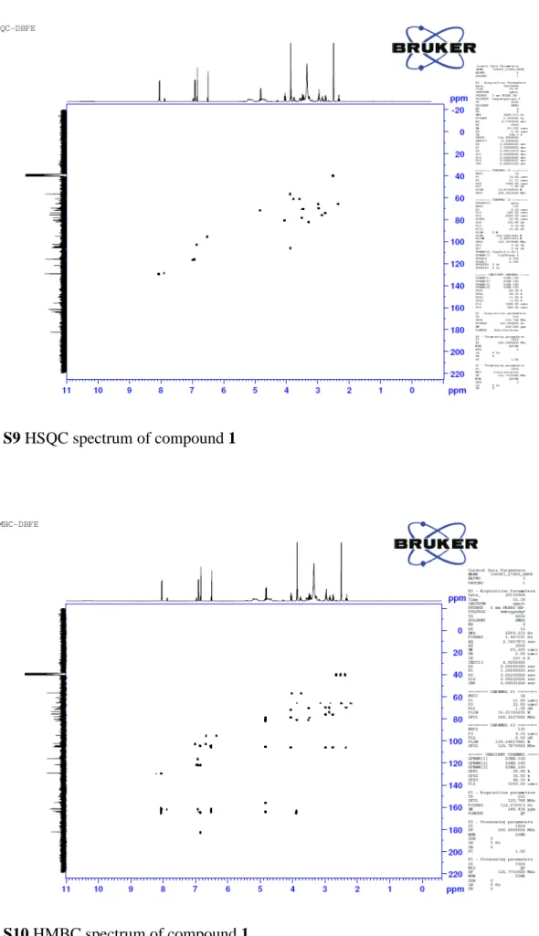
6 Fig. S9 HSQC spectrum of compound 1
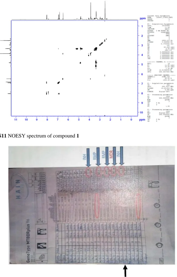
7 Fig. S11 NOESY spectrum of compound 1