Pulmonary Toxoplasmosis, a Rare but Severe
Manifestation of a Common Opportunistic
Infection in Late HIV Presenters: Report of
Two Cases
H. Kovari, C. Ebnöther, A. Schweiger, N. Berther, H. Kuster, H.F. Günthard
AbstractPulmonary toxoplasmosis is rare, particularly in the era of highly active antiretroviral therapy (HAART). Here, we describe two severe cases in patients not known to be HIV-infected. In both patients, early diagnosis and therapy led to a favourable outcome. Pulmonary toxoplasmosis should be considered in the differential diagnosis in potentially HIV-infected patients with respiratory symptoms.
Infection 2010; 38: 141–144 DOI 10.1007/s15010-009-9367-5
Introduction
Highly active antiretroviral therapy (HAART) and anti-biotic prophylaxis against opportunistic infections have resulted in a dramatic decrease of morbidity and mortality [1–3]. However, patients infected with HIV – also in developed countries – may still present with severe opportunistic infections. Most of these events are among those newly diagnosed with HIV at a late stage of disease [4–6].
Toxoplasmosis is the most common parasitic oppor-tunistic infection of the central nervous system in AIDS patients, who are not on appropriate prophylaxis [7]. Extracerebral sites may be involved with or without toxoplasmic encephalitis [8–11]. HIV-related pulmonary toxoplasmosis was first described in 1984. It was respon-sible for less than 1% of the pulmonary complications of HIV infection [12]. In a French national survey, the prevalence of extracerebral toxoplasmosis among HIV-infected patients in the pre-HAART era was 1.5–2%. Of 199 patients with extracerebral toxoplasmosis, the lungs were involved in 26% and were the second most common extracerebral site after involvement of the eyes [9]. Here, we present two cases with pulmonary toxoplasmosis.
Case 1
A 26-year-old Brazilian heterosexual patient presented to the emergency department because of a three-day history of malaise, rapid progressive dyspnoea, chest pain and fever, but no cough. In addition, he complained about diarrhoea and weight loss of
4 kg during the past four months. His medical history was unremarkable. He was not known to be HIV-positive. On examination, the patient was alert and oriented, but in respira-tory distress. His temperature was 37.8 °C, blood pressure 110/ 45 mmHg, pulse 127 min–1; the respirations were 50–60 breaths per minute, with an oxygen saturation of 50% without supple-mental oxygen. Chest examination did not reveal rhonchi or crackles. The axillary and inguinal lymph nodes were enlarged; the remainder of the examination was normal. A chest radio-graph showed extensive diffuse bilateral alveolar infiltrates (Figure 1a). An electrocardiogram was unremarkable. Labora-tory test results included a mild normocytic anaemia with a haemoglobin level of 11.1 g/dl, a lymphocytopaenia with 410 cells/ll, C-reactive protein (CRP) of 175 mg/l (<5), an ele-vated lactate dehydrogenase (LDH) of 2,640 U/l (240–420) and an aspartate aminotransferase (AST) of 130 U/l (10–50). A blood gas analysis with room air revealed a partial pressure of O2
(pO2) of 5.1 kPa, a partial pressure of CO2(pCO2) of 3.5 kPa
and a pH of 7.47. An HIV test turned out to be positive. The patient was transferred to the intensive care unit. Besides cef-triaxone and clarithromycin, high-dose trimethoprim-sulpha-methoxazole and prednisone were started. During the next day, his condition worsened, with increasing dyspnoea and respiratory exhaustion and mechanical ventilation was required. His CD4 cell count was 21 cells/ll and an HIV-1 viral load was 348,000 copies/ml. A bronchoalveolar lavage (BAL) was per-formed. In Giemsa and indirect immunofluorescence stains, toxoplasma tachyzoites were identified (Figure 1b, c). There was no evidence of Pneumocystis jirovecii and acid-fast bacilli. Toxoplasma serology turned out to be highly positive with IgG > 549 IU/l and negative IgM antibodies. Pulmonary toxo-plasmosis with acute respiratory distress syndrome (ARDS) in a patient with advanced HIV infection was diagnosed. Therapy was changed to sulphadiazine and pyrimethamine plus folinic acid. After three days, the patient was extubated and he subsequently
H. Kovari (corresponding author), N. Berther, H. Kuster, H.F. Günthard Division of Infectious Diseases and Hospital Epidemiology, University Hospital Zurich, University of Zurich, 8091 Zurich, Switzerland, e-mail: helen.kovari@usz.ch
C. Ebnöther
Stadtspital Triemli, Zurich, Switzerland A. Schweiger
Institute of Parasitology, University of Zurich, Zurich, Switzerland Received: October 6, 2009 Æ Revision accepted: December 7, 2009 Published online: March 29, 2010
Infection
Case Reportrecovered rapidly. He was discharged from the hospital four days later.
Case 2
A 38-year-old immigrant from Nigeria was admitted with low-grade temperature, productive cough lasting for 14 days, pro-gressive headache for a few days and agpro-gressive behaviour on the day of admission. His medical history was unremarkable.
On admission, his body temperature was 36.7 °C, blood pressure 110/77 mmHg, heart rate 60 min–1, frequency of respi-ration 14 min–1and pulmonary examination revealed generalised wheezing. The patient was confused and somnolent, with a fluctuating Glasgow Coma Scale (GCS) score between 5 and 13. He had ataxia and dysarthria, but no meningeal symptoms.
Significant laboratory values were as follows: microcytic anaemia with a haemoglobin level of 10.3 g/dl, lymphopaenia with 440 cells/ll, CRP 15 mg/l (<5), LDH 636 U/l (150–420), AST 85 U/l (<50) and alanine aminotransferase (ALT) 80 U/l (<50). HIV serology turned out to be positive. The CD4 cell count was 5 cells/ll and HIV1-RNA 1,620,000 copies/ml. Assays for toxo-plasma antibodies were positive for IgG (not quantified) and negative for IgM. An electrocardiogram was normal. Chest X-ray revealed bilateral, diffuse patchy infiltrates and increased interstitial striation. Chest computed tomography (CT) scan also showed bilateral, patchy infiltrates (Figure 1d) and in a cranial CT scan, edema and midline shift was found. One day later, cranial magnetic resonance imaging (MRI) revealed multiple abscesses. Central nervous system (CNS) lymphoma was ruled
142
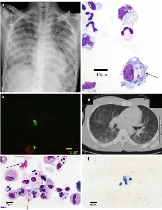
Infection 38 Æ 2010 Æ No. 2Figure 1. Chest radiograph and bronchoalveolar fluid of case 1: diffuse bilateral alveolar infiltrates (a), Giemsa staining (b) and indirect
immunofluorescence staining (c) with toxoplasma tachyzoites (black arrow). Chest computed tomography (CT) scan and bronchoalveolar fluid of case 2: extensive bilateral, patchy infiltrates (d), Giemsa staining (e) revealing toxoplasma tachyzoites (black arrow) and Pneumocystis jirovecii (red arrow) and toluidine blue O staining with P. jirovecii (f).
out by positron emission tomography (PET) scan. A BAL was performed: the Giemsa-stained specimen revealed tachyzoites of Toxoplasma gondii, together with cysts of P. jirovecii (Figure 1e) that were confirmed by toluidine blue O staining (Figure 1f) and immunofluorescence staining (not shown). Pulmonary and cerebral toxoplasmosis with contemporaneous pulmonary pneumocystis infection was diagnosed. Treatment with sulph-adiazine, pyrimethamine, folinic acid and atovaquone was started. The former drug was prescribed instead of trimethoprim-sulphamethoxazole to minimise sulphonamide toxicity. In the following several days, the patient’s condition progressively im-proved. After 17 days, he was discharged from the hospital.
Discussion
Here, we present two cases of pulmonary toxoplasmosis. This is a very rare manifestation of a common opportu-nistic infection in AIDS patients. Strikingly in both pa-tients, the HIV status was unknown when they presented to the emergency room.
The first case illustrates that pulmonary toxoplasmo-sis can rapidly progress to a life-threatening state. Toxo-plasma pneumonitis is also known to cause severe systemic infections with ARDS [13].
The second patient presented with toxoplasmosis manifesting in two locations and, in addition, contempo-raneously suffered from a second opportunistic infection, a P. jirovecii pneumonia. Toxoplasmosis involving several locations have been described [8, 10, 11]. In a French case series, 11 of 64 patients (17%) presented with toxoplas-mosis including the lungs and the brain [14]. In addition, there are rare published reports of patients with concur-rent pulmonary toxoplasmosis and pneumocystis infection [8, 14]. However, these reports originated from the pre-HAART era, when, in general, the incidence of oppor-tunistic infections was higher by magnitudes in developed countries when compared to today [15].
Usually, T. gondii pneumonia manifests with fever, dyspnoea and non-productive cough. The most common finding on chest radiographs is bilateral diffuse interstitial infiltrates [8, 14, 16]. The clinical and radiological appearance may be indistinguishable from the far more common P. jirovecii pneumonia. As compared with pneumocystis pneumonia, the clinical onset and the evo-lution of symptoms are more rapid in toxoplasma pneu-monia [8]. A blood level of LDH > 600 U/l is more likely to be associated with toxoplasmosis than pneumocystis pneumonia [17]. A toxoplasma antibody titre of >150 U/ ml is an important predictor of cerebral and extracerebral toxoplasmosis [18]. Finally, pulmonary toxoplasmosis oc-curs mainly in patients with severe immunodeficiency, with a CD4 cell count of 40 ± 75 cells/ll [14].
Mortality in immunosuppressed patients with pul-monary toxoplasmosis is high: in the French survey, 37% of HIV-infected patients with toxoplasma pneumonia died [14] and in a meta-analysis, mortality among immunosuppressed persons with pulmonary toxoplas-mosis was 40% [16]. As toxoplasma pneumonitis is
clinically difficult do diagnose and is associated with a considerable mortality, it is necessary to force the diag-nosis by the microbiological examination of respiratory samples. Especially in patients with severe clinical pic-tures or not responding to therapy, it is judicious to perform a BAL with cytological examination of BAL fluid, using appropriate special stains to detect toxo-plasma organisms. In both of our cases, early diagnosis led to a favourable outcome.
In resource-poor countries, for example, in Africa with a toxoplasmosis seroprevalence of up to 78% [19] and more HIV-infected patients with very low CD4 cell counts, the prevalence of pulmonary toxoplasmosis might be higher than in industrialised countries. However, in these countries, diagnostic possibilities are often very limited and in the absence of respiratory samples, clini-cians need to consider an empiric toxoplasmal therapy, particularly in the presence of risk factors (see above) and non-responsiveness to therapy.
In summary, pulmonary toxoplasmosis is an infre-quent but severe opportunistic infection in HIV-positive patients that still occurs in the era of HAART. The two cases emphasise the importance of considering toxoplas-mosis early in the differential diagnosis in (potentially) HIV-infected patients presenting with respiratory symp-toms and bilateral pulmonary infiltrates. Early diagnosis and initiation of specific therapy is essential.
Conflict of interest statement. None declared by all authors.
References
1. Antiretroviral Therapy Cohort Collaboration: Life expectancy of individuals on combination antiretroviral therapy in high-income countries: a collaborative analysis of 14 cohort studies. Lancet 2008; 372: 293–299.
2. Egger M, Hirschel B, Francioli P, Sudre P, Wirz M, Flepp M, Rickenbach M, Malinverni R, Vernazza P, Battegay M: Impact of new antiretroviral combination therapies in HIV infected patients in Switzerland: prospective multicentre study. Swiss HIV Cohort Study. BMJ 1997; 315: 1194–1199.
3. Palella FJ Jr, Delaney KM, Moorman AC, Loveless MO, Fuhrer J, Satten GA, Aschman DJ, Holmberg SD: Declining morbidity and mortality among patients with advanced human immunodefi-ciency virus infection. HIV Outpatient Study Investigators. N Engl J Med 1998; 338: 853–860.
4. Sabin CA, Smith CJ, Gumley H, Murphy G, Lampe FC, Phillips AN, Prinz B, Youle M, Johnson MA: Late presenters in the era of highly active antiretroviral therapy: uptake of and responses to antiretroviral therapy. AIDS 2004; 18: 2145–2151.
5. Battegay M, Fluckiger U, Hirschel B, Furrer H: Late presentation of HIV-infected individuals. Antivir Ther 2007; 12: 841–851. 6. Girardi E, Sabin CA, Monforte AD: Late diagnosis of HIV
infec-tion: epidemiological features, consequences and strategies to encourage earlier testing. J Acquir Immune Defic Syndr 2007; 46 Suppl 1: S3–S8.
Infection 38 Æ 2010 Æ No. 2
143
7. Porter SB, Sande MA: Toxoplasmosis of the central nervous system in the acquired immunodeficiency syndrome. N Engl J Med 1992; 327: 1643–1648.
8. Oksenhendler E, Cadranel J, Sarfati C, Katlama C, Datry A, Marche C, Wolf M, Roux P, Derouin F, Clauvel JP: Toxoplasma gondii pneumonia in patients with the acquired immunodefi-ciency syndrome. Am J Med 1990; 88: 18N–21N.
9. Rabaud C, May T, Amiel C, Katlama C, Leport C, Ambroise-Thomas P, Canton P: Extracerebral toxoplasmosis in patients infected with HIV. A French National Survey. Medicine (Baltimore) 1994; 73: 306–314.
10. Eza DE, Lucas SB: Fulminant toxoplasmosis causing fatal pneumonitis and myocarditis. HIV Med 2006; 7: 415–420. 11. Signorini L, Gulletta M, Coppini D, Donzelli C, Stellini R, Manca
N, Carosi G, Matteelli A: Fatal disseminated toxoplasmosis during primary HIV infection. Curr HIV Res 2007; 5: 273–274. 12. Murray JF, Felton CP, Garay SM, Gottlieb MS, Hopewell PC,
Stover DE, Teirstein AS: Pulmonary complications of the acquired immunodeficiency syndrome. Report of a National Heart, Lung, and Blood Institute workshop. N Engl J Med 1984; 310: 1682–1688.
13. Gandhi S, Lyubsky S, Jimenez-Lucho V: Adult respiratory distress syndrome associated with disseminated toxoplasmosis. Clin Infect Dis 1994; 19: 169–171.
14. Rabaud C, May T, Lucet JC, Leport C, Ambroise-Thomas P, Canton P: Pulmonary toxoplasmosis in patients infected with
human immunodeficiency virus: a French National Survey. Clin Infect Dis 1996; 23: 1249–1254.
15. Sterne JA, Hernán MA, Ledergerber B, Tilling K, Weber R, Sendi P, Rickenbach M, Robins JM, Egger M, Swiss HIV Cohort Study: Long-term effectiveness of potent antiretroviral therapy in preventing AIDS and death: a prospective cohort study. Lancet 2005; 366: 378–384.
16. Pomeroy C, Filice GA: Pulmonary toxoplasmosis: a review. Clin Infect Dis 1992; 14: 863–870.
17. Pugin J, Vanhems P, Hirschel B, Chave JP, Flepp M: Extreme elevations of serum lactic dehydrogenase differentiating pul-monary toxoplasmosis from pneumocystis pneumonia. N Engl J Med 1992; 326: 1226.
18. Belanger F, Derouin F, Grangeot-Keros L, Meyer L: Incidence and risk factors of toxoplasmosis in a cohort of human immuno-deficiency virus-infected patients: 1988–1995. HEMOCO and SEROCO Study Groups. Clin Infect Dis 1999; 28: 575–581. 19. Lucas SB, Hounnou A, Peacock C, Beaumel A, Djomand G,
N’Gbichi JM, Yeboue K, Hondé M, Diomande M, Giordano C, Doorly R, Brattegaard K, Kestens L, Smithwick R, Kadio A, Ezani N, Yapi A, De Cock KM: The mortality and pathology of HIV infection in a west African city. AIDS 1993; 7: 1569–1579.