Biological Functions of Ribonuclease 1 and Angiogenin
by
Emily R. Garnett
B.S. Biochemistry, Cell, and Molecular Biology, B.A. Chemistry Drake University, 2012
Submitted to the Department of Chemistry
in Partial Fulfillment of the Requirements for the Degree of DOCTOR OF PHILOSOPHY
IN BIOLOGICAL CHEMISTRY
at the
Massachusetts Institute of Technology June 2018
2018 Massachusetts Institute of Technology. All rights reserved.
Signature of Author: Department of Chemistry May 18, 2018
Signature redacted
Certified by: Ron d Raines Firmenic Profes of Chvmistry Thesis SupervisorSignature redacted
Accepted by:Robert W. Field Haslam and Dewey Professor of Chemistry Chairman, Departmental Committee on Graduate Students
MASSACHUSETTS INSTITUTE OF TECHNOLOGY
This doctoral thesis has been examined by a committee of professors from the Department of Chemistry as follows:
Signature redacted
Elizabeth Nolan Associate Professor of Chemistry Thesis Committee Chair
Signature redacted
Rormd Raines Firm\ich Profes r of Chemistry Thesis SupervisorSignature redacted
David Bartel Professor of Biology Thesis Committee MemberBiological Functions of Ribonuclease 1 and Angiogenin
by
Emily R. Garnett
Submitted to the Department of Chemistry on May 18, 2018 in Partial Fulfillment of the Requirements
for the Degree of Doctor of Philosophy in Biological Chemistry
Abstract
Pancreatic-type ribonucleases (ptRNases) are a large family of vertebrate-specific secretory endoribonucleases. They catalyze degradation of many RNA substrates, mediating a variety of biological functions. The homology shared by ptRNases has enabled extensive biochemical characterization and evolutionary study of these enzymes, yet understanding of their biological roles is still incomplete. The goal of this thesis is to identify novel physiological functions for two ptRNases, RNase 1 and angiogenin, through characterization of murine model systems. In Chapter 1, I introduce the ptRNase superfamily and highlight evidence of biological function for RNase 1 and angiogenin that has motivated and informed our study of these enzymes.
Extracellular RNA drives blood coagulation, which is preventable by administration of RNase A. In Chapter 2, I demonstrate that loss of RNase 1, a nonspecific and extracellular ptRNase similar to RNase A, results in the potentiation of blood coagulation by activation of coagulation factors in mice.
Angiogenin is a ptRNase with unique angiogenic activity and suggested biological function in cancer and amyotrophic lateral sclerosis, as well as in cellular growth and quiescence. My studies demonstrate a fundamental role for angiogenin. In Chapter 3, I find that this enzyme is essential for the development of mice, with heterozygosity for angiogenin resulting in impaired vascularization of the placenta and reduced survival of offspring.
Biological study of ptRNases is hampered by the high degree of conservation of the family, which engenders antibody nonspecificity. In Chapter 4, I describe efforts to generate novel specific anti-ptRNase antibodies by producing tagged ptRNases for use in an antibody phage-display workflow. Finally, Chapter 5 outlines future directions for the study of RNase 1 and angiogenin.
Taken together, this thesis reveals a more complete picture of the physiological niches of these two enzymes, confirming some previously suspected roles, ascribing new ones, and providing groundwork for future characterization of the biology of these and other ptRNases.
Thesis Supervisor: Ronald T. Raines Title: Firmenich Professor of Chemistry
Acknowledgments
Many, many people have encouraged me and supported me on my journey through graduate school.
First and foremost, I have to thank my parents for always providing opportunities for me to learn, and for unconditional love and support. They instilled in me a love of learning and exploration, and have always pushed me to be independent and to go further - which is what drove me to pursue graduate education in the first place. I also have to thank my entire family for their constant encouragement and infectious attitude - I have truly been lucky to grow up surrounded by support, positivity, and a sense of delight in what the world has to offer.
I have to thank my undergraduate advisors, Julia Moffitt and Marc Busch, as well as Amanda
Jepson, for being excellent coaches as I was first learning to do research. It was under their guidance that I really learned how "science" is done in academic laboratories, and the experience was invaluable in orienting myself once I began working independently. I also have to thank Mark Vitha and Vibs Petersen for pushing my boundaries, driving me to think critically, and encouraging me to see myself as a scientist.
I have to thank the people who got me on my feet as a student of Molecular and Cellular
Pharmacology (MCP) at the University of Wisconsin. Patty Keely and Vincent Cryns got me through the initial hurdles of "how to be a graduate student," and the students of the MCP program (dozens over the five years I spent at UW) gave me a sense of community and a
sounding board. I also have to thank Jon Audhya and Kristin Cooper for all their support as administrators of the MCP program, and the program as a whole for funding me through NIH
T32 GM008688.
I have to thank my graduate advisor, Ron Raines, who has given me complete freedom to pursue
my research in directions totally unfamiliar to our group, and the support I needed to do so. In forging ahead into biological spheres unknown, I was able to develop an independence and confidence in my work that has served me extremely well so far, and will be a valuable asset as I move forward. I also have to thank Ron for making sure our research was never wanting for funding - my work was supported by NIH R01 CA073808.
I have to thank the Raines lab as a whole for creating such a great environment to grow
personally and professionally. Even though my research was outside of the general repertoire of our group, I never felt alone, and I loved the enthusiasm and the thoughtful feedback that came
from all of you. I especially have to thank Kristen Andersen, Joelle Lomax, Chelcie Eller, and Trish Hoang for their guidance as I got started, and for being amazing role models as women scientists.
I have to thank my undergraduate assistants, Conner Feldman and Katharine Tippins, for being
such skilled and dedicated managers of our mouse colony. Moving a mouse project forward without their assistance would have been impossible, and I am proud to have been able to watch their growth as scientists. I also have to thank the animal care staff, veterinarians, and
administrators who helped keep our mice safe and healthy, and taught me how to navigate the regulations associated with performing government-funded research.
I have to thank my collaborator, John Sheehan, for excellent advice and encouragement about
my lab work and my career, as well as for teaching me the intricacies of the blood coagulation cascade from an expert (and physician's) perspective. I owe much of what I understand about this subject to him. I also have to thank Deane Mosher, Pam Westmark, and the Molecular Hematology group at the University of Wisconsin for their instruction and feedback.
I have to thank my former thesis committee members at the University of Wisconsin, Emery Bresnick, Beth Weaver, and Lara Collier, for their excellent advice and their support of me as I pursued fellowships, grants, and postdoctoral appointments.
I have to thank the people who supported me in the move to the Massachusetts Institute of Technology: The Kiessling lab students and postdocs, who moved from Wisconsin to
Massachusetts with us, have become friends and commiserators, the Women in Chemistry and the Buchwald lab made me feel especially welcome, Brian Pretti has been superhuman in helping reassemble our lab, and Jennifer Weisman has been instrumental in getting me oriented as a Chemistry student. I am particularly grateful to Liz Nolan and David Bartel for being such attentive and thoughtful readers of my thesis and acting as my new thesis committee at MIT.
I have to thank a number of people who supported me in my journey towards a fellowship in
Clinical Chemistry. Sarah Hackenmueller introduced me to the field and ultimately helped me edit my postdoctoral applications, and I received encouragement about my career choice by many clinical chemists and fellows-in-training who took the time to chat with me over the last few years. I especially have to thank Sridevi Devaraj and the Clinical Chemistry program at Texas Children's Hospital for giving me the opportunity to continue my training after my time at MIT is finished.
I have to thank Lauren Zasadil and Val Ressler for being incredible friends and roommates
during the last few years of graduate school - you inspired me with your drive, thoughtfulness, generosity, and creativity. I also have to thank Chris Evans for the support, encouragement, and for bringing many new adventures into my life. I have to thank all of these people also for the very specific type of support that comes from knowing what it's like to wrap up a dissertation while far away from loved ones.
Finally, I have to thank the contributors to the freefood and vultures mailing lists, the Graduate Student Council, and the Chemistry Education office for always keeping me apprised of opportunities to find food on campus. I have never eaten so well for so cheaply.
Table of Contents: A bstract ... 3 Acknowledgm ents ... 4 List of figures ... 9 List of tables ... 10 List of equations ... 10 Abbreviations ... 11 Chapter 1 ... 14
Emerging biological functions of pancreatic-type ribonucleases... 14
1. 1: Introduction ... 15
1.2: The pancreatic-type ribonuclease superfam ily ... 15
1.2.1: Characteristic biochem ical features of ptRNases... 16
1.2.2: Known biological functions of the ptRNase superfam ily ... 18
1.3: Ribonuclease 1... 19
1.3.1: Early studies of the biology of RNase 1... 20
1.3.2: Extracellular RNA - key to the function of an extracellular RNase ... 21
1.3.3: N ew biological roles for RNase 1... 22
1.4 Angiogenin ... 26
1.4.1 A ptRNase with unique structural and signaling properties ... 26
1.4.2 Angiogenin-m ediated cell growth ... 28
1.4.3 Angiogenin-m ediated protection from cell stress ... 30
1.5 Connections to vertebrate physiology ... 32
1.6 Prospectus... 35
Chapter 2 ... 36
Loss of ribonuclease 1 produces a hypercoagulable phenotype... 36
2. 1 Abstract ... 37
2.2: Introduction ... 38
2.3: Experim ental procedures... 39
2.3. 1: M aterials...39
2.3.2: Generation of Rnase I knockout m ice ... 40
2.3.3: Quantitative PCR ... 42
2.3.4: Gross phenotypic analysis ... 43
2.3.5: Preparation of plasm a sam ples ... 43
2.3.6: Quantitation of RNA in plasm a ... 43
2.3.7: Ribonucleolytic activity assay ... 44
2.3.8: Ribonuclease zym ogram assay ... 44
2.3.9: Kinetic coagulation assay... 45
2.3.10: ELISA and im m unoblotting ... 46
2.3.11: Throm bin-antithrom bin com plex formation assay ... 47
2.3.12: Tail-vein bleeding tim e assay ... 47
2.3.13: Saphenous vein hem ostasis assay ... 47
2.3.14: FeCl3-induced throm bosis assay ... 48
2.3.15: Factor activity assay... 48
2.3.16: Statistical analyses ... 49
2.4: Results... 50
2.4.3: Rnasel m ice are viable and appear physically norm al ... 54
2.4.4: Loss of Rnasel results in increased plasm a RN A ... 55
2.4.5: Rnasei -plasma clots more quickly than does RnaseK plasma ... 57
2.4.6: Rnase] - mice do not exhibit coagulation abnormalities in vivo... 59
2.4.7: Rnase - plasm a exhibits elevated FXII and FXI activity... 59
2.5: D iscussion ... 61
2.6: Supplem entary inform ation... 65
Chapter 3 ... 67
Angiogenin is necessary for embryonic development in mice... 67
3.1 Abstract ... 68
3.2 Introduction ... 69
3.3 Experim ental procedures... 70
3.3.1: M aterials... 70
3.3.2: Expansion of AngV - m ice and genotyping ... 70
3.3.4: Gross phenotyping analysis... 71
3.3 5: Developm ental viability studies...71
3.3.6: Quantitative PCR ... 72
3.3.7: Ribonuclease zym ogram assay... 73
3.3.8: Im m unoblotting ... 73
3.3.9: Statistical analyses... 74
3.4 Results... 74
3.4.1: Angl] m ice have reduced Ang] gene expression... 74
3.4.2: Angl - plasma exhibits reduced angiogenin expression and activity... 76
3.4.3: Ang ]> m ice are not produced from Ang]1' intercrosses ... 76
3.4.4: Angl' m ice experience partial litter loss ... 77
3.4.5: Ang]"' dams exhibit impaired uterine vascularization and elevated incidence of embryonic loss during pregnancy ... 77
3.5 D iscussion ... 79
3.6: Supplem ental inform ation ... 83
Chapter 4 ... 85
Towards novel anti-secreted ribonuclease antibodies using phage display... 85
4.1 A bstract ... 86
4.2 Introduction ... 87
4.3 Experim ental procedures... 89
4.3.1: M aterials... 89
4.3.2: Design of tagged ribonuclease constructs ... 89
4.3.3: Cloning of tagged ribonucleases... 90
4.3.4: Production of tagged ribonucleases ... 91
4.3.5: Biotinylation of tagged ribonucleases ... 91
4.3.6: Ribonucleolytic activity assays... 92
4.3.7: TEV protease cleavage assays ... 92
4.3.8: A cetylation of RN ase ... 93
4.3.9: Statistical analyses ... 93
4.4 Results... 93
4.4.1: Evolutionarily divergent residues in ptRNases are not present near the amino acid term ini .. 93
4.4.2: Tagged ptRNases express in E. coli, are of expected molecular weight, and retain ribonuclease activity ... 94
4.4.4: m RNase 1 is successfully acetylated... 99
4.5 Discussion ... 100
Chapter 5 ... 103
Conclusions and future directions ... 103
5.1 Conclusions ... 104
5.2: Future directions ... 106
5.2. 1: Roles for RNase 1 in growth ... 106
5.2.2: Roles for RNase 1 in cancer ... 107
5.2.3: Roles for RNase 1 in inflam m ation...108
5.2.4: Fundam ental roles for nonspecific, extracellular ribonucleolytic activity ... 109
5.2.5: Mechanisms of impaired fertility and embryonic development in Ang]' mice...110
5.2.6: Characterization of anti-ptRNase antibodies...111
Appendix 1... 113
Evaluation of metabolic and inflammatory phenotypes in Rnase1-P mice... 113
6.1 Abstract ... 114
6.2 Introduction ... 115
6.3 Experim ental procedures...116
6.3.1: M aterials...116
6.3.2: Body weight and length m easurem ents...116
6.3.3: Food and water consum ption studies ... 117
6.3.4: Urinary protein and glucose excretion studies...117
6.3.5: Fecal lipid content studies ... 117
6.3.6: Plasm a cholesterol m easurem ent ... 118
6.3.7: Visceral adipose tissue m easurem ent...118
6.3.8: Quantitative PCR ... 119
3.3.9: Statistical analyses ... 120
6.4 Results...120
6.4.1: Rnase 1 m ice are heavier and larger than Rnase1*'* m ice...120
6.4.2: Rnasef' m ice are m etabolically sim ilar to Rnase1*'* m ice...120
6.4.3: Rnasefr' mice do not exhibit elevated plasma cholesterol or increased body fat...123
6.4.4: Rnase'1 mice do not have altered inflammatory factor expression relative to Rnase'* mice ... ... 124
List of figures
1.1: Structures of RN ase 1 and RI... 16
1.2: Putative mechanism for the transphosphorylation and hydrolysis of RNA catalyzed by p tR N ase s ... 17
1.3: Ribbon diagram of the structure of RNase 1 ... 20
1.4: Schematic of RNase 1 and angiogenin activities and substrates ... 25
1.5: Ribbon diagrams of the structure of angiogenin...27
1.6: Phylogenetic tree of species by ptRNase homology ... 33
2.1: Generation of Rnase] knockout mice...51
2.2: Verification of the loss of RNase 1 in Rnasell mice ... 52
2.3: General phenotyping and longevity studies of Rnasell mice...54
2.4: Effect of Rnasel on coagulation in vitro ... 56
2.5: Effect of Rnasel on coagulation in vivo...58
2.6: Effect of Rnase] on the levels of FXII and FXI ... 60
S2.1: Generation of Rnase] knockout mice: strategy and results...65
S2.2: Bar graph showing the tissue-specific expression of relevant genes in Rnasel-l mice relative to that in Rnase]+'* littermates as measured by qPCR...66
3.1: Verification of the reduction of Ang in A ng1]' mice ... 75
3.2: Effect of Ang] heterozygosity on litter size and survival...76
3.3: Embryonic development and effect of Ang] heterozygosity on uterine vascularization ... 7 8 S3.1: Genotyping for the Ang] allele ... 83
S3.2: Effect of Angi heterozygosity on expression of other angiogenin genes ... 84
4.1: PyMOL renderings of ptRNases, with evolutionary conservedness mapped by residue ... 9 5 4.2: Design schematic for tagged ribonuclease constructs...96
4.3: Tagged ribonucleases are readily purified and retain activity ... 97
4.4: Gel showing purity of biotinylated ribonucleases... 98
4.5: TEV protease cleaves the c-terminal peptide tag of ribonucleases...99
4.6: Mass spectrum of mRNase 1 acetylation...100
4.7: Reactivity of an anti-RNase 1 antibody across species ... 102
6.1: Effect of RnaseI on mouse body weight and length...121
6.2: Effect of Rnasel on consumption and metabolism...122
6.3: Effect of Rnase] on plasma lipids and body fat...123
List of tables
2.1: Prim ers used for qPCR ... 42
3.1: Prim ers used for qPCR ... 72
4.1: Sequence and structure identity and similarity between ptRNases...94
4.2: Molecular weights of tagged ribonucleases... 97
6.1: Prim ers used for qPC R ... 119
List of equations
2.1: Calculation of ribonuclease concentration from activity...442.2: Calculation of coagulation factor activity from a standard curve ... 49
4.1: Calculation of ribonucleolytic activity from concentration...92
Abbreviations ALS Ang ApoE aPTT APAFI bp C. albicans Cas9 Cl2 CD34 cDNA CHO CMV CNS CpA CRAMP CRISPR CT DEXA DNA DNase dsRNA F. coli ELISA eRNA ES FAM FPLC FSL FIX FXI FXIa FXII FXIIa Gapdh hAng HCC HEPES HIV HPSC Hprt] hRNase Icam-1 IFNg
Amyotrophic lateral sclerosis Angiogenin
Apolipoprotein E
Activated partial thromboplastin time Apoptotic peptidase activating factor 1 Base pair
Candida albicans
CRISPR-associated system 9
Chemokine C-C motif ligand 2/Monocyte chemoattractant protein 1 Cluster of differentiation protein 34
Complementary deoxyribonucleic acid Chinese hamster ovary
Cytomegalovirus Central nervous system Cytidylyl(3'--+5')adenosine
Cathelin-related antimicrobial peptide
Clustered regularly interspaced short palindromic repeats Cycle threshold
Dual-energy x-ray absorptiometry Deoxyribonucleic acid
Deoxyribonuclease
Double-stranded ribonucleic acid
Escherichia coli
Enzyme-linked immunosorbent assay Extracellular RNA
Embryonic stem Fluorescein
Fast protein liquid chromatography Factor sensitive and lupus
Coagulation factor IX Coagulation factor XI
Activated coagulation factor XI Coagulation factor XII
Activated coagulation factor XII
Glyceraldehyde 3-phosphate dehydrogenase gene Human angiogenin
Hepatocellular carcinoma
4-(2-Hydroxyethyl)- 1 -piperazineethanesulfonic acid Human immunodeficiency virus
Hematopoetic progenitor/stem cells
Hypoxanthine phosphoribosyltransferase 1 gene Human ribonuclease
Intracellular adhesion molecule 1 Interferon gamma
IgG IL-lbeta IL-6 IL-8 IL-10 IL-12 iNOS KD kDa LC-MS lncRNA LPS MALDI mAng MBP mRNA mRNase miRNA NFkB NKT NLS nt PC:PS:C PCR piwiRNA PNGase F Poly(A) Poly(C) Poly(I) Poly(U) pRNA ptRNase qPCR SDS-PAGE SEM sTF ssRNA rDNA RI RNA RNase Rpl13a rRNA SEM SLE sncRNA Immunoglobulin G Interleukin 1 beta Interleukin 6 Interleukin 8 Interleukin 10 Interleukin 12
Inducible nitric oxide synthase Equilibrium dissociation constant Kilodaltons
Liquid chromatography-mass spectrometry Long noncoding ribonucleic acid
Lipopolysaccharide
Matrix-assisted laser desorption-ionization Mouse angiogenin
Maltose binding protein Messenger ribonucleic acid Mouse ribonuclease
Micro-ribonucleic acid
Nuclear factor kappa-light-chain enhancer of activated B cells Natural killer T
Nuclear localization signal Nucleotide(s)
Phosphatidylcholine:phosphatidylserine:cholesterol Polymerase chain reaction
Piwi-interacting ribonucleic acid Peptide-N-glycosidase F
Poly(adenylic acid) Poly(cytidylic acid) Poly(inosinic acid) Poly(uridylic acid) NoRC associated RNA Pancreatic-type ribonuclease
Quantitative polymerase chain reaction
Sodium dodecylsulfate polyacrylamide gel electrophoresis Standard error of the mean
Soluble tissue factor
Single-stranded ribonucleic acid Ribosomal deoxyribonucleic acid Ribonuclease inhibitor
Ribonucleic acid Ribonuclease
60S ribosomal protein L 13a gene
Ribosomal ribonucleic acid Standard error of the mean Systemic lupus erythematosus Small noncoding ribonucleic acid
S. pneumoniae TACE TAMRA TAT TBST TEV tiRNA TLR7 TM TNFalpha TNFaR1 TNFaR2 TOF tRNA Vcam-1 VE-cadherin VEGF Streptococcus pneumoniae
Tumor necrosis factor alpha converting enzyme 5-Carboxytetramethylrhodamine
Thrombin-antithrombin
Tris-buffered saline with Tween Tobacco etch virus
Transfer RNA halves Toll-like receptor 7 Template modeling
Tumor necrosis factor alpha
Tumor necrosis factor alpha receptor 1 Tumor necrosis factor alpha receptor 2 Time of flight
Transfer ribonucleic acid
Vascular cell adhesion protein 1 Vascular endothelial cadherin Vascular endothelial growth factor
Chapter 1
1.1: Introduction
Ribonucleases (RNases) are omnipresent enzymes, fulfilling a variety of functions that are essential for the proper processing of RNA. These RNAs act in many biological niches: RNA primers that promote the replication of DNA are ultimately removed by RNase H, tRNAs for the ribosomal translation of proteins are matured by RNase P, and miRNAs that modulate expression of other RNA transcripts are processed by Dicer and Drosha. All of these functions are highly conserved in eukaryotes and are essential (1-4). Vertebrates express a unique RNase superfamily, termed the vertebrate secreted or pancreatic-type ribonucleases (ptRNases). ptRNases are
secreted enzymes, and in contrast to the well-defined function of most intracellular RNases, our understanding of the biological function of ptRNases is still evolving. In recent years, we have begun to appreciate that RNAs in the extracellular space exert biological function as
inflammatory mediators and enzyme activators. We have also learned more about the evolution and expression of the ptRNase family tree - which, taken together, have enabled new
conclusions to be drawn about the biological roles of ptRNases.
1.2: The pancreatic-type ribonuclease superfamily
Pancreatic-type ribonucleases are perhaps the most well-studied enzyme family of the past century. This familiarity with ptRNases is owed to extensive studies of the prototypical member of this enzyme family, Ribonuclease A (RNase A), which was first isolated from pancreatic tissue of pigs and cows in the early 2 0th century, and was favored as a model protein during the
"golden age of biochemistry." During this time, studies of RNase A were used to pioneer many areas of biochemical research - it was the first enzyme for which an amino acid sequence was
determined (5), the first enzyme for which a mechanism of catalysis was identified (6), the third enzyme for which a crystal structure was determined (7), and was the subject of three Nobel-prize-winning studies on protein folding (8), protein chemical structure (9), and protein solid-phase synthesis (10). In addition to their contributions to biochemistry as a field, these studies informed the biochemical properties of other ptRNases that were discovered in the years that followed.
1.2.1: Characteristic biochemical features ofptRNases
ptRNases are small enzymes. RNase A is composed of 124 amino acid residues, with a molecular mass of 13.7 kDa, which is similar to that of other family members (11). Each ptRNase contains a signal peptide that directs secretion of the protein (12). Despite the low sequence identity between ptRNase genes (11), the protein structure of their encoded enzymes is
A B
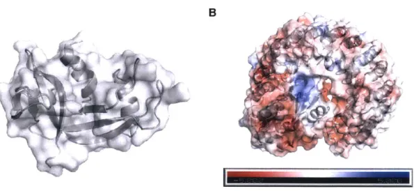
FIGURE 1.1. Structures of RNase I and RI. PyMOL renderings are based on PDB entry 1 z7x. A, Ribbon diagram of RNase 1, with surface shown. This structure is conserved among
ptRNases. B, Complex of RNase 1 with RI. Surface charges of the protein complex are shown on a scale from highly anionic (red) to highly cationic (blue), using the APBS plugin for PyMOL
(13).
16
01
highly conserved (Figure 1.1, panel A). An invariant catalytic triad (His12, Lys41, and His 119)
endows ptRNases with nonspecific endonucleolytic activity, cleaving and hydrolyzing the P-O' bond on the 3' side of pyrimidine residues, with a general preference for cytidine over uridine
(14,15) (Figure 1.2). ptRNases are cationic in character, and are also highly thermostable, owing
to the presence of four intramolecular disulfide bonds (Figure 1.3) (16).
Additionally, all ptRNases interact with - and have their ribonucleolytic activity blocked by -ribonuclease inhibitor (RI), a 50-kDa protein that is likewise conserved in vertebrates (17) and is ubiquitously and abundantly expressed in the cytosol of cells (18). RI is comprised of
leucine-Cyt 5 'RNA or Ura 0 O Q, / NH H NH I HnP -O-P= O ' N 1 HiSl2 + H- His12, W 0N RNAV' HN-J/ Hisl19 Cyt W'RNA or Ura 0 7 N NH
C66
H' His12 N: H-0. His1 19 Cyt 5 'RNA or Ura 0 O 0, P '-60 / N:H-O HN--/ RNA3' His119 Cyt 5'RNA or Ura 0 O OH HO-P=O + H -O / N' HN1FIGURE 1.2. Putative mechanism for the transphosphorylation and hydrolysis of RNA catalyzed by ptRNases. Figure is adapted from reference (14).
/ NH
+N=
rich repeats (19) and is anionic, facilitating coulombic interactions with ptRNases (20). The two proteins form a 1-1 complex with a KD value on the order of 10-15 M, forming one of the tightest known protein-protein interactions (21). This interaction blocks the activity of ptRNases (22,23). (Figure 1.1, panel B). Consequently, RI is capable of protecting the cell from the action of free ptRNases, which would otherwise degrade cellular RNAs and drive cell death (24).
Based on these characteristics, ptRNases have been identified in all vertebrate species evaluated to date. Humans express 13 such enzymes, clustered near one another on chromosome 14 (12), with eight classified as "canonical" ptRNases (25). The remaining five exhibit more unusual features, such as the lack of one or more catalytic residues and lack of cationic character (11). Other species also express multiple ptRNases, with gene duplication resulting in larger numbers of enzymes in some species - mice express about 20 ptRNases, owing to duplication of the ribonuclease 2 and 3 and ribonuclease 5 (angiogenin) subfamilies (11).
1.2.2: Known biological functions of the ptRNase superfamily
Understanding of the biological roles of ptRNases has increased substantially since the first report on the function of RNase A in 1969, which claimed that the enzyme participated in ruminant digestion but was vestigial in non-ruminants (26). Now, we appreciate that ptRNases have other roles, with many family members exhibiting functions in innate immunity.
Antimicrobial activity has been described for a cluster of closely related ptRNases, including ribonucleases 2 and 3, as well as 6, 7, and 8 (25), which are expressed in eosinophil granules, placenta, and skin and exhibit relatively broad-spectrum cytotoxicity (27-31). Angiogenins derive their name from their well-documented angiogenic activity, but some subfamily members also been described to exhibit immunomodulatory activity. Ribonucleases 9 and 10,
non-canonical members of the ptRNase superfamily, play roles in the maturation of sperm (32,33). For other members of the ptRNase superfamily, however, biological functions are still emerging. Recent research on two family members in particular, RNase 1 and angiogenin (RNase 5), has revealed numerous new functions for these enzymes in various biological niches, ranging from control of vascular homeostasis and inflammation to cellular growth and quiescence. Here, we survey these newly identified biological roles.
1.3: Ribonuclease
1
Ribonuclease 1 is the most catalytically-active member of the ptRNase superfamily in humans. Its activity against ssRNA is the highest among ptRNases, and it is uniquely nonspecific in its substrate preference, showing the ability to degrade poly(C), poly(U), and poly(A) as well as dsRNA and RNA:DNA hybrids where other ptRNases degrade only a subset of these species
(15). In contrast to the restricted expression of other ptRNases, RNase 1 is also very
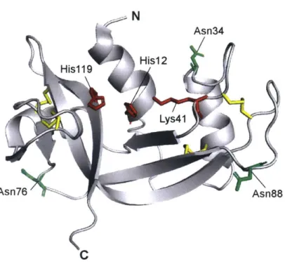
widely-expressed, with Rnase] mRNA being detectable in all tissue types (34). RNase 1 circulates in serum at a concentration of 250 ng/mL in healthy individuals (35), and this RNase 1 is produced predominantly by vascular endothelial cells (36,37). RNase 1 is secreted constitutively, but also accumulates in Weibel-Palade bodies in endothelial cells (38). Additionally, RNase I can be N-glycosylated at Asn34, Asn76, and Asn88 (39), a phenomenon that appears to vary with the tissue of origin (40,41) but has as-of-yet unknown biological implications (Figure 1.3). RNase 1 also interacts with multiple cell-surface glycans with micromolar affinity (42), exhibiting an especially specific interaction with Globo H (43).
N Asn34 His12 His1 19 Au Lys41 Asn76 Asn88 C
FIGURE 1.3. Ribbon diagram of the structure of RNase 1. PyMOL rendering is based on
PDB entry lz7x. Important side chains are shown as sticks. Active-site residues His12, Lys4l, and His1 19 are shown in red. The three N-glycosylation sites at Asn34, Asn76, Asn88 are shown in green. The four intramolecular disulfide bonds are highlighted in yellow.
1.3.1: Early studies of the biology of RNase 1
The first studies of RNase 1 in humans to draw widespread attention identified its upregulation in the plasma of patients with pancreatic cancer (44), which spurred excitement about the use of RNase 1 as a biomarker. Later studies showed that RNase 1 expression was elevated not only in pancreatic cancer, but in other malignant states (35,45,46). The idea of RNase 1 as a biomarker was ultimately abandoned, as it is nonspecific - RNase 1 is upregulated in a wide variety of disease states, including bacterial and viral infection (47), smoking (48), burn injury (49), surgery (50), myocardial infarction (51), ageing (52), Alzheimer's disease (53), malnutrition
Yet, RNase 1 returned to the forefront of cancer research with the identification of a homolog in the northern leopard frog (Rana pipiens) that exhibited toxicity against cancer cells (56). This cytotoxicity was attributed to its ability to enter the cytosol and degrade cellular RNA, while being insensitive to binding by human RI (57). Variants of RNase 1 were endowed with the ability to evade association with RI, and showed promise as anti-cancer cytotoxic agents (58), using the same mechanism outlined for the amphibian enzyme, without immunotoxicity
(42,58,59). Clinical trials are underway to bring ptRNase therapy to the clinic based on this
mechanism of action (60,61).
During these studies, the function of endogenous RNase 1 remained elusive. It was only with the appreciation that RNA exists in the extracellular space - where RNase 1 is free to act without inhibition by RI - that new ideas began to emerge as to biological roles for RNase 1.
1.3.2: Extracellular RNA - key to the function of an extracellular RNase
RNA was long thought to be unstable outside of the cellular environment - ptRNases had been known to exist in the extracellular space for many years, and were thought to immediately
degrade any extracellular RNA (eRNA) species (62). More recent studies showed that RNA can indeed persist in serum long enough to permit analysis - EBERI RNAs from Epstein-Barr virus, as well as tyrosinase mRNA from melanoma cells, were successfully isolated from serum and plasma (63,64). Characterization of eRNAs has revealed that almost every known class of RNA exists outside the cell. Whereas the bulk of eRNA are miRNAs, mRNA, and tRNA, other types of RNA, such as rRNA, piwiRNA, IncRNA, and sncRNA have also been detected in varying
proportions (65-67). These eRNAs are carried in a variety of ways, including in membrane vesicles such as microvesicles and apoptotic bodies (68), ribonucleoprotein complexes (69), and high-density lipoproteins (70).
We now appreciate that eRNA can be actively released from cells (71), with cancer cells releasing RNA at a significantly higher rate than noncancerous cells (72,73). This eRNA has been reported to act as a messenger between cells, driving changes in gene expression in paracrine via delivery of mRNA or miRNA (74,75). eRNA does not need to be coding RNA to exert biological function, as it has been demonstrated to act as a potent inflammatory mediator. eRNA stimulates lymphocyte adhesion to smooth muscle by upregulating expression of adhesion molecules such as Vcam-I, Icam-1, P-selectin, and Ccl2 in smooth muscle cells (76), and drives macrophage polarization to an Ml (proinflammatory) phenotype, increasing expression of inflammatory markers such as TNFx, IL-1
P,
IL-6, IL-12, and iNOS (77). eRNA also potentiatesblood coagulation by acting as an anionic scaffold for the activation of intrinsic coagulation proteases FXI, FXII, and plasma prekallikrein (78,79). At least for its procoagulant activities, the
size and secondary structure of eRNA appears to be just as important as its sequence, with coagulation protease activity only potentiated with RNAs greater than 100 nt in size, and more strongly potentiated with RNAs forming a stable hairpin structure (80). Poly(G) and poly(I) RNAs have been reported to be the best activators of intrinsic proteases in vitro (81).
1.3.3: New biological roles for RNase I
In light of these discoveries, studies on RNase 1 highlighted new roles for the enzyme that were mediated through degradation of these eRNAs rather than cell internalization. Interestingly, in
parallel with other studies demonstrating the anti-cancer properties of modified RNase 1, wild-type RNase A was capable of reducing tumor growth in mice when administered exogenously
(73). Rather than direct cytotoxicity, the slowing of tumor growth was due to the prevention of
pro-inflammatory activity of eRNA. eRNA was found to drive TNFa expression via activation of TNFa-converting enzyme (TACE) in macrophages (76), thereby producing an inflammatory environment conducive to tumor growth.
Other studies have shown similar effects of RNase 1 on inflammation in other contexts. In a mouse model of atherosclerosis, eRNA accumulated in atherosclerotic plaques and was able to recruit macrophages to these plaques (77). Elevated plasma eRNA was also associated with the incidence of ischemia-reperfusion injury in patients undergoing heart surgery (82), and with edema and tissue death in a mouse model of myocardial infarction (83). These negative effects of eRNA were blocked by administration of exogenous RNase 1, although notably, RNase 1
therapy was reported to be most effective when given prophylactically, as changes engendered
by eRNA exposure were irreversible by RNase-after injury occurred (84). Additionally, a
TLR7-overexpression mouse model of systemic lupus erythematosus suffered fewer
inflammatory symptoms when RNase A was knocked-in (85), resulting in extended lifespan, reduced myeloid cell number, and reduced inflammatory deposits in the kidneys and liver of mice. Pre-treatment of an ischemic murine stroke model with RNase A also resulted in less edema, smaller brain lesions, and reduced vascular permeability in treated animals than controls
(86,87). Treatment of rats undergoing heart transplantation surgery with RNase 1 also resulted in
RNase 1 has been shown to be beneficial in a mouse model of arterial thrombosis, with treatment resulting in clearance of eRNA from the thrombus, delay of vascular occlusion, and reduction in thrombus size (79). Studies in our laboratory have also demonstrated that loss of RNase 1 results in a procoagulant phenotype, with absence of the enzyme causing accumulation of eRNA in the plasma and activation of intrinsic coagulation factors. The mechanism of action of eRNA in blood clotting is activation of intrinsic coagulation factors FXII and FXI, as well as the upstream protease plasma kallikrein (79,81). This process is closely linked to inflammation, as plasma kallikrein is a protein that, when cleaving plasma kininogen, releases bradykinin, a potent vasodilator and vascular permeabilizing agent. Interestingly, eRNA alone spurs vascular permeabilization in a manner mediated through VEGF and nitric oxide synthase, leading to disintegration of tight junctions in cells (87).
Given the myriad pro-inflammatory functions ascribed to eRNA and the studies conducted to date on RNase 1, the biological function of this enzyme appears to be more complex and
multifaceted than appreciated previously. Early studies demonstrating the upregulation of RNase 1 in many disease-states likely point to a general role for RNase 1 as a regulator of inflammatory processes, particularly in the cardiovascular system (Figure 1.4). A role for RNase 1 in this niche reflects its high compatibility with the pH of the vascular environment (42), and its
colocalization in endothelial cells with other vascular regulators, such as von Willebrand factor and P-selectin (38). RNase 1 could modulate the effects of RNA accumulating in the
extracellular space secondary to tissue damage, malignancy, or death, and thus prevent excess activation of macrophage activity, blood coagulation, and vascular permeabilization. Future
studies of RNase 1 are likely to uncover not only the functions of the enzyme, but the biological roles of many poorly characterized RNA substrates on which this nonspecific enzyme acts.
Fibrin generation TACE M1 polarizaton Platelet activation TNF/ Adhesion factors Ang Globo H Receptor R11 (PKC )Stress tORNA
RI/ tRNA Stress
granules Apoptosome Translation PRNA mRNA rDNA f RNase 1 Angiogenin eRNA blood coagulation cell stress response tRNA tiRNA pRNA cell proliferation
inflammation
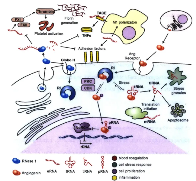
FIGURE 1.4. Schematic of RNase 1 and angiogenin activities and substrates. RNase 1
acts primarily on targets outside of the cell, where it degrades extracellular RNAs (eRNA). These eRNA would otherwise act as activators of coagulation and inflammatory pathways. Angiogenin, in contrast, mediates its effects intracellularly, driving both cellular proliferation via pRNA degradation and cellular quiescence in response to stress via tRNA degradation.
CQQWO
1.4 Angiogenin
Angiogenin is named for its ability (unique among ptRNases) to stimulate blood-vessel growth
(89). This activity coincides with other unusual features. For example, angiogenin has activity 104-fold lower than that of RNase 1 and other ptRNases (90). This deficiency has been attributed
to the occlusion of the enzymic active site by Glnl 17 (91). Notably, however, mutation of this residue does not restore RNase 1-like activity to angiogenin, suggesting that other
conformational differences exist (92). The substrates preferred by angiogenin are also somewhat restricted, as it is unable to cleave typical ptRNase substrates such as poly(C) and poly(U). Angiogenin does cleave preferentially at CpA sequences, and has demonstrated activity against tRNA (93) and pRNA, a small noncoding RNA that associates with the NoRC chromatin remodeling complex (94).
1.4.1 A ptRNase with unique structural and signaling properties
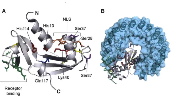
Angiogenin has other unique structural features (Figure 1.5, panel A), including a receptor-binding site at residues 60-68, which is essential for its internalization into cells (95). Angiogenin was determined to act through a cell-surface receptor, as the circulating
concentration of the enzyme (~400 ng/mL) is much higher than the concentration required to stimulate its angiogenic activity (96) - yet, this activity occurs in normal cells in a density-dependent manner (97). Several different proteins have been identified as receptors that mediate angiogenin activity, including Syndecan-4 in astrocytes (98) and Plexin B2 in multiple cell types
B
A N
NLS
His1 14 His1 3 Ser37
G~n17 Lys40 Ser,87
Receptor binding
FIGURE 1.5. Ribbon diagrams of the structure of angiogenin. PyMOL renderings are
based on PDB entries 1ang and 1a4y. Important side chains are shown as sticks. A,
Ribbon diagram of angiogenin. Active-site residues Hisl2, Lys4l, and Hisl19 are shown in red. GIn1 17, which occludes the active site, is shown in blue. The three intramolecular disulfide bonds are highlighted in yellow. The nuclear localization signal (NLS) (RRRGL) is shown in orange. The receptor binding domain (KNGNPHRE) is shown in green.
Phosphorylation sites at Ser28, Ser37, and Ser87 are shown in purple. B, Ribbon diagram of angiogenin in complex with RI, shown in cyan. Note that binding of angiogenin to RI occludes the active site and nuclear localization signal of angiogenin, and that the three phosphorylated serine residues are in contact with the RI-binding interface.
(99). Angiogenin also contains a nuclear-localization signal (RRRGL, at positions 31-35) (100),
which drives the accumulation of the enzyme in the nucleolus and is required for its angiogenic activity (101).
Nuclear localization of angiogenin would normally be prevented due to the association of the enzyme with cytosolic RI, to which angiogenin binds with subfemtomolar affinity (21). Angiogenin avoids association with RI by disruption of its Coulombic interactions through phosphorylation, which is known to occur at Ser28, Ser37, and Ser87, and is mediated through protein kinase C and cyclin-dependent kinase (Figure 1.5, panel B). This phosphorylation event
has also been demonstrated to be essential for nuclear localization and proliferative activity of angiogenin (94).
In the nucleus, angiogenin has been shown to cleave pRNA, which normally associates with TIP5, a member of the NoRC chromatin remodeling complex. pRNA is responsible for recruitment of TIP5 and NoRC to the ribosomal DNA (rDNA) promoter, which results in repression of rDNA transcription (102,103). Cleavage of pRNA prevents its association with TIP5 and impairs recruitment of NoRC to the rDNA promoter, which results in derepression of rDNA and cell growth (94). This signaling pathway is unique among transcription factors, as no other proteins are known to reach the nucleus from the extracellular space and effect
transcriptional change.
1.4.2 Angiogenin-mediated cell growth
The angiogenic ability of angiogenin has been studied in the context of cancer for over 50 years, as it was initially identified as a factor capable of stimulating blood vessel growth from human adenocarcinoma (89). Angiogenin has since been shown to be secreted by multiple types of tumors, including prostate cancer (104), oral squamous cell carcinoma (105), and melanoma
(106). Its expression from cancer cells appears to be regulated by HIF-la (105), suggesting a
mechanism of action for tumor neovascularization in response to oxygen starvation. Angiogenin has also been studied as a biomarker in human colorectal cancer (107), bladder cancer (108), and breast cancer (109), as its expression has been correlated with tumor aggressiveness.
Consequently, angiogenin is being explored as a therapeutic target for the treatment of cancer. Knockdown of angiogenin in bladder cancer cells using siRNA was successful in reducing tumor
growth in a mouse xenograft model (110), and antibody neutralization of angiogenin reduced in
vitro migration of triple-negative breast cancer cells (111). An experimental small-molecule
drug, (8-amino-5-(4'-hydroxybiphenyl-4ylazo)naphthalene-2-sulfonate), that inhibits catalysis
by angiogenin, has also been developed, and shows promising results in inhibition of tumor
growth and vascularization in mouse xenograft models (112).
Angiogenin has also been demonstrated to act in the placenta, endometrium, and ovarian follicle, tissues that normally undergo extensive angiogenesis in adults (113). Angiogenin is present in ovarian follicular fluid, with expression stimulated by human chorionic gonadotropin - a hormone that promotes luteinization and angiogenesis during formation of the corpus luteum (114). In studies of ovarian follicle transfer, angiogenin was identified to be a crucial factor in mediating the angiogenic and follicle-preserving effect of co-transplanted mesenchymal stem cells (115), underscoring the importance of the enzyme for follicle survival and function. Angiogenin expression in the endometrium reflects the menstrual cycle, with expression approximately doubling in the mid-to-late secretory phases relative to the proliferative phase
(116). In pregnancy, angiogenin expression increases over time in the human placenta, with
maximal expression at term (117,118). In the placenta, angiogenin is found alongside markers of early vasculogenesis, such as VE-cadherin, CD34, and von Willebrand factor, and is present in developing fetal blood vessels (119).
Angiogenin can act on cells other than in vascular endothelium to promote cell growth. This versatility was first observed in the context of cancer - HeLa cells both secrete angiogenin and take it up, with angiogenin localizing to the nucleus and stimulating cell proliferation
independent of cell density (120). Angiogenin has also demonstrated this activity in PC-3 prostate cancer cells (104). In addition, angiogenin appears to promote growth of hepatocellular carcinoma cells (HCCs) in paracrine. HCCs secrete angiogenin, which is taken up by tumor-associated hepatic stellate cells, spurring their activation, which in turn promotes the growth and metastasis of HCCs by modulation of the extracellular matrix (121,122). Angiogenin also
appears to modulate the growth of neurons, as it is expressed in the nervous system during embryonic development in mice and is localized to axonal growth cones, where it appears to promote axonal growth and neurite pathfinding (123).
1.4.3 Angiogenin-mediated protection
from
cell stressIn addition to stimulating neuronal growth, angiogenin has been also been observed to have a protective effect on motoneurons in the context of amyotrophic lateral sclerosis (ALS). Similarly to the action of angiogenin in hepatocellular carcinoma, this activity is mediated in paracrine, by astrocyte uptake of motoneuron-secreted angiogenin (98). Angiogenin-stimulated astrocytes are then able to protect motoneurons against excitotoxic stress through a mechanism that involves protein expression changes in astrocytes largely related to modulation of the extracellular matrix (124). This finding helps to explain many earlier studies that linked mutations in angiogenin to
ALS, many of which are missense mutations that interfere with the catalytic activity of the
enzyme (125,126). These studies did not, however, identify specific effector RNA targets for angiogenin.
A possible effector substrate for angiogenin in neurons is tRNA, which has been known to be an
cleaved tRNA were identified to arrest protein translation. Yet, it has only recently been
appreciated that tRNA cleaved at the anticodon loop, termed tiRNA, are a product of catalysis by angiogenin that is induced by cellular stress (127,128). These tiRNAs do promote the formation of stress granules and inhibit protein synthesis, which is suggested to occur via displacement of the initiation factor eIF4G/A from mRNA (129). Nonetheless, tiRNAs also exert a pro-survival effect in cells by binding to apoptotic protease-activating factor 1 (APAF 1) in a manner that competitively blocks the binding of cytochrome C and formation of the apoptosome (130).
Angiogenin has also been implicated to play roles in stem cell self-renewal. In hematopoetic stem/progenitor cells (HPSCs), angiogenin has been found to generate tiRNA and reduce protein synthesis, which is suggested to promote sternness and quiescence of these cells, while having a proliferative effect on myeloid progenitor cells (131). The mechanism by which angiogenin
differentially regulates these two cell types is unknown.
Additionally, some antimicrobial and anti-inflammatory activity has been demonstrated for angiogenin. It is an acute-phase protein and was initially thought to contribute to angiogenesis as part of the tissue repair process (132), but more recent studies have reported that angiogenin is also toxic to C. albicans and S. pneumoniae. Closely related proteins in the angiogenin subfamily in mice appear to have broader-spectrum antimicrobial activity (133). Further, angiogenin
suppresses the effects of TNFalpha in fibroblasts, reducing expression of NFKB, TNFaR1 and TNFcR2, IL6, and IL8 (134) and exerting a net anti-inflammatory effect.
Emerging research on angiogenin suggests that in addition to its well-characterized roles in vascular growth, this enzyme could play broader roles in cell growth and survival (Figure 1.4).
Future research on angiogenin might offer a better understanding of the mechanisms that determine whether angiogenin drives proliferation or quiescence in a cell. Angiogenin has been an attractive drug target for both antiangiogenic therapy in cancer (112) or protection from cellular stress in neurodegeneration (135), and more knowledge of the regulators of its activity could allow more selective targeting of the enzyme in specific biological niches.
1.5 Connections to vertebrate physiology
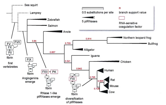
New roles identified for RNase 1 and angiogenin cluster broadly into the areas of regulating vascular homeostasis and cell growth/survival. Interestingly, some of these roles are tied to the innate physiology of vertebrates, where ptRNase expression is exclusively present - the only enzyme family for which this is the case (136). The distribution of ptRNase subfamilies within vertebrates does vary, however. Lower vertebrate species such as fish express only angiogenin-like enzymes (137,138), and the diversification of the RNase 2 and 3 family occurs only in rodents (11). Some highlights of this evolutionary path are depicted in Figure 1.6.
Angiogenin has been suggested to be the primordial ptRNase family member. This thought is supported by the presence of ptRNase enzymes in fish that are more similar to human angiogenin than to RNase 1 or other ptRNases (138). Additionally, angiogenin has three intramolecular disulfide bonds in contrast to the four in other ptRNases (25). This fourth disulfide bond is more readily reduced than the other three (139), suggesting it is less essential to the enzyme and was likely a later addition (140). Angiogenin, then, is likely as old as vertebrates themselves
brain highlight this centrality, as the neural crest, which gives rise to protective glial cells, is not present in invertebrates (141).
One of the other hallmarks of vertebrate physiology is the presence of a closed circulatory system with a continuous endothelial barrier (153), which requires a specialized blood clotting
--- Sea squirt
n
- --- Lamprey 0.5 substitutions per site branch support value
Zebrafish 5 ptRNases RNA-sensitive
Salmon coagulation factor
Anole
0.914 Northern leopard frog
F0.703 Bullfrog F11 0.857 Alligator fibrin Iguana first C vertebrates P fbrinHmn Angiogenins - F 0.795 Rat em erge 7t M-use fibrin 0.
RNase 1-like fibrin Cow
ptRNases emerge diversification of ptRNases
FIGURE 1.6. Phylogenetic tree of species by ptRNase homology, annotated with blood coagulation factors. Protein sequences of RNase 1 homologs from Uniprot entries P07998 (human), P00683 (mouse), P00684 (rat), P61823 (cow), P30374 (chicken), P80287 (iguana),
AQA1 51 LY34 (alligator), P11916 (bullfrog), Q8UVX5_LITPI (northern leopard frog),
H9GD73_ANOCA (anole), A0A1S3RRZ8 (salmon), and A5HAKO (zebrafish) were used to
generate a phylogenetic tree using the program phylogeny.fr (142-146). The branch support value is shown in red for each junction, and the number of substitutions per site is represented
by line length. Line weight at the termini of each leaf represents the number of ptRNase proteins
reported to be expressed in each organism (11,137,138,147-150). Species without identified ptRNases are represented by dashed lines and are not to scale; they are included to highlight the emergence of vertebrate coagulation factors. The emergence of angiogenins in fish, RNase
1-like proteins in amphibians, and RNA-sensitive coagulation factors in amphibians and mammals is highlighted at the appropriate junctions (151,152)
system (152). Lower vertebrates share the common thrombin-catalyzed coagulation pathway present in humans, but lack intrinsic factor proteases such as FXII, FXI, and prekallikrein (154). Intrinsic factors emerged with the evolution of amphibians, with FXI appearing only in mammals
(151). Interestingly, the emergence of these RNA-sensitive coagulation factors mirrors the
presence of RNase 1. RNase 1 is more highly expressed in reptiles and birds than in fish that do not express these contact factors, and much more highly expressed in mammals (155),
suggesting that the evolution of RNA-responsive coagulation proteins and a cognate extracellular ribonuclease to regulate this activity were simultaneous.
Vertebrates also exhibit a much more specialized immune system than that found in
invertebrates, with the emergence of major histocompatibility complex proteins, T-cell receptor, and immunoglobulins. The rapid evolution of ptRNases has been used to suggest that these enzymes also are part of the vertebrate-specific immune response (156), as many family members (including angiogenin) are cytotoxic against a variety of microorganisms. RNase 1 alone, however, is not cytotoxic - instead, it modulates a broad array of cytokines and affects the interplay between innate and adaptive immunity via control of eRNA-mediated macrophage polarization. These macrophages can then can influence T and B cell activity (88). The polarization of macrophages also appears to be a vertebrate-specific phenomenon as well, as
polarization has been observed in fish but not lower animals (157). These effects represent multiple possible avenues by which eRNA (and by extension, RNase 1) might regulate the inflammation associated with cancer, autoimmune disease, and infectious disease.
1.6 Prospectus
Numerous new biological functions of pancreatic-type ribonucleases have been outlined in the past decade. Still, our appreciation of the function of these enzymes in their endogenous
environment is lacking. The effects of RNase 1 as an exogenous agent have been characterized in multiple models of cancer, vascular homeostasis, and injury. Angiogenin has been more
thoroughly studied, but little information about the phenotype of constitutive knockouts are available. Further study on the biological functions of these enzymes would advance our
knowledge of the roles of the ptRNase superfamily and the functions of novel RNA populations, and guide their use as therapeutic targets.
The central aim of this thesis is to describe biological functions for RNase 1 and angiogenin through the study of knockout mouse models lacking these enzymes. I identify that RNase 1 is a natural anticoagulant enzyme in mice, and that angiogenin plays an important role in
reproduction and embryonic viability. I have also have worked to generate tools for further study of these enzymes in biological systems by producing proteins for use in a phage-display
antibody-generation system. Together, the studies in this thesis provides a previously
unappreciated perspective on the biological functions of these two ptRNases, and sets the stage for future study of these dynamic proteins in physiological contexts.
Chapter 2
Loss of ribonuclease
1
produces a hypercoagulable phenotype
Contributions: J.E. Lomax performed the work to generate Rnasel-k mice and designed the genotyping strategy. J.P. Sheehan was involved in experimental design and data interpretation for kinetic coagulation, saphenous vein bleeding, and factor activity assays. D. Gailani and B. Mohammed performed plasma immunoblots and aided in data interpretation. Technical assistance in dissection/surgery was provided by R. Sullivan, T. Hacker, and G. Diarra, and in mouse colony management by C. Feldman and K. Tippins. I designed and carried out all other experiments, and wrote the manuscript.
This chapter has been submitted as Loss ofribonuclease 1 produces a hypercoagulable
2.1: Abstract
Ribonuclease 1 (RNase 1) is a small secretory enzyme with unknown biological function. In contrast, recent work has established biological roles for extracellular RNAs (eRNAs). In
particular, eRNAs can initiate blood coagulation via activation of intrinsic factor proteases (such as FXII and FXI), and studies using the bovine homolog of RNase 1 (RNase A) have revealed its ability to prevent blood vessel occlusion in mouse models of thrombosis and stroke. We sought to evaluate the biological role of RNase 1 in an organismal context by generating and analyzing RNase 1 knockout (Rnaseh') mice. These mice are viable, fertile, and appear generally healthy, but Rnase1- plasma contains more RNA than does the plasma of Rnase*/* mice. In addition, the plasma of Rnase-' mice form fibrin clots more rapidly. Yet, Rnase-' mice do not exhibit perturbations in thrombin-antithrombin complex formation, tail or saphenous vein bleeding, or iron-chloride thrombosis assays in vivo. These data suggest activation of intrinsic pathway factors, and FXII and FXI activity is indeed elevated in Rnase-' mice. This phenotype is consistent with known roles of eRNA and the intrinsic pathway in blood coagulation, and
mirrors the largely healthy phenotype of FXII knockout mice. Our work identifies RNase 1 as an endogenous regulator of blood coagulation.
2.2: Introduction
Ribonuclease 1 (RNase 1) is a secretory protein with robust and nonspecific ribonucleolytic activity. The physical and catalytic properties of this enzyme are well understood due to its similarity to RNase A, which is a bovine homolog of RNase 1 that served as a model protein for seminal studies in biological chemistry during the 2 0th century (14,158-160). RNase 1 is
conserved in vertebrates and is part of a large enzyme superfamily termed the pancreatic-type ribonucleases (ptRNases) or vertebrate secretory ribonucleases, many of which have been identified to play diverse biological roles (25).
No distinct biological function has been identified for RNase 1. The earliest hypothesized function of RNase 1 was in digestion, as RNase A is highly expressed in the pancreas of cows, and RNase 1 was thought to have negligible function in non-ruminant animals (26). More recent research has challenged this hypothesis, demonstrating a wide range of non-digestive activities for exogenously-administered RNase 1 in vitro and in vivo, including as an anti-viral agent
(161,162) and cancer chemotherapeutic agent (163,164).
Still, the function of endogenous RNase 1 remains elusive. RNase 1 has the highest catalytic activity and the widest expression pattern of any of the ptRNases, and is present in many bodily fluids (165,166). In both humans and mice, RNase 1 circulates at ~0.5 pg/ml (35). The major source is the vascular endothelium (36,3 8), and the pH optimum for its enzymatic activity is 7.3, which is close to the pH of blood (42). Thus, we reasoned that the endogenous enzyme could play a role within the vascular system.
Recent studies have highlighted the ability of RNase A to exert biological effects via degradation of extracellular RNA (eRNA), which is known to play roles in immunity, cancer, and blood coagulation (73,76,79). RNA has been shown to potentiate the activation of intrinsic coagulation pathway factors XII (FXII) and XI (FXI) via physical interaction with these enzymes (79). Further study suggests that RNA is a particularly effective activator in the conversion of FXII to FXIIa by plasma kallikrein and in the conversion of FXI to FXIa by thrombin (81). Indeed, previous analyses of RNase A have highlighted its ability to reduce eRNA-mediated thrombosis in murine models of carotid thrombosis (79) and stroke (86). eRNA secondary structure is
thought to be important for its procoagulant activity (80), and RNase 1, which has higher activity against dsRNA than does RNase A, might be better adapted as an endogenous regulator of eRNA (42).
Evaluation of RNase 1 in an organismal context is indicated to determine its biological function. Here, we report the generation of Rnase] knockout mice and characterize the impact of the loss of RNase 1 on blood coagulation in vitro and in vivo. Our results reveal activation of intrinsic coagulation factors upon loss of RNase 1. These data indicate that RNase 1 serves as an endogenous regulator of blood coagulation.
2.3: Experimental procedures
2.3.1: Materials-Murine ribonuclease 1 (RNase 1) and ribonuclease A were produced in E. coli
as described previously (167). PNGase F was from New England BioLabs. Human FXI-, FXII-, and PK-deficient plasma was from George King Bio-Medical. Murine FXI- and FXII-deficient plasma was obtained as described previously (168,169), and murine FIX-deficient plasma was