HAL Id: tel-01663080
https://tel.archives-ouvertes.fr/tel-01663080
Submitted on 13 Dec 2017HAL is a multi-disciplinary open access archive for the deposit and dissemination of sci-entific research documents, whether they are pub-lished or not. The documents may come from teaching and research institutions in France or abroad, or from public or private research centers.
L’archive ouverte pluridisciplinaire HAL, est destinée au dépôt et à la diffusion de documents scientifiques de niveau recherche, publiés ou non, émanant des établissements d’enseignement et de recherche français ou étrangers, des laboratoires publics ou privés.
Functions of the transcription factor Lyl-1 in the
development of the macrophage lineage
Shoutang Wang
To cite this version:
Shoutang Wang. Functions of the transcription factor Lyl-1 in the development of the macrophage lineage. Embryology and Organogenesis. Université Paris Saclay (COmUE), 2017. English. �NNT : 2017SACLS457�. �tel-01663080�
Fonctions du facteur de transcription
Lyl-1 dans le développement du
lignage macrophagique
Thèse de doctorat de l'Université Paris-Saclay,
préparée à l'Université Paris-Sud
École doctorale n°577 :
Structure et dynamique des systémes vivants (SDSV)
Spécialité: Sciences de la vie et de la santé
Thèse présentée et soutenue à Villejuif, le 27 Novembre 2017, par
Shoutang WANG
Composition du Jury :
Dr. Sophie CREUZET Présidente
Université Paris-Saclay
Dr. Valérie PINET Rapporteur
IGMM, Montpellier
Dr. Julien BERTRAND Rapporteur
Université de Genève
Dr. Elisa GOMEZ-PERDIGUERO Examinatrice
Institut Pasteur
Dr. Michel MALLAT Examinateur
UPMC, Paris
Dr. Isabelle GODIN Directrice de thèse
NNT : 2 01 7 S A C L S4 5 7
UNIVERSITÉ PARIS-SUD
École doctorale n°577 "Structure et Dynamique des Systèmes Vivants"
Spécialité : Sciences de la Vie et de la Santé
THÈSE DE DOCTORAT
Soutenue le 27 Novembre 2017
Par Shoutang WANG
Fonctions du facteur de transcription Lyl-1 dans le
développement du lignage macrophagique
Composition du jury:
Directrice de thèse: Dr. Isabelle GODIN Université Paris Sud
Rapporteurs: Dr. Valérie PINET IGMM, Montpellier
Dr. Julien BERTRAND Université de Genève Examinateurs: Dr. Elisa GOMEZ-PERDIGUERO Institut Pasteur
ACKNOWLEDGEMENTS
This work presented here was done at the Institut Gustave Roussy, under the supervision of Dr. Isabelle GODIN. It is my pleasure to thank the many people who helped me during the PhD study.
First of all, I would like to thank my supervisor, Dr. Isabelle GODIN, for giving me this opportunity to work in the team U1170. Thank you for all your systematic guidance, helpful suggestions, kind support and patience throughout the course of this thesis.
I would like to thank all the members of the jury, Dr. Valerie PINET, Dr. Julien BERTRAND, Dr. Elisa GOMEZ-PERDIGUERO, Dr. Michel MALLAT and Dr. Sophie CREUZET for taking time out of teaching/research/life to give critical reading of my thesis. This thesis would not have been possible without the financial support of China Scholarship Council and Société Française d’Hématologie.
I would like to thank Dr. William VAINCHENKER, for always being prepared to help and advice; Dr. Isabelle PLO, for the general support. I am also thankful to the past and present colleagues in this lab. To Gabriel MATHERAT for the helpful discussion and continuous encouragement. To Deshan REN for the scientific assistance and friendship. I am quite happy to work in this friendly and cheerful group.
I want to acknowledge all of the students, staff and faculty members in Institut Gustave Roussy for providing a productive working atmosphere and for their scientific, administrative and moral support.
Last but not least, I would like to express the deepest gratitude to my parents for their love, care and constant support throughout the past years. Also thanks to my girl friend Tingting LEI for her understanding, accompany and endless encouragement.
Index
1 INTRODUCTION ... 1
1.1. Ontogeny of the hematopoietic system ... 3
1.1.1 Hematopoietic stem cells ... 3
1.1.2. Hierarchical structure of mouse hematopoietic system ... 5
1.1.3. The regulation of hematopoiesis ... 6
1.1.4. Development of hematopoietic system ... 8
1.1.5. Distinct waves of embryonic hematopoiesis ... 15
1.1.6. The macrophage lineage ... 22
1.2. Development and function of microglia in mice ... 32
1.2.1 Myeloid cell types in the central nervous system ... 32
1.2.2. The development of microglia ... 34
1.2.3. Factors that regulate the development of microglia ... 37
1.2.4. Physiological roles of microglia ... 38
1.2.5 Microglial dysfunction is involved in neurodevelopmental defects ... 43
1.3. Transcription factor Lyl-‐1 in hematopoiesis ... 46
1.3.1 Lyl-‐1 function in leukemia ... 46
1.3.2 Functional roles of Lyl-‐1 in hematopoiesis ... 48
1.3.3 The expression pattern of Lyl-‐1 during hematopoietic ontogeny ... 49
2 RESULTS ... 51
2.1. Summary ... 52
2.1.1. Lyl-‐1 expression characterizes primitive macrophage progenitors from the first of YS-‐derived wave. ... 52
2.1.2. Lyl-‐1 expressing primitive MΦ give rise to embryonic microglia. ... 56
2.1.3. Lyl-‐1 inactivation impairs MΦ maturation at early embryonic stage. ... 57
2.1.4. Lyl-‐1 controls embryonic microglia pool at two developmental stages. ... 58
2.1.5. Lyl-‐1-‐defective microglia induce social anxiety disorders ... 60
2.2. Result 1: Lyl-‐1 links microglia development to neuropsychiatric disease (Article in
preparation) ... 62
2.3. Result part 2: Additional data ... 112
2.3.1. Lyl-‐1 does not influence macrophage production from AGM-‐HSC ... 112
2.3.2. Identification of the differences in genes expression between YS-‐derived primitive and definitive macrophage by RNA sequencing ... 113
2.3.3. Lyl-‐1 expressing primitive MΦ colonize the fetal liver ... 115
2.3.4. Lyl-‐1 deficiency leads to a reduction of the microglia pool at E12 ... 117
2.3.5. Lyl-‐1 inactivation results in a reduction of inflammatory-‐related genes expression ... 119
2.3.6. Bibliographic and database information evidencing a possible involvement of Lyl-‐1 in human microglia in heath and diseases. ... 120
3 GENERAL DISCUSSION AND PERSPECTIVES ... 126
3.1 Lyl-‐1 expression discriminates the YS-‐derived primitive and definitive macrophages. ... 127
3.2 YS-‐derived primitive macrophages (Lyl-‐1 positive) give rise to the embryonic microglia. 129 3.3 Lyl-‐1 regulates the development and functions of microglia and is involved in neuro-‐developmental diseases. ... 131
4 RÉSUMÉ DE LA THÈSE EN FRANÇAIS ... 134
Figure list
Figure 1: The hierarchy of hematopoietic cells. ... 4
Figure 2: Schematic of the heptad transcription factor complexes bound to DNA. ... 8
Figure 3: Schematic representation of the successive hematopoietic sites during mouse ontogeny. ... 11
Figure 4: Migratory and circulatory routes of hematopoietic cells in the murine embryo over developmental time. ... 14
Figure 5: Different hematopoietic waves during ontogeny. ... 17
Figure 6: Comparison between primitive and definitive erythropoiesis. ... 19
Figure 7: Developmental stages followed by the 3 distinct MΦ waves, characterized by their phenotype, origin, differentiation potential, and lineage relationship. ... 21
Figure 8: Localization and functions of macrophage and monocyte subpopulations. ... 23
Figure 9: A Schema of the constitutive and inducible conditional gene targeting approaches. ... 25
Figure 10: Fate-‐mapping systems of macrophages and other hematopoietic cell. ... 27
Figure 11: Three Different Models of Fetal Macrophage Ontogeny. ... 29
Figure 12: The localization and genetic signature of CNS macrophages. ... 33
Figure 13: Microglial origin and development. ... 36
Figure 14: Main microglial functions during CNS development. ... 42
Figure 15: Schematic comparison of the two targeted alleles of Lyl1. ... 48
Figure 16: Lyl-‐1 does not influence the production of MΦ from AGM-‐HSC. ... 113
Figure 17: Lyl-‐1+ primitive MΦ progenitors are present in the fetal liver at E10. ... 116
Figure 18: Lyl-‐1 deficiency leads to a reduction of the microglia pool at E12.5. ... 118
Abbreviations
β-Gal:
β-Galactosidase
AGM:
Aorta-Gonad-Mesonephros
AML:
Acute Myeloblastic Leukemia
ASD:
Autism Spectrum Disorder
BDNF:
Brain-Derived Neurotrophic Factor
BFU-Es:
Burst-forming Units-erythroid
bHLH:
basic Helix-Loop-Helix
BM:
Bone Marrow
CLP:
Common Lymphoid Progenitor
CMP:
Common Myeloid Progenitor
CNS:
Central Nervous System
cpMΦ:
Choroid Plexus Macrophages
CREB1:
Cyclic AMP Receptor Element Binding Protein 1
CSF1R:
Colony Stimulating Factor 1 Receptor
DAP12:
DNAX activation protein of 12 kDa
DCs:
Dendritic Cells
E:
Embryonic Day
EMp:
Erythro-Myeloid Progenitors
ER:
Estrogen Receptor
EryP-CFCs:
Primitive Erythroid Colony-forming Cells
ETPs:
Early T Lineage Progenitors
FACS:
Fluorescence-activated Cell Sorting
FL:
Fetal Liver
Flt3:
FMS-like Tyrosine Kinase 3
GFP:
Green Fluorescent Protein
GM:
Granulo-Monocytic
HSCs:
Hematopoietic Stem Cells
IGF-1:
Insulin-like Growth Factor-1
IL-1R:
Interleukin-1 Receptor
IRF8:
Interferon Regulatory Factor 8
Lmo2:
LIM domain only 2
LMPP:
Lymphoid-primed Multipotent Progenitors
LSK:
Lin
-Sca-1
+c-Kit
+LT-HSC:
Long-Term Repopulating Hematopoietic Stem Cells
LTR:
Long-Term Reconstitution Assay
Lyl-1:
Lymphoblastic leukemia 1
MeCP2:
Methyl-CpG-Binding Protein 2
Mer:
Modified Estrogen Receptor
mMΦ:
Meningeal Macrophages
MΦ:
Macrophages
MPP:
Multi-potent Progenitor
MEP:
Megakaryocyte/Erythroid Progenitor
OrgD1:
Organ Cultured for 1 Day
P:
Postnatal
P-Sp:
Paraaortic Splanchnopleura
pvMΦ:
Perivascular Macrophages
Runx1:
Runt-related transcription factor 1
S:
Somite
Scl/Tal-1:
Stem Cell Leukemia/T-ALL 1
ST-HSC:
Short-Term Repopulating Hematopoietic Stem Cells
STMN1:
Stathmin
T-ALL:
T-cell Acute Lymphoblastic Leukemia
TNFs:
Tumor Necrosis Factors
Objective and Organization of the thesis
Transcription factors, such as SCL/Tal-1, Lmo2, Runx1 and GATA-2, are proved to control hematopoietic cells fate decision, cellular phenotypes, and differentiation. Lyl-1 is a bHLH transcription factor that is closely related to SCL/Tal-1, which is required for hematopoietic development in the embryo. Moreover, we previously showed using the Lyl-1LacZ strain that Lyl-1 expression pattern during ontogeny largely overlaps with that of SCL/Tal-1, indicating that Lyl-1 may play a role during developmental hematopoiesis. My thesis mainly focuses on the Lyl-1 function in developmental hematopoiesis, which is still unknown.
Two transgenic mouse models, Lyl-1LacZ and Cx3cr1GFP, in the laboratory contributed to the studies. The key points of the studies are: (1) the effects of Lyl-1 inactivation in hematopoiesis during early embryonic development; (2) Lyl-1 expression pattern in yolk sac-derived primitive and definitive macrophage lineage; (3) the contribution of yolk sac-derived Lyl-1 positive MΦ population to microglia development; (4) the defects of macrophage/microglia development in Lyl-1 mutants; (5) the characterization of neurogenesis and behavior in adult Lyl-1mutants.
During the course of my thesis, I had to gather information on different fields, developmental biology, hematopoiesis and its regulation by transcription factors, macrophage and microglia development and neuro-development.
I will first give a general background about part of these fields related to my research in the introduction. I will then summarize the experiments design and the results I obtained. Altogether, I expect that this work will contribute to a better understanding of the functions of Lyl-1 in developmental hematopoiesis and neurogenesis.
1.1. Ontogeny of the hematopoietic system
1.1.1 Hematopoietic stem cells
In adult mammals, blood cells are constantly produced from progenitors in the bone marrow (BM) to replenish short-lived mature blood cells throughout life (Cumano and Godin, 2007). This constant process initiates from a rare population of progenitors, the hematopoietic stem cells (HSCs). HSCs reside in the BM in adult mammals and can produce a series of multi-lineage progenitors, then to lineage-committed precursors. This progressive differentiation process ultimately gives rise to all types of mature blood cells, including erythrocytes, megakaryocytes, myeloid cells (monocyte/macrophage and granulocytes), dendritic cells (DCs), B and T lymphocytes and natural Killer cells (Fig 1) (Clements and Traver, 2013; Orkin, 2000; Weissman, 2000). Analysis of blood system regeneration in vivo revealed that HSCs possess two main properties, multi-potency and self-renewal, which are also the only hematopoietic cells that contain both potential (Ema et al., 2005; Hock, 2010; Morita et al., 2010; Osawa et al., 1996; Wagers et al., 2002). Multi-potency makes HSCs able to produce a differentiated progeny comprising all the different blood cell types. Self-renewal allows the maintenance of the HSC pool without cell differentiation through asymmetric cell division (Jones et al., 2015; Wilson et al., 2004). After committed differentiation, the progeny of HSCs progressively loose self-renewal capacity and becomes restricted to single lineages. BM transplantation has shown that a single HSC is capable of repopulating the entire hematopoietic system of a recipient (Dyksta et al., 2007; Kiel et al., 2005; Osawa et al., 1996; Sieburg et al., 2006; Smith et al., 1991).
Figure 1: The hierarchy of hematopoietic cells.
Abbreviations: LT-HSC, long-term repopulating HSC; ST-HSC, short-term repopulating HSC; MPP, multi-potent progenitor; CMP, common myeloid progenitor; CLP, common lymphoid progenitor; MEP, megakaryocyte/erythroid progenitor; GMP, granulocyte-macrophage progenitor. The encircled pluripotent population, LT-HSC, ST-HSC and MPP are Lin-Sca-1+c-kit+ as shown (Larsson et al., 2005).
1.1.2. Hierarchical structure of mouse hematopoietic system
Progress in investigating phenotypic and functional characterization of HSCs and other precursors has made the hematopoietic system a paradigm in stem cell biology (Orkin and Zon, 2008; Rowe et al., 2016). Experimental evidences showed that HSCs can be isolated from multi-committed progenitors and BM cells by fluorescence-activated cell sorting (FACS) based on specific phenotypic markers (Table 1) (Adolfsson et al., 2001; Christensen and Weissman, 2001; Kiel et al., 2005). In the mouse, HSCs have been initially extensively purified from BM by utilizing cell-surface markers, Lin-Sca-1+c-Kit+ (LSK) (Lin -corresponding to the fraction that does not express markers specific for mature cells for the various lineages, such as CD11b for macrophage, Gr1 for granulocyte, etc). But it was later shown that this fraction also contain restricted progenitors. The self-renewal has been better defined though in vivo functional assay named long-term reconstitution assay (LTR). This experimental approach makes use of congenic cell surface markers that differ between donor and host (e.g. CD45.1/CD45.2) to follow the progeny of multi-potential hematopoietic cells
in vivo after transferring the HSCs into the irradiated recipient over six months. HSCs are
now defined phenotypically as Flk2-CD34- LSK cells, also named Long-Term (LT)-HSCs (Yang et al., 2005). In vitro and in vivo assays performed to determine the differentiation potential of HSCs and of other progenitors have suggested a hierarchical structure in hematopoietic development in which self-renewal is maintained by particular subsets and multi-potency is progressively restricted (Fig 1) (Akashi et al., 2000; Kondo et al., 1997; Nakorn et al., 2003). It is generally accepted that LT-HSCs partially lose their self-renewal potential and transit to Short-Term (ST)-HSCs (Flk2-CD34+) (Akashi, 2005; Hock and Orkin, 2005). Then ST-HSCs give rise to the multi-potent progenitors (MPPs), which no longer possess self-renewal ability yet keeping full-lineage differentiation potential. Recent evidences showed that MPPs are still heterogeneous, which is considered as a fully multi-potent but lineage-biased population (Pietras et al., 2015). Different phenotypic MPP subsets are produced in parallel by ST-HSCs at variable levels depending on hematopoietic
progenitors (CMPs) or lymphoid lineage by generating common lymphoid progenitors (CLPs). Collectively these committed progenitors then give rise to all the mature cells of the hematopoietic system, such as CMPs produce megakaryocyte/erythrocyte progenitors (MEPs) to follow the megakaryocyte/erythrocyte lineage or produce granulocyte-macrophage progenitors (GMPs) to follow the granulocyte/macrophage lineage. Interestingly, the major subsets of DCs can be derived either form CMP and CLP (Geissmann et al., 2010).
Table 1: Surface marker phenotypes to separate mouse stem and progenitor cell subsets (Galen et al., 2014)
1.1.3. The regulation of hematopoiesis
Hematopoietic progenitors features, such as cell fate decision, progenitor maintenance and differentiation, etc. are regulated by a transcription factor network that comprises, amongst others, Stem Cell Leukemia/T-ALL 1 (Scl/Tal-1), Runt-related transcription factor 1 (Runx1), LIM domain only 2 (Lmo2), which are the major regulators of hematopoietic progenitors development during ontogeny (Pina and Enver, 2007; Wilson et al., 2010). Scl/Tal-1, Lmo2 and Runx1 are believed to orchestrate the formation of the HSC pool during embryonic development. Either Scl/Tal-1 or Lmo2 (Shivdasani et al., 1995; Wilson et al., 2009) were shown to be essential for hematopoietic cell fate specification since knock out embryos exhibited early lethality due to a complete absence of blood cells. The initial step of
hematopoietic development (yolk sac hematopoiesis, see chapter 1.5) is occurring normally in Runx1 knock out mice, but the generation of fetal HSCs is blocked in this model (Speck, 2001). However, conditional deletion of these factors in adult hematopoietic cells showed that they are not required for the maintenance of HSCs in the BM. Instead, more lineage-specific defects were seen. Conditional Runx1 ablation inhibited CLP production, blocked B-cell and T-cell maturation, and reduced platelet formation. Similarly, conditional deletion of Scl/Tal-1 in adult HSCs did not affect HSC engraftment, self-renewal and differentiation into myeloid and lymphoid lineages. However, differentiation into erythroid and megakaryocytic precursors was perturbed. Taken together, such comparative findings from conventional and conditional transcription factor knockout mice indicate that there is a clear difference in the requirement for different factors in the establishment versus maintenance of the HSC pool. In addition, regarding the macrophage (MΦ) lineage central to this work, the formation of the earliest myeloid transcriptional network essentially depends on Pu.1 (also called Spi1). Pu.1 deletion in mice resulted in a fatal defect in fetal liver (FL) and/or newborn hematopoiesis, including the complete absence of B cells and MΦ (Lichanska et al., 1999; Olson et al., 1995). The expression of Pu.1 is regulated by other hematopoietic transcription factors (e.g., Runx1). Runx1 directly binds to the upstream regulatory element of the Pu.1 gene and modulates its expression during embryonic and adult hematopoiesis (Imperato et al., 2015).
Combining transcription factor gene expression data in HSCs with genome-wide ChIP-seq analysis using the hematopoietic progenitor cell line HPC-7 revealed that Scl/Tal-1, Runx1 and Lmo2 belong to a heptad transcriptional complex, which also includes the Lymphoblastic leukemia 1 (Lyl-1) (Wilson et al., 2010) (Fig 2), suggesting that Lyl-1 may play roles in regulation of HSCs function. Sequence analyses showed that Lyl-1 protein closely relates to Scl/Tal-1 that is mandatory for numerous decisions in embryonic and adult hematopoiesis (Curtis et al., 2012), as they share 82% of amino acid identity in the basic helix-loop-helix (bHLH) regions, providing the possibilities that these two proteins might
outside the bHLH region of Lyl-1 and Scl/Tal-1 are largely divergent (Chan et al., 2007; San-Marina et al., 2008). Moreover, single-cell expression analysis of HSCs displayed a distinct expression pattern in HSCs between these two transcription factors (Ramos et al., 2007), and ChIP-seq analysis demonstrated that Lyl-1 has a very different pattern of transcription factor binding with Scl/Tal-1, with the majority of binding peaks over intragenic or intergenic sites rather than within 61 kb of transcription start sites, suggesting that Lyl-1 may exert specific functions during hematopoiesis (Wilson et al., 2010). The evidence pointing to Lyl-1 function during developmental hematopoiesis will be developed latter.
Figure 2: Schematic of the heptad transcription factor complexes bound to DNA. The links between RUNX1 and SCL/LYL1/GATA2/ERG are indicated by green arrows. The order of proteins shown is for illustrative purposes rather than reflecting a particularly common arrangement of binding sites (adapted from Wilson et al, 2010).
1.1.4. Development of hematopoietic system
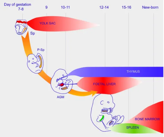
1.1.4.1. The main stepsHematopoietic cells are generated during embryonic development and sequentially colonize the FL, the thymus, then the spleen, and finally the BM (Fig 3) (Cumano and Godin, 2007). The first hematopoietic site is the yolk sac (YS) blood islands, where the hematopoietic
activity initiates from early organogenesis (soon after gastrulation) and hematopoietic progenitors are generated in situ (Haar and Ackerman, 1971). Because of this early generation, for years researchers considered the YS to be the source of HSCs that colonize further hematopoietic sites. However, the YS origin of HSCs was challenged by chick/quail chimeras construction experiments (quail embryo body grafted into a chicken YS) that showed YS-derived cells are not able either to produce hematopoietic cells sustainably or to give rise to lymphoid cells (Beaupain et al., 1979; Dieterlen-Lievre, 1975; Lassila et al., 1982). Hence, YS progenitors lack of multipotential and long-term maintenance, therefore, the YS is not the initial source of HSCs. In addition, similar chick/quail chimeras experiments showed that HSCs come from the embryo body, precisely the aorta region. In the mouse, Godin et al demonstrated that at embryonic day 9 (E9), the region that contains the developing aorta, known as the paraaortic splanchnopleura (P-Sp), was able to give rise to B cells upon transfer into irradiated mice, whereas the YS precursors could not (Godin et al., 1995). Other groups pointed to the same region named the aorta-gonad-mesonephros (AGM) at slightly later stage (E10.5) as containing stem cells capable of long-term multilineage repopulation in adult recipients (Medvinsky et al., 1996; Muller et al., 1994). The P-Sp and AGM are identical regions at two different development time points and can be referred to as P-Sp/AGM.
The in situ origin of HSCs in the mouse dorsal aorta was established through organ culture of the embryonic site that will give rise to the P-Sp/AGM. This part of the embryo (called Splanchnopleura) was isolated at E8, before any hematopoietic cells are generated in the embryo body, and also before blood cells circulate from the YS to the embryo (a connection that occurs at the 4-5 somite [S] stage) (McGrath et al, 2003). In vitro and in vivo analyses of the hematopoietic cells grown for the Sp-explants and from the corresponding YS showed that hematopoietic progenitors with multilineage potential (including lymphoid) and LTR-activity could develop after organ culture from the Sp region that will later give rise to the P-Sp/AGM, indicating in situ generation of HSCs. In contrast, the corresponding YS,
erythro-myeloid reconstitution potential in vivo (Cumano et al., 1996, 2001). The model of HSCs development during embryonic life is the same in human embryos (Ivanovs et al., 2014; Tavian et al., 2001).
These findings together established the fundamental notion that embryonic hematopoiesis initiates in two independent sites, first in the YS and later in the P-Sp/AGM that is the first place for HSCs emerge (Cumano and Godin, 2007; Cumano et al., 2001; Godin et al., 1999). As a consequence, the YS-derived hematopoiesis is independent on HSCs, and it does not follow the same differentiation processes as in the adult hierarchy (Fig 1). I will first give the information about HSCs development, then describe later the details of the development of YS-derived hematopoiesis, which is more related to my thesis project.
Figure 3: Schematic representation of the successive hematopoietic sites during mouse ontogeny.
An illustration of a mouse shows the hematopoietic sites and organs during embryonic development (two independent hematopoietic sites: yolk sac and P-Sp/AGM; intermediate site: fetal liver; hematopoietic organs: the first being the thymus, then the spleen and, finally, the bone marrow; the placenta has not been considered to be a hematopoietic organ,). P-Sp/AGM: paraaortic splanchnopleura/aorta-gonad-mesonephros region.
1.1.4.2. HSCs generation and further contribution to hematopoietic development
HSCs are generated in the P-Sp/AGM region but do not differentiate in situ (Godin et al., 1999; de Brujin et al., 2000; Kumaravelu et al., 2002; Lancrin et al., 2009). In vitro assays
(Godin et al., 1999). In addition, the circulating blood is found to contain multipotent hematopoietic progenitors as early as E10 when the production of HSCs has not reached the maximum level (Delassus and Cumano, 1996). These observations suggest that P-Sp/AGM should be considered as a generation site of HSCs but not a site of hematopoietic differentiation. Moreover, beside the aorta, other arterial regions (vitelline and umbilical vessels) were found to be sites for HSCs emergence (de Brujin et al., 2000; Inman and Downs, 2007), suggesting a close relationship between the developing hematopoietic and vascular systems.
Intraembryonic HSCs were phenotypically characterized as c-Kit high, CD41 medium, CD45 low to undetectable (Bertrand et al., 2005a). They also share a large number of markers with endothelial cells, such as CD31, CD34, AA4.1, VE-Cadherin, Tie2, etc. Because of the close physical relationship between developing HSCs and the artery wall, and also because HSCs and endothelial cells share so many surface markers and transcription factors (Scl/Tal-1, Lmo2 Runx1, GATA-2) that are important for the production of hematopoietic progenitors, a common progenitors for both lineages was considered (hemangioblast for the YS and hemogenic endothelium for the P-Sp/AGM). The model considered now for HSCs generation, following evidences gathered in zebrafish (Bertrand et al., 2010; Kissa and Herbomel, 2010), chicken (Jaffredo et al., 1998) and mouse (Boisset et al., 2010) models, is that they develop from already differentiated endothelial cells through a mechanism called endothelia to hematopoietic transition.
Once HSCs are generated, they rapidly enter circulation and reach the different hematopoietic organs (Fig 4). HSCs activity was found in the mouse placenta starting at E10.5-E11.0 and the placental HSCs pool rapid expands between E11.5 and 12.5 (Gekas et al., 2005). As a result, the placenta harbors significant numbers of HSCs that is over 15-fold more than does the AGM region or the YS, suggesting that the placenta may provide a unique microenvironment for HSC development. Placental HSCs could be generated in situ or be colonized by AGM- or FL-HSCs though circulation, or both, which still needs to be comprehensively studied.
The properties of HSCs in each site vary in function, most probably depending on diverse niches that modulate HSCs expansion and/or differentiation and ensure intrinsic characteristics of HSCs at each stage (Cordeiro Gomes et al., 2016; Martinez-Agosto et al., 2007; Wang and Wagers, 2011). The FL is considered as the major site of hematopoietic site during embryonic development, where the size of hematopoietic cell pool largely amplifies to fulfill the requirement of embryonic development (Godin and Cumano, 2002; Mikkola and Orkin, 2006). Exogenous hematopoietic cells colonize the FL to initiate hematopoiesis (Müller et al., 1994; Sánchez et al., 1996; Kieusseian et al., 2012). Considering the limited number of HSCs produced in the P-Sp/AGM (500-1000 cells) (Godin et al., 1999) and the extent of hematopoietic activity observed at early stages in FL, it is likely that the majority of cells that initially colonize the FL are erythroid/myeloid cells from the YS, which has generated numerous hematopoietic cells and establishes the first vascular connections to the FL through vitelline vessels (Cumano and Godin, 2007). HSCs are detected in the FL at E10 (Kieusseian et al., 2012). In contrast to adult BM, where HSCs are largely quiescent (Oguro et al., 2013; Reagan and Rosen, 2016), in the FL, HSCs are actively cycling. Indeed, FL provides an unique microenvironment that allows the rapid expansion of HSCs. HSCs expand in numbers from E12.5 to E16.5 and acquire the final characteristic phenotypic makers that define them in the adult BM around E15 (Morrison et al., 1995). In addition to supporting HSCs expansion, the FL is also the main hematopoietic site for the differentiation of erythrocytes, myeloid cells and lymphocytes (Kieusseian et al., 2012). It is generally stated that FL HSCs migrate to the BM and become the main contributors to adult hematopoiesis. Transplantation assays in mice have demonstrated that FL HSCs start to colonize the BM during late period of gestation (Medvinsky et al., 2011).
Figure 4: Migratory and circulatory routes of hematopoietic cells in the murine embryo over developmental time.
The first hematopoietic progenitors are found in the YS blood islands at E8.0-E8.5. Once circulation is established, blood cells colonize other developing hematopoietic organs through vitelline artery. Around E9.5, the P-Sp/AGM initiate the generation of blood precursors that, together with YS cells, colonize to the FL rudiment. The FL is the major hematopoietic site where blood progenitors expand and/or mature. Finally, the BM is colonized by precursors from the FL before birth and remains the main hematopoietic niche throughout adult life (from Costa et al., 2012).
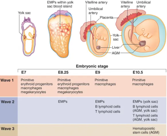
1.1.5. Distinct waves of embryonic hematopoiesis
1.1.5.1. The three wavesDuring embryonic development, the hematopoietic system is established in three successive waves that are temporally and spatially restricted, with each producing specific blood progenitors (Fig 5) (McGrath et al., 2016; Palis, 2016; Yoder, 2014). The first wave of blood production is termed “primitive” and initially arises in the blood islands of the developing YS at E7.5 (Palis, 2014). The initial primitive hematopoiesis process is considered to be unilineage, while the adult-type hematopoiesis that is termed “definitive” is multilineage. Studies in the spatial and temporal kinetics of early embryonic hematopoiesis potential indicate that primitive hematopoiesis is restricted to the erythroid, megakaryocyte, and MΦ lineages and does not produce lymphoid cells or HSCs (Palis et al., 1999; Tober et al., 2006, 2008; Yoshimoto et al., 2011). I will give more information on this wave in the following chapter.
The second wave of hematopoiesis occurs in the YS between E8.0 and E8.5 with the appearance of “erythro-myeloid progenitors” (EMp) that continue to be formed until E10.5 (Frame et al., 2013, 2015; Lux et al., 2008; McGrath et al., 2015a). Clonogenic analysis has confirmed that EMp are able to give rise to definitive erythroid cells and megakaryocyte, and to broad myeloid lineage (MΦ, and various types of granulocytes, as well as their common granulo-monocytic [GM] progenitors), but do not contain lymphoid potential (McGrath et al., 2015b), meaning that the primitive and EMp-derived definitive hematopoiesis share erythro-myeloid potential. Contrary to EMp that give rise to the mature cells through a progressive differentiation process with intermediate progenitors (GM and EMk), mature MΦ in the first primitive wave appear to lack of the intermediate progenitors.
Because EMp-derived hematopoiesis wave lacks the long-term repopulating potential and produces mature cells in a differentiation pathway similar to the adult, this wave has been called the ‘‘transient definitive’’ wave, distinguishing it from the earlier primitive
hematopoiesis that is composed of transient wave of monopotent hematopoietic progenitors and the third which is the HSCs-derived definitive wave of hematopoiesis (McGrath et al., 2016). McGrath et al showed that YS-derived EMp have a unique immune-phenotype (McGrath et al., 2015a): at E8.5, EMp express significant c-Kit and CD41 on their cell surface, while primitive erythroid progenitors (primitive erythroid colony-forming cells [EryP-CFCs]) reside in the c-KitloCD41lo population. By E9.5, EMp can be prospectively identified as the surface marker of c-Kit+CD41+CD16/32+, distinguishing from maturing populations of c-Kitneg CD16/32+ CD45hi primitive MΦ and c-Kitneg CD41hi Gp1bβ+ primitive megakaryocytes. At E10.5, EMp are found to remain present in the YS and the blood circulation, but are more highly enriched in the FL, where they expand and differentiate into multiple cell lineages (McGrath et al., 2015a).
Recent evidences showed that during the second wave of hematopoiesis, B and T lymphoid progenitors also arise from the YS, as well as in the embryo proper around E9.0 (Boiers et al., 2013; Cumano et al., 1996; Yoshimoto et al., 2012). These B or T lymphocyte-restricted progenitors migrate to and differentiate in the early FL, and are thought to provide unique subsets of lymphoid cells that persist in the adult. However, although this wave of hematopoiesis contains erythro-myeloid and lymphoid potential, it is not involved in adult-type HSCs.
The third wave begins at E10.5 with the emergence of HSCs in the AGM regions and in other embryonic arterial vessels (Cumano and Godin, 2007). As mentioned above, these precursors then colonize the FL where they begin to expand in numbers and establish definitive hematopoiesis.
Figure 5: Different hematopoietic waves during ontogeny.
The first wave originates primarily in the YS to produce primitive erythrocyte, MΦ and megakaryocytes. A second wave consists of erythroid-myeloid progenitors (EMp) and lymphoid-biased progenitors that can reconstitute lethally irradiated recipients after experimental transplantation, but spontaneously disappear before adulthood in their native environment. The third wave is that conventional fetal HSCs give rise to the quiescent adult HSCs that sustain post-natal hematopoiesis (Yoder, 2014).
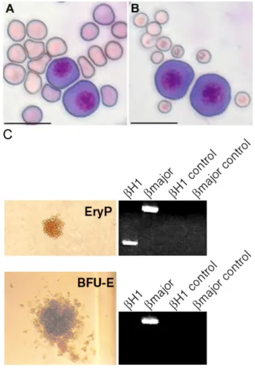
1.1.5.2. YS-derived waves: erythropoiesis in the primitive and transient definitive waves Studies of adult hematopoiesis provide an assumption that all circulating blood cells are ultimately generated by HSCs. This paradigm was also applied to the embryonic hematopoiesis in early studies. However, as mentioned above, while the emergence of HSCs begins at E10 in the AGM region of the embryo proper, a functional hematopoietic circulatory system, involving embryonic (primitive) erythroid cells, definitive erythrocytes, as well as platelets and hematopoietic progenitors, has already established in the murine embryo (Palis, 2016). In fact, early erythropoiesis is essential for survival and growth of the murine embryo, as the newly forming bloodstream need to be filled by hemoglobinizing primitive erythroblasts (Baron et al, 2012, 2013; Ferkowicz et al, 2003; Palis et al., 2001). The first few EryP-CFCs appear in the YS blood island of the late streak mouse embryo at E7.25 and produce large, nucleated βH1-globin-expressing erythroid cells. EryP-CFCs transiently expand in numbers by E8.5, and subsequently differentiate into a cohort of primitive erythroblasts in the bloodstream with the onset of the cardiac contractions at E8.25 (Kingsley et al., 2004). Just soon after the appearance of EryP-CFCs, the overlapping wave of definitive erythroid progenitors (burst-forming units-erythroid [BFU-Es]) begins to emerge, also in the YS, accompanied temporally and spatially by megakaryocyte and myeloid cells (Ferkowicz et al, 2003). In contrast with primitive erythroblasts, definitive erythroblasts are smaller, anucleate and express adult globin genes, which are similar to those found later in the FL and adult BM (Fig 6) (Baron et al, 2012). BFU-Es increase in numbers by E9.5 within the YS, then transit to the newly forming liver, prior to the HSCs colonization, and rapidly set up the definitive erythropoiesis (McGrath et al., 2011). Thus, continuous production of erythrocytes derived by BFU-Es ensures the requirement of the dramatically growing mid-gestation mouse embryo and provides an essential bridge between transient primitive erythropoiesis and HSC-derived definitive erythropoiesis. Though the primitive and definitive erythropoiesis have been well characterized, little is known about the differences between primitive and definitive megakaryocytes, as well as primitive and definitive MΦ in terms of specific function for embryonic development.
Figure 6: Comparison between primitive and definitive erythropoiesis.
(A) Morphology of primitive and definitive erythroblasts. EryP-CFCs (E10.5) were mixed with fetal (E17.5) or (B) maternal peripheral blood erythrocytes. Cytospun cells stained with Giemsa stain. Scale bar: 20 um (Baron et al, 2012); (C) Colony morphology and analysis of hemoglobin mRNA expression. Photomicrograph of an E8.5 yolk sac primitive EryP and a definitive BFU-E. Panels to the right of the photomicrographs depict the results of RT-PCR analysis of plucked erythroid colonies and reveal that EryP contain mRNA for βH1 (embryonic hemoglobin) and β globin major (adult hemoglobin) while BFU-E express mRNA for only β globin major. Negative control samples were tested using no reverse transcriptase (Ferkowicz et al, 2003).
1.1.5.3. Early ontogeny of YS-derived MΦ
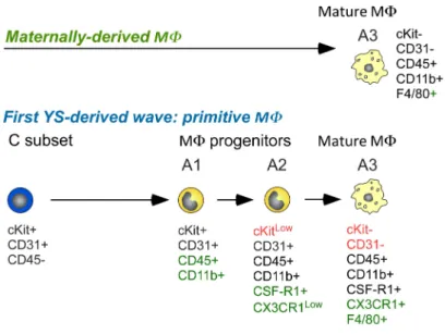
CX3CR1 is the receptor for chemokine CX3CL1/fractalkine and is prominently expressed in the mononuclear myeloid compartment (Jung et al., 2000). By using a mouse strain model in which the CX3CR1 chemokine receptor gene was replaced by a green fluorescent protein (GFP) reporter gene (referred to as CX3CR1GFP mice), Bertrand et al described three different pathways leading to the establishment of MΦ populations in the YS during early embryonic development, according to the correlation of progenitor phenotypes and differentiation potentials (Fig 7) (Bertrand et al., 2005b). The first MΦ population detected in the YS is maternally derived-MΦ that colonize the mesoderm layer from E7.5 on and display a mature phenotype (CD45+CD11b+F4/80+). These maternally derived cells transiently contribute to the MΦ population pool and cannot be detected in the YS after E9.5. A first wave of YS-derived MΦ is produced by monopotent MΦ progenitors (YS-MΦ), which appear slightly later at E8 (from the first somite stage). These YS-MΦ display a CD45-c-Kit+ phenotype and generate primitive MΦ population through a differentiation pathway (A1, CD45+c-Kit+CD11b+; A2, CD45+c-KitloCD11b+CX3CR1lo; A3, CD45+c-Kit-CD11b+CX3CR1+F4/80+), which is similar to that found in the adult. Later, progenitors displaying erythro-myeloid potential (YS-EMp) give rise to the second YS-derived MΦ wave, which is a transient definitive population, as well as granulocytes and monocytes. These YS-EMp-derived MΦ progenitors arise independently in the YS at around the 5S stage and share a similar phenotype (CD45-c-Kit+) and MΦ differentiation pathway to the previous one. Consistent with these observations, utilizing the MΦ lineage-tracing approaches, Hoeffel et al (Hoeffel et al., 2015) recently identified two independent YS-derived MΦ waves during the transient definitive stage. A first wave of called “early” EMp-like cells mainly generates MΦ population at E7.5, which might represent primitive MΦ, and the second wave of “late” EMp at E8.25 gives rise to YS MΦ in situ. Then these precursors migrate to the FL through blood circulation at E9.5, where they generate progenitors with broader myeloid cell potential, including FL monocytes. Both of these successive YS-derived MΦ populations have potential to contribute to the establishment of
resident tissue MΦ and participate in embryonic developmental events by their phagocytic nature.
Figure 7: Developmental stages followed by the 3 distinct MΦ waves, characterized by their phenotype, origin, differentiation potential, and lineage relationship.
1.1.6. The macrophage lineage
MΦ are multifunctional cell types that are found in most tissues in mice and play a central role in tissue homeostasis and repair, as well as immunity (Okabe and Medzhitov, 2016; Wynn et al., 2013; Wynn and Vannella, 2016). During embryonic development, they have mainly been involved in the clearing of dead cell through their phagocytic activity, which make them important for the shaping of the developing embryo (Mosser et al., 2017; Veiga-Fernandes and Pachnis, 2017). But now, they appear to play more specialized functions during development since recent genetic lineage-tracing experiments demonstrated that early HSC-independent hematopoiesis contributes to long-lived tissue-resident MΦ populations such as brain microglia, skin Langerhans cells as well as liver Kupffer and lungs alveolar MΦ, that persist into adulthood (Davies et al., 2012; Gomez Perdiguero and Geissmann, 2015). Most of them are maintained by local self-renewal, without replenishment by adult HSCs, under healthy condition. Thus, both HSC-derived and HSC-independent MΦ compartments are co-existing in the adult blood system. Understanding the unique features of fetal-derived MΦ cells will be necessary to fully decode the intrinsic disease processes involving these MΦ.
1.1.6.1. Tissue-resident macrophages
Tissue-resident MΦ include spleen red-pulp MΦ, lung alveolar MΦ, epidermal Langerhans cells, brain microglia, liver Kupffer cells, large peritoneal MΦ, and F4/80bright pancreatic, kidney and cardiac MΦ, etc (Fig 8). For ages, it was believed that tissue-resident MΦ were continuously repopulated by blood-circulating monocytes that are produced by HSCs-derived myeloid progenitors in the adult BM. However, in recent years, the knowledge for the origin and maintenance of tissue-resident MΦ has changed dramatically. Several studies have now revealed that except for the mucosal/border tissues such as the intestine, the dermis and the heart, in which circulating monocytes are constantly
replenishing the aging tissue-resident MΦ pool, the majority of tissue-resident MΦ populations are embryonically derived and can be self-maintained by local proliferation throughout adulthood, without a contribution from BM-derived precursors in the steady state (Bain et al., 2016; Ensan et al., 2016; Epelman et al., 2014; Gibbings et al., 2017; Hashimoto et al., 2013; Hoeffel et al., 2012; Sheng et al., 2015; van de Laar et al., 2016). The current understanding of tissue-resident MΦ ontogeny is largely depended on lineage tracing approaches in vivo driven by genes generally involved in myeloid differentiation (Csf1r or
Cx3cr1) or hematopoietic emergence (Runx1 or Tie2), which provide strong evidences for
the source of tissue-resident MΦ populations from the different hematopoietic waves.
Figure 8: Localization and functions of macrophage and monocyte subpopulations. AM, alveolar macrophage; KC, Kupffer cell; LC, Langerhans cell; LPM, large peritoneal
1.1.6.2. Strategies for the fate-mapping system
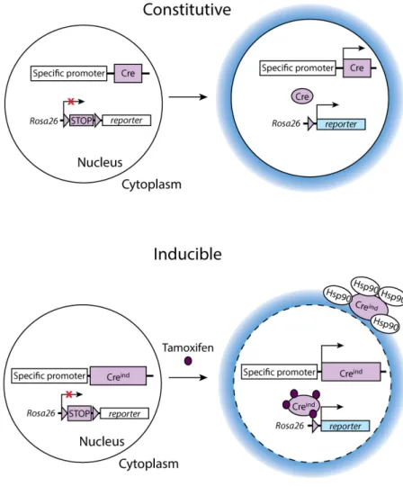
Despite several shortages for temporal restriction and promoter usage in different hematopoietic waves and their progeny, lineage tracing approach is still a very powerful tool to assess the fate of progenitor cells in vivo. Generally, in conditional gene targeting mouse model, Cre is inserted by site-specific homologous recombination into the selected locus through genetic manipulations. The principle of target gene selection is that its expression in mice should be restricted to a particular developmental stage or certain cell lineage, or both. Conditional gene expression has been achieved by engineering loxP-flanked stop sequences located between the promoter and a cDNA gene encoding a fluorescent marker, such as EGFP, tdRFP, EYFP, LacZ, TomatoRed, or another spectral variant (Fig 9). The expression of selected target gene is simultaneously activating the Cre-recombinase protein expression that irreversibly removes the loxP-flanked stop codon, resulting in turning on the permanent expression of fluorescent protein. The Rosa26 locus is one of the commonest targets to be used as a reporter gene driver because of its strong and ubiquitous expression pattern (Zambrowicz et al., 1997). In addition, some specific signature genes of cell type are also widely used as a target reporter, such as Cx3cr1 in the MΦ lineage (Wolf et al., 2013),
FMS-like tyrosine kinase 3 (Flt3) in stem cells (Boyer et al., 2011), etc. Because
Cre-recombinase is constitutively active, once it is expressed at a particular stage, or in a lineage, all daughter cells will be continuously labelled. For example, because BM LSK precursors have high-level Flt3 expression, the majority of myeloid and lymphoid lineages progenies are marked in Flt3Cre knock-in adult mice (Boyer et al., 2012).
Moreover, in order to temporally control Cre activity to study different hematopoietic waves of developing cells, or to be able to study the effects of gene deletion at late development stages or in the adult when the mutant dies at early stage of embryonic development, inducible gene targeting mice model has been created by fusing the ligand-binding domains of steroid hormone receptors, such as estrogen receptor (ER) and modified ER (Mer), to chimeric recombinases (Metzger et al., 1995; Metzger and Feil, 1999). Specifically, in the absence of the inducing agent (tamoxifen; metabolized to 4-hydroxytamoxifen, the active
compound), the heat shock protein Hsp90 forms heterodimers with Cre-recombinase protein in the cytoplasm and prevents Cre from removing the loxP-flanked stop codon to activate reporter gene expression. Under injection of tamoxifen, Cre-recombinase protein is released from the inhibitory complex and translocated into the nucleus, finally leading to heritable labeling of the targeted cell (Fig 9).
Figure 9: A Schema of the constitutive and inducible conditional gene targeting approaches.
1.1.6.3. Lineage tracing of the origin of macrophage in different tissues
Several transgene mouse models of inducible gene targeting, such as the Runx1-Mer-Cre-Mer, the Tie2-Mer-Cre-Mer (Tie2-iCre) and the c-Kit-Mer-Cre-Mer (c-Kit-iCre) mice, have been exploited for the MΦ lineage tracing studies, as well as other hematopoietic cells (Fig 10) (Ginhoux et al., 2010; Gomez Perdiguero et al., 2015; Sheng et al., 2015). Runx1 plays a pivotal role for the emergence of embryonic hematopoiesis and loss-of-function Runx1 is lethal to the embryo (Hirschi, 2012). Runx1 is first expressed in hematopoietic cells as they emerge in the YS. Therefore, in Runx1MerCreMer mice (Ginhoux et
al., 2010), in which Mer-Cre-Mer recombinase is induced by tamoxifen under the control of
Runx1, cells labeled in the YS at E7.5, a time when Runx1 expression is limited to the
primitive hematopoiesis, will only give rise to primitive MΦ, as well as primitive EryP-CFC and megakaryocytes, and tamoxifen application at few hours later can lead to either EMp-derived definitive MΦ (at E8.5) or HSCs-derived definitive MΦ (at E9.5) labeling in this system (Fig 10A).
In contrast, Tie2 is expressed on all the endothelial-like precursors of hematopoiesis by E7.5 and then is turned off in hematopoietic cells after they differentiation (McGrath et al., 2015b). Thus, all endothelial cells and their progeny (both non-hematopoietic and hematopoietic cells) will be labeled after tamoxifen injection in the Tie2-iCre model. Consequently, an early tamoxifen injection (at E7.5) will target all hematopoietic waves-derived MΦ that are produced before the time of analysis, whereas a late injection (at E10.5) will only target the latest HSCs-derived definitive MΦ (Gomez Perdiguero et al., 2015) (Fig 10B).
Moreover, all hematopoietic progenitors, but not endothelial cells, express c-Kit (Ivanova et al., 2002). Thus, an early tamoxifen injection (at E7.5) in the c-Kit-iCre model will restrict the labeling to primitive wave (Sheng et al., 2015). However, progenitors produced by each hematopoietic wave coexist in the embryo after E8.5 and these progenitors still express c-kit. A later tamoxifen injection (at E8.5 and E9.5) might thus result in the cumulative labeling of
undifferentiated primitive MΦ progenitors and EMp-derived MΦ precursors, as well as both EMp- and HSCs-derived MΦ precursors, and their mature progenies (Fig 10C).
Figure 10: Fate-mapping systems of macrophages and other hematopoietic cell. Green color represents labeling cells (Hoeffel and Ginhoux, 2015).
1.1.6.4. The three models on the origin of tissue resident macrophages
Only recently, by using the fate-mapping system, researchers have proposed three different models for the contribution of embryonic progenitors to adult tissue resident MΦ populations (Fig 11). Using Tie2-dependent inducible mice models, Gomez Perdiguero et al (Gomez Perdiguero et al., 2015) suggested that YS-derived EMp are a common origin for the majority of tissue resident MΦ in liver, brain, epidermis and lung (Fig 11, Model 1). The authors found that there is a higher proportion of adult tissue resident MΦ YFP labeling than that of leukocytes when tamoxifen is injected upon Tie2-iCre:Rosa26YFP mice at E7.5; a similar YFP labeling proportions of both cell types at E8.5 injection; and a relatively more
frequently YFP labeled in leukocyte populations than tissue resident MΦ from E9.5 injection. These findings, thus, indicated that a progenitor cell type that expresses Tie2 at E7.5 but not after E9.5, gives rise to the majority of fetal and adult MΦ, and the authors proposed this progenitor to be YS EMp.
In another model, researchers (Hoeffel et al., 2015) proposed that except for brain microglia, which derives from YS MΦ, the most other adult tissue resident MΦ, including skin, liver, kidney, lung, gut and spleen, derive predominantly from fetal monocytes generated by YS c-myb+ EMp (Fig 11, Model 2). Using Runx1- and Csf1r-dependent inducible mice models, as well as Rosa26R26-EYFP/R26-EYFP, Hoeffel et al (Hoeffel et al., 2015) identified two temporally and functionally distinct waves of EMPs emerging in the YS between E7.5 and E8.5. The first wave of “early” CSF-1Rhic-Myb- "EMp" emerges in the YS from E7.5 and gives rise mostly to brain microglia. The “late” CSF-1Rloc-Myb+ EMp are produced in the YS from E8.5 and most of these cells migrate into the FL through the blood circulation at E9.5. After colonized the FL, these cells differentiate into multiple lineages, including FL monocytes that give rise to most tissue resident MΦ through a monocytic intermediate. In the third Model, Sheng et al recently concluded that all adult tissue resident MΦ, such as Kuppffer cell, alveolar MΦ, red pulp MΦ, kidney F4/80hi MΦ, dermis MHCIIloF4/80hi MΦ, as well as colon F4/80hi MΦ and peritoneum F4/80hi MΦ, are progenies of fetal classical HSCs with the exception of microglia and partially epidermal Langerhans cells, which originate from YS progenitors (Fig 11, Model 3) (Sheng et al., 2015). The authors have generated a c-Kit-iCre:Rosa26-loxP-STOP-loxP-eYFP mice model in which the administration of tamoxifen induces irreversible tagging of c-Kit expressing cells, as well as their progenies. In this fate-mapping system, researchers found that adult microglia and partial epidermal Langerhans cells can be robustly labeled when tamoxifen is injected at E7.5, but other tissue resident MΦ, as well as peripheral hematopoietic cells, are labeled very poorly. In contrast, tamoxifen injection at E8.5 or E9.5 efficiently labels all adult peripheral hematopoietic cells including adult tissue resident MΦ (about 40%-60%). These results therefore led to the hypothesis that definitive fetal HSCs give rise to all the adult tissue
resident MΦ, with the exception of microglia and, partially, epidermal Langerhans cells, which arise from YS-derived primitive MΦ progenitors.
Figure 11: Three Different Models of Fetal Macrophage Ontogeny.
The Model 1 corresponds to the work of Gomez Perdiguero et al. (2015), the Model 2 to the work of Hoeffel et al. (2015), and the Model 3 to Sheng et al. (2015). Red arrow indicates the proposed major path of ontogeny and differentiation in each model. Cell colors are matched to their proposed origins (Ginhoux and Guilliams, 2016).
1.1.6.5. The origin of embryonic microglia
Microglia are CNS resident MΦ that play pleiotropic functions in the normal and pathological brain. Impaired microglial development and function are thought to be involved in the onset and development of various neurodevelopmental and neurodegenerative diseases (Colonna and Butovsky, 2017; Prinz and Priller, 2014). Thus, understanding of the origin of microglia may provide new therapeutic approaches for the treatment of these diseases. In fact, studies in the past decades have been carried out to dissect the origin of microglia (Alliot et al., 1999; Epelman et al., 2014; Ginhoux et al., 2010; Gomez Perdiguero et al., 2015; Hashimoto et al., 2013; Hoeffel et al., 2015; Kierdorf et al., 2013; Schulz et al., 2012). As stated above, although there is a controversy for the origin of some tissue MΦ, all fate mapping experiments agreed that microglia originates from early YS-derived MΦ. Similarly, by combining vital staining with in situ hybridization, Herbomel et al found that, in zebrafish embryos, early MΦ (equivalent to primitive MΦ in mice [Herbomel et al., 1999]; for a review on hematopoietic cell development in the zebrafish embryo, see Bertrand and Traver, 2009) colonize the brain and differentiate into microglia through a CSF1R-dependent invasive process (Herbomel et al., 2001). Rossi et al further demonstrated that in the zebrafish, it is the slc7a7+ primitive MΦ subset produced early during development in the anterior lateral plate mesoderm region that will give rise to embryonic microglia (Rossi et al., 2015).
However, in contrast to zebrafish embryos, in which primitive and definitive hematopoiesis are separated by temporally and spatially (Bertrand and Traver, 2009), in mice, primitive and EMp-derived MΦ subsets appear overlapped and coexist in the YS so that their respective contribution to microglia was naturally questioned. Primitive hematopoietic populations have been characterized as independent of c-Myb-expression for their development, contrary to their definitive counterpart (McGrath et al., 2015b; Gomez Perdiguero and Geissmann, 2013; Tober et al., 2008). The normal amount of microglial cells found in mice lacking the c-Myb transcription factor argues for a primitive origin of microglia progenitors (Kierdorf et al., 2013; Schulz et al., 2012). Additionally, Ginhoux et al. reached a similar conclusion because
the induction of Runx1 expression at E7.25-7.5 that leads to microglia labeling, targets progenitors present at the latest E8.5 (Ginhoux et al., 2010), when EMp-derived MΦ progenitors are not yet produced. However, this protocol leads to 30-40 % microglia labeling, and although this level is likely to be underestimated, an additional contribution from a slightly later source, namely EMp-derived MΦ progenitors, might be considered (Ginhoux et al., 2010). Conversely, two sets of investigations suggested that EMp-derived definitive MΦ population provide microglia (Gomez Perdiguero et al., 2015; Kierdorf et al., 2013). Sorted c-Kit+CD45low progenitors from E8-YS were shown to give rise to erythro-myeloid cells in vitro and to microglial cells upon culture on microglia-depleted hippocampal slice cultures (Kierdorf et al., 2013). In the second study (Gomez Perdiguero et al., 2015), the induction of csf-1rCre at E8.5 resulted in the labeling of c-Kit+CD45low MΦ progenitors in E9.5 YS and of head MΦ at E10.5. However, EMp and primitive MΦ progenitors share the same c-Kit+CD45low phenotype and are both present in the YS at the stages analyzed (Bertrand et al., 2005b), so that in both instances, the identification of the MΦ/microglia progenitors is still unclear (McGrath et al., 2015b). The question of the respective contribution of primitive and EMp-derived definitive MΦ progenitors to the developing microglia thus remained open.
1.2. Development and function of microglia in mice
1.2.1 Myeloid cell types in the central nervous system
The central nervous system (CNS) contains a heterogeneous class of myeloid populations that play distinct roles in maintaining tissue homeostasis and pathological response in different tissue compartments during development and adulthood (Prinz et al., 2011; Ransohoff and Cardona, 2010). According to their localization, morphology, as well as surface-marker expression and in vitro responses, the myeloid cells of the CNS have been characterized and classified into five groups: parenchymal microglia, non-parenchymal meningeal MΦ (mMΦ), CNS perivascular MΦ (pvMΦ), MΦ of the choroid plexus (cpMΦ) and disease-associated monocytes (Fig 12) (Prinz et al., 2017). Specifically, microglia are the only myeloid cell type present in the CNS parenchyma where they are surrounded by neurons, astrocytes and oligodendrocytes; subdural mMΦ are in close proximity to meningeal fibroblasts; pvMΦ are located between the laminin+ endothelial and glial basement membranes; cpMΦ are mainly found on the apical side of the choroid plexus epithelium facing the cerebrospinal fluid and in the stroma (Prinz and Priller, 2014; Ransohoff and Cardona, 2010).
In addition, CNS MΦ have been recently shown to possess a unique feature: In most organs, such as the heart, spleen, lungs and intestine, tissue MΦ are regularly replaced by blood-derived monocytes (Goldmann et al., 2016). In contrast, except cpMΦ, other CNS MΦ, including microglia, mMΦ and pvMΦ, have been shown to originate entirely from embryonic YS-precursors without any significant input from the circulating blood cells or BM-derived monocytes during adulthood (Goldmann et al., 2016). In these locations, MΦ are maintained by their longevity and capacity of self-renewal (Soucie et al., 2016). Interestingly, these MΦ have unique phenotypes in the healthy CNS, in addition to MΦ-related surface markers common among the resident MΦ, such as Iba-1, F4/80, CD11b, CX3CR1 and MERTK. Microglia display a down-regulated phenotype with a low expression of the transmembrane tyrosine phosphatase CD45, of Fc receptors and major
histocompatibility complex class II (MHC class II) compared to other CNS MΦ. Several microglia-specific genes such as P2ry12, Fcrls, Hexb, Tmem119, Tgfbr1 and Sall1 have also been identified in large gene-expression studies (Bennett et al., 2016; Buttgereit et al., 2016; Gautier et al., 2012; Gosselin, et al., 2014; Lavin et al., 2014). Furthermore, mMΦ and pvMΦ selectively express the endocytic pattern-recognition receptor CD206 (Goldmann et al., 2016). Moreover, pvMΦ, as well as part of mMΦ and cpMΦ, were found to express the scavenger receptor CD163 (Goldmann et al., 2016). Recent single-cell RNA-sequencing studies further showed that pvMΦ specifically express Cd163, Hpgd, Mrc1, Slc40a1 and
F13a1 (Goldmann et al., 2016; Mass et al., 2016; Zeisel et al., 2015). The discovery of
specific expression signatures in the various MΦ subsets in the CNS may be due to the profound effect of the local microenvironment, pointing to the different roles of CNS MΦ in homeostasis and defence. In this part, I will focus on recent findings in regulation of microglia development and the role of microglia during neurodegenerative disease.
Figure 12: The localization and genetic signature of CNS macrophages.
CNS MΦ can be distinguished from monocytes by the expression of Ly6C, a marker present on monocytes but not on CNS MΦ (Prinz et al., 2017).
1.2.2. The development of microglia
The population of parenchymal microglia in the adult murine CNS accounts for 5% to 12% of the total number of glial cells and varies the different brain regions analyzed (from 22 to 165 cells/mm2) (Aguzzi et al., 2013). Contrary to neurons and glia that derive from the neuroectoderm, microglial cells originate from the YS mesoderm. By crossing the Runx1iCre mice with floxed Rosa26 reporter mice and performing tamoxifen-mediated induction at different time points Ginhoux et al (Ginhoux et al., 2010) strongly demonstrated that the YS-derived primitive MΦ population that initially present at E7.5 is the major source of the resident adult microglia (Fig 13). The transcription factor c-Myb is required for the development of definitive hematopoiesis (Tober et al., 2008), and for the maintenance and renewal of HSC-derived myeloid cells (Schulz et al., 2012). Nevertheless, microglial pool is unaffected in c-Myb-deficient embryos, confirming that they derive from the earlier YS population (Schulz et al., 2012). It was further proved that the development of
c-Myb-independent microglia requires the transcription factor Pu.1, as well as Csf1r
(Ginhoux et al., 2010; Kierdorf et al., 2013).
Microglia populate the CNS during early embryogenesis: MΦ progenitors are initially found in the neuroepithelium of mice at E8.5-10, with substantial numbers being detected in the fourth ventricle at E10.5. From E11.5, they start to invade the cortex (Swinnen et al., 2013). Specifically, MΦ/microglia cells cluster in the pial surface of the cortex and within the lateral ventricles at E12.5, the onset of this migration process. Afterwards, they become randomly distributed throughout the cortical wall, except for the cortical plate region in later embryonic ages (Swinnen et al., 2013). However, to date, little is known about how microglial migration, recruitment and positioning into the cortex are regulated during embryonic CNS development. Recent studies showed that the ablation of the basal progenitors that express the chemokine SDF1/Cxcl12+ in the subventricular zone affects CX3CR1+ microglia recruitment into the ventricular/subventricular zone (Arnò et al., 2014), suggesting that neural progenitor cells may orchestrate the migration and positioning of