V. Geenen 9 B. Goxe 9 H. Martens 9 E. Vandersmissen Y. Vanneste 9 I. Achour 9 O. Kecha 9 RJ. Lefebvre
Cryptocrine signaling in the thymus network and T cell education
to neuroendocrine self-antigens
Received: 24 October 1994 ! Accepted: 4 July 1995
Abstract Both during phylogeny and ontogeny the thy-
mus appears as a nodal point between the two major sys- tems o f cell-to-cell signaling, the neuroendocrine and immune systems. This review presents the experimental observations which support a dual role in T cell selection played by the thymic repertoire of neuroendocrine poly- peptide precursors. Through the mode of cryptocrine in- tercellular signaling thymic neuroendocrine-related pre- cursors synthesized in thymic epithelial cells have been shown to influence the early steps in T cell differentia- tion. In addition, thymic neuroendocrine-related poly- peptides are a source of self-antigens which are present- ed by the major histocompatibility system of the thymic epithelium. Preliminary data also suggest that the intra- thymic T cell education to neuroendocrine self-antigens is not strictly superimposible to the antigen presentation by dedicated presenting cells. Insulin-like growth factor- II (IGF-II) was identified as one dominant member of the insulin family expressed by thymic epithelial and nurse cells. The intrathymic presentation of IGF-II or IGF-II derived self-antigens is under current investigation. If further confirmed, the central tolerogenic properties of IGF-II could be considered in the elaboration of a strate- gy for an efficient and safe prevention of insulin-depen- dent diabetes.
K e y w o r d s Thymus 9 Cryptocrine signaling 9 Neuroen- docrine self-antigens. Molecular evolution 9 Develop- mental biology 9 T cell tolerance
A b b r e v i a t i o n s I D D M Insulin-dependent diabetes 9 I G F Insulin-like growth factor 9
I G F B P IGF-binding protein - TCR T cell antigen receptor 9
M H C Major histocompatibility complex 9 OT Oxytocin 9 TEC Thymic epithelial cell 9 TNC Thymic nurse cell 9 VP Vasopressin
V. Geenen ( ~ ) 9 B. Goxe 9 H. Martens 9 E. Vandersmissen Y. Vanneste 9 I. Achour 9 O. Kecha 9 EJ. Lefebvre Department of Medicine-Endocrinology,
Institute of Pathology CHU B-23, University of Libge, B-4000 Li~ge-Sart Tilman, Belgium
Introduction
Although the thymus has long been considered as a gland, there is still difficulty in applying the model of endocrine cell-to-cell signaling to the intrathymic pro- cess of T cell differentiation. This may be due to the im- portance of the thymus as a central organ of the immune system, so that its immune properties have overshad- owed its endocrine role. These two functions are, howev- er, intimately interlinked, and neuroendocrine-immune interactions in T cell education have important physio- logical and pathological implications.
Thymic development of T lymphocytes
The thymus shapes the T cell repertoire in two distinct ways. The most specific role of the thymus is the induc- tion of central immunological self-tolerance which fol- lows the negative selection o f self-reactive T cells. The existence of such "forbidden" self-reactive T cell clones has never been demonstrated, but theoretically they could emerge during the random recombination of the gene segments coding for the chains of the T cell antigen receptor (TCR) [31]. The clonal deletion or developmen- tal arrest of self-reactive T cells is thought to follow the high-affinity binding of TCRs that recognize self-anti- gens presented by thymic major histocompatibility com- plex (MHC) derived proteins. The generic terms self-an- tigen or self-peptide are used in this review to designate the short eight to ten amino acid sequences derived from endogenous proteins that are effectively presented to T cells by MHC-related molecules in the thymic stroma. Another role of the thymus concerns the developmental program and the positive selection of the peripheral T cell repertoire. This dual physiological function of the thymus remains a paradox in contemporary immunology [2, 5]. The objective o f the present review is to show how neuroendocrine-related polypeptide precursors syn- thesized in the thymic stroma may explain, at least par- tially, the paradox of thymic physiology.
The neurohypophysial hormone family
The neurohypophysial hormones constitute a family of nonapeptides that have been highly conserved through- out evolution [1]. They can be divided in two lineages corresponding to the oxytocin (OT)-like and vasopressin (VP)-like peptides. Both of the lineages may have fol- lowed the duplication of one ancestral gene. These pep- tides all consist of nine amino acids with cysteine resi- dues in positions 1 and 6 forming a disulfide bridge. In mammalian vertebrates OT-like peptides are implicated in the control of reproduction whereas VP-like peptides regulate water homeostasis as well as some cardio-vas- cular functions. All known neurohypophysial hormones are synthesized as larger precursors which all possess a 10-kDa neurophysin-like domain in their structure. Both at the peptide and genetic levels, members of this family have recently been demonstrated in molluscs and insects (for a complete review, see [19]).
Despite their high conservation during evolution the physiological role of the neurophysins remains obscure. In the so-called higher vertebrates, the neurophysins bind and transport the active nonapeptides OT and VP along axons of the hypothalamo-neurohypophysial neurons [3, 10, 28]. However, the expression of neurohypophysial genes in various species devoid of a neurohypophysial system strongly suggests another physiological role of neurophysins which remains to be deciphered.
OT as a cryptocrine signal in intrathymic
T cell development
As early as 1910 Ott and Scott showed that thymic ex- tracts can induce milk ejection when injected into the goat. It was only in the 1950's that the biochemical prin- ciple of milk ejection was identified as OT by du Vign- eaud's group. Thymic epithelial and nurse cells (TEC/TNC) from various species synthesize neurohypo- physial-related precursors, with a marked dominance of OT and its associated neurophysin [4, 21-23, 33, 37, 40, 41]. Using a 3" RACE polymerase chain reaction proto- col, the two neurohypophysial OT and VP genes were demonstrated to be transcribed in human and murine thy- muses [27]. Since OT is the dominant peptide of the family expressed by TEC/TNC, the discrepancy in the data between the thymic cDNA and peptide levels sup- ports the existence of posttranscriptional modifications. After several years of thorough research we arrived to the conclusion that the model of neurosecretion cannot explain the processing and the fate of neuropeptides syn- thesized in thymic epithelium [23]. The intrathymic OT gene expression and OT synthesis are not correlated with secretion of the nonapeptide or its neurophysin in the su- pernatant of primary cultures of TEC/TNC. A recent ul- trastructural study has further demonstrated that ir-OT and neurophysin are expressed by TEC only (and not by immature T cells). Thymic ir-OT is not located in secre-
tory granules but is diffuse in the cytosol, clear vacuoles, and the juxtamembranar space of murine TEC [49]. Such ultrastructural features were also recently reported for OT and VP synthesized by murine spleen eosinophil-like cells [32]. The model of cryptocrine cell-to-cell signal- ing has been advanced by Funder [18] to describe the transmembrane exchanges of chemical informations be- tween large nursing cells and immature elements which migrate and differentiate at their contact. On the basis of the above findings we hypothesised that thymic OT me- diates a cryptocrine-type signaling between TEC and pre-T cells.
As an important argument supporting an effective cryptocrine signaling in vivo in the thymus, neurohypo- physial receptors are expressed in the rat thymus, by rat thymocytes (pre-T cells) [13, 16], by a murine pre-T cell line (RL12-NP), and by murine cytotoxic T cells [34, 47]. A molecular maturation of the neurohypophysial re- ception system expressed by T cells seems to occur in parallel with T cell differentiation since pre-T cells ex- press neurohypophysial V]-type receptors whereas cyto- toxic T cells express neurohypophysial receptors of the OT-type [34]. These receptors are functional since they transduce neurohypophysial signals according to the rules established in other cellular systems. The interac- tion between neurohypophysial signals and their pre-T cell receptors is followed by the phosphorylation of focal adhesion kinases [43]. This event could play an impor- tant role in promoting the T cell interactions with the thymic microenvironment which are important for their developmental program (unpublished data). Thus the bulk of available data strongly suggest that thymic OT mediates a functional cryptocrine signaling which could serve as an accessory pathway in the positive selection of T cells. In medical pathology the neoplastic transforma- tion of TEC (as seen in thymic carcinoma and in some thymomas) is sometimes associated with a clinical syn- drome of water intoxication [42]. This syndrome may be due in fact to an overexpression of thymic ir-OT, leading to a release in the peripheral bloodstream and to a subse- quent interaction with neurohypophysial V 2 receptors ex- pressed by the kidney tubular cells.
OT as the self-antigen of the neurohypophysial
hormone family
In the thymus cryptocrine signaling is intimately associ- ated with presentation of the self-molecular structure to developing T cells. This action was long thought to be mediated by interdigitating thymic cells only, but there is increasing evidence that TEC/TNC are actively involved in the thymic induction of immunological self-tolerance [9]. Since OT and its neurophysin are coexpressed by TEC/TNC, we hypothesized a processing of the thymic neurohypophysial-related precursor that could be related to the one involved in antigen presentation. Following the appropriate methodology, human thymic stromal cell membranes were purified and solubilized in a nonionic
detergent. The solution was passed on an immunoaffinity column prepared with a monoclonal antibody directed to the monomorphic part of human MHC class I molecules (monoclonal antibody B9.12) [39]. The choice of this class I specific monoclonal antibody was justified by the fact that MHC class I proteins usually present antigenic sequences derived from endogenous proteins. To avoid MHC-antigen complex dissociation, the column was eluted with diethylamine in basic pH conditions. After analysis using sodium dodecyl sulfate-polyacrylamide gel electrophoresis, instead o f the expected 45-kDa frac- tions (MW of MHC class I heavy chains), immunoblot analyses revealed a 55-kDa fraction which could be la- beled by monoclonal antibody B9.12 as well as by an an- tiserum against the central highly conserved region of neurophysins [26]. Since antineurophysin antiserum and monoclonal antibody B9.12 exhibit no cross-reactivity, the most plausible explanation for these data is that the thymic 55-kDa precursor is a chimeric or a hybrid pro- tein bearing both a neurophysin-like (10-kDa) and a MHC class I heavy chain-related (45-kDa) domain [26, 27]. The MHC class I domain is most probably implicat- ed in the membrane translocation of this chimeric/hybrid protein while its neurophysin domain could bind OT for presentation to pre-T cells. The precise biochemical mechanisms leading to the synthesis of this hybrid neu- rohypophysial/MHC class I protein remain of course to be further deciphered. Since the three exons o f neurohy- pophysial genes are transcribed in the thymus, the origin o f this protein should reside at a posttranscriptional level (such as a trans-splicing-like event), or even at a post- translational level (as the ATP-dependent covalent bind- ing to ubiquitin of proteins targeted to proteolysis).
For another experimental argument for the role of thy- mic OT as the self-antigen of the neurohypophysial fami- ly we investigated the effects of the immune recognition of neurohypophysial antigens on the cytokine profile se- creted by human TEC in primary cultures. Only antibod- ies directed to OT (but neither anti-VP antibodies nor different preparations of Igs) were able to stimulate the TEC production o f interleukin 6 and leukemia-inhibitory factor as measured by specific EASIAs (Medgenix Diag- nostics, Fleurus, Belgium; Martens et al., manuscript submitted).
The thymic repertoire of neuroendocrine self-antigens
Based upon these observations, a model (Fig. 1) has been advanced proposing that neuroendocrine-related thymic peptides ("X") engage into two distinct types of interac- tions with pre-T cells depending on their intervention as
signals or as self-antigens of their respective family. This model can be easily applied to various neuroendocrine hormone families. A kind o f economical principle ap- pears in the organization o f the thymic peptide reper- toire. TEC are not the site of expression of all members of one given family, but a representative member is dom- inantly expressed by TEC/TNC (Table 1). From our
T h y m i c a n t l g e n - p r e s e n t i n g cell |TEC, TNC o r IDC)
Cryptocr/ne slgnallln~ Physiology
Accessory signal in T-cell differentiation/activation Pharmacology Irnmunomodulation by "X" agonist s/antagonists S e l f education Physiology
Central T-cell tolerance of "X"-related endocrine fuactiorts Pharmacology
Blockade of "X"- specific autoammunity
Fig. 1 Neuroendocrine-related polypeptide precursors ("X") syn-
thesized in TEC/TNC exert two types of physiological actions in T cell differentiation. Through cryptocrine signaling they interact with neuroendocrine-type receptors ("X" receptor) expressed by target pre-T cells and may constitute accessory signals in the pro- cess of T cell development. In relationship with the thymic MHC system, the highly conserved sequences of neuroendocrine fami- lies are presented as self-antigens (self-"X') to pre-T cells and could induce the negative selection of T cells bearing a randomly rearranged TCR directed against their respective families. An effi- cient pharmacological manipulation of both types of interactions may be expected in the near future
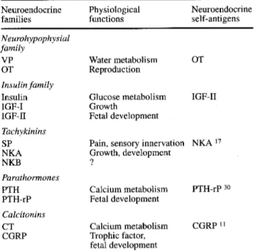
Table 1 The organization of the thymic repertoire of neuroendo- crine self-peptide precursors
Neuroendocrine Physiological Neuroendocrine
families functions self-antigens
Neurohypophysial family VP OT Insulin family Insulin IGF-I IGF-II Tachykinins SP NKA NKB Parathormones PTH PTH-rP Calcitonins CT CGRP Water metabolism OT Reproduction Glucose metabolism Growth Fetal development IGF-II
Pain, sensory innervation NKA 17 Growth, development ? Calcium metabolism PTH-rP 3o Fetal development Calcium metabolism Trophic factor, fetal development CGRP ~1
studies, a difference also appears between neuroendo- crine autoantigens expressed by the peripheral tissues tackled by an autoimmune response and their homolo- gous self-antigens expressed in thymic epithelium. This difference is important to take into account since autoan-
Table 2 The insulin peptide family B domain A domain 1GF-H Human Cow Rat Mouse IGF-I Human Cow Rat Mouse Insulin Lymn~ea Bombyx Hagfish Guinea pig Rat Human Leydig I-L Relaxin Porcine R P S E T L C G G E L V D T L Q F V C G D R G F Y F S G G S GPETLCGAELVDALQFVCGDRGFYFNKPT P P PHRRGVCGSALADLVDFACSS SNQPAMV QAVHTTCGRHLARTLADLCWEAGVD RTTGHLCGKDLVNALTIACGVRGFFYDPTLM FVSRHLCGSNLVETLTSVCQDDGFFYIPKD FVNQHLCGSHLVEALYLVCGERGFFYTPLT FVNQHLCGSHLVEALYLVCGERGFFYTPLT PAQEAPEKLCGHHFVRALVRLCGGPRWSPEDG NDFIKACGRELVRLWVEICGVWS G I V E E C C F R S C D L A L L E T Y C A GIVDECCFRSCDLRRLEMYCA NIVCECCMKPCTLSELRQYCP GIVDECCLRPCSVAVLLSYC GIVEQCCHKRCSIYNLQNYCN GIVDQCCTGTCTRHQLQSYCN GIVDQCCTSICSLYQLENYCN GIVEQCCTSIC SLYQLENYCN NPARHCCLSGCTRQDLLTLCPH TLSEKCCEVGCIRKDIARLC
tigens are known to activate autoreactive immune cells whereas, at least theoretically, self-antigens should de- lete or anergize autoreactive cells. In the neurohypophy- sial family there is now good evidence that OT is the neurohypophysial self-antigen. Thus a strong immuno- logical tolerance protects the OT lineage, more than the VP one, from an eventual autoimmune aggression. In- deed some cases of idiopathic diabetes insipidus have been shown to result from an autoimmune hypothalami- tis oriented toward VP-producing neurons [29, 44]. Giv- en the importance of the OT lineage in the control of the reproductive process at several levels (parturition, mater- nal behavior, lactation, and paracrine regulation of go- nadal functions), a stronger tolerance of this lineage ap- pears to be crucial for the preservation of the species. Therefore in the neurohypophysial family OT behave as the self-antigen while VP is strongly suspected to be the target autoantigen of autoimmune process. This conclu- sion is also supported by the frequence and the titers of antibodies induced by active immunization against neu- rohypophysial peptides (VP>OT>VT). An infiltration of the hypothalamo-neurohypophysial tract by inflammato- ry mononuclear cells has been observed repeatedly, both after active immunization against VP [15] and in sponta- neous autoimmune diabetes insipidus [29]. Altogether, these data strongly support the idea that hypothalamic magnocellular neurons express on their surface antigenic markers specific of their neurosecretory activity.
In the insulin family many authors have shown that insulin is an autoantigen involved in the diabetogenic au- toimmune process. Thymic epithelium is the site of a marked expression of IGF-II [25], although IGF-II is not secreted by primary cultures of human or rat TEC. A marked hyperplasia of the thymus has been observed in transgenic mice for the Igf2 gene [48]. Following the alignment of amino-acid sequences of insulin-related
members (Table 2) one may observe that the sequences of residues 7-15 in B domains and residues 1-10 in A domains are highly conserved throughout evolution of the insulin family. Therefore those sequences might be considered as self-antigens of insulin-related polypep- tides. Interestingly, these sequences closely correspond to the target antigen of cytotoxic T cells oriented against insulin [45]. Such a correspondence between highly con- served sequences and dominant antigens has also been noticed recently by others [36]. We have identified and further characterized ir-IGF-II in the cytosol and mem- brane preparations from human thymuses (Achour et al., submitted). This result is surprising because pro-IGFs possess no transmembrane domain. We are currently in- vestigating the mechanisms involved in the intrathymic presentation of IGF-II. A molecular defect in thymic T cell education to IGF-II or to IGF-II-derived self-pep- tides could play a role in the physiopathology of insulin- dependent diabetes (IDDM). IGF-II is very homologous but not identical to insulin, and this biochemical differ- ence could elicit completely opposite immune responses. Indeed, while insulin has been shown to activate autore- active cytotoxic T cells [45], IGF-II could program their tolerant state. These differences in the biochemical iden- tity and immunological responses elicited by insulin-re- lated antigens could be fundamental for the design of an efficient and secure prevention of autoimmune IDDM.
A new physiological role for neurophysin
and other binding proteins?
For a long time the binding of neurohypophysial nona- peptides to their associated neurophysins for their axonal transport has been a useful model for understanding the interactions between small peptides and larger proteins
[1, 3, 10, 28]. Interestingly, the residue tyrosine in posi- tion 2 of OT and VP had been shown to play an impor- tant role in this binding [28]. Interestingly, the residue tyrosine in the same position has been shown to play a crucial role in the binding of antigens to some MHC class I alleles [35]. According to our data, even if MHC class I pathways are implicated in the process, the thy- mic neurophysin domain seems to be the final step in the presentation of OT by TEC/TNC to immature T cells. A very close functional analogy thus exists for neurophys- ins between, on one hand, the binding and transport of OT along the neurohypophysial axons until the nerve endings of the posterior pituitary and, on the other, the binding and presentation of the self-antigen OT to imma- ture T cells at the TEC membrane. Other authors also have observed a dissociation between thymic T cell edu- cation to self and T cell recognition of antigens [46]. The major implication of this new physiological role of neu- rophysin in T cell education is that the central T cell tol- erance to the neurohypophysial self-antigen OT is n o t tightly restricted by MHC class I alleles. Another selec- tive advantage resides in the potential presentation to pre-T cells of the s t r u c t u r e characteristic of neurohypo- physial-related peptides. Because of the ring structure a classic presentation by MHC class I alleles is indeed ex- cluded [38]. The absence of a tight MHC allelic restric- tion of central T cell tolerance to neuroendocrine self-an- tigens has important implications for the prevention of autoimmune endocrine diseases. At last, the role of neu- rophysin in the presentation of the neurohypophysial self-antigen is more appropriate with their high conser- vation throughout evolution.
In the insulin-related peptide system, the role of bind- ing and transport proteins is assumed by IGF-binding proteins (IGFBPs). In contrary to neurophysins, IGFBPs are not part of IGF precursors but are encoded by sepa- rate genes. Interestingly, some IGFBPs are anchored to cell membranes, but the hypothesis of a relationship with MHC has never been explored. The putative presentation of IGF-II by a membrane-anchored IGFBP could explain why it is detected in membranes but not in the superna- tants of TEC cultures. This hypothesis is currently being investigated in our department.
Developmental and evolutionary aspects
Both in ontogeny and phylogeny a continuum of interac- tions appears between the neurohypophysial family and the Ig/MHC/TCR superfamily. At the cellular level in the thymus TNCs have been shown to constitute a crucial microenvironment in which such interactions take place [22]. This continuum of neuroendocrine-immune interac- tions culminates at the biochemical level with the identi- fication in thymic membranes of a hybrid neurohypophy- sial/MHC class I-related 55-kDa protein. Although the posttranscriptional mechanisms leading to this protein remain to be further explored, the existence of this hy- brid protein argues strongly for a common ancestral ori- gin of two families implicated in cell-to-cell signaling
and molecular recognition. It is also noteworthy that the diversification of both families occurred at about the same time as the emergence of early vertebrates. With regard to the Ig superfamily, its extreme diversification is catalyzed by the recombinases (RAG 1 and 2) recently identified [7]. Since the existence of neurohypophysial precursors has been established in molluscs and insects, it is tempting to speculate that some biochemical proper- ties of this family [14] may have used as structural guides during further evolution of the Ig/MHCFFCR su- perfamily.
The immune system has evolved primarily to protect the integrity of self against aggression from nonself-in- fectious invaders. Given the common peptide nature of most allo-, auto-, and self-antigens, the immune system must be educated to recognize and to tolerate the molec- ular structure of self. Although peripheral tolerogenic pathways are being increasingly established [20], the thymus is recognized as playing the central role in allow- ing T cells to recognize self-antigens. Since differentia- tion of the whole T cell repertoire involves recombina- tion at random of gene segments coding for the antigen receptor (TCR) chains, the emergence of self-reactive T cells may naturally follow this highly hazardous biologi- cal phenomenom. The thymus thus exerts a radical "anti- hazard" constraint by purging the immune system of self-reactive T cells which otherwise could represent a serious threat for survival. In the same global perspec- tive, pathological autoimmunity can be considered as the tribute paid by mammalian species for the higher com- plexity and efficiency of their immune defenses.
If a defect exists in the molecular mechanisms ruling intrathymic presentation of neuroendocrine self-antigens, self-reactive or intolerant T cells would migrate continu- ously from the thymus and could play a major role in au- toimmune diseases. As early as in 1962 Burnet and Mac- kay [12] hypothesized that the thymus plays a homeo- static role by deleting forbidden self-reactive lymphocyte clones. The therapeutic benefit of thymectomy in a vari- ety of autoimmune diseases (such as IDDM or myasthe- nia gravis) can be explained by the removal of the defec- tive thymic self-censorship. Since insulin per se is not expressed in TEC/TNC, the presence of anti-insulin au- toantibodies and autoreactive T cells in normal individu- als is not surprising. It is well known, however, that the pathogenetic power of anti-insulin immune effectors is low, although they constitute good markers and perhaps predictors of the autoimmune process tackling islet [3 cells [6]. At this point one can reasonably ask: In the case of a defect in thymic IGF-II presentation, what could be the pathogenetic role of IGF-II-reactive T cells in the development of IDDM?
Pharmacological implications
The existence of cryptocrine signaling between thymic OT and functional neurohypophysial receptors expressed by T cells led us to investigate the possibility of a phar- macological manipulation of T cell activity by OT recep-
tor antagonists. U s i n g the m e t h o d o l o g y o f h u m a n whole b l o o d cell cultures, we have shown that novel OT hexa- peptide antagonists (developed by M e r c k Sharp & D o h m e R e s e a r c h Laboratories) inhibit the p r o d u c t i o n o f interleukin-1 [3 and interleukin-6 elicited b y T cell activa- tion with anti-CD3 [24]. T h e design o f specific OT im- m u n e receptor antagonists could offer a therapeutic ben- efit in certain c i r c u m s t a n c e s , such as the postpartum pe- riod, during w h i c h an e n h a n c e m e n t o f nonspecific im- m u n e reactivity is d e t e r m i n e d by the i m m u n o s t i m u l a t o r y lactation-inducing h o r m o n e s prolactin and OT.
T h e treatment o f a u t o i m m u n e disorders remains high- ly nonspecific, a l t h o u g h significant progress has been ac- c o m p l i s h e d in recent years. We are n o w waiting for the design o f innovative therapeutic procedures based on the m e c h a n i s m s o f self-tolerance and on the w a y s to rein- duce i m m u n o l o g i c a l tolerance once it has been broken. T h e experimental feasibility o f this tolerogenic approach has been k n o w n since 1953 [8]. If the prevention o f auto- i m m u n e diabetes insipidus is not a p r i m a r y goal, given the low o c c u r r e n c e o f the disease and its easy treatment, the situation is radically different with regard to the cure and prevention o f I D D M . Preliminary approaches using insulin as a p r o p h y l a c t i c agent have provided some p r o m i s i n g results in children at high risk for I D D M . However, one m a y logically question both the duration and the practical aspects o f such a preventive approach. T h e identification o f t h y m i c I G F - I I as a potential source o f self-antigens for the w h o l e insulin family should al- low m o r e definitive and safer tolerogenic strategies for I D D M p r e v e n t i o n (such as vaccination procedures based on the negative selective effect o f self-antigens). F r o m our studies and those o f others, it is also increasingly ap- parent that the tolerogenic properties o f T E C are impor- tant and rather underestimated. U n d o u b t e d l y in the near future these p h y s i o l o g i c a l properties should be m o r e ex- ploited both in transplantation and in the prevention o f a u t o i m m u n e diseases.
Acknowledgements This manuscript is dedicated to the memory of our Colleague and Friend, Paul Franchimont, with our greatest respect and our warmest gratefulness. Deceased on 9 August 1994, at the age of 60 years, Paul Franchimont was a pioneer of radio-immunology in Europe and he founded the laboratory where most of the present studies have been performed. The stage of ad- vancement of this work is greatly indebted to Paul Franchimont's encouragement and support since its very early initiation. These studies were supported by the Fund for Scientific Medical Re- search of Belgium (grants 3.4562.90, 7.4611.91, and 7.4548.93) and by the Juvenile Diabetes Foundation International. V. Geenen is Research Associate Professor of the National Fund of Scientific Research (Belgium). B. Goxe is PhD of the University of Paris VI and is supported by the Association de la Recherche contre le Cancer (France).
References
I. Acher R (1993) Neurohypophysial peptide systems: process- ing machinery, hydroosmotic regulation, adaptation and evolu- tion. Regul Pept 45:1-13
2. Allen PM (1994) Peptides in positive and negative selection: a delicate balance. Cell 76:593-596
3. Ando S, McPhie P, Chaiken IM (1987) Sequence redesign and the assembly mechanism of the oxytocin/bovine neurophysin I biosynthetic precursor. J Biol Chem 262:12962-12969 4. Argiolas A, Gessa GL, Melis MR, Stancampiano R, Vaccari A
(1990) Effects of neonatal and adult thyroid dysfunction on thymic oxytocin. Neuroendocrinology 52:556-559
5. Ashton-Rickardt PG, Tonegawa S (1994) A differential-affini- ty model for T-cell selection. Immunol Today 15:362-366 6. Bach JF (1994) Insulin-dependent diabetes as an autoimmune
disease. Endocr Rev 15:516-542
7. Bartl S, Baltimore D, Weissman IL (1994) Molecular evolu- tion of the vertebrate immune system. Proc Natl Acad Sci USA 91 : 10769-10771
8. Billingham RE, Brent L, Medawar PB (1953) Actively ac- quired tolerance of foreign cells. Nature 172:603-605
9. Bonomo A, Matzinger P (1993) Thymic epithelium induces tissue specific tolerance. J Exp Med 177:1153-1164
10. Breslow E, Burman S (1990) Molecular, thermodynamic, and biological aspects of recognition and function in neurophysin- hormone systems: a model system for the analysis of protein- peptide interactions. Adv Enzymol 63:1-67
11. Bulloch K, Radjocic T, Yu R, Hausman J, Lenhard L, Baird S (1993) The distribution and function of calcitonin gene-related peptide in the mouse thymus and spleen. Prog Neuroendocrin- immunol 4:186-194
12. Burnet FM, Mackay IR (1962) Lymphoepithelial structures and autoimmune disease. Lancet II: 1030-1033
13. Caldwell J, Musiol IM, Walker CH, Pedersen CA, Mason GA (1993) Effects of mating and estrogen on thymic oxytocin re- ceptors. Ann NY Acad Sci 689:573-577
14. Capra JD, Cheng KW, Friesen HG, North WG, Walter R (1974) Evolution of neurophysin proteins: the partial sequence of human neurophysin-I. FEBS Lett 46:71-74
15. Cau P, Rougon-Capuzzi G (1979) Autoimmune alterations in the neurohypophysis of rabbits immunized against vasopres- sin. Brain Res 177:265-271
16. Elands J, Resink A, de Kloet ER (1990) Neurohypophysial hormone receptors in the rat thymus, spleen, and lymphocytes. Endocrinology 126:2703-2710
17. Ericsson A, Geenen V, Robert F, Legros JJ, Vrindts-Gevaert Y, Franchimont P, Bren6 S, Persson H (1990) Expression of pre- protachykinin A and neuropeptide Y messenger RNA in the thymus. Mol Endocrinol 4:1211-1218
18. Funder JW (1990) Paracrine, cryptocrine, acrocrine. Mol Cell Endocrinol 70:C21-C24
19. Gainer H, Wray S (1993) Cellular and molecular biology of oxytocin and vasopressin. In: Knobil E, Neill JD (eds) The physiology of reproduction, 2nd edn. Raven Press, New York, pp 1099-1129
20. Geenen V, Kroemer G (1993) Multiple ways to cellular im- mune tolerance. Immunol Today 14:573-575
21. Geenen V, Legros JJ, Franchimont P, Baudrihaye MF, Def- resne MP, Boniver J (1986) The neuroendocrine thymus: coex- istence of oxytocin and neurophysin in the human thymus. Science 232:508-511
22. Geenen V, Defresne MP, Robert F, Legros JJ, Franchimont P, Boniver J (1988) The neurohormonal thymic microenviron- ment: Immunocytochemical evidence that thymic nurse cells are neuroendocrine cells. Neuroendocrinology 47:365-368 23. Geenen V, Robert F, Martens H, Benhida A, Degiovanni G,
Defresne MP, Boniver J, Legros J J, Martial J, Franchimont P (1991) At the cutting edge. Biosynthesis and paracrine/cryp- tocrine actions of "self' neurohypophysial-related peptides in the thymus. Mol Cell Endocrinol 76:C27-C31
24. Geenen V, Martens H, Robert F, Vrindts-Gevaert Y, De Groote D, Franchimont P (,l 992) Immunomodulatory properties of cy- clic hexapeptide oxytocin antagonists. Thymus 20:217-226 25. Geenen V, Achour I, Robert F, Vandersmissen E, Sodoyez JC,
Defresne MP, Boniver J, Lefbbvre PJ, Franchimont P (1993) Evidence that insulin-like growth factor-II (IGF-II) is the dom- inant thymic peptide of the insulin superfamily. Thymus 21:115-127
26. Geenen V, Vandersmissen E, Cormann-Goffin N, Martens H, Legros JJ, Degiovanni G, Benhida A, Martial J, Franchimont P (1993) Membrane translocation and relationship with MHC class I of a human thymic neurophysin-like protein. Thymus 22:55-66
27. Geenen V, Vandersmissen E, Martens H, Goxe B, Kecha O, Legros JJ, Lef~bvre PJ, Benhida A, Rentier-Delrue F, Martial JA (1995) Cellular and molecular aspects of thymic T-cell edu- cation to neurohypophysial principles. In: Saito T, Yoshida S, Imura H (eds) (1995) Proceedings of the First Joint World Congress of Neurohypophysis and Vasopressin. Elsevier, Am- sterdam (in press)
28. Griffin JH, Alazard R, Cohen P (1973) Complex formation be- tween bovine neurophysin-I and oxytocin, vasopressin and tri- peptide analogs of their NH2-terminal region. J Biol Chem 248:7975-7978
29. Imura H, Nakao K, Shimatsu A, Ogawa Y, Sando, T, Fujisawa I, Yamabe H (1993) Lymphocytic infundibuloneurohypophysi- tis as a cause of central diabetes insipidus. N Engl J Med 329:683-689
30. Kramer S, Reynolds FHJr, Castillo M, Valenzuela DM, Thori- kay M, Sorvillo JM (1991) Immunological identification and distribution of parathyroid hormone-like protein polypeptide in normal and malignant tissues. Endocrinology 128: 1927-1937
31. Kruisbeek AM (1993) Development of ~ T cells. Curr Opin Immunol 5:227-234
32. Kumamoto K, Matsuura T, Amagai T, Kawata M (1995) Oxy- tocin-producing and vasopressin-producing eosinophils in the mouse spleen: immunohistochemical, immuno-electron-micro- scopic and in situ hybridization studies. Cell Tiss Res 281:1-10
33. Markwick AJ, Lolait S J, Funder JW (1986) Immunoreactive arginine vasopressin in the rat thymus. Endocrinology 119: 1060-1064
34. Martens H, Robert F, Legros J J, Geenen V, Franchimont P (1992) Expression of functional neurohypophysial peptide re- ceptors by immature and cytotoxic T-cell lines. Prog NeuroEn- docrinImmunol 5:31-39
35. Maryanski JL, Romero P, van Pel A, Boon T, Salemme FR, Cerrottini JC, Corradin G (1991 ) The identification of tyrosine as a common key residue in unrelated H-2K d restricted anti- genic peptides. Int Immunol 3:1035-1042
36. Mertz AKH, Daser A, Skurnik M, Weissmtiller KH, Braun J, Appel H, Batsford S, Wu P, Distler A, Sieper J (1995) The evolutionary conserved ribosomal protein L23 and the cationic urease w of Yersinia enterocolotica 0:3 belong to the immunodominant antigens in Yersinia-triggered reactive ar- thritis: implications in autoimmunity. Mol Med 1:44-55
37. Moll UM, Lane BL, Robert FR, Geenen V, Legros JJ (1988) Abundant occurrence of oxytocin-, vasopressin- and neuro- physin-like peptides in human thymic epithelial cells. Histo- chemistry 89:385-390
38. Rammensee HG, Falk K, R6tzschke O (1993) Peptides natu- rally presented by MHC class I molecules. Annu Rev Immu- nol 11:213-244
39. Reba'i N, Malissen B (1983) Structural and genetic analyses of HLA class I molecules using monoclonal xenoantibodies. Tis- sue Antigens 22:107-117
40. Robert F, Geenen V, Schoenen J, Burgeon E, De Groote D, Defresne MR Legros JJ, Franchimont P (1991) Colocalization of immunoreactive oxytocin, vasopressin and interleukin 1 in human thymic epithelial neuroendocrine cells. Brain Behav Immun 5:102-112
41. Robert F, Martens H, Cormann N, Benhida A, Schoenen J, Ge- enen V (1992) The recognition of hypothalamo-neurohypo- physial functions by developing T cells. Dev Immunol 2:131-140
42. Robertson GL (1987) Posterior Pituitary. In: Felig R Baxter JD, Broadus AE, Frohman LA (eds) Endocrinology and me- tabolism. McGraw-Hill, New York, pp 338-385
43. Schaller MD, Borgman CA, Cobb BS, Vines RR, Reynolds AB, Parsons JT (1992) p125 FAK, a structurally distinct protein- tyrosine kinase associated with focal adhesions. Proc Natl Acad Sci USA 89:5192-5196
44. Scherbaum WA, Bottazzo GF (1983) Autoantibodies to vaso- pressin cells in idiopathic diabetes insipidus: evidence for an autoimmune variant. Lancet I:897-901
45. Sheil JM, Shepherd SE, Klimo GF, Paterson Y (1992) Identifi- cation of an autologous insulin B chain peptide as a target an- tigen for H-2Kb-restricted cytotoxic T lymphocytes. J Exp Med 175:545-552
46. Simpson E, Robinson PJ, Chandler R Millrain MM, Pircher HE Br~indle D, Tomlinson P, Antoniou J, Mellor A (1994) Separation of thymic education from antigen presenting func- tions of major histocompatibility complex class I molecules. Immunology 81:132-136
47. Tortes BA, Johnson HM (1988) Arginine vasopressin (AVP) replacement of helper cell resuirement in IFN-g production. Evidence for a novel AVP receptor on mouse lymphocytes. J Immunol 140:2179-2183
48. Van BuuI-Offers SC, de Haan K, Reijnen-Gresnigt MG, Mein- sma D, Jansen M, Oei SL, Bonte EJ, Sussenbach JS, Van den Brande JL (1995) Overexpression of human insulin-like growth factor-II in transgenic mice causes increased growth of the thymus. J Endocrinol 144:491-502
49. Wiemann M, Ehret G (1993) Subcellular localization of im- munoreactive oxytocin within thymic epithelial cells of the male mouse. Cell Tissue Res 273:79-87