Mikrochimica Acta
9 Springer-Verlag 1997 Printed in AustriaEnhanced Visualization of Polysaccharides from Aqueous Suspensions
Charles-Philippe Lienemann, Denis Mavrocordatos, and Didier Perret*
Institut de Chimie Min6rale et Analytique, Universit6 de Lausanne, BCH, CH-1015 Lausanne, Switzerland
Abstract. Aqueous suspensions of polysaccharides such as those prepared for domestic and industrial applications or present in natural waters, although difficult to visualize by conventional transmission elec- tron microscopy (TEM) because of their poor electron density, can be characterized at the ultrastructural level by using mild en b l o c staining and contrast en- hancement by energy-filtered TEM (EF-TEM). The advantages and drawbacks of the proposed method are discussed in relation to the different parameters controlling the quality of final images. It is shown, with synthetic polysaccharides, purified algal fibrils and lacustrine exocellular polymers as key examples, that optimizing specimen preparation and visualization parameters allows unbiased identification of organic substructures never revealed or strongly degraded by classical microscopic procedures.
Key words: fibrillar polysaccharides, contrast enhancement, energy- filtered transmission electron microscopy (EF-TEM), densitometry, stain, uranyl acetate.
Carbohydrates in nature show high diversity and com- plexity in their composition, structure and function [1,2]. They are produced as neutral or acidic com- pounds by all living species, from micro-organisms to higher animal cells, and it is not surprising to detect them in our environment (soils, waters), as a conse- quence of exocellular release (extracellular polysac- charides, EPS) or degradation process in the natural reservoirs [3].
Because of their thickening properties, native or derivatized polysaccharide biopolymers from botan- ical and microbial sources have found widespread
* To whom correspondence should be addressed
applications for industrial, domestic and environ- mental uses where rheology, gelling or pseudoplasti- city is of concern [4, 5].
The behaviour of polysaccharides in synthetic for- mulations and natural suspensions will primarily de- pend on their macromolecular structure, varying from totally linear (cellulose) to strongly branched (sphero- colloids like amylopectin or gum arabic) compounds; due to minimal steric hindrances, the former have a strong tendency to pack into water-insoluble thick fibres, while the latter are usually highly hydrated (hydrocolloids) and form gels which can contain up to 99.5% water [6].
Polysaccharides have a high complexing capacity for di- and trivalent ions [7, 8]. Furthermore, the hy- droxyl groups of polysaccharides tend to interact with the hydrated surfaces of dispersed mineral particles (oxyhydroxides) and with hydrophilic organic material by hydrogen binding, and to form biofilms on sub- merged surfaces [9, 10]. These properties are demon- strated in soils, where fungal polysaccharides act as binding/aggregating agents between clay particles and as water stabilizers [11-13]. Polysaccharides are used in water treatment plant for the removal of undesirable substances by cross-linking, micro-gel formation and flocculation. However, the role of EPS on the stability of suspended mineral particles and on the transport processes of micropollutants in freshwaters and marine environments still remains unclear, mainly because of a lack of information on the structure of polysac- charidic fibrils at the ~tm to nm scale.
In order to address this lack, the present study focuses on the energy-filtered transmission electron microscopic (EF-TEM) tools used for the identifica- tion and visualization of fibrillar polysaccharides (hy- drocolloids) in synthetic or natural media, a domain
level, without introducing artefacts.
Experimental
Polysaccharides
Synthetic suspensions of potysaccharides were prepared with ultra- pure ultrafiltered UV-irradiated water (Elgastat UHQ-II). Experi- ments were carried out under a laminar-flow hood in order to avoid contamination on TEM grids. Three different sources of polysac- charides were used as models: Commercial xanthan, purified algal fibrils and natural aquatic suspensions.
Xanthan (MW > 2 x 10⁶ dalton; Flocon 4800C, courtesy of Pfizer), which is widely used in oil recovery procedures, is a concentrated gel (ca. 90 g/l) produced by fermentation of Xanthomonas campestris.
Dilute suspensions of this polysaccharide (ca, 0.2 mg/t) were stirred for 24 h prior to use.
The second source is a purified algal suspension of Scenedesmus
acutus (hereafter refered to as "isolated fibrils") at a mature stage of
fibril production ([14], strain B4; gift from Prof. J. Kushner, Univer- sity of Toronto). Polysaccharides were isolated by centrifugation of the crude suspension (3500 rpm, RCF = 2500g, 15 min) in order to separate cells from free fibrils. The concentration ofpolyuronic acids [15] in the initial crude suspension was below 20gmol/ml, and analyses of the supernatant (free fibrils) yielded 2 mg/l of Total Organic Carbon (Shimadzu 5000; ca. 5 mg/1 when converted into C6I-I1206).
For several experiments, monodisperse haematite microparticles (~-Fe203, 70 _+ 5 nm; 0.1 mg/l) were added as model mineral micro- particles to xanthan or isolated fibrils (0.2 mg/1) in order to simulate mineral-organic networks observed in natural systems [ 12, 16, 17].
Natural aquatic samples containing polysaccharides were col- lected in surface waters (0 20 m) of Lake Lugano (Ticino, Switzer- land) during the 1994 summer season (higher concentrations of photosynthetic products). Continuous precentrifugations were di- rectly processed on shore in order to eliminate particles with sizes larger than 1 gm and densities higher than 1.2 g/cm 3 (11000 rpm, RCF = 9500g).
Preparation of TEM Specimens
A0.2 gm filtered, 2% aqueous stock solution of uranyl acetate was used to stain the polysaccharides. Small aliquots of this solution were added directly to the suspensions of polysaccharides (en bloc staining [18]) prior to preparation of TEM specimens in order to obtain the desired stain concentrations.
Specimens were prepared on TEM grids (Cu 200 mesh; collodion covered, carbon coated) by a non-disturbing ultracentrifugation procedure described elsewhere [ 17]. Suspensions were directly ultra- centrifuged above TEM grids in a swing-out rotor (17000rpm, RCF = 50000 g, 15 min). After removal of supernatant and recovery of grids, post-coating of specimens was achieved by adding 5 IA of a dilute Nanoplast FB101 resin (hydrophilic embedding melamine resin [19]; resin: HzO = 1:10) on grids deposited on the horizontal disk of a home-made microcentrifuge (10 s centrifugation at 7000 rpm 1-20]).
scattered electrons that have lost discrete amounts of energy (AEIo~) during their interaction with the specimen; a series of characteristic AEIos~ edges arises for each element present in the analysed specimen [21-23]. In previous studies [18, 24] we have shown with aquatic suspensions (mineral particles and organic matter) that EF-TEM can be applied to (i) elemental analysis (spectrum mode; electron energy loss spectrometry, EELS-TEM), (ii) elemental mapping (imaging mode; electron spectroscopic Imaging, ESI-TEM), and (iii)
the visualization of organic matter (structure-sensitive mode; contrast tuning, CT-TEM). The last technique has been selected and optimized in this work to enhance contrasting of polysaccharide fibrils.
Densitometric measurements of TEM prints were carried out with Wincam, a software dedicated to analysis of thin layer chromatog- raphy plates, after digitizing of images recorded with a CCD camera (Cybertech CS-1).
Results and Discussion
Contrast enhancement of poorly electron-dense or- ganic matter by conventional TEM can be achieved when heavy metal compounds are used as stains [25, 26]. However, ultimate visualization of ultrastruc- tures of fine polysaccharidic fibrils without producing artefact is usually difficult to achieve, mainly because of non-specific precipitation of stains when the aqueous matrix of the sample is complex [277 . On the other hand, it has been observed that contrast enhancement of EF-TEM is improved with stained specimens when visualized in CT-TEM mode below the carbon K- ionisation edge [23].
Figure 1 is a spectacular example of the increase in contrast which is obtained by CT-TEM under optimal conditions for a synthetic network of slightly stained polysaccharides. Below the carbon ionization edge, the increase in contrast is produced by the quantitative difference in number of electrons scattered from (/) the supporting film and (ii) heavy metal enriched organic material (higher atomic mass) plus the supporting film. The advantages of CT-TEM for this kind of speci- men are clearly demonstrated; while ultrafine fibrils (see arrows) are hardly, if at all, visible in the conven- tional image, they are sharply revealed in the filtered image. It is however necessary to determine (i) the optimal energy AE~os~ where contrast enchancement of organic matter is obtained and
(ii)
the concentration of stain leading to unbiased fibril architecture.Fig. 1. Bright field TEM (a) and CT-TEM (b; acquired at AElos~ = 250 eV) of a synthetic mixture of xanthan polysaccharides (0.2 mg/1) and haematite micro- particles (0.1 rag/l) prepared by ultracentrifugation of suspension with en bloc staining (uranyl acetate 0.1%). Because of the procedure used in CT-TEM for the contrast enhancement of organic fibrils, microparticles appear as white blurred spots on the images. Bar = 0.5 gm
C T - T E M images have been acquired between AEloss = 0 eV and AE1o~s = 300 eV with an energy resol- ution of 15 eV; this range includes the carbon K- ionization edge (284 eV). X a n t h a n and isolated fibrils have been used for this optimization in order to deter- mine the effect of the nature of species on the o p t i m u m AE~os~ value. A print was recorded at each AE~o~ and the differences in grey levels were measured den- sitometrically between the background surrounding selected fibrils and the fibrils themselves. The results of these measurements are presented in Fig. 2.
Small scale inhomogeneities in the background and in the fibrils explain the high variability within individ- ual measurements ( m a x i m u m 10% for three selected segments along the fibrils). On the other hand, den-
3 0 , , j i i , 2O >,. ~ 1 0 0 I r I I ~ I i I t I ~ I 0 5 0 1 CO 1 5 0 2 0 0 2 5 0 3 0 0 A Eloss (eV)
Fig. 2. Densitometric measurements of CT-TEM images of syn- thetic xanthan (O) and isolated fibrils (I) recorded at different AElos~ values. For each fibril, 3 measurements were achieved on different segments of the polysaccharide. Densitometric values are calculated as the difference in grey levels between the fibril segment and its surrounding. Error bars represent standard deviations
sitometric measurements show a relatively large range of AE1 .... between ca. 170 eV and ca. 270 eV, where the contrast is higher. The optimal contrast is obtained between 230 and 250 eV (around 250 eV for isolated fibrils and around 230 eV for xanthan).
These results indicate that the intimate composition of polysaccharides could slightly influence their con- trast. The degree of interaction between stain and polysaccharides might also be responsible for the ob- served differences in contrast. These differences would be minimized with a lower energy resolution. Nonethe- less, our results on individual fibrillar material are in agreement with observations on m o r e classical tissue sections of biological samples [28, 29].
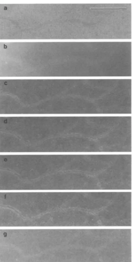
Figure 3 shows the drastic differences in contrast between the b a c k g r o u n d (collodion and carbon film) and a network of slightly stained xanthan fibrils, as obtained by C T - T E M at different energies.
The thickness of material under the electron b e a m is usually an i m p o r t a n t factor for proper E F - T E M analy- sis [30]. We have, however, observed that removal of the collodion supporting film by solvent dissolution did not increase the quality of the final filtered image for stained and unstained specimens. This is explained by the negligible mass effect of the very thin (ca. 2 - 6 n m , measured according to [21]) and poorly electron-dense collodion film, as c o m p a r e d with the stain-loaded fibrils (ca. 5 - 1 0 nm) deposited on the carbon film (ca. 6 - 1 0 nm).
The above results show that C T - T E M is a valuable and powerful method for the highly contrasted visual- ization of individual polysaccharide fibrils, but that observation of ultra-fine fibrils is not possible without the use of stains. Uranyl acetate is a non-specific m a r k e r usually employed to increase the contrast of
Fig. 4. Bright field TEM (a-c) and CT-TEM (d-f) images of xanthan- haematite specimens prepared by en bloc staining with uranyl acetate at 0.2% (a), 0.1% (b, d), 0.05% (e,e) and 0.01% (f/. Bar = 0.5 gm
Fig. 3. Assembling of bright field TEM (a) and CT-TEM (b g) of a network of xanthan (uranyl acetate 0.05% en bloc), b 50 eV, e 140 eV, d 200 eV, e 250 eV, f 270 eV, g 300 eV. Bar = 0.5 gm
T E M images for organic material in biological samples. The use of this stain at conventional concen- trations (1-2%) has, however, been shown to drasti- cally hamper the detection of the ultrastructure of polysaccharides [27], because of the formation of grainy deposits along the fibrils, masking nodes and intersections within networks.
It is thus necessary to determine the optimum con- centration of stain which fulfils the compromise be- tween high C T - T E M contrast and low deleterious effect of stain. Concentrations between 0.5 and 0.01% were added to suspensions for e n b l o c staining just prior to ultracentrifugation above grids. Comparison of conventional T E M and C T - T E M results, illustrated in Fig. 4, shows that the compromise is optimal be- tween 0.1 and 0.05% final stain concentration, where the ultrastructure of polysaccharides is maintained on C T - T E M images. At lower stain concentrations, fibrils are difficult to visualize by conventional TEM, while grainy deposits are observed at higher concentrations.
F o r stain concentrations below 0.01%, only thick aggregates of fibrils are revealed (Fig. 4t).
When optimum staining conditions are chosen, polysaccharide fibrils are homogeneously and repro- ducibly contrasted. Figure 5 is a combination of opti- mal T E M (conventional image), C T - T E M (contrast enhancement of organic matter) and ESI-TEM (elemental mapping of uranium, recorded at the
0 4 , 5 ionization edge of this element [31]). Uranyl stain, visualized as uranium on Fig. 5c, is exclusively and smoothly distributed at the surface of fibrils, without non-specific deposits on the carbon film, thus validat- ing the use ofuranyl acetate stain at low concentration. One might envisage using ESI-TEM images of the uranium stain instead of C T - T E M to record contras- ted images of polysaccharide fibrils; however, such images result in a loss of information for ultra-fine substructures of fibrils and are more fastidious to acquire (risk of specimen drift during recording of background and edge images).
When studying fragile specimens, it is necessary to prevent them from being physically or chemically transformed prior to T E M investigation. F o r instance, natural suspensions sampled from anoxic water layers are highly redox sensitive; they are usually subject to fast air oxidation leading to precipitation of iron oxi- hydroxide particles from dissolved Fe 2 +, which causes factitious structures to appear. On the other hand, flexible polysaccharidic fibrils can shrink during air drying on specimen grids. In order to avoid biased
Fig. 5. Bright field TEM (a), CT-TEM (b; acquired at
•Eloss = 250eV) and ESI-TEM (c; acquired at uranium 04. 5 edge,
| AElos~ = 115 eV; subtracted background extrapolated from images acquired at AElos~ = 80 eV and 90 eV) ofa polysaccharide suspension with 0. t % uranyl acetate en bloc | staining. Bar = 0.5 gm
microscopic information, physico-chemical protection of specimen is achieved by post-coating of grids with hydrophilic N a n o p l a s t resin. Figure 6 shows that
N a n o p l a s t protective film does not lower the contrast of stained fibrils when they are visualized by C T - T E M , The sum of the above results obtained on industrial xanthan fibrils demonstrate that mild staining pro- cedures combined with the use of E F - T E M in contrast tuning m o d e lead to unbiased information on the architecture of this network-forming polysaccharide at the ultrastructural level. O u r a p p r o a c h contributes and strengthens accurately documented studies on conformational and structural characterization of xan- than [32, 33]. It is therefore also possible to demon- strate the applicaton of our proposed methods for the characterization and understanding of two other sys- tems with different origins (purified algal suspensions; lacustrine samples).
In order to study the ability of freshwater algae to produce EPS and to understand the influence of trace metals on algal growth, fresh cultures of S c e n e d e s m u s a c u t u s , were exposed to various concentrations of Cu and Ni (D. J. Kushner, personal communication). Polysaccharide fibrils produced by different strains of this alga were then isolated and studied by C T - T E M , The highly contrasted images obained by C T - T E M highlight valuable features at the ultrastructural level (Fig. 7). F o r instance, small substructures are observed at high magnification along the fibrils with a periodic- ity of ca. 35 nm. While the origin of these nanoexcres- cences is currently unknown, their position along the chain could be the consequence of a ternary structure of individual sugar moieties, The systematic C T - T E M characterization of the m o r p h o l o g y of algal fibrillar polysaccharides, currently under investigation, might
Fig. 6. Effect of hydrophilic Nanoplast embedding resin on the quality of CT-TEM images of xanthan (0.2 mg/1) and haematite (0.1 mg/1) mixtures (0.1% uranyl acetate en bloc, staining), a Without and b with post-protective Nanoplast film. Bar = 0.5 gm
Fig. 7a, b. CT-TEM images of purified fibrils isolated from
Scenedesmus acutus (0.1% uranyl acetate en bloc staining; AElo~ = 250 eV). Arrows on b point to nanoexcrescences exhibiting a high spatial periodicity. Bar = 05 gm
acetate was not added to aqueous suspensions, because organic matter of lacustrine or marine origin might already be naturally and slightly stained. This results from the complexation of dissolved cations (pre- dominantly Ca 2 § > Mg 2 + > Na + > K § ) by organic species [4].
When visualized by C T - T E M (Fig. 8), specimens exhibit highly contrasted networks of organic fibrillar material. Mainly because of technical limitations, these networks have rarely been observed or documented in natural waters, while C T - T E M allows an accurate description of their morphological properties at the ultrastructural level, even in the absence of stain. F o r instance, careful examination of Fig. 8 reveals that it is possible to distinguish between intimately connected (single arrows) and overlapping (double arrows) fibrils. Combined with stereoscopic images (TEM equipped with a goniometer stage), C T - T E M should help in gaining a better insight into the complex 3-dimensional structure of organic networked macromolecules pres- ent in natural waters. Amongst others, one can clearly show by C T - T E M that mineral microparticles (clays, SiO2, F e O O H being the most relevant) strongly inter- act with EPS networks; evidence of these organo-
Conclusions
E F - T E M visualization in contrast tuning mode allows identification of ultra-fine substructures of fibrillar polysaccharides in water suspensions, provided that these are labelled with a sufficient amount of marker. Uranyl acetate, a non-specific stain, can be used at low concentration to fulfil this requirement. When the ionic strength of the sample is low (i.e. synthetic suspensions) en bloc staining with uranyl acetate at ca. 0.05-0.1% prior to deposition of fibrils by ultracentrifugation on T E M grids has no deleterious effect on the morphology and thickness of polysaccharides while their network arrangements are retained. In the case of natural aqua- tic samples, visualization is even possible in the absence of stain. The highest contrast of fibrillar material is obtained at AEIo~s between 230 and 250 eV. Nanoplast post-protection of specimen can be effectively used in conjunction with C T - T E M imaging ofpolysaccharides without fading the contrast of micro-fibrils.
The proposed method can be usefully applied to natural and synthetic aqueous samples; it provides new opportunities for the physico-chemical characteriz- ation of fibrillar polysaccharides at the ultrastructural level, for a better understanding and modelling of their intimate architecture. The undisturbed characteriz- ation ofpolysaccharides in unstable and highly hetero- geneous natural aquatic samples should contribute to studies on the importance of EPS in the control of the transport of mineral particulates and micropollutants, while simultaneously avoiding the strong structural modifications induced by physical or chemical bias in the usual procedures.
Fig. 8. CT-TEM image of Lake Lugano surface water polysacchar- ides in a heterogeneous mixture of mineral microparticles. Single arrows indicate naturally connected fibrils, while double arrows point to overlapping fibrils. Bar = 0.5 jam
Acknowledgements. We gratefully acknowledge Prof. D. J. Kushner (University of Toronto, Canada) for providing fresh algal cultures, Dr. S. Fakan and his team (Center of Electron Microscopy, Univer- sity of Lausanne) for access on EM facilities, and E. Leipzig-Pagani (Institute of Inorganic and AnaIytical Chemistry, University of Lausanne) for his help on densitometric measurements. This work was supported by grants from the Swiss National Science Founda- tion (project 21-36593.92) and the Swiss Prioritary Program Envi- ronment (Project Dytrac 50-39145.93)
References
E 1] A. Varki Glycobiotogy 1993, 3, 97.
[2] G.G. Leppard, ScL Tot. Environ. 1995, 165, 103.
[31 J. Buffle, Complexation Reactions in Aquatic Systems, an Analytical Approach, Ellis Horwood, Chichester, 1988. [4] P. A. Sandford, J. Baird, in: The Polysaccharides, Vol. 2, (G. O.
Aspinal, ed.), Academic press, New York, 1983, p.411. [5] R.L. Whistler, J. N. BeMiller, Industrial Gums, 2nd Ed., Aca-
demic Press, New York, 1973.
[61 D.A. Rees, in: Advances in Carbohydrate Chemistry and Bio- chemistry, Vol. 24, (M. L. Wolfrom, R. S. Tipson, eds.), Aca- demic press, New York, 1969, p.267.
[71 D. Cozzi, P. G. Desideri, L. Lepri, G. Ciantelli, J. Chromatogr. 1968, 35, 396.
[8] D. Cozzi, P. G. Desideri, L. Lepri, G. Ciantelli, J. Chromatogr. 1968, 35,405.
[9] D.G. Allison, Microbiol. Europe, 1993, Nov.-Dec., 16. [10] H. C. Jones, I. L. Roth, W. M. Sanders, J. Bacteriol. 1969, 99,
316.
[111 C. Chenu, Soil Biol. Biochem. 1989, 21,299. [12] C. Chenu, Geoderma 1993, 56, 143.
[13] R. N. Yong, D. Mourato, Can. Geotech. J. 1990, 27, 774. [14] T. Strycek, J. Acreman, A. Kerry, G. G. Leppard, M. V. Nermut,
D. J. Kushner, Microb. Ecol. 1992, 23, 53. [15] T. Bitter, H. M. Muir, Anal. Biochem. 1962, 4, 330.
[16] M. Fletcher, G. D. Floodgate, J. Gen. Microbiol. 1973, 74, 325. [17] D. Perret, M. E. Newman, J.-C. N6gre, Y. Chen, J. Buffie, War.
Res. 1994, 28, 91.
[18] D. Mavrocordatos, C.-P. Lienemann, D. Perret, Mikrochim. Acta 1994, 117, 39.
E19] D. Fr6sch, C. Westphal, Electron Microsc. Rev. 1989, 2, 231.
[20] D. Perret, G. G. Leppard, M. Miiller, N. Belzile, R. DeVitre, J. Buffle, War. Res. 1991, 25, 1333.
[21] R. F. Egerton, Electron Energy-Zoss Spectroscopy in the Elec- tron Microscope, Plenum, New York, 1986.
[22] L Fink, in: Advances in Electronics and Electron Physics, Vol. 75 (P. W. Hawkes, ed.), Academic Press, San Diego, 1989, p.121. [23] L. Reimer, I. Fromm, P. Hirsch, U. Platte, R. Rennekamp,
Ultramicroscopy, 1992, 46, 335.
[24] D. Perret, C.-P. Lienemann, D. Mavrocordatos, Microsc. Microanal. Microstruct. 1995, 6, 41.
[25] G.G. Leppard, in: Environmental Particles, VoL 1. (J. Bufite, H. P. Van Leeuwen eds.), Lewis, Chelsea, 1992, p.231. [26] A.W. Robards, A.J. Wilson, Procedures in Electron Micro-
scopy, Wiley, Chichester, 1993.
[27] K.J. Wilkinson, S. Stoll, J. Buffle, Fresenius J. Anal. Chem. 1995, 351, 54.
[28] C. W. J. Sorber, A. A. W. De Jong, N. J. Den Breejen, W. C. De Bruijn, Ultramicroscopy 1990, 32, 55.
E29] J. Colliex, C. Mory, A. L Olins, D. E. Olins, M. Tence, J. Microsc. 1989, 153, 1.
[30] R. Door, K. D. H~iberle, R. Martin, J. Microsc. 1994, 174, 183. [31] L. Reimer, U. Zepke, J. Moesch, S. Schulze-Hillert, M. Ross- Messemer, W. Probst, E. Weimer, EEL Spectroscopy : a Refer- ence Handbook of Standard Data for Identification and Interpre- tation of Electron Energy Loss Spectra and for Generation of Electron Spectroscopic Images, Carl Zeiss, Oberkochen, 1992. [32] B.T. Stokke, A. Elgsaeter, O. Smidsrg)d, lnt. J. Biol. Macromol.
1986, 8, 217.
[33] B.T. Stokke, A. EIgsaeter, D. A. Brant, S. Kitamura, Macro- molecules 1991, 24, 6349.