DOI 10.1007/s00405-011-1664-1
R H I N O L O G Y
The value of
18
F-FDG-PET/CT imaging for sinonasal malignant
melanoma
S. K. Haerle · M. B. Soyka · D. R. Fischer ·
K. Murer · K. Strobel · G. F. Huber · D. Holzmann
Received: 7 February 2011 / Accepted: 31 May 2011 / Published online: 29 June 2011
© Springer-Verlag 2011
Abstract The aim this study was to evaluate imaging Wndings using position emission tomography (PET) in combination with computed tomography (CT) and 18F-Xuorodeoxyglucose (18F-FDG) in sinonasal malignant mel-anoma (SNMM) of the head and neck in a retrospective analysis of a consecutive cohort of patients. 18F-FDG-PET/ CT examinations were performed for initial staging and compared with CT or magnetic resonance tomography (MRI), and 18F-FDG-PET alone. Medical records were reviewed retrospectively with regard to the location and the size of the tumor. Furthermore, locoregional and distant metastases with a consecutive change in therapy detected by 18F-FDG-PET/CT were assessed. Ten patients suVering from sinonasal malignant melanoma were staged and fol-lowed by 18F-FDG-PET/CT imaging. A total of 34 examin-ations were obtained. 18F-FDG-PET/CT depicted all primary tumors adequately. Aside from one cerebral metasta-sis all regional and distant metastases were truly identiWed by using this method. In summary, if available, 18 F-FDG-PET/CT is a valuable imaging modality for staging and re-staging sinonasal malignant melanoma to evaluate expan-sion of the primary tumor, locoregional disease, and distant metastases.
Keywords 18F-FDG-PET/CT · Imaging · Staging · Head and neck · Sinonasal malignant melanoma
Introduction
Mucosal malignant melanoma (MMM) is a very rare tumor counting for approximately 0.8–1.3% of all melanomas [1, 2]. The most common region for MMM is the head and neck, whereas other sites, such as the rectum and the anus, the urinary system, and the female genitalia may also be involved [1, 2]. The most prevalent sites of MMM in the head and neck are the nasal cavity, oral cavity, and the par-anasal sinuses [1]. Sinonasal malignant melanoma (SNMM) accounts roughly for 4% of all head and neck melanomas and sinonasal malignancies alike [3]. Although the incidence of cutaneous melanoma is increasing, the incidence of MMM remains stable [1].
Positron emission tomography (PET) with 18F-Xuorode-oxyglucose (18F-FDG) is increasingly used for initial stag-ing for head and neck malignancies. It is also an established tool for initial staging and follow-up imaging of cutaneous malignant melanomas [4]. In most centers, the standard imaging technique for MMM in the head and neck at initial staging is computed tomography (CT) or magnetic reso-nance imaging (MRI) if there is suspicion for skull base inWltration. The role of 18F-FDG-PET in the management of MMM has been validated in a previous report by our institution [5]. Meanwhile, technology has evolved, and high-resolution multi-slice CT scanners are integrated in 18F-FDG-PET- systems. The aim of this study was to eluci-date imaging and the possible additional value of 18 F-FDG-PET/CT on therapeutic management of patients with MMM, particularly SNMM, when using it at initial staging or follow-up for locoregional or distant disease.
S. K. Haerle (&) · M. B. Soyka · G. F. Huber · D. Holzmann Department of Otolaryngology-Head and Neck Surgery, University Hospital Zurich, Frauenklinikstrasse 24, 8091 Zurich, Switzerland
e-mail: [email protected]
D. R. Fischer · K. Strobel
Department of Nuclear Medicine, University Hospital Zurich, 8091 Zurich, Switzerland
K. Murer
Department of Otolaryngology-Head and Neck Surgery, Kantonsspital St. Gallen, 9000 St. Gallen, Switzerland
Materials and methods
We retrospectively reviewed the charts of all patients treated for MMM of the head and neck at the Department of Otolaryngology-Head and Neck Surgery, University Hospi-tal Zurich, Switzerland . We revealed 10 patients, with sin-onasal involvement only, examined by 18F-FDG-PET/CT since the introduction of this technique at the Division of Nuclear Medicine, University Hospital Zurich, Switzer-land, has been introduced in March 2001. If patients were referred to our institution with a CT or MRI scan done by the referring institution, these data were blindly re-evalu-ated by our double board certiWed radiologist and nuclear physician. In case of patients without previous imaging, the CT part from the fused 18F-FDG-PET/CT was evaluated in a blind way without looking at the 18F-FDG-PET part. All suspicious lesions detected by physical examination (endoscopy) and imaging (CT, MRI or 18F-FDG-PET/CT) were further investigated by histological work- up which served as the standard of reference. The study was con-ducted in accordance with the local guidelines established by the ethics committee for retrospective evaluations. Each patient was followed by imaging using 18F-FDG-PET/CT at 6-month intervals.
18F-FDG-PET/CT acquisition
For this study, we used a combined PET/CT system (Discov-ery STE or Discov(Discov-ery RX, GE Health Systems, Milwaukee, WI). This device integrates a PET scanner with a multi-slice helical CT (16 or 64 slices; slice thickness: 2.5 mm) and per-mits the acquisition of coregistered CT and PET images in the same session. Patients fasted for at least 4 h prior to scan-ning, which started approximately 60 min after the injection of a standard dose of approximately 350 MBq of 18F-FDG. Patients were examined in the supine position. The CT scan was acquired during breath holding in the normal expiratory position. Immediately following the CT acquisition, the PET emission scan was acquired. The CT data were used for the attenuation correction and the images were reconstructed using a standard iterative algorithm (ordered subset expecta-tion maximizaexpecta-tion (OSEM)) for 3D PET reconstrucexpecta-tion. The acquired images were postprocessed with a dedicated soft-ware (AW workstation, GE Health Systems) providing mul-tiplanar reformatted images of 18F-FDG-PET alone, CT alone, and fused 18F-FDG-PET/CT with linked cursors. 18F-FDG-PET/CT evaluation
18F-FDG-PET/CT images were retrospectively analyzed by a double board certiWed radiologist and nuclear physician with 6 years of experience (K. S.) in reading 18F-FDG-PET/ CT`s of head and neck cancer patients. The 18F-FDG-PET
images were evaluated with regard to the presence and nature of focal lesions with increased 18F-FDG- uptake. Attenuation-corrected 18F-FDG-PET images were used for analysis. Lesions were interpreted as a malignancy if the uptake was higher than the uptake of the surrounding back-ground tissue. It was a visual interpretation and not based on standardized uptake value (SUV) measurements. Fur-thermore, morphological Wndings of suspicious lesions pro-vided by the low-dose CT part were also used for interpretation. 18F-FDG- uptake in muscles, glands, brown fat or pulmonary inWltration were not interpreted as tumor or metastases. Four patients were initially examined after resection and therefore attribution of 18F-FDG-uptake and histological Wnding was either equivocal or no more uptake was observed. Two patients received intravenous and oral contrast agent (Ultravist 300, GastrograWn, Bayer AG Swit-zerland) for obtaining either a diagnostic CT of the neck or of the whole body.
Semiquantitative analysis of 18F-FDG-uptake in all sus-picious lesions was performed by measuring the maximum standardized uptake value (SUV max).
Results
Clinical and histopathologic Wndings
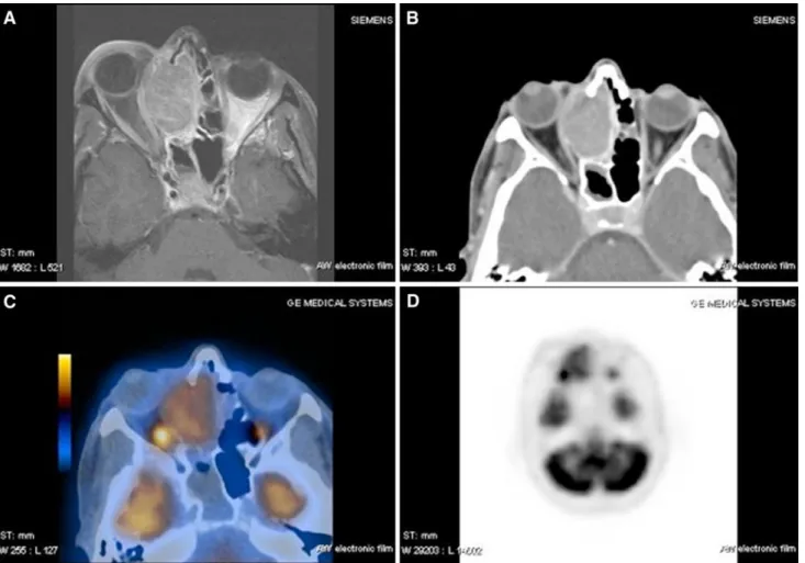
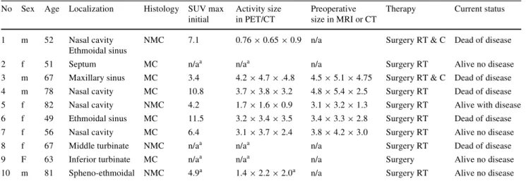
The charts of ten consecutive patients (4 males, 6 females) were reviewed. The median age was 65 years (range, 49–82 years). At initial staging, no patient had clinical evi-dence of a primary melanoma elsewhere in the body. The MMM involved the nasal cavity and/or the adjacent ethmoidal and maxillary sinus in all cases (Table1). Most fre-quent symptoms at initial presentation were unilateral nasal hemorrhagic discharge, nasal obstruction, and facial pain. After clinical investigation including nasal endoscopy, a CT or MRI scan was acquired. In six patients (patient nos. 1, 3, 4, 5, 6, 7, Table1), an additional 18F-FDG-PET/CT scan for initial staging was obtained (Fig.1a–d). In patient nos. 2, 8, 9, and 10 an additional 18F-FDG-PET/CT after initial resection for re-staging was performed.
SNMM was conWrmed histologically and immunohisto-logically in all patients. Four tumors (nos. 1, 5, 8, 10; Table1) showed the histologic pattern of an amelanotic mel-anoma. The primary tumor was surgically removed in all patients; postoperative percutaneous radiotherapy (intensity modulated radiotherapy, IMRT) was given to all but one patient (patient no. 9, Table1) with a mean of 58 Gy. Two patients underwent adjuvant chemotherapy because of distant metastases detected during follow-up (patient nos. 1, 3; patient no. 1 was included in a study protocol receiving inter-feron alpha (IFNa; 3MU s.c., three times a week) plus Dacar-bazine (DTIC 850 mg/m2 via intravenous (i.v) infusion on
day 1 every 28 days during the Wrst 6 months; patient no. 3 was included in a diVerent study (SAKK 50/70) receiving Bevacizumab (Avastin® 10 mg/kg via i.v. infusion every 2 week) and Temozolomid (Temodal® 150 mg/m2 daily for 7 days every second week)). The median follow- up period was 17.5 months (range, 8–34 months). Five patients died as a result of locoregional recurrence and distant metastases (Table2). Four Patients are alive with no evidence of dis-ease, whereas one patient is known to have bone and pulmonary metastases (Table2).
18F-FDG-PET/CT Wndings versus other imaging Wndings
18F-FDG-PET/CT was performed at the time of diagnosis before surgical resection in six patients, within 1 week after surgery (n = 4), and during follow-up (n = 24). A total of 34 18F-FDG-PET/CT were analyzed for this study.
The follow-up examinations were performed between 5 and 33 months after diagnosis. Increased 18F-FDG-uptake
was observed in all patients with present tumors. All the 18F-FDG-PET/CT Wndings with regard to the detection of the primary tumor were also detected by CT or MRI-scans and were well correlated in size. The contrast-enhanced (ce) CT part of the 18F-FDG-PET/CT was able to approve the 18F-FDG-PET Wndings. One cerebral metastasis was not detected either by 18F-FDG-PET or by ceCT scanning and could only be found on MRI. Three suspicious lesions revealed by native CT (one osteolytic lesion in the spine; one small lesion in the lung; one enlarged lymph node in the mediastinum) showed none, or very little 18F-FDG activity. During follow-up, these lesions turned out to be false-positive lesions as no growth or onset of symptoms could be shown.
Increased activity in soft tissue was located in brown fat tissue in one patient. Patient no. 10 showed repeatedly local 18F-FDG activity at the site of resection (SUV 4.4-4.7). The fused CT did not approve a growing mass at this position and malignancy was Wnally ruled out by biopsy.
Fig. 1 56-year-old female suVering from a sinonasal malignant mela-noma originating from the right nasal cavity involving the ethmoid si-nus, the dura, and the periorbit. a T1 weighted MRI with Gadolinium,
axial section. b Bone window CT with contrast agent, axial section. c Fused 18F-FDG-PET-CT, axial section. d 18F-FDG-PET, axial section
Discussion In general
Primary mucosal malignant melanoma (MMM) is a rare disease and occurs in older patients of both genders. Inci-dence of nasal cavity MMM was reported to be 0.018/10 [5] per year [6]. Only around 4% of all head and neck mela-nomas arise in the nasal cavity or adjacent paranasal sinuses [3]. The most common site of origin is the nasal septum, closely followed by the lateral nasal wall and the middle and inferior turbinates [3, 7]. Lymph node metasta-ses are generally noted in about one-third of mucous mem-brane head and neck melanomas at initial presentation, implying a worse prognosis [1, 8]. The poorer prognosis for mucous membrane melanoma has been related to the tumor bulk (size), delay in diagnosis resulting from the non-spe-ciWc nature of the presenting symptoms, diYcult visual examination, and technical inaccessibility of the primary tumor [9].
Sinonasal malignant melanomas have, like cutaneous malignant melanomas, an increased glucose metabolism. The role of 18F-FDG-PET in staging mucosal malignant melanomas of the head and neck has already been aYrmed [5]. 18F-FDG-PET imaging is also known to be more sensi-tive and speciWc than CT for the detection of metastasis in melanoma patients [10].
Advantages of18F-FDG-PET/CT
At initial staging, all our patients showed increased 18F-FDG uptake at the tumor site and could thus be easily identiWed.
18F-FDG-PET-only imaging is lacking anatomical infor-mation. Complementary anatomical imaging is often required, especially for surgical therapy planning in this complex anatomical area. Coregistred 18F-FDG-PET/CT has been available for a few years and provides such anatometabolic datasets in a single examination. 18 F-FDG-PET/CT has been found to be superior to CT alone and PET alone in staging and evaluation of therapy response in several oncological diseases including malignant mela-noma [11–13].
18F-FDG-PET/CT as a functional and anatomical imaging method provides additional information for sur-gical therapy planning. Especially in the nasal cavity and paranasal sinuses, where anatomy is diYcult and diVer-ent vital structures are in close proximity, 18F-FDG-PET/ CT appears to be superior to 18F-FDG-PET alone. Partic-ularly in recurrent tumors it seems essential to distin-guish scar-tissue from melanoma masses and predicts the extent of aVection, which is not possible by CT alone. 18F-FDG-PET without the additional information of the fused CT on the other hand is not able to give enough spatial resolution to perform exact surgical planning in the setting of the pre-operated nasal cavities and parana-sal sinuses, where CT data add valuable information on fused images.
In a previous study by Department of Otolaryngology-Head and Neck Surgery, University Hospital Zurich, Switzerland, it could be shown that local tumor control in SNMM can be achieved in many cases and the majority of patients die from distant metastasis [7]. Hence, 18 F-FDG-PET/CT is in our opinion the imaging of choice during the follow-up period.
Table 1 Patient and tumor characteristics
f Female, m Male, NMC non-melanotic melanoma, MC melanotic melanoma, MRI magnetic resonance imaging, CT computed tomography, SUV standardized uptake value, RT radiotherapy, C chemotherapy
Sizes indicated in anterio-posterior £ cranio-caudal £ right-left distances (in cm) a After resection
No Sex Age Localization Histology SUV max initial
Activity size in PET/CT
Preoperative size in MRI or CT
Therapy Current status
1 m 52 Nasal cavity Ethmoidal sinus
NMC 7.1 0.76 £ 0.65 £ 0.9 n/a Surgery RT & C Dead of disease
2 f 51 Septum MC n/aa n/aa n/a Surgery RT Alive no disease
3 m 67 Maxillary sinus MC 3.4 4.2 £ 4.7 £ .4.8 4.5 £ 5.1 £ 4.75 Surgery RT & C Dead of disease 4 m 78 Nasal cavity MC 10.8 3.7 £ 3.8 £ 3.2 4.8 £ 5.4 £ 2.5 Surgery RT Dead of disease 5 f 82 Nasal cavity NMC 4.2 1.7 £ 1.6 £ 0.9 3.1 £ 3.2 £ 1.3 Surgery RT Alive with disease 6 f 49 Ethmoidal sinus MC 11.5 3.2 £ 3.4 £ 3.5 3.4 £ 3.3 £ 2.8 Surgery RT Dead of disease 7 f 56 Nasal cavity MC 6.4 3.1 £ 3.7 £ 2.4 3.8 £ 4.2 £ 3.0 Surgery RT Alive no disease
8 f 67 Middle turbinate NMC n/aa n/aa n/a Surgery RT Dead of disease
9 F 63 Inferior turbinate MC n/aa n/aa n/a Surgery Alive no disease
Limitations of18F-FDG-PET/CT
A known limitation of 18F-FDG-PET imaging is the identi-Wcation of brain metastases due to the high background metabolism as well as small melanoma metastases, where the spatial resolution of 18F-FDG-PET/CT- imaging is not small enough [14]. We, too, did encounter these diYculties, as one cerebral metastasis was not seen during follow-up 18F-FDG-PET/CT scans and was only identiWed by MRI.
Fused 18F-FDG-PET/MRI might possibly eliminate this problem in the future.
In a study by Gulec et al. [14] all of the 29 lesions larger than 1 cm showed 18F-FDG uptake, whereas only two out of 15 lesions smaller than 1 cm were detected by 18F-FDG-PET alone14. A theoretical three-dimensional spatial resolution of 4 mm can be achieved by modern 18F-FDG-PET/CT scanners. However, due to motion and the tumor related uptake ratio, a spatial resolution of 6-8 mm can be expected realistically [15].
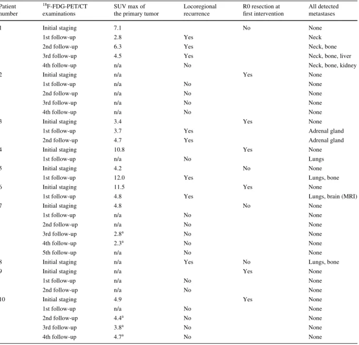
Table 2 Clinical and radiological information for every patient given at time of 18F-FDG-PET/CT examinations
R0 resection with neither macroscopic nor microscopic evidence of residual tumor, SUV max maximum standardized uptake value, n/a: no focal uptake a False-positive uptake Patient number 18F-FDG-PET/CT examinations SUV max of the primary tumor
Locoregional recurrence
R0 resection at
Wrst intervention All detected metastases
1 Initial staging 7.1 No None
1st follow-up 2.8 Yes Neck
2nd follow-up 6.3 Yes Neck, bone
3rd follow-up 4.5 Yes Neck, bone, liver
4th follow-up n/a No Neck, bone, kidney
2 Initial staging n/a Yes None
1st follow-up n/a No None
2nd follow-up n/a No None
3rd follow-up n/a No None
4th follow-up n/a No None
3 Initial staging 3.4 Yes None
1st follow-up 3.7 Yes Adrenal gland
2nd follow-up 4.7 Yes Adrenal gland
4 Initial staging 10.8 Yes None
1st follow-up n/a No Lungs
5 Initial staging 4.2 No None
1st follow-up 12.0 Yes Lungs, bone
6 Initial staging 11.5 Yes None
1st follow-up 4.8 Yes Lungs, brain (MRI)
7 Initial staging 4.8 No None
1st follow-up n/a No None
2nd follow-up n/a No None
3rd follow-up 2.8a No None
4th follow-up 2.3a No None
5th follow-up n/a No None
8 Initial staging n/a Yes No Lungs, bone
9 Initial staging n/a Yes None
1st follow-up n/a No None
2nd follow-up n/a No None
10 Initial staging 4.9 Yes None
1st follow-up n/a No None
2nd follow-up 4.4a No None
3rd follow-up 3.8a No None
Veit-Haibach et al. [16] stated that 18F-FDG-PET/CT show low sensitivities at initial staging of patients suVering from malignant cutaneous melanomas and therefore meta-bolic imaging should only be recommended as a Wrst-line diagnostic tool for selected melanoma patients in high-risk groups. Although none of our patients was found to have regional or distant metastasis upon Wrst imaging workup, all of the primary sites could be identiWed adequately. This is also due to late presentation of the oligosymptomatic patient in a late stadium with large tumor size, as stated above. As a rule, we believe it is advantageous performing the same fol-low-up imaging modality as that used for primary workup. The cost-eVectiveness of18F-FDG-PET/CT
DiVerent authors have described the cost-eVectiveness of 18F-FDG PET/CT in staging head and neck squamous cell carcinoma (HNSCC) [17, 18]. The drawback of staging patients with 18F-FDG PET/CT is the relatively high num-ber of false-positive Wndings. Although many of the posi-tive 18F-FDG PET/CT Wndings will have an impact on the patient treatment, the costs of subsequent investigations and the burden caused to patients by additional investigations are considerable. Therefore, the indication for metabolic imaging should be handled reluctantly. A recent study showed 18F-FDG PET/CT is the most eVective pretreat-ment screening method for distant metastases in HNSCC patients with risk factors [19]. It is our opinion, that in future, when integrated 18F-FDG PET/CT and MRI scan-ners become routine, this will be the most cost-eVective imaging method for SNMM.
The role of CT
Three 18F-FDG- negative lesions (spine, lung, and medi-astinum) detected and found to be suspicious by native CT were shown to be unsuspicious on 18F-FDG-PET scanning. All of these Wndings were proven not to be malignant upon follow-up examination. These lesions represent false-positive Wndings on native CT scans; therefore, CT scanning should be ultimately performed ce, and, furthermore, 18F-FDG negativity shows the potential to identify unreliable CT Wndings. On the other hand it can be challenging, especially in the head and neck area, to distinguish physiological or inXammatory uptake from malignant tissue which can be downsized by the ceCT part [20].
As malignant mucosal melanoma is a very rare condi-tion, it is diYcult to show signiWcant improvements in stag-ing with 18F-FDG-PET/CT because of the small number of patients. However, we know from previous reports on 18 F-FDG-PET/CT imaging that only few false-negative results are obtained, leading to a high negative predictive value,
which is also supported by the current study. As modern 18F-FDG-PET/CT scanners are providing a better resolu-tion of the added CT informaresolu-tion, one can also expect less false-positive Wndings. As shown, it is important to perform a ceCT part, which is not routine in many centers.
The role of MRI
To evaluate dural or intracranial involvement, MRI seems to be indisputably the imaging modality of choice. How-ever, the tumor’s appearance is dependent on the amount of melanin found within. Melanotic tumors usually show hyperintensity on T1 images, whereas hypomelanotic or amelanotic melanomas may appear intermediate in signal intensities [21–23]. In our series 4/10 SNMM were amela-notic. In previous studies of mucosal melanoma, the fre-quency of amelanotic melanoma ranged from 25 to 42.9% [22]. All of the tumors in our series had increased 18F-FDG uptake independent of their melanin contents which is in accordance with the literature.
Conclusion
In addition to the previously published experience con-cerning 18F-FDG-PET imaging for the detection of MMM, the updated data presented in this study suggest that 18F-FDG-PET/CT is superior to other imaging, e.g., CT or MRI scans, with its known limitations. Owing to the combined 18F-FDG-PET and CT part, the visibility of SNMM is not solely dependent on 18F-FDG activity anymore.
Therefore, if available, contrast-enhanced 18 F-FDG-PET/CT, in our opinion, is the adequate staging and re-staging imaging modality to respond to the question of the expansion of the primary tumor, locoregional disease, or distant metastases.
If dural or intracranial involvement is clinically suspected, we believe that, additional MRI is required. However, it will never replace 18F-FDG-PET/CT scanning as a staging modality unless MRI is fused with 18F-FDG-PET.
ConXict of interest None.
References
1. Chang A, Karnell L, Menck H (1998) The National Cancer Data Base report on cutaneous and noncutaneous melanoma: a summary of 84, 836 cases from the past decade. The American College of Surgeons Commission on Cancer and the American Cancer Society. Cancer 83:1664–1678
2. Andersen L, Berthelsen A, Hansen H (1992) Malignant melanoma of the upper respiratory tract and the oral cavity. J Otolaryngol 21:180–185
3. Thompson L, Wieneke J, Miettinen M (2003) Sinonasal tract and nasopharyngeal melanomas: a clinicopathologic study of 115 cases with a proposed staging system. Am J Surg Pathol 27:594–611 4. Steinert H, Huch Böni R, Buck A et al (1995) Malignant
mela-noma: staging with whole-body positron emission tomography and 2-[F-18]-Xuoro-2-deoxy-D-glucose. Radiology 195:705–709 5. Goerres G, Stoeckli S, von Schulthess G, Steinert H (2002) FDG
PET for mucosal malignant melanoma of the head and neck. Laryngoscope 112:381–385
6. Chiu N, Weinstock M (1996) Melanoma of oronasal mucosa. Population-based analysis of occurrence and mortality. Arch Otolaryngol Head Neck Surg 122:985–988
7. Roth TN, Gengler C, Huber GF, Holzmann D (2010) Outcome of sinonasal melanoma: clinical experience and review of the litera-ture. Head Neck 32(10):1385–1392
8. Lengyel E, Gilde K, Remenár E, Esik O (2003) Malignant mucosal melanoma of the head and neck. Pathol Oncol Res 9:7–12 9. Brandwein M, Rothstein A, Lawson W, Bodian C, Urken M
(1997) Sinonasal melanoma. A clinicopathologic study of 25 cases and literature meta-analysis. Arch Otolaryngol Head Neck Surg 123:290–296
10. Swetter S, Carroll L, Johnson D, Segall G (2002) Positron emis-sion tomography is superior to computed tomography for meta-static detection in melanoma patients. Ann Surg Oncol 9:646–653 11. Strobel K, Dummer R, Husarik D, Pérez Lago M, Hany T, Steinert H (2007) High-risk melanoma: accuracy of FDG PET/CT with added CT morphologic information for detection of metastases. Radiology 244:566–574
12. Strobel K, Skalsky J, Steinert H et al (2007) S-100B and FDG-PET/CT in therapy response assessment of melanoma patients. Dermatology 215:192–201
13. Reinhardt M, Joe A, Jaeger U et al (2006) Diagnostic performance of whole body dual modality 18F-FDG PET/CT imaging for N- and M-staging of malignant melanoma: experience with 250 consecu-tive patients. J Clin Oncol 24:1178–1187
14. Gulec S, Faries M, Lee C et al (2003) The role of Xuorine-18 deoxyglucose positron emission tomography in the management of patients with metastatic melanoma: impact on surgical decision making. Clin Nucl Med 28:961–965
15. Von Schulthess G, Hany T (2008) Imaging and PET–PET/CT imaging. J Radiol 89:438–447 (quiz 48)
16. Veit-Haibach P, Vogt F, Jablonka R et al (2009) Diagnostic accu-racy of contrast-enhanced FDG-PET/CT in primary staging of cutaneous malignant melanoma. Eur J Nucl Med Mol Imaging 36:910–918
17. Valk PE, Pounds TR, Tesar RD, Hopkins DM, Haseman MK (1996) Cost-eVectiveness of PET imaging in clinical oncology. Nucl Med Biol 23:737–743
18. Hollenbeak CS, Lowe VJ, Stack BC Jr (2001) The cost-eVective-ness of Xuorodeoxyglucose 18-F positron emission tomography in the N0 neck. Cancer 92:2341–2348
19. Uyl-de Groot CA, Senft A, de Bree R, Leemans CR, Hoekstra OS (2010) Chest CT and whole-body 18F-FDG PET are cost-eVective in screening for distant metastases in head and neck cancer pa-tients. J Nucl Med 51:176–182
20. Goerres G, Von Schulthess G, Hany T (2002) Positron emission tomography and PET CT of the head and neck: FDG uptake in nor-mal anatomy, in benign lesions, and in changes resulting from treatment. AJR Am J Roentgenol 179:1337–1343
21. Yousem D, Li C, Montone K et al (1996) Primary malignant mel-anoma of the sinonasal cavity: MR imaging evaluation. Radio-graphics 16:1101–1110
22. Yoshioka H, Kamada T, Kandatsu S et al (1998) MRI of mucosal malignant melanoma of the head and neck. J Comput Assist To-mogr 22:492–497
23. Uchiyama Y, Murakami S, Kawai T, Ishida T, Fuchihata H (1998) Primary malignant melanoma in the oral mucosal membrane with metastasis in the cervical lymph node: MR appearance. AJNR Am J Neuroradiol 19:954–955