ORIGINAL ARTICLE
Limited value of
18
F-FDG PET/CT and S-100B tumour
marker in the detection of liver metastases from uveal
melanoma compared to liver metastases
from cutaneous melanoma
K. Strobel&B. Bode&R. Dummer&P. Veit-Haibach&D. R. Fischer&L. Imhof&S. Goldinger&
Hans C. Steinert&G. K. von Schulthess
Received: 9 February 2009 / Accepted: 8 May 2009 / Published online: 4 June 2009 # Springer-Verlag 2009
Abstract
Purpose The objective of this study was to evaluate the value of18F-FDG PET/CT and S-100B tumour marker for the detection of liver metastases from uveal melanoma in comparison to liver metastases from cutaneous melanoma. Methods A retrospective evaluation was conducted of 27 liver metastases in 13 patients with uveal melanoma (UM) (mean age: 56.8, range: 30–77) and 43 liver metastases in 14 patients (mean age: 57.9, range: 40–82) with cutaneous melanoma (CM) regarding size and FDG uptake by measur-ing the maximum standardized uptake value (SUVmax). S-100B serum tumour markers were available in 20 patients. Cytology, histology, additional morphological imaging and follow-up served as reference standard. In nine patients liver metastases were further evaluated histologically regarding GLUT-1 and S-100 receptor expression and regarding epithelial or spindle cell growth pattern.
Results Of 27 liver metastases in 6 of 13 patients (46%) with UM, 16 (59%) were FDG negative, whereas all
liver metastases from CM were positive. Liver metasta-ses from UM showed significantly (p<0.001) lower SUVmax (mean: 3.5, range: 1.5–13.4) compared with liver metastases from CM (mean: 6.6, range: 2.3–15.3). In four of six (66.7%) patients with UM and liver metastases S-100B was normal and in two (33.3%) increased. All PET-negative liver metastases were detect-able by morphological imaging (CT or MRI). S-100B was abnormal in 13 of 14 patients with liver metastases from CM. S-100B values were significantly higher (p= 0.007) in the CM patient group (mean S-100B: 10.9μg/l, range: 0.1–115 μg/l) compared with the UM patients (mean: 0.2 μg/l, range: 0.0–0.5 μg/l). Histological work-up of the liver metastases showed no obvious difference in GLUT-1 or S-100 expression between UM and CM liver metastases. The minority (36%) of patients with UM had extrahepatic metastases and the majority (86%) of patients with CM had extrahepatic metastases, respectively. There was a close to significant trend to better survival of UM patients compared with CM patients (p=0.06).
Conclusion FDG PET/CT and serum S-100B are not sensitive enough for the detection of liver metastases from UM, whereas liver metastases from cutaneous melanoma are reliably FDG positive and lead regularly to increased S-100B tumour markers. The reason for the lower FDG uptake in UM liver metastases remains unclear. We recommend to perform combined contrast-enhanced PET/CT in order to detect FDG-negative liver metastases from UM.
Keywords Liver metastases . Uveal melanoma . Cutaneous melanoma
K. Strobel (*)
:
P. Veit-Haibach:
D. R. Fischer:
H. C. Steinert:
G. K. von SchulthessDivision of Nuclear Medicine, Department of Medical Radiology, University Hospital Zurich,
Raemistr. 100,
8091 Zurich, Switzerland e-mail: klaus.strobel@usz.ch B. Bode
Department of Pathology, University Hospital Zurich, Zurich, Switzerland
R. Dummer
:
L. Imhof:
S. GoldingerDepartment of Dermatology, University Hospital Zurich, Zurich, Switzerland
Introduction
FDG PET/CT is increasingly used for initial staging and restaging of patients with cutaneous melanoma (CM). PET/ CT has the highest impact in patients with high-risk melanoma. We define patients with CM as “high-risk” if one of the following criteria is fulfilled: Breslow tumour thickness > 4 mm, Clark level III or IV or known resected metastases in the prior history. FDG PET/CT showed a high accuracy for the detection of metastases from CM, helps to select patients for surgery and has impact on patient survival [1–4].
Uveal melanoma (UM) is a rare type of melanoma but the most common primary intraocular malignancy in adults. Over the last decades a stable incidence of UM in the USA and Europe with four to seven cases per million has been observed [5, 6]. The pattern of metastatic spread differs from CM. Because of the absence of lymphatic drainage of the eye UM metastasizes haematogeneously predominantly into the liver [7]. Despite the common origin of both melanoma types deriving from melanocytes, both melano-mas show marked differences in their metastatic potential, clinical response to treatments, immune response and genetic alterations. The genetic alterations in cutaneous melanomas are well described but not much is known about the genetic alterations associated with the development of UM because of the small number of biopsy samples [8]. It has been demonstrated that abnormalities related to chro-mosomes 3, 6 and 8 are strongly related to this tumor and are associated with a reduction of survival from 95 to less than 50% [9].
The reason for the high affinity of UM metastases to the liver is not known. Factors such as tumour size, ciliary body involvement, cell type, extravascular matrix patterns and cytogenetics seem to be related to the presence of distant metastases [10]. Detection of liver metastases is crucial because they are the life-limiting component of the disease in the majority the patients and treatment improves survival in selected patients. Prognosis of metastatic UM is very poor with a median survival of 19 months without liver metastases and only 7 months with liver metastases. Patients with resectable liver metastases may profit from surgical resection [11]. Systemic therapies have poor response rates (1%) in unresectable cases. Chemoemboliza-tion seems to be effective, inducing responses in about 36% of the patients [12–15]. New treatment options like treatment with90Y-microspheres are promising [16].
In patients with CM serum S-100B is a useful tumour marker, indicating the presence of distant metastases and reflecting the tumour burden. In addition, S-100B has prognostic implications [17–22]. The literature on the prognostic and diagnostic value of S-100B in patients with UM is controversial [23–25].
The aim of this study was to evaluate the value of F-FDG PET/CT and the tumour marker S-100B regarding the detection of liver metastases from uveal melanoma in comparison to liver metastases from cutaneous melanoma.
Materials and methods Patients
This retrospective study contains 27 melanoma patients (12 women, 15 men); 13 patients (mean age: 56.8 years, range: 30–77 years) had a history of UM and 14 patients (mean age: 57.9 years, range: 40–82 years) had a history of CM. All patients had proven liver metastases of UM or CM. A FDG PET/CT scan was performed in all patients for staging or restaging between November 2002 and March 2008. No systemic treatment was performed before the PET/CT investigations. Patient characteristics are summarized in Table1.
We received approval from our Institutional Review Board to undertake this retrospective study.
Tumour marker S-100B measurement
The determination of serum S-100B was performed with a commercially available enzyme-linked immunosorbent assay (ELISA) kit (Sangtec 100 ELISA, Diasorin Inc., Stillwater, MN, USA) according to the instructions of the manufacturer. A normal cut-upper limit was defined as 0.2 μg/l (the 95th percentile of blood donor samples). Values ≥0.3 μg/l were taken to be an indication for melanoma metastases. The detection limit is 0.03 μl/ l (BO+3 SD). Intra-assay and inter-assay precision was estimated by analysis of variance (ANOVA). The“within run” and “total run” reproducibility was within 10%.
In all patients S-100B levels were determined before therapy at the time point of the PET/CT investigation (less than 7 days interval between S-100B measurement and PET/CT.
Reference standard
In eleven patients the presence of liver metastases was proven by cytology (n=1) or histology (n=10). In the remaining 17 patients a combined reference was used: all patients additionally had morphological imaging (contrast-enhanced CT and/or MRI of the liver). In 19 patients follow-up imaging with PET/CT was available. Altogeth-er, 71 follow-up PET/CT examinations were performed (mean: 3.7, range: 0–13). In the surviving patients we had a mean clinical follow-up time of 18 months (range: 3–37 months).
T able 1 Characteristics of 27 patients with UM and CM No. Age T ype of melanoma Number of PET/CT scans performed Number and size of liver mets FDG uptake SUV max S-100B (μg/l) Proof of liver metastases Extrahepatic metastases T reatment Outcome 1 5 7 U M 4 1 (15 mm) No 2.4 n.a. Histology No Resection Exitus after 19 months 2 6 6 U M 1 1 (19 mm) No 1.8 n.a. Cytology No Unknown n.a. 3 5 7 U M 6 1 (39 mm) Y es 3.1 0.0 CT , follow-up Lung, bone, soft tissue Chemo Exitus after 37 months 4 3 0 U M 5 2 (14, 27 mm) No 1.8, 1.7 0.1 Histology No Resection, chemo Exitus after 59 months 5 3 8 U M 6 1 (50 mm) Y es 3.2 0.5 MRI, follow-up Bone, LN R T spine, chemo Alive after 20 months 6 6 4 U M 3 4 (10 –170 mm) Y es 4.0 –7.1 0.1 CT , follow-up No Chemoembolization Alive after 21 months 7 7 1 U M 3 4 (10 –42 mm) No 1.5 –1.7 0.5 MRI, CT , follow-up No Unknown Alive after 6 months 84 6 U M 3 5 (6 –28 mm) No 1.6 –1.7 0.1 CT , MRI, angio., follow-up No SIR T Alive after 6 months 9 5 3 U M 1 1 (24 mm) Y es 9.8 n.a. Histology Lung Resection Alive after 6 months 10 77 UM 1 1 (152 mm) Y es 6.0 n.a. Histology No SIR T Alive after 3 months 1 1 55 UM 3 3 (8 –63 mm) No/yes 2.4 –4.8 n.a. Histology No Resection Alive after 15 months 12 61 UM 1 2 (3, 21 mm) No/yes 2, 2.7 n.a. Histology No Resection Exitus after 17 months 13 63 UM 1 1 (128 mm) Y es 13.4 n.a. Histology Peritoneal mets Chemo Exitus after 2 months 14 57 CM 5 6 (7 –21 mm) Y es 9.1 –10.9 0.6 Follow-up LN mets Chemo Exitus after 6 months 15 66 CM 3 1 (54 mm) Y es 14.6 2.2 Follow-up Bone, LN, lung Chemo Exitus after 6 months 16 72 CM 5 2 (14, 14 mm) Y es 5.4, 6.0 0.6 Follow-up Lung, soft tissue, LN Chemo Exitus after 12 months 17 41 CM 1 1 (16 mm) Y es 3.7 0.5 Follow-up Lung, brain, soft tissue Chemo Exitus after 4 months 18 43 CM 2 5 (13 –27 mm) Y es 2.3 –5.8 0.6 Follow-up Bone, LN Chemo Exitus after 4 months 19 52 CM 1 1 4 (12 –27 mm) Y es 5.7 –9.5 14.3 Histology Spleen, soft tissue, LN, adrenals Chemo Exitus after 27 months 20 40 CM 12 1 (37 mm) Y es 8.7 1.7 Follow-up Adrenals, LN Chemo Alive after 36 months 21 60 CM 4 5 (7 –13 mm) Y es 2.9 –3.6 0.3 Follow-up LN mets Chemo Exitus after 7 months 22 70 CM 2 2 (14 –22 mm) Y es 3.4, 6.5 0.3 Follow-up No Chemo Exitus after 9 months 23 42 CM 3 5 (9 –14 mm) Y es 3.1 –3.7 1 1 5 Follow-up Bone, LN Chemo Exitus after 7 months 24 82 CM 1 5 (13 –102 mm) Y es 10.3 –15.3 3.3 Follow-up Lung, bone, LN Chemo Exitus after 1 months 25 60 CM 4 1 (20 mm) Y es 4.0 0.5 Histology Brain, lung, LN Chemo, R T brain Alive after 37 months 26 61 CM 1 5 (1 1– 15 mm) Y es 4.5 –5.2 1.6 Follow-up Brain, spleen, bone Chemo Exitus after 2 months 27 64 CM 4 1 (32 mm) Y es 10.8 0.1 Histology No Resection Alive after 30 months SUV max maximum standardized uptake value, UM uveal melanoma, CM cutaneous melanoma, LN lymph node, mets metastases, n.a. not available, RT radiotherapy ,chemo chemotherapy
PET/CT imaging protocol
All data were acquired on a combined PET/CT in-line system (Discovery LS, Discovery RX, Discovery STE, GE Health Systems, Milwaukee, WI, USA).
Patients fasted for at least 4 h prior to the scanning, which started approximately 60 min after the injection of 350–400 MBq of 18
F-FDG. In patients with primary CM distally to the groin a whole-body PET/CT was performed; in the remaining patients a partial body PET/CT was performed with exclusion of the legs. All patients were tested for a normal glucose level before scanning. Patients with elevated glucose levels were rescheduled and scanned with normal glucose levels. PET/CT imaging was per-formed as described previously [26].
Overall, 96 PET/CT scans were performed in these 27 patients. For the evaluation only the PET/CT images were used, which were performed at the time point of the diagnosis of liver metastases.
PET/CT interpretation and measurement of SUVmax The PET/CT images were reviewed and analysed by an experienced nuclear radiology physician without knowl-edge of the results of other imaging studies. The PET images and the corresponding CT images of the PET/CT study were analysed for the presence and nature of focal lesions with an increased 18F-FDG uptake in the liver or outside the liver. For all patients, the attenuation-corrected PET images were analysed. Lesions were interpreted as metastases if the uptake was higher than the uptake of the surrounding background tissue and thus a focal lesion was clearly depictable. FDG uptake in physiological or benign variants such as uptake into muscles or pulmonary infiltration were excluded from the analysis.
Semiquantitative analysis of FDG uptake in all suspi-cious lesions was performed by measuring the maximum standardized uptake value (SUVmax). SUVmax measure-ments were performed as previously described [27]. Histological evaluation of liver metastases
The tumour samples were retrieved from the archive of the Institute of Surgical Pathology of our institution. All tumour tissues were fixed in buffered 4% formalin, embedded in paraffin and 2 thick slides were cut for standard haematoxylin and eosin (H&E) staining (Figs.1,2,3 and4).
Immunohistochemistry
Immunohistochemistry was performed on 2-μm thick paraffin sections using the Ventana Benchmark automated staining system (Ventana Medical Systems, Tucson, AZ,
USA) with Ventana reagents for the entire procedure. Primary antibodies against protein S-100 (DAKO Cytoma-tion, Glostrup, Denmark; dilution 1:1000) and GLUT-1 (Chemicon Intl. Inc; dilution 1:1000) were detected using the iVIEW DAB or Ventana AP PAR detection kit (yielding a brown or red reaction product, respectively). Slides were counterstained with haematoxylin. The quality of the reactions was controlled on tissue slides with known reaction patterns stained in parallel with the probes examined. Expression of GLUT-1 and S-100 was graded visually with a 4-point scale (0=negative, 1= discretely positive, 2= moderately positive, 3= markedly positive). Statistical analysis
Data were analysed on a patient basis using SPSS 15 for Windows (SPSS Inc.). Statistical significance was assessed with the sign test. A pvalue<0.05 was considered to indicate a significant difference. The log-rank test was calculated for the overall survival.
Results
FDG uptake in liver metastases
Of 27 liver metastases in 6 out of 13 patients (46%) with UM, 16 (59%) were FDG negative (Figs.1and2), whereas all liver metastases (100%) from CM were positive in all patients (100%) (Figs. 3). Liver metastases from UM showed significantly (p=0.008) lower SUVmax (mean: 3.5, range: 1.5–13.4) compared with liver metastases from CM (mean: 6.6, range: 2.3–15.3, p<0.001). All PET-negative liver metastases were detectable by morphological imaging. Table1
shows results and Table2box plots of SUVmax values. S-100B values
S-100B serum measurements were available in six patients with UM metastases. S-100B was normal in four of six patients with UM and liver metastases and increased in two. S-100B was abnormal in 13 of 14 patients with liver metastases from CM. S-100B values were significantly higher in the CM patient group (mean S-100B: 10.9 μg/l, range :0.1–115 μg/l) compared with the UM patients (mean: 0.2μg/l, range: 0.0–0.5 μg/l, p=0.007).
Histological evaluation of liver metastases
The two FDG-negative liver metastases from UM showed a spindle cell-like growth pattern. The two FDG-active liver metastases from UM showed epithelial growth patterns like all the histological findings from CM. All histological
findings from UM and CM showed positivity for S-100. One liver metastasis from UM and two liver metastases from CM showed no GLUT-1 expression despite FDG uptake on the PET/CT images.
Extrahepatic metastases
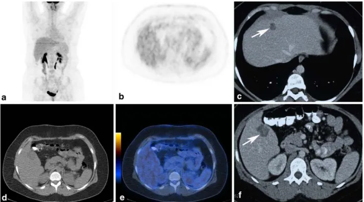
Of 13 patients with UM, 9 (69%) had no extrahepatic metastases. Of 13 patients with UM, 4 (31%) had Fig. 2 a Liver metastasis
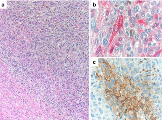
(patient 4) of a predominantly spindle cell, partially pigmented malignant melanoma of the uvea. Residual liver tissue at lower left. H&E staining, original magnification ×100. b Strong immunohistochemical positivity (red reaction product) for the melanocytic marker S-100, original magnification ×200. c Immunohistochemical negativity of the melanoma cells for GLUT-1. Internal positive control of erythrocytes (red). Original magnification ×200
Fig. 1 A 30-year-old female patient (patient 4) 4 years after therapy for an uveal melanoma. FDG PET/CT (a, b, d, e) images without pathological uptake in the liver. Serum S-100B was normal.
Contrast-enhanced CT (c, f) showing two hypodense lesions in the liver. The liver lesions were resected and metastases from the UM were confirmed
extrahepatic metastases: one patient with FDG-active liver metastases had multiple FDG-active lung metasta-ses. One patient with FDG-active liver metastases had additional FDG-active bone and lymph node metastases. One patient with an FDG-active liver metastasis had an additional histologically proven solitary lung metastasis. Another patient with an FDG-active liver metastasis had additional FDG-active peritoneal metastases. The combination of inactive liver metastases and FDG-active extrahepatic deposits occurred in none of the patients.
Only 2 (14%) of 14 patients with CM had no extrahepatic metastases. The other 12 (86%) patients with CM had extrahepatic metastases in the following local-izations: lymph nodes (10 patients), bone (5), lung (5), soft tissue (3), spleen (2), adrenal (2) and brain (1).
Outcome
There was an almost significant trend for a better outcome of the patients with liver metastases from UM compared to CM (p=0.06). Of 12 patients with UM liver
metastases, 5 (42%) died during follow-up and 11 of 14 (79%) patients with CM liver metastases died during follow-up. The mean overall survival of patients with UM liver metastases was 26.8 months (range: 2–59) and 6.5 months (range: 2–27) in the CM group. Two of seven survivors with UM are in complete remission with normal PET/CT examinations and S-100B serum markers after 15 and 6 months of follow-up. Two of three survivors with CM liver metastases are alive in complete remission with normal PET/CT examinations and S-100B serum markers after 36 and 37 months follow-up. One patient with UM was lost to follow-up.
Discussion
Our data show that FDG PET has limited value for the detection of liver metastases from uveal melanoma. This evidence is new and our findings contradict the experience of other authors in patients with uveal melanoma: Francken and co-authors reported on 17 patients with ocular melanoma and liver metastases, 9 confirmed by histology. patient (patient 27) 5 years after
resection of a CM at the ear (Breslow 0.71 mm). FDG PET/ CT images (a, b, c, d) show a solitary liver lesion (arrows) with high FDG uptake (SUVmax:
10.8). S-100B value was nor-mal. No extrahepatic metastases were detectable. The liver lesion was resected and metastasis of a CM was confirmed
Four patients had negative PET scans, supported by negative anatomical imaging or uneventful follow-up. Sensitivity for the detection of liver metastases was 100%, specificity 67% and accuracy 90%. Kurli and co-workers imaged 20 patients with suspected choroidal melanoma metastases with FDG PET/CT and found metastases in 8 (40%) patients. The liver was involved in all eight patients with metastases. No false-negative PET finding was reported in this study [28]. Other authors used FDG PET/CT as a screening tool in patients with choroidal melanoma for initial staging. The same group found metastases in 2 of 52 (3.8%) patients with choroidal
melanoma at initial staging before treatment [29]. In this study again no false-negative PET/CT was observed.
Comparing the cited studies with our results and searching for a reason for the marked difference in sensitivity of PET regarding UM liver metastases the following points can be made: there was contemporane-ously morphological imaging with CT and MRI in the majority of the cited studies and follow-up of at least 6 months available as sufficient reference standard. The PET/CT imaging protocol and injected doses were similar in the cited studies. It seems that in the two studies of Kurli et al. only patients with PET-positive lesions underwent
Table 2 Box plots of SUVmaxvalues of liver metastases from UM and CM
UM= uveal melanoma, CM=cutaneous melanoma, SUV= standard uptake value. * SUV max. of patient No.13. ° SUV max. of patient No. 9. Fig. 4 Liver metastasis
(patient 27) of a predominantly epitheloid, pigmented dermal malignant melanoma. Residual liver tissue at lower left (a). H&E staining, original magnification ×100. b Immuno-histochemical positivity (red reaction product) for the melanocytic marker S-100, original magnification ×200. c Focal immunohistochemical positivity of the melanoma cells for GLUT-1 (brown reaction product). Original magnification ×200
biopsy but not patients with suspicious CT scans but negative PET. With the experience of our study a negative PET in UM cannot“overrule” a suspicious liver finding on MRI or CT and this different approach might explain the different results of the studies. Otherwise we found no obvious explanation for the apparent differences.
Furthermore, our results indicate that S-100B serum maker is not sensitive enough for the detection of metastases in UM patients: we found four patients with normal values of S-100B tumour markers with proven liver metastases from UM. This evidence is also new and contradicts previous reports showing that elevated S-100B markers correlate well with metastatic UM to the liver [23]. Other authors found no prognostic value of S-100B serum concentrations in patients with UM [25]. Also other serum markers like osteopontin (OP) and melanoma inhibitory activity (MIA) are discussed contro-versially in the literature for the detection of UM metastases [23]. Other blood tests like gamma-glutamyl transpeptidase and alkaline phosphatase showed no sufficient accuracy in the detection of liver metastases from UM [30]. In our experience there is no reliable serum marker for the detection of UM liver metastases.
Our comparison of tumour marker, FDG uptake and outcome in patients with UM and CM underlines that these are markedly different melanoma entities with different biological behaviour. Research is ongoing to better understand the molecular characteristics of UM liver metastases [31,32]. It has been shown in vivo and in vitro in different tumour entities that the expression of GLUT transporters on tumour cell membranes can correlate with FDG uptake [33–36].
Histopathological evaluation of the resected liver metastases with GLUT-1 transporters gave no clear explanation for the decreased FDG uptake in our patients with UM liver metastases. There was no striking difference in GLUT-1 transporter or tumour cell density between patients with PET-negative and PET-positive metastases. We had only a limited number of histological results available and further investiga-tion of other transporters or hexokinase might correlate better with the FDG uptake like shown in cholangiocellular carcino-ma [37]. Whether the trend to more epithelial growth in FDG-inactive liver metastases from uveal melanoma observed in two patients of our study has an influence on FDG uptake must be evaluated further in larger patient numbers.
Like in other tumours the FDG uptake seems to correlate with the aggressiveness of the tumour biology as patients with liver metastases of CM had a significantly worse outcome compared with UM patients with liver metastases [38]. Obviously, as shown before, the liver is often the first organ involved in UM while in CM the liver is often involved together with multiple other organs in a more advanced stage [7], another reason for the bad outcome.
MR might be the most accurate imaging modality in FDG-negative liver metastases from uveal melanoma and
we recommend that MRI be performed to evaluate the whole extent of liver involvement prior to therapy in particular cases, especially if resection of liver metastases is considered [39].
Our study has several limitations. This is a retrospective study. S-100B measurements were not available in all patients, and histopathological proof is obviously not available for all metastases. We think that our reference standard with close clinical up and PET/CT follow-up in many patients is sufficient.
Our results have direct impact on our PET/CT imaging protocol. On the basis of these findings we have started to add a contrast-enhanced CT of the liver in patients with UM to increase the sensitivity for the detection of liver metastases of PET/CT in order to also detect FDG-inactive metastases. In patients with CM we continue to perform a native low-dose CT as part of the PET/CT examination.
In conclusion, FDG-PET/CT and S-100B are not sensitive enough for the detection of liver metastases from UM, whereas liver metastases from cutaneous melanomas are reliably FDG positive and lead regularly to increased S-100B tumour markers. The pathophysiological reasons for the lower FDG uptake in UM liver metastases remain unclear. We recommend that a contrast-enhanced PET/CT protocol be performed in patients with UM in order to avoid false-negative findings in the liver.
References
1. Dalrymple-Hay MJ, Rome PD, Kennedy C, Fulham M, McCaughan BC. Pulmonary metastatic melanoma—the survival benefit associated with positron emission tomography scanning. Eur J Cardiothorac Surg 2002;21:611–4. doi:10.1016/S1010-7940 (02)00026-X. discussion 614-615.
2. Reinhardt MJ, Joe AY, Jaeger U, Huber A, Matthies A, Bucerius J, et al. Diagnostic performance of whole body dual modality 18F-FDG PET/CT imaging for N- and M-staging of malignant melanoma: experience with 250 consecutive patients. J Clin Oncol 2006;24:1178–87. doi:10.1200/JCO.2005.03.5634. 3. Strobel K, Dummer R, Husarik DB, Pérez Lago M, Hany TF,
Steinert HC. High-risk melanoma: accuracy of FDG PET/CT with added CT morphologic information for detection of metastases. Radiology 2007;244:566–74. doi:10.1148/radiol.2442061099. 4. Strobel K, Skalsky J, Kalff V, Baumann K, Seifert B, Joller-Jemelka
H, et al. Tumour assessment in advanced melanoma: value of FDG-PET/CT in patients with elevated serum S-100B. Eur J Nucl Med Mol Imaging 2007;34:1366–75. doi:10.1007/s00259-007-0403-8. 5. Singh AD, Topham A. Incidence of uveal melanoma in the United
States: 1973–1997. Ophthalmology 2003;110:956–61. doi:10.1016/ S0161-6420(03) 00078-2.
6. Virgili G, Gatta G, Ciccolallo L, Capocaccia R, Biggeri A, Crocetti E, et al. Incidence of uveal melanoma in Europe. Ophthalmology 2007;114:2309–15. doi:10.1016/j.ophtha.2007.01.032.
7. Albert DM, Ryan LM, Borden EC. Metastatic ocular and cutaneous melanoma: a comparison of patient characteristics and prognosis. Arch Ophthalmol 1996;114:107–8.
8. Belmar-Lopez C, Mancheno-Corvo P, Saornil MA, Baril P, Vassaux G, Quintanilla M, et al. Uveal vs. cutaneous melanoma. Origins and causes of the differences. Clin Transl Oncol 2008;10:137–42. doi:10.1007/s12094-008-0170-4.
9. Baggetto LG, Gambrelle J, Dayan G, Labialle S, Barakat S, Michaud M, et al. Major cytogenetic aberrations and typical multidrug resistance phenotype of uveal melanoma: current views and new therapeutic prospects. Cancer Treat Rev 2005;31:361–79. doi:10.1016/j.ctrv.2005.05.001.
10. Damato B. Developments in the management of uveal melanoma. Clin Experiment Ophthalmol 2004;32:639–47. doi:10.1111/ j.1442-9071.2004.00917.x.
11. Hsueh EC, Essner R, Foshag LJ, Ye X, Wang HJ, Morton DL. Prolonged survival after complete resection of metastases from intraocular melanoma. Cancer 2004;100:122–9. doi:10.1002/ cncr.11872.
12. Bedikian AY, Legha SS, Mavligit G, Carrasco CH, Khorana S, Plager C, et al. Treatment of uveal melanoma metastatic to the liver: a review of the M. D. Anderson Cancer Center experience and prognostic factors. Cancer 1995;76:1665–70. doi:10.1002/1097-0142 (19951101) 76:9<1665::AID-CNCR2820760925>3.0.CO;2-J. 13. Feldman ED, Pingpank JF, Alexander HR Jr. Regional treatment
options for patients with ocular melanoma metastatic to the liver. Ann Surg Oncol 2004;11:290–7. doi:10.1245/ASO.2004.07.004. 14. Sharma KV, Gould JE, Harbour JW, Linette GP, Pilgram TK,
Dayani PN, et al. Hepatic arterial chemoembolization for management of metastatic melanoma. AJR Am J Roentgenol 2008;190:99–104. doi:10.2214/AJR.07.2675.
15. Vogl T, Eichler K, Zangos S, Herzog C, Hammerstingl R, Balzer J, et al. Preliminary experience with transarterial chemoemboliza-tion (TACE) in liver metastases of uveal malignant melanoma: local tumor control and survival. J Cancer Res Clin Oncol 2007;133:177–84. doi:10.1007/s00432-006-0155-z.
16. Sato KT, Lewandowski RJ, Mulcahy MF, Atassi B, Ryu RK, Gates VL, et al. Unresectable chemorefractory liver metastases: radio-embolization with 90Y microspheres–safety, efficacy, and survival. Radiology 2008;247:507–15. doi:10.1148/radiol.2472062029. 17. Abraha HD, Fuller LC, Du Vivier AW, Higgins EM, Sherwood RA.
Serum S-100 protein: a potentially useful prognostic marker in cutaneous melanoma. Br J Dermatol 1997;137:381–5. doi:10.1111/ j.1365-2133.1997.tb03742.x.
18. Andrés R, Mayordomo JI, Zaballos P, Rodino J, Isla D, Escudero P, et al. Prognostic value of serum S-100B in malignant melanoma. Tumori 2004;90:607–10.
19. Bánfalvi T, Gilde K, Boldizsár M, Kremmer T, Ottó S. Serum levels of S-100 protein and 5-S-cysteinyldopa as markers of melanoma progression. Pathol Oncol Res 1999;5:218–22. doi:10.1053/ paor.1999.0218.
20. Domingo-Domènech J, Molina R, Castel T, Montagut C, Puig S, Conill C, et al. Serum protein s-100 predicts clinical outcome in patients with melanoma treated with adjuvant interferon–comparison with tyrosinase rt-PCR. Oncology 2005;68:341–9. doi:10.1159/ 000086973.
21. Guo HB, Stoffel-Wagner B, Bierwirth T, Mezger J, Klingmüller D. Clinical significance of serum S100 in metastatic malignant melanoma. Eur J Cancer 1995;31A:1898–902. doi: 10.1016/0959-8049(95)00087-Y.
22. Hamberg AP, Korse CM, Bonfrer JM, de Gast GC. Serum S100B is suitable for prediction and monitoring of response to chemo-immunotherapy in metastatic malignant melanoma. Melanoma Res 2003;13:45–9. doi:10.1097/00008390-200302000-00008. 23. Barak V, Frenkel S, Kalickman I, Maniotis AJ, Folberg R, Pe’er J.
Serum markers to detect metastatic uveal melanoma. Anticancer Res 2007;27:1897–900.
24. Missotten GS, Korse CM, van Dehn C, Linders TC, Keunen JE, Jager MJ, et al. S-100B protein and melanoma inhibitory activity protein in uveal melanoma screening. A comparison with liver function tests. Tumour Biol 2007;28:63–9. doi:10.1159/000099151. 25. Missotten GS, Tang NE, Korse CM, Hurks HM, de Wolff-Rouendaal D, Keunen JE, et al. Prognostic value of S-100-beta serum concentration in patients with uveal melanoma. Arch Ophthalmol 2003;121:1117–9. doi:10.1001/archopht.121.8.1117.
26. Strobel K, Dummer R, Steinert HC, Conzett KB, Schad K, Lago MP, et al. Chemotherapy response assessment in stage IV melanoma patients-comparison of 18F-FDG-PET/CT, CT, brain MRI, and tumormarker S-100B. Eur J Nucl Med Mol Imaging 2008;35:1786–95. doi:10.1007/s00259-008-0806-1.
27. Strobel K, Skalsky J, Steinert HC, Dummer R, Hany TF, Bhure U, et al. S-100B and FDG-PET/CT in therapy response assessment of melanoma patients. Dermatology 2007;215:192–201. doi:10.1159/ 000106575.
28. Kurli M, Reddy S, Tena LB, Pavlick AC, Finger PT. Whole body positron emission tomography/computed tomography staging of metastatic choroidal melanoma. Am J Ophthalmol 2005;140:193– 9.
29. Finger PT, Kurli M, Reddy S, Tena LB, Pavlick AC. Whole body PET/CT for initial staging of choroidal melanoma. Br J Ophthalmol 2005;89:1270–4. doi:10.1136/bjo.2005.069823. 30. Hicks C, Foss AJ, Hungerford JL. Predictive power of screening
tests for metastasis in uveal melanoma. Eye 1998;12(Pt 6):945–8. 31. Meir T, Dror R, Yu X, Qian J, Simon I, Pe’er J, et al. Molecular characteristics of liver metastases from uveal melanoma. Invest Ophthalmol Vis Sci 2007;48:4890–6. doi:10.1167/iovs.07-0215. 32. Shields CL. The hunt for the secrets of uveal melanoma. Clin
Experiment Ophthalmol 2008;36:277–80. doi: 10.1111/j.1442-9071.2008.01717.x.
33. Chung JK, Lee YJ, Kim C, Choi SR, Kim M, Lee K, et al. Mechanisms related to [18F]fluorodeoxyglucose uptake of human colon cancers transplanted in nude mice. J Nucl Med 1999;40:339– 46.
34. Ong LC, Jin Y, Song IC, Yu S, Zhang K, Chow PK. 2-[18F]-2-deoxy-D-glucose (FDG) uptake in human tumor cells is related to the expression of GLUT-1 and hexokinase II. Acta Radiol 2008;49:1145–53. doi:10.1080/02841850802482486.
35. Hamada K, Tomita Y, Qiu Y, Zhang B, Ueda T, Myoui A, et al. 18F-FDG-PET of musculoskeletal tumors: a correlation with the expression of glucose transporter 1 and hexokinase II. Ann Nucl Med 2008;22:699–705. doi:10.1007/s12149-008-0173-9. 36. Higashi T, Saga T, Nakamoto Y, Ishimori T, Mamede MH,
Wada M, et al. Relationship between retention index in dual-phase (18) F-FDG PET, and hexokinase-II and glucose transporter-1 expression in pancreatic cancer. J Nucl Med 2002;43:173–80.
37. Paudyal B, Oriuchi N, Paudyal P, Higuchi T, Nakajima T, Endo K. Expression of glucose transporters and hexokinase II in chol-angiocellular carcinoma compared using [18F]-2-fluro-2-deoxy-D-glucose positron emission tomography. Cancer Sci 2008;99:260–6. doi:10.1111/j.1349-7006.2007.00683.x.
38. Downey RJ, Akhurst T, Gonen M, Park B, Rusch V. Fluorine-18 fluorodeoxyglucose positron emission tomographic maximal stan-dardized uptake value predicts survival independent of clinical but not pathologic TNM staging of resected non-small cell lung cancer. J Thorac Cardiovasc Surg 2007;133:1419–27. doi:10.1016/j. jtcvs.2007.01.041.
39. Maeda T, Tateishi U, Suzuki S, Arai Y, Kim EE, Sugimura K. Magnetic resonance screening trial for hepatic metastasis in patients with locally controlled choroidal melanoma. Jpn J Clin Oncol 2007;37:282–6. doi:10.1093/jjco/hym018.