ORIGINAL ARTICLE
Diagnostic accuracy of contrast-enhanced FDG-PET/CT
in primary staging of cutaneous malignant melanoma
Patrick Veit-Haibach&Florian M. Vogt&
Robert Jablonka&Hilmar Kuehl&Andreas Bockisch&
Thomas Beyer&Gerlinde Dahmen&
Sandra Rosenbaum&Gerald Antoch
Received: 10 September 2008 / Accepted: 5 December 2008 / Published online: 21 January 2009 # Springer-Verlag 2009
Abstract
Purpose To evaluate the diagnostic accuracy of contrast-enhanced FDG-PET/CT (ce-PET/CT), PET-only, and CT-only in patients with newly diagnosed and resected cutaneous malignant melanoma.
Methods A final group of 56 patients (mean age 62 years, range 23–86 years; 29 women, 27 men) were staged with ce-PET/CT after resection of the primary tumour. Histopa-thology as well as clinical follow-up (mean 780 days, range 102–1,390 days) served as the standards of reference. Differences between the staging modalities were tested for statistical significance with McNemar’s test.
Results All imaging procedures provided low sensitivities in the detection of lymph nodes (sensitivity N-stage: PET/ CT and PET-only 38.5%; CT-only 23.1%) and distant metastases (sensitivity M-stage: PET/CT 41.7%, PET-only 33.3%, CT-only 25.0%) in initial staging after resection of the primary tumour. No statistically significant differences were detected between the imaging procedures (p>0.05). PET/CT resulted in an alteration in further treatment in two patients compared to PET-only and in four patients compared to CT-only.
Conclusion All imaging modalities had a low sensitivity on initial staging of patients with malignant melanoma. Thus, close patient follow-up must be considered mandatory.
Keywords ce-PET/CT . Cutaneous melanoma . Staging . Follow-up . Diagnostic accuracy
Introduction
The incidence of melanoma has been increasing worldwide, especially in the Caucasian population [1]. In 2007 approximately 60,000 new cases of melanoma were diagnosed and over 8,000 patients were expected to die from this disease in the United States. The patients’ prognosis strongly depends on the tumour depth (tumour thickness and Breslow index), potential ulceration, and the presence of metastases [2, 3]. Cutaneous melanoma is considered curable in patients if the tumour is <2 mm deep, is without ulceration, and is without lymph node metasta-ses. Tumours of higher tumour stages are considered to have an advanced likelihood of metastatic spread. Thus, overall survival is heavily dependent on the stage of the primary tumour [4,5].
P. Veit-Haibach (*)
:
F. M. Vogt:
H. Kuehl:
G. AntochDepartment of Diagnostic and Interventional Radiology and Neuroradiology, University Hospital Essen, Hufelandstrasse 55,
45122 Essen, Germany
e-mail: [email protected] R. Jablonka
Department of Dermatology, University Hospital Essen, Essen, Germany
A. Bockisch
:
T. Beyer:
S. RosenbaumDepartment of Nuclear Medicine, University Hospital Essen, Essen, Germany
G. Dahmen
Institute of Medical Biometry and Statistics, University at Luebeck,
Luebeck, Germany Present address: P. Veit-Haibach
Department of Medical Radiology, Division of Nuclear Medicine, University Hospital Zurich,
Rämistrasse 100, 8091 Zurich, Switzerland
Several imaging methods are considered appropriate for imaging patients with malignant melanoma and potential metastatic spread. Computed tomography (CT) is widely accepted and recommended for detection of organ metas-tases [6,7]. However, CT has a limited sensitivity for the detection of lymph node metastases because of its strict morphological nature. Locoregional ultrasonography has been used for assessment of lymph nodes, but is investiga-tor-dependent and strictly morphological.
To overcome the lack of functional data, positron emission tomography (PET) using [18F]-fluoro-2-deoxy-D -glucose (FDG) as a radioactive tracer has gained wide acceptance in patients with malignant melanoma in partic-ular in patients with clinical suspicion of systemic meta-static spread [8,9]. However, the accuracy of FDG-PET in initial staging of malignant melanoma has rarely been assessed. PET-only imaging can be impaired based on its lack of anatomical resolution. Hence, complementary anatomical imaging is often required, especially for surgical therapy planning. Coregistered PET/CT has been available since 2001 and provides such anatometabolic datasets in a single examination. PET/CT has been found to be superior to CT alone and PET alone in staging and evaluation of therapy response in several oncological diseases including malignant melanoma [10–12]. However, as mentioned above, there is currently only little knowledge concerning the accuracy of FDG-PET/CT compared to CT alone and PET alone for initial staging of malignant melanoma. Thus, the aim of our study was (1) to evaluate the accuracy of FDG-PET/CT, CT-only, and PET-only for initial staging of malignant melanoma using long-term follow-up and histopathology as the standards of reference, and (2) to assess the potential impact of FDG-PET/CT on patient management.
Materials and methods
Patients
Seventy-four patients were included based on their order of referral without further selection. All patients were referred for a combined FDG-PET/CT examination after surgical resection of a primary malignant melanoma. This prospec-tive study was performed in accordance with the regu-lations of the local institutional review board and ethics committee. All patients were included consecutively based on the time of referral without further selection. Patients were excluded from the analysis if no sufficient follow-up data were available (e.g. patient did not attend for follow-up examinations or decided to have follow-up at another institution). Informed consent was obtained from all patients prior to the examination.
FDG-PET/CT imaging procedure
Patients were instructed to fast for at least 4 h prior to the PET/CT procedure. Glucose levels in all patients were measured prior to FDG injection to ensure they were in the normal range. PET/CT imaging was conducted on a Biograph Duo PET/CT system (Siemens Molecular Imag-ing, Hoffman Estates, IL). The system integrates a dual-slice CT scanner (Somatom Emotion, Siemens Medical Solutions, Forchheim, Germany) and a full-ring, BGO-based PET tomograph (Siemens Molecular Imaging). The axial field-of-view of the PET scanner is 15.5 cm per bed position, and the in-plane spatial resolution is 4.6 mm, respectively. The average FDG activity administered 60 min prior to the PET/CT examination was 330–350 MBq. During the uptake time, 1,500 ml of a water-based, water-equivalent oral contrast agent was administered to all patients for small-bowel distension [13].
During the whole-body CT examination (part of the PET/CT examination), 140 ml of iodinated contrast agent (300 mmol/ml, Xenetix 300; Guerbet, Sulzbach, Germany) was administered intravenously according to a standardized protocol [14]. The CT scan was performed in the caudocranial direction. A start delay of 50 s was chosen for the CT acquisition after the start of the contrast agent injection. The first 90 ml of contrast agent were injected at a rate of 3 ml/s, and the remaining 50 ml were injected at a rate of 1.5 ml/s. The dual-phase injection was intended to ensure fully diagnostic (portal venous phase) CT data in the abdomen. The contrast-enhanced CT scan was used for attenuation correction of the PET data. The PET acquisition time per bed position was 3–5 min, depending on the weight of the patient. PET images were corrected for scatter and attenuation based on the available CT transmission images. Corrected PET images were reconstructed itera-tively (FORE-OSEM, two iterations, eight subsets, 128× 128 matrix with 5-mm gaussian smoothing). CT images as well as PET data sets were viewed separately (CT-only, PET-only), and in fused mode (PET/CT) on a commer-cially available computer workstation (Siemens Molecular Imaging). Therefore, all imaging modalities compared were derived from the same dataset.
Image evaluation
N-staging and M-staging
The T-stage was documented from the histological speci-mens from the surgical resection, which was performed not more than 1 week prior to the imaging procedure. N-staging and M-staging evaluation were performed for CT-only, PET-only, and coregistered FDG-PET/CT. The PET images were evaluated with and without attenuation correction by
two nuclear medicine specialists in consensus. The CT images were evaluated by two radiologists in consensus. Contrast-enhanced PET/CT (ce-PET/CT) images were evaluated by a different radiologist and nuclear medicine specialist in consensus. The participating readers were informed about the patient-specific clinical background (first diagnosis of melanoma, postsurgical resection status, location of the resection site), but blinded to the results of histopathology of the primary tumour, and blinded to the other imaging procedures and to clinical examination.
Distant metastases were assessed based on the detec-tion of soft-tissue masses (or focal cutaneous thickening) with contrast enhancement in different body compart-ments and in conjunction with focally increased glucose metabolism above the surrounding tissue level on FDG-PET/CT. The diagnosis of a distant metastasis was also supported by an maximum standardized uptake value (SUVmax) of at least 1.5 for cutaneous lesions, 2.5 for other extrahepatic lesions, and 3.5 for intrahepatic lesions [15]. However, the SUVmax was not taken as the absolute threshold to differentiate between malignant and benign findings. In fact, the qualitative assessment was taken as the most important parameter. If a lesion showed clear focal FDG avidity but displayed a lower SUVmax (e.g. due to small size), the lesion was rated malignant. In cases of malignant findings on CT-only without focally in-creased glucose metabolism, the lesions were evaluated based on CT criteria (see below). Lymph nodes were assessed for metastatic spread based on an increased glucose metabolism and independent of their size on PET/CT images.
On CT-only images, detection of soft-tissue masses (or focal cutaneous thickening) with contrast enhancement characterized malignancy. Lymph node assessment was based on lesion size: a short-axis diameter threshold of 1.5 cm was used for jugulodigastric lymph nodes and precarinal lymph nodes. A short-axis diameter threshold of 1 cm was used for all other lymph nodes of the neck, thorax, and abdomen [16]. Central necrosis was defined as a sign of malignancy as well, independent of lymph node size. Furthermore, according to standard CT criteria, a fatty hilum and calcifications were used as benign criteria on CT-only images.
PET-only images were assessed qualitatively and quan-titatively for areas of increased FDG uptake. Lesions (distant metastasis and lymph nodes) were called malignant if the glucose utilization exceeded the surrounding tissue or blood pool level. As in evaluation of the PET/CT images, the diagnosis of metastases was also supported by a SUVmax of more than 1.5 for cutaneous lesions, more than 2.5 for extrahepatic lesions, and more than 3.5 for intrahepatic lesions. The N-stage and M-stage in all patients
were assessed based on the current AJCC criteria for all imaging modalities [17].
The impact of FDG-PET/CT imaging on patient man-agement as compared to PET-only and CT-only was assessed in consensus by the referring physicians and a radiologist and nuclear medicine specialist each, and evaluation was based on international clinical guidelines [6,18].
Standard of reference
Initial clinical staging derived from histopathological examination of the primary tumour (T-stage) after resection, from sentinel lymph node resection within 4 weeks of PET/CT imaging and all other available clinical studies and imaging studies (MRI, radiography, ultrasonography, tumour markers). Because PET/CT imaging was the modality to be evaluated, the results of the PET/CT imaging were not taken into account for the definition of the initial clinical stage. Sentinel lymph node imaging and resection were performed within 4 weeks of initial PET/CT imaging. In all patients with suspected metastases on imaging, histopathological eval-uation and the resected surgical specimen (tumours and/ or lymph nodes) of at least one metastatic site served as the standard of reference for both N-stage and M-stage during the clinical course. For all other patients, clinical follow-up including all clinically available data (imaging, tumour markers, physical examination) served as the standard of reference.
Statistical analysis
The primary endpoint of the study was the correct classification of the N-stage and M-stage using CT-only or PET-only in comparison to fused PET/CT. Differences in the assessment of the N-stage and M-stage between the different imaging procedures were tested for significance by McNemar’s test (exact). Bonferroni correction was applied to account for multiple comparisons. To maintain a global significance level of 0.05 the nominal significance level to evaluate the four hypotheses of the primary analysis had to be 0.0125. We calculated 95% confidence intervals (CI) according to the method of Tango for the difference in correlated proportions of the correct N-stage and M-stage [19]. Sensitivities, specificities, negative predictive values (NPV), positive predictive values (PPV), and accuracies (with exact 95% confidence intervals) for all modalities were determined for N-stage assessment and for M-stage assessment using histology and/or clinical follow-up as the standard of reference.
Statistical analyses were performed with SAS statistical software (version 9.1; SAS Institute, Cary, NC).
Results Patients
Included in this prospective study were 74 consecutive patients who underwent combined PET/CT imaging in a University Hospital setting. Of these 74 patients, 18 were excluded because of a lack of sufficient follow-up, leaving 56 patients for final evaluation (mean age 62 years, range 23–86 years; 29 women, 27 men) (Table1). The T-stage of the primary, melanoma location, histological type of the melanoma and the clinical stages according to the standard of reference are given in Table 1 for the 56 evaluated patients. Overall, the mean follow-up time was 780 days (range 102–1,390 days). A total 18 patients had metastases, in 12 detected at initial staging (clinical stage III or IV), and in the other 6 during the clinical course. Sentinel lymph node imaging and resection was performed within 4 weeks of the initial PET/CT procedure in 14 patients. Overall, at least one suspicious lesion was confirmed histologically in all patients suspected of harbouring metastases in lymph nodes or distant organs during the clinical course. During the follow-up 28 patients remained disease-free. Four patients died during the clinical course. In one patient, a secondary cancer (breast cancer) was detected at initial staging. All patients tolerated the PET/CT procedures well.
Metastases at initial staging
At initial staging, 12 patients were diagnosed with lymph node and/or distant metastases. The initial clinical stages were stage IV in eight patients and stage III in four patients. Combining the T-stage from histopathology with the staging results from FDG-PET/CT, PET-only and CT-only, the tumour stage was correctly classified in six patients with PET/CT, in five patients with PET, and in three patients with CT. No statistically significant difference was detected concerning the N-stage and the M-stage when comparing PET/CT and PET-only (p>0.05), PET/CT and CT-only (p>0.05) in this subgroup of patients.
Overall N-staging/M-staging
A comparison of the N-stages and M-stages for all imaging modalities are shown in Table 2. Overall, no statistically significant differences were detected concerning the N-stage between PET/CT and PET-only (difference 0%; 95% CI−10– 10%; p>0.05) or between PET/CT and CT-only (difference 4%; 95% CI−15–7%; p>0.05). Additionally, no statistically significant differences were found concerning the M-stage between PET/CT and PET-only (difference −2%; 95% CI −12–8%; p>0.05) or between PET/CT and CT-only (difference−2%; 95% CI −12–8%; p>0.05).
Therapy alteration, occurrence of metastases and side findings
N-stage
In two patients, PET/CT and PET-only were superior to CT-only because lymph nodes with focal FDG avidity were not evaluated as malignant on CT-only due to size criteria. In these two patients these findings were thera-peutically relevant, because both lymph node metastases were identified and treated consecutively after the initial scan.
Overall (during the whole clinical course including initial staging), eight patients were falsely staged as negative on PET/CT and PET-only concerning lymph node metastases and ten patients were falsely staged as negative on CT-only (Table 2). Two of these patients had micro-metastases at initial staging which were overlooked on all imaging modalities but detected by sentinel lymph node resection after PET/CT imaging.
M-stage
Overall, in four patients, therapeutically relevant advan-tages arose from PET/CT imaging concerning the M-stage. In one patient, PET/CT detected small pulmonary nodules without FDG activity, which were evaluated as lung metastases based on the CT part of the PET/CT scan. However, these lesions were overlooked on PET-only (but detected by CT-only), based on their size and lack of FDG activity. Pulmonary metastases were confirmed on clinical follow-up.
In another patient, PET-only imaging showed an increased FDG uptake in an adrenal gland, but the CT component of the PET/CT scan showed no morphological correlate. Thus, on the PET/CT image, the readers evaluated this finding as not pathological, while on the PET-only image the readers considered the possibility of adrenal gland metastases. During the following clinical course, no adrenal gland metastasis was detected in this patient.
PET/CT had a therapeutically relevant advantage in another patient, in whom a pathologically enlarged inguinal lymph node was evaluated as malignant on CT-only. PET/ CT showed no increased glucose metabolism. Thus, the lymph node was evaluated as normal on PET/CT which was confirmed during the clinical course. PET/CT had a therapeutically relevant advantage in another patient in whom (vice versa) the readers detected increased glucose metabolism in a lymph node that was not pathologically enlarged on PET/CT. The PET/CT image was evaluated correctly as malignant, while the CT-only image was evaluated falsely as negative (Fig.1).
Table 1 Patient characteristics, locations of the primary tumour, histological type of melanoma, and clinical stage
Patient no. Age (years) Location Histological type S-100 (mg/l)a Clinical stageb
1 59 Left back Nodular 0.11 I A
2 53 Right breast Superficial spreading 0.15 I A
3 58 Left shoulder Superficial spreading 0.057 III
4 48 Left thigh Nodular 0.016 I B
5 69 Right lower leg Superficial spreading II A
6 45 Left lower leg Superficial spreading 0.168 II A
7 69 Left upper arm Superficial spreading 0.132 II A
8 79 Left face Lentigo maligna 0.027 I A
9 71 Right upper arm Nodular IV
10 47 Right thigh Superficial spreading I A
11 83 Right foot Nodular 0.093 II A
12 35 Right forearm Lentigo maligna 0.042 II A
13 69 Right knee Superficial spreading 0.07 I B
14 70 Right hand Superficial spreading 0.136 IV
15 86 Right lower leg Nodular 0.402 IV
16 59 Right upper arm Nodular IV
17 46 Right lower back Superficial spreading <0.15 IV
18 48 middle back Lentigo maligna 0.024 I B
19 69 Right thorax Nodular 0.087 II A
20 65 Right thorax Nodular IV
21 57 Right abdomen Superficial spreading I A
22 65 Right upper arm Superficial spreading 0.078 I A
23 46 Left back Superficial spreading IV
24 48 Right knee Nodular 0.066 II C
25 53 Left back Superficial spreading 0.038 I B
26 73 Right face Nodular 0.075 II B
27 39 Left hip Superficial spreading I A
28 83 Left forearm Nodular II A
29 79 Right back Superficial spreading I A
30 41 Right lower back Superficial spreading I B
31 42 Left thorax Superficial spreading 0.045 I A
32 59 Left thorax Nodular 0.095 IV
33 57 Right thorax Nodular IV
34 77 Right forearm Nodular 0.116 I B
35 65 Right thorax Superficial spreading IV
36 23 Right lower leg Superficial spreading 0.117 I A
37 82 Right groin Nodular II B
38 62 Right upper arm Nodular I B
39 79 Right foot Acrolentiginous II B
40 75 Left abdomen Superficial spreading 0.139 II B
41 66 Right thigh Nodular III
42 76 Right back Superficial spreading 0.056 I B
43 79 Left back Acrolentiginous III
44 68 Right back Superficial spreading 0.12 I B
45 70 Right thorax Superficial spreading 0.092 IV
46 56 Left knee Nodular I B
47 73 Right shoulder Superficial spreading 0.086 II A
48 71 Right lower leg Superficial spreading 0.133 IV
49 55 Right lower leg Nodular II A
50 74 Left shoulder Superficial spreading 0.075 IV
51 60 Left upper arm Superficial spreading I A
52 40 Right abdomen Superficial spreading I B
53 59 Left thigh Lentigo maligna III
54 84 Left upper arm Nodular 0.08 I B
55 32 Right thorax Superficial spreading IV
56 49 Left thigh Nodular II C
a
Standard tumour marker measured at initial diagnosis.
b
Overall, 18/56 patients (32%) had either metastases at initial staging (12 patients) or developed metastases during the clinical course. Of these 18 patients, 12 (21%) presented initially with clinical stage III or IV, and 6 (11%) initially staged as clinical stage I or II, developed local lymph node metastases and/or distant metastases (Fig.2).
Overall, in patients classified as false-negative, lymph node metastases were detected after a mean follow-up time of 150 days (range 15–425 days), and distant metastases were detected after a mean follow-up time of 204 days (range 71–558 days).
Discussion
Both functional and morphological imaging procedures have a low sensitivity when assessing patients with cutaneous malignant melanoma for locoregional and distant metastases at initial diagnosis. In metastasis-negative patients this has to be taken into account and should mandate a close patient follow-up.
Currently there is only little knowledge about FDG-PET/ CT and its role as a first-line diagnostic tool or follow-up imaging modality in malignant melanoma. However, different studies have found a benefit of PET-only when
compared with CT imaging in patients with recurrent melanoma [8]. Fuster et al. reported an FDG-PET sensitiv-ity and specificsensitiv-ity of 74% and 86% for lesion detection in patients with melanoma compared with 58% and 54% for standard imaging [20]. Crippa et al. found an even higher accuracy of FDG-PET imaging. In their study the sensitiv-ity, specificity and accuracy, and the PPV and NPV for detection and characterization of lymph node metastases were 95%, 84%, 91%, 92%, and 89%, respectively [21]. In another patient population with a primary diagnosis of melanoma, which may thus be comparable to our popula-tion, a lesion-based sensitivity of 94.9% was found [22]. These overall promising results of PET imaging were confirmed by Reinhardt et al. who evaluated a mixed patient population comprising 250 patients with a primary diagnosis, therapy control, recurrence, and follow-up on FDG-PET/CT imaging [10]. The sensitivity, specificity, and accuracy for the N-stage and M-stage in the entire patient population ranged between 95% and 100% for PET/CT imaging. The substantial differences between previously reported sensitivities and the sensitivities in our study can be attributed to several factors. Most previous studies included a limited follow-up time. We followed our patients over a mean period of over 2 years, compared to 1 year in the study by Reinhardt et al. and most of the other studies.
Table 2 Staging results
Stage Modality Correctly
staged
Overstaged Understaged Sensitivity
(95% CI) (%) Specificity (95% CI) (%) Accuracy (95% CI) (%) NPV (95% CI) (%) PPV (95% CI) (%) N PET/CT 48/56 0/56 8/56 38.5 (14–68) 100 (92–100) 85.6 (74–94) 84.3 (71–93) 100 (48–100) PET-only 48/56 0/56 8/56 38.5 (14–68) 100 (92–100) 85.7 (74–94) 84.3 (71–93) 100 (48–100) CT-only 46/56 0/56 10/56 23.1 (5–53) 100 (92–100) 82.1 (70–91) 81.1 (68–91) 100 (48–100) M PET/CT 46/56 3/56 7/56 41.7 (15–72) 93.2 (81–99) 82.1 (70–91) 85.4 (72–94) 62.5 (24–91) PET-only 44/56 4/56 8/56 33.3 (9–65) 90.9 (78–97) 78.6 (66–88) 83.3 (70–93) 50.0 (16–84) CT-only 44/56 3/56 9/56 25.0 (5–57) 93.2 (81–99) 78.6 (66–88) 82.0 (69–91) 50.0 (12–88)
Fig. 1 A 66-year-old female after resection of a malignant melanoma of the right thigh. a A small lymph node that is not pathologically enlarged is seen on the CT-only image dorsal to the right external iliac vessels. However, based on size criteria, the lymph node was not evalu-ated as malignant. b On the corresponding PET/CT image, the same lymph node is seen as FDG avid and was thus evalu-ated as metastasis
It is well known that with an increase in follow-up time, the incidence of metastases increases. Metastases which oc-curred during the clinical course and were not detected on initial FDG-PET/CT imaging were rated as false-negative in our analysis. We chose this approach because the primary melanoma had already been resected. Metastases developing in the further course of disease must, therefore, have been present as micrometastases before. Small metastatic cell deposits need to have a certain size to be detected by FDG-PET or FDG-PET/CT imaging. It has been shown that cell conglomerates of 78 mm3are needed for PET-only imaging to provide a sensitivity of 90% [23]. As the detectors of the PET component of PET/CT are similar to those in PET-only no additional sensitivity of FDG-PET/CT over FDG-PET can be expected. However, rating the imaging findings as false-negative in the presence of micrometastases has been a matter of controversy, as metastases detected at a later time may have resulted from small cell deposits in other organs. Yet metastatic cells might circulate over a certain period of time, probably depending on the immunological status of the patient. In this case, detection of lymph nodes or organ metastases must be considered impossible with current imaging techniques. However, this would still result in a
false-negative assessment for metastatic spread on initial staging and stresses the need for a close follow-up in metastasis-negative patients.
Another major difference compared to the study of Rheinhardt et al. was the finding of metastases in 35% of the 76 patients at initial staging, while in our study only 21% of the patients (12/56) had metastases by the time of first diagnosis. Consequently, therapy changes occurred in a significantly higher percentage (43% in patients with primary diagnosis of melanoma) compared to our patient population. It is already known from PET imaging studies that the impact on therapy and staging increases in higher clinical stages [24]. However, there are also studies in the literature that have shown low sensitivities of FDG-PET in malignant melanoma. In a study including patients with early-stage melanoma, the sensitivity to detect lymph node metastases was only 21% [25]. Based on these results FDG-PET was not recommended as a first-line tool to stage malignant melanoma. Also in this study, the follow-up time was significantly longer than 1 year (>41 months).
As sentinel lymph node imaging and biopsy should be considered in stages I and II and is definitively recom-mended by staging guidelines in higher stages, it should be applied at least in patients with advanced clinical stages [6].
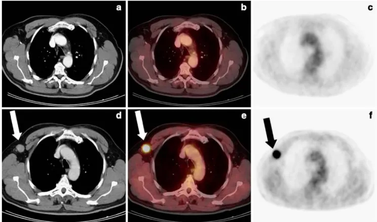
Fig. 2 A 59-year-old male after resection of a malignant melanoma of
the left dorsal thorax (initial clinical stage II). a–c On initial staging 12
days after resection of the primary tumour, no right axillary distant
metastases are seen on the CT-only (a), PET/CT (b) or PET-only (c)
images. d–f During follow-up, a distant metastasis in the right axilla is
If positive, the next staging step and follow-up could be combined PET/CT imaging (PET imaging is recommended in staging guidelines anyway) for several reasons. First, it has already been shown (also partly in our study), that PET/CT imaging can correctly change the subsequent therapy and therefore might decrease rates of morbidity compared to CT-only and PET-CT-only [10,26]. PET/CT imaging has also been found to be useful for the follow-up of patients with resected melanoma because it provides detection of metastases, even when tumour markers are negative, and it can furthermore improve the detection of melanoma metastases compared to PET-alone [11, 12]. An efficient follow-up imaging algo-rithm (as proposed here) has to be collaboratively developed because 11% of our patients developed metastases despite the initially low clinical stage.
Our study has several limitations concerning compara-bility with other studies. Our whole-body CT protocol was different from that used in the study by Reinhardt et al. in which no intravenous contrast medium was administered. However, since PET/CT is a combined imaging modality, we believe that adequate image quality should be provided in both imaging parts (CT and PET). An additional reason for the differences was the relatively low number of patients with metastases at initial staging and during the clinical course in our study. However, we included our patients prospectively with no further selection thus, reflecting the clinical routine in our department.
Sentinel lymph node biopsy was not available in every patient because there are no strict guidelines recommending this in every melanoma case (see guidelines and discussion above). However, there is evidence that sentinel lymph node imaging and resection is more sensitive than PET imaging alone, especially in stage I and II disease [27–30]. Currently there is no study available comparing the sensitivity of sentinel lymph node imaging and PET/CT imaging (there are studies comparing sentinel lymph node imaging to PET-only), but since the CT part in PET/CT generally does not increase the sensitivity in lymph node detection, it can be assumed that these data are still valid.
We did not have the standard tumour marker available in every patient (S-100, see Table 1) because only lactate dehydrogenase is optionally included in a guideline-oriented standard work-up in advanced clinical stages. It has already been demonstrated, that the S-100 marker also has a limited ability to detect melanoma metastases compared to PET/CT imaging [12].
Conclusion
Based on our results, ce-FDG-PET/CT can only be recommended as a first-line diagnostic tool for selected melanoma patients in the high-risk group. Generally, ce-FDG-PET/CT and FDG-PET have low sensitivities on
initial staging of patients with malignant melanoma. Thus, close patient follow-up must be considered mandatory.
References
1. Jemal A, Siegel R, Ward E, Murray T, Xu J, Thun MJ. Cancer
statistics, 2007. CA Cancer J Clin 2007;57 1:43–6.
2. Balch CM, Soong SJ, Gershenwald JE, Thompson JF, Reintgen DS, Cascinelli N, et al. Prognostic factors analysis of 17,600 melanoma patients: validation of the American Joint Committee on Cancer
melanoma staging system. J Clin Oncol 2001;19 16:3622–34.
3. Breslow A. Thickness, cross-sectional areas and depth of invasion in the prognosis of cutaneous melanoma. Annals of surgery Nov 1970;172 5:902–8.
4. Balch CM, Buzaid AC, Soong SJ, Atkins MB, Cascinelli N, Coit DG, et al. Final version of the American Joint Committee on Cancer staging system for cutaneous melanoma. J Clin Oncol 2001;19 16:3635–48.
5. Day CL Jr, Sober AJ, Lew RA, Mihm MC Jr, Fitzpatrick TB, Kopf AW, et al. Malignant melanoma patients with positive nodes and relatively good prognoses: microstaging retains prognostic significance in clinical stage I melanoma patients with metastases
to regional nodes. Cancer 1981;47 5:955–62.
6. National Comprehensive Cancer Network. Fort Washington, PA. http://www.nccn.org.
7. Tsao H, Atkins MB, Sober AJ. Management of cutaneous
melanoma. N Engl J Med 2004;351 10:998–1012.
8. Friedman KP, Wahl RL. Clinical use of positron emission tomography in the management of cutaneous melanoma. Semin
Nucl Med 2004;34 4:242–53.
9. Gambhir SS, Czernin J, Schwimmer J, Silverman DH, Coleman RE, Phelps ME. A tabulated summary of the FDG PET literature. J Nucl Med 2001;42 5 Suppl:1S–93S.
10. Reinhardt MJ, Joe AY, Jaeger U, Huber A, Matthies A, Bucerius J, et al. Diagnostic performance of whole body dual modality 18F-FDG PET/CT imaging for N- and M-staging of malignant melanoma: experience with 250 consecutive patients. J Clin Oncol
2006;24 7:1178–87.
11. Strobel K, Dummer R, Husarik DB, Perez Lago M, Hany TF, Steinert HC. High-risk melanoma: accuracy of FDG PET/CT with added CT morphologic information for detection of metastases.
Radiology 2007;244 2:566–74.
12. Strobel K, Skalsky J, Steinert HC, Dummer R, Hany TF, Bhure U, et al. S-100B and FDG-PET/CT in therapy response assessment of
melanoma patients. Dermatology 2007;215 3:192–201.
13. Antoch G, Freudenberg LS, Stattaus J, Jentzen W, Mueller SP, Debatin JF, et al. Whole-body positron emission tomography-CT: optimized CT using oral and IV contrast materials. AJR Am J
Roentgenol 2002;179 6:1555–60.
14. Beyer T, Antoch G, Blodgett T, Freudenberg LF, Akhurst T, Mueller S. Dual-modality PET/CT imaging: the effect of respiratory motion on combined image quality in clinical oncology. Eur J Nucl Med Mol Imaging 2003;30 4:588–96. 15. Delbeke D, Martin WH, Sandler MP, Chapman WC, Wright JK Jr,
Pinson CW. Evaluation of benign vs malignant hepatic lesions
with positron emission tomography. Arch Surg 1998;133 5:510–5.
16. Valk PE, Bailey DL, Townsend DW, Maisey MN. Positron emission tomography: basic science and clinical practice. Berlin: Springer; 2003.
17. American Joint Committee on Cancer. Cancer staging manual. 6th edition. Heidelberg: Springer; 2002.
18. European Society for Medical Oncology. Viganello-Lugano,
19. Tango T. Equivalence test and confidence interval for the difference in proportions for the paired-sample design. Stat Med
1998;17 8:891–908.
20. Fuster D, Chiang S, Johnson G, Schuchter LM, Zhuang H, Alavi A. Is 18F-FDG PET more accurate than standard diagnostic proce-dures in the detection of suspected recurrent melanoma? J Nucl Med 2004;45 8:1323–7.
21. Crippa F, Leutner M, Belli F, Gallino F, Greco M, Pilotti S, et al. Which kinds of lymph node metastases can FDG PET detect? A
clinical study in melanoma. J Nucl Med 2000;41 9:1491–4.
22. Rinne D, Baum RP, Hor G, Kaufmann R. Primary staging and follow-up of high risk melanoma patients with whole-body 18F-fluorodeoxyglucose positron emission tomography: results of a
prospective study of 100 patients. Cancer 1998;82 9:1664–71.
23. Wagner JD, Schauwecker DS, Davidson D, Wenck S, Jung SH, Hutchins G. FDG-PET sensitivity for melanoma lymph node metastases is dependent on tumor volume. J Surg Oncol 2001;77
4:237–42.
24. Tyler DS, Onaitis M, Kherani A, Hata A, Nicholson E, Keogan M, et al. Positron emission tomography scanning in malignant
melanoma. Cancer 2000;89 5:1019–25.
25. Wagner JD, Schauwecker D, Davidson D, Logan T, Coleman JJ 3rd, Hutchins G, et al. Inefficacy of F-18 fluorodeoxy-D-glucose-positron
emission tomography scans for initial evaluation in early-stage
cutaneous melanoma. Cancer 2005;104 3:570–9.
26. Schoder H, Larson SM, Yeung HW. PET/CT in oncology: integration into clinical management of lymphoma, melanoma, and gastrointestinal malignancies. J Nucl Med 2004;45 Suppl 1:72S–81S.
27. Belhocine T, Pierard G, De Labrassinne M, Lahaye T, Rigo P. Staging of regional nodes in AJCC stage I and II melanoma: 18FDG PET imaging versus sentinel node detection. Oncologist
2002;7 4:271–8.
28. Havenga K, Cobben DC, Oyen WJ, Nienhuijs S, Hoekstra HJ, Ruers TJ, et al. Fluorodeoxyglucose-positron emission tomogra-phy and sentinel lymph node biopsy in staging primary cutaneous
melanoma. Eur J Surg Oncol 2003;29 8:662–4.
29. Macfarlane DJ, Sondak V, Johnson T, Wahl RL. Prospective evaluation of 2-[18F]-2-deoxy-D-glucose positron emission to-mography in staging of regional lymph nodes in patients with
cutaneous malignant melanoma. J Clin Oncol 1998;16 5:1770–6.
30. Wagner JD, Schauwecker D, Davidson D, Coleman JJ 3rd, Saxman S, Hutchins G, et al. Prospective study of fluorodeox-yglucose-positron emission tomography imaging of lymph node basins in melanoma patients undergoing sentinel node biopsy. J Clin Oncol 1999;17 5:1508–15.