HAL Id: hal-02006693
https://hal-amu.archives-ouvertes.fr/hal-02006693
Submitted on 20 Mar 2019
HAL is a multi-disciplinary open access
archive for the deposit and dissemination of
sci-entific research documents, whether they are
pub-lished or not. The documents may come from
teaching and research institutions in France or
abroad, or from public or private research centers.
L’archive ouverte pluridisciplinaire HAL, est
destinée au dépôt et à la diffusion de documents
scientifiques de niveau recherche, publiés ou non,
émanant des établissements d’enseignement et de
recherche français ou étrangers, des laboratoires
publics ou privés.
Distributed under a Creative Commons Attribution| 4.0 International License
Histoplamosis in an immunocompetent man returning
from Brazil: A diagnostic challenge helped by 18 FDG
PET CT
Clémentine Montagnac, Carole Eldin, Anais Thouret, Stéphane Ranque,
Philippe Brouqui
To cite this version:
Clémentine Montagnac, Carole Eldin, Anais Thouret, Stéphane Ranque, Philippe Brouqui.
Histo-plamosis in an immunocompetent man returning from Brazil:
A diagnostic challenge helped
by 18 FDG PET CT. Travel Medicine and Infectious Disease, Elsevier, 2019, 27, pp.136-138.
�10.1016/j.tmaid.2018.10.004�. �hal-02006693�
Contents lists available atScienceDirect
Travel Medicine and Infectious Disease
journal homepage:www.elsevier.com/locate/tmaidHistoplamosis in an immunocompetent man returning from Brazil: A
diagnostic challenge helped by 18 FDG PET CT
Clémentine Montagnac
a, Carole Eldin
a,∗, Anais Thouret
a, Stéphane Ranque
a, Philippe Brouqui
baAix Marseille Univ, IRD, AP-HM, SSA, VITROME, IHU-Méditerranée Infection, Marseille, France bAix Marseille Univ, IRD, AP-HM, MEPHI, IHU-Méditerranée Infection, Marseille, France
A R T I C L E I N F O Keywords: Histoplasma capsulatum Brazil Travel Immunocompetent
A 43-year-old French man presented with fever, arthralgia, head-ache, night sweats and dry cough, 3 weeks after returning from Brazil. Physical examination was normal. He reported a visit at the botanic garden of Rio de Janeiro during a 7-day-trip. Blood tests revealed lymphocytosis (5.28G/L), C-reactive protein at 50mg/L, and cholestatic hepatitis (ASAT 148UI/L, ALAT 319UI/L, gamma-GT: 523UI/L, alka-line phosphatase: 234UI/L). Microbiological investigations were nega-tive for the following: blood cultures, malaria quick test, Legionella
pneumophila, Streptococcus pneumonia antigenuria; serology for HIV, Histoplasma, Brucella, Leptospira, blood PCR for C. burnetii, Histoplasma, Leptospira, culture and PCR for Mycobacterium tuberculosis. Chest
CT-Scan showed bilateral lower lobes pulmonary condensations, two upper left supracentimetric nodules, a right hilar lymphadenopathy and a minimal bilateral pleural effusion.
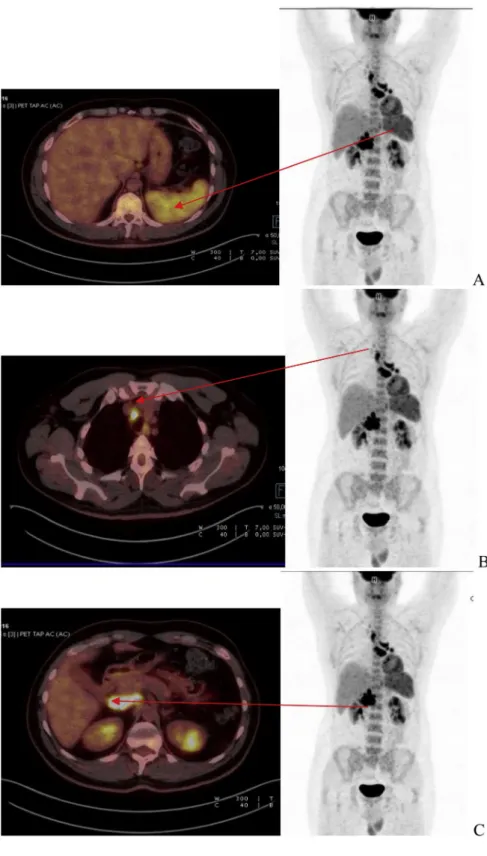
Because of no improvement after 2 weeks, malignancy was sus-pected, and 18-FDG-PET-CT was performed (Fig. 1a) showing intense FDG uptake affecting mediastinal and hilar lymph nodes, pulmonary nodules, hepatic hilar lymph nodes, spleen, and bone marrow. A
bronchoalveolar lavage and transbronchic lymph node aspiration were performed. The lymph node showed a lymphocytosis and a positive
Histoplasma capsulatum-specific PCR. Culture on Sabouraud
chlor-amphenicol gentamicin agar yielded one fluffy white colony identified as H. capsulatum via both MALDI-TOF and ITS2 rRNA region sequen-cing. The patient condition improved under itraconazole. After a 12-week treatment, 18-FDG-PET-CT (Fig. 1b) showed a complete regres-sion of pathologic uptake.
H. capsulatum (a dimorphic fungus) is endemic in America, spread
by droppings of birds of bats. It is transmitted to humans by the in-halation of contaminated spores. It induces disseminated infection in immunocompromised hosts and is symptomatic in 10% of cases in immunocompetent patients. In this report where malignancy was sus-pected, 18-FDG-PET-CT helped guiding the diagnosis. Clinicians in Europe should consider histoplamosis in the differential diagnosis of hypermetabolic lymphadenopathy or lung nodules in im-munocompetent travelers returning from endemic countries.
https://doi.org/10.1016/j.tmaid.2018.10.004
Received 16 May 2018; Accepted 4 October 2018
∗Corresponding author. VITROME, Institut Hospitalo-Universitaire Méditerranée Infection, 19-21 Boulevard Jean Moulin, 13385, Marseille Cedex 05, France. E-mail address:carole.eldin@ap-hm.fr(C. Eldin).
Travel Medicine and Infectious Disease 27 (2019) 136–138
Available online 06 October 2018
1477-8939/ © 2018 Elsevier Ltd. All rights reserved.
Fig. 1. Legend
1a
18F FDG PET CT scan at diagnosis. A. Intense splenic FDG uptake
B. Intense mediastinal lymph node uptake C. Intense hilar hepatic lymph node uptake 1b
18F FDG PET CT scan after treatment A. Regression of splenic uptake B. Regression of mediastinal uptake
C. Regression of hilar hepatic lymph node uptake.
C. Montagnac et al. Travel Medicine and Infectious Disease 27 (2019) 136–138
Acknowledgments
This work was supported by the French Government under the « Investissements d'avenir » (Investments for the Future) program
managed by the Agence Nationale de la Recherche (ANR, fr: National Agency for Research), (reference: Méditerranée Infection 10-IAHU-03). The funders had no role in study design, data collection and analysis, decision to publish, or preparation of the manuscript.
Fig. 1. (continued)
C. Montagnac et al. Travel Medicine and Infectious Disease 27 (2019) 136–138