Stem Cells and the Stem Cell Niche in the Breast:
An Integrated Hormonal and Developmental Perspective
Cathrin Brisken&Stephan DussPublished online: 30 June 2007 # Humana Press Inc. 2007
Abstract The mammary gland is a unique organ in that it undergoes most of its development after birth under the control of systemic hormones. Whereas in most other organs stem cells divide in response to local stimuli, to replace lost cells, in the mammary gland large numbers of cells need to be generated at specific times during puberty, estrous cycles and pregnancy to generate new tissue structures. This puts special demands on the mammary stem cells and requires coordination of local events with systemic needs. Our aim is to understand how the female reproductive hormones control mammary gland develop-ment and influence tumorigenesis. We have shown that steroid hormones act in a paracrine fashion in the mammary gland delegating different functions to locally produced factors. These in turn, affect cell–cell interactions that result in changes of cell behavior required for morphogenesis and differentiation. Here, we discuss how these hormonally regulated paracrine interactions may impinge on stem cells and the stem cell niche and how this integration of signals adds extra levels of complexity to current mammary stem cell models. We propose a model whereby the stem cell niches change depending on the developmental stages and the hormonal milieu. According to this model, repeated hormone stimulation of stem cells and their niches in the
course of menstrual cycles may be an important early event in breast carcinogenesis and may explain the conundrum why breast cancer risk increases with the number of menstrual cycles experienced prior to a first pregnancy. Keywords Stem cells . Breast cancer . Mammary gland
Mammary Gland Development
The mammary gland is a unique organ in that it undergoes most of its development after birth. In utero, mammary placodes form in the ventral skin of the embryo through epithelial–mesenchymal interactions. The placodes give rise to buds that grow into the underlying stroma and subsequently sprout to form rudimentary ductal systems, which are embedded in specialized stroma, the mammary fat pads. Until puberty, the mammary glands grow isometrically with the rest of the body [1,2].
With the onset of puberty and ovarian function, development of the ductal system accelerates dramatically. The tips of the ducts enlarge to form club-shaped structures, called terminal end buds (TEBs), which contain highly proliferative cells. The ducts penetrate the fat pad by branching dichotomously until they reach the edge of the fat pad [1]. With sexual maturity and the establishment of regular estrous cycles, the ductal system gains further complexity through the addition of side branches. Lateral branching is further enhanced during pregnancy. Later during pregnancy, little saccular outpouchings called alveoli bud all over the ductal system. This leads to a substantial increase in functional surface and enables the mammary gland to produce copious amounts of milk when the pups are born and begin to suckle. Once the pups are weaned,
C. Brisken (*)
:
S. Duss NCCR Molecular Oncology,Swiss Institute for Experimental Cancer Research (ISREC), 155 Chemin des Boveresses,
CH 1066 Epalinges s/Lausanne, Switzerland e-mail: [email protected]
C. Brisken
Ecole Polytechnique Fédérale de Lausanne, Lausanne, Switzerland
involution begins involving massive apoptosis and loss of alveolar structures [1].
Hormonal Control
Ovarian estrogens and progestins are pivotal regulators of female reproductive function including breast development. Upon maturation of the hypothalamic-anterior pituitary– ovarian axis, in response to episodic release of gonadotro-pin-releasing hormone, surges in secretion of pituitary follicle stimulating hormone (FSH) and luteinizing hor-mone (LH) trigger cyclic maturation ovarian follicles and of ovarian steroid production. The menstrual cycle in humans begins with the follicular phase characterized by FSH induced maturation of ovarian follicles that is accompanied by high estrogen secretion. The LH surge at mid cycle triggers the release of the egg whereas the remnant follicle will become the corpus luteum and secrete increasing amounts of progesterone characteristic of the post ovulato-ry, luteal phase of the cycle. During the first year and a half, cycles are irregular, anovulatory and have no luteal phase.
In mice, the situation is similar. Puberty begins at 3 weeks of age, with the ovaries secreting increasing amounts of estrogens. Usually around 8 weeks of age, regular estrous cycles are established with cyclic peaks in serum progester-one levels, sexual maturity is reached. In the mammary gland, at this stage, the ducts have reached the edge of the fat pad, subsequent proliferation results in the formation of side branches.
When a pregnancy is established, the corpus luteum increases progesterone synthesis and secretion until the placenta takes over this steroidogenic function to ensure the characteristically high progestin levels of pregnancy. Later in pregnancy, pitutitary prolactin becomes important for the formation of the secretory alveoli and differentiation of the mammary epithelial cells into milk secreting cells [3].
Stem Cells and the Stem Cell Niche: Concepts and Assays
Mouse mammary stem cells have been functionally defined as cells that are able to reconstitute an epithelium-divested mammary fat pad. For this assay, the nipple-near half of the inguinal mammary gland of 3-week-old, prepubertal, female mice that contains the rudimentary ductal tree is surgically removed leaving behind stroma devoid of epithelium [4]. Primary mammary epithelial cells injected into this “cleared fat pad” will reassemble and grow out behaving like the endogenous epithelium with the only difference being that the newly formed ductal system does
not connect to the nipple and hence secretions cannot be released. Combining this assay with the power of fluores-cence activated cell sorting (FACS), a series of elegant studies have recently characterized cell populations in the mammary gland that are enriched for stem cells [5–8], as reviewed elsewhere in this issue.
A description of stem cells based on morphological hallmarks has been elaborated by G. Smith and various collaborators. Their studies have shown that small light cells (SLC) bearing morphological characteristics of undif-ferentiated cells exist in both mice and rats [9–11]. The percentage of SLCs in the cell population does not change even during pregnancy as cell number increases manifold [12]. The number of SLCs decreases as epithelium is transplanted serially and looses reconstitution potential. Moreover, SLCs are located basally and never touch the lumen; hence, they are likely to be comprised in the stem cell enriched populations identified by immunotyping and FACS as the Lin− SCA-1low CD24med, CD49f high com-partment that shows characteristics of basally located cells [6, 10]. All these findings make the SLCs attractive candidates, but the formal proof that they are indeed the reconstituting cells remains to be provided.
The stem cell niche is the microenvironment surrounding stem cells that maintains their stemness and prevents them from differentiating [13–15]. It comprises signaling cells, characteristic extracellular matrix (ECM) and the stem cell [9,16]. The most impressive evidence of the power of the mammary gland stem cell niche was provided by the recent demonstration that it is able to redirect spermatogenic fate [17]. When genetically marked cells from adult seminifer-ous tubules were mixed with single cell suspensions of mammary epithelial cells and injected into cleared fat pads, they contributed to all aspects of the reconstitution. Moreover, the reprogrammed testicular cells were able to reconstitute upon serial transplantation [17].
The identity of stem cell niches within the mouse mammary gland has not been defined for lack of molecular markers; in the human breast, however, several markers have recently be ascertained [18].
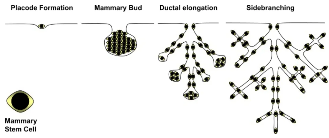
Similarly, the frequency at which stem cells and/or stem cell niches occur in the mammary gland is still a matter of contention. Independent of the developmental stage, mam-mary epithelium taken from any area of the mammam-mary gland is able to fully reconstitute a ductal tree when grafted to cleared fat pads indicating that stem cells and their niches are distributed at regular intervals throughout the ductal system throughout development (Fig.1). Estimates of how frequent stem cells are in the mouse mammary epithelium, based on serial dilution experiments in the fat pad reconstitution assay, vary between 1 mammary repopulating unit (MRU) in 200 dissociated cells [19] to 1 in 5,000 dissociated cells [5].
Control of Stem Cells and the Stem Cell Niche by Hormones and Cell–Cell Interactions
Our laboratory is interested in understanding how the female reproductive hormones control mammary gland development and influence breast tumorigenesis. Combin-ing the use of hormone receptor deficient mice with tissue recombination techniques, we and others have revealed that the hormones act sequentially on the mammary gland through their respective receptors in the epithelium (Fig.2).
Estrogens and progestins, drive ductal elongation and side branching respectively; both processes involve exten-sive cell proliferation. We propose that the two hormones signaling through their respective receptors expressed on a subset of luminal epithelial cells induce the expression of local factors that trigger the assembly and the activation of the stem cell niche. In this way the activity of stem cells and their niches respond to a systemic stimulus, which in turn reflects systemic requirements.
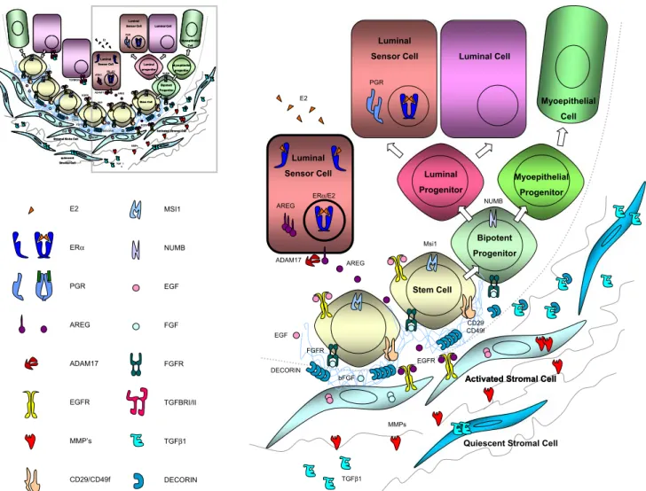
During estrogen-driven ductal outgrowth, stem cells at the tip of the growing ducts, called cap cells, are in direct contact with the stroma because the basal lamina is disrupted in this zone. Thus, stromal cells can be recruited to fulfill the niche cell function (Fig.3). During
progester-one-induced lateral branching, the basal lamina may become thinner but remains intact, and myoepithelial cells are recruited to form the niche (Fig. 4). Thus the composition of the niche varies related to the requirements of the specific developmental stage. Both models will be discussed below in the context of recent work from our lab. Estrogen and the Stem Cell Niche
Role of Estrogen Signaling in the Mammary Gland The role of estrogens in mammary gland development is illustrated by the finding that pubertal ductal outgrowth comes to a halt when the ovaries are removed. Outgrowth is restored when 17-β-estradiol is administered locally by means of slow release pellets grafted to the mammary gland [20]. Consistent with estrogens driving ductal outgrowth, in mammary glands of mice deficient for the estrogen receptor α (ERα), the prime mediator of estrogen function, a normal rudimentary ductal system is formed but subsequent development is blocked. When ERα deficient epithelium is grafted to cleared fat pads of wild type (wt) mice it does not develop indicating that ERα signaling in the mammary epithelium is essential for ductal outgrowth.
estrogen progesterone prolactin
rudimentary ductal elongation/bifurcation sidebranching alveologenesis ductal system lactogenic differentiation
Fig. 2 Schematic representation of mammary gland development (black) and hormonal control (red) of different morphogenetic steps. Tissue recombination experiments with hormone receptor deficient mouse
strains revealed that the female reproductive hormones estrogens, progesterone, and prolactin act sequentially during mammary gland development through their respective receptors in the mammary epithelium Placode Formation Mammary Bud Ductal elongation Sidebranching
Mammary Stem Cell Fig. 1 Stem cell distribution at
different stages of mammary gland development. Schematic representation of distinct stages of mammary gland development with respective distribution of stem cells, not drawn to scale. Mammary epithelium taken from any area of the mammary gland is able to fully reconstitute a ductal tree when grafted to cleared fat pads indicating that stem cells and their niches are distributed at regular intervals throughout the mammary gland
When chimaeric epithelia are generated using wt and ERα deficient mouse mammary epithelial cells to recon-stitute cleared fat pads, ERα deficient cells, identified based on the expression of the marker gene, contribute extensively to the ductal outgrowth. The mutant cells are found both in the outer cap cell layer and in the subtending body cell compartment of the TEBs of the growing ducts as well as in the luminal and the myoepithelial cell layer of mature ducts [21].
Two conclusions can be drawn. First, ERα deficient mammary epithelia contain stem cells. Second, the ERα deficient stem cells can only unfold their potential when assisted by ERα positive cells.
The first conclusion is in line with various findings indicating that mammary stem cells are ERα negative. The observation that transplantation of the mammary anlage (see, Fig.1), which only contains ERα negative cells [22],
results in full reconstitution of the mammary gland [23], implicates that mouse mammary stem cells are steroid receptor negative. Recent characterization of stem cell enriched populations by FACS, established that stem cells in the adult mammary gland are enriched within a cell population that does not express ERα [8]. Sleeman et al. demonstrated that even large numbers of steroid receptor positive cells identified as CD24highand CD133high(prominin) population by FACS sorting, fail to reconstitute a mammary gland when transplanted into a cleared fat pad whereas prominin negative cells give rise to ductal outgrowth [7].
Thus, mammary stem cells appear to be ERα negative throughout development, yet, they require the presence of ERα positive cells, that we will name the “sensor cells”. The “sensor cells” detect the systemic estrogen stimulus and translate it to subsequent local events that result in the stem cells dividing and unfolding their potential.
Activated Stromal Cell
TGFβ1 Luminal Sensor Cell ERα/E2 ADAM17 AREG AREG Luminal Cell Luminal Sensor Cell PGR EGFR MMPs NUMB bFGF Bipotent Progenitor Luminal Progenitor Msi1 Myoepithelial Progenitor EGF Stem Cell Myoepithelial Cell E2 DECORIN FGFR CD29 CD49f
Activated Stromal Cell
Quiescent Stromal Cell E2 ERα PGR AREG ADAM17 EGFR MMP’s CD29/CD49f MSI1 NUMB EGF FGF FGFR TGFBRI/II TGFβ1 DECORIN
Activated Stromal Cell
quiescent Stromal Cell TGF ≤ 1 Luminal Sensor Cell ER ± /E2 ADAM17 AREG AREG Luminal Cell Luminal Sensor Cell PGR EGFR MMPs
Stromal Niche Cell
TGFBRI/II NUMB CD29/ CD49f FGFR EGF bFGF Bipotent Progenitor Luminal progenitor Msi1 Myoepithelial progenitor Stem Cell Myoepithelial Cell EGFR E2 NICD
DECORIN Activated Stromal Cell
quiescent Stromal Cell TGF ≤ 1 Luminal Sensor Cell ER ± /E2 ADAM17 AREG AREG Luminal Cell Luminal Sensor Cell PGR EGFR MMPs
Stromal Niche Cell
TGFBRI/II NUMB CD29/ CD49f FGFR EGF bFGF Bipotent Progenitor Luminal progenitor Msi1 Myoepithelial progenitor Stem Cell Myoepithelial Cell EGFR E2 NICD DECORIN
Fig. 3 Model of the pubertal stem cell niche. Shown is a schematic representation of a terminal end bud (TEB) during estrogen-driven ductal outgrowth. Estrogens acting on the luminal sensor cell induce the secretion of amphiregulin. The growth factor is released by epithelial ADAM17 and acts on EGFR on stromal cells. The stromal cells in turn
release FGFs and the TGF-β inhibitor, decorin. Activation of FGF signaling in the stem cells and downmodulation of TGF-β signaling allow for stem cell proliferation a prerequisite for ductal elongation. Note, that the basal lamina is disrupted and the stem cells are in direct contact with stromal cells that act as niche cells at this stage
Amphiregulin as Downstream Mediator of Estrogen Signaling
How does estrogen elicit its paracrine effects? How can estrogen stimulate the ERα negative stem cells and their daughter cells? Epidermal growth factor (EGF), like 17- β-estradiol, restores ductal outgrowth when administered locally in the mammary glands of ovariectomized pubertal mice [24] implicating that EGF receptor (EGFR) signaling is important downstream of ERα signaling. Amphiregulin is the only EGF family member the transcription of which is induced by estrogen at a time of exponential expansion of the ductal system in the mammary glands of pubertal mice [25]. Infact, estrogens induce amphiregulin through the ERα
and require amphiregulin to induce proliferation of the mammary epithelium. Consequently, amphiregulin deficient mammary epithelia do not form TEBs and do not invade the fat pad in virgin mice. Like ERα deficient cells, amphir-egulin−/−mammary epithelial cells are rescued by neighbour-ing wt cells. In chimeric epithelia, mutant cells proliferate and contribute to all epithelial cell compartments of the ductal outgrowth [25]. These observations indicate that amphiregulin, like estrogens signals in a paracrine fashion and are consistent with amphiregulin being the important clue provided to the stem cells and their progeny by an ERα positive neighbouring cell.
When two differentially marked wt cell populations are used to reconstitute cleared fat pads, distinct patterns of chimerism are found. In one type, entire ductal segments are made of cells expressing one single marker, in the other type, ducts displayed a patchwork of the two markers (L. Ciarloni et al., unpublished observations). On the other hand, when either ERα or amphiregulin deficient cells are mixed with wt cells, the mutant cells never give rise to an entire ductal segment but are only observed in the patchy type of chimerism, never more than 3–4 cell diameters away from wt cells. This strongly suggests that close interactions are required during ductal outgrowth to relegate the estrogen-induced signals to ERα negative stem and/or progenitor cells. Whether direct cell–cell contact is required or signals can travel over several cell diameters remains to be addressed.
It is tempting to speculate that the high number of stem cells necessitated by the large cell number expansion that the mammary gland undergoes during development, ren-ders this organ particularly prone to carcinogenesis. Model of the Estrogen-Driven Stem Cell Niche
Is amphiregulin the stimulus that makes ERα negative stem cells divide? Intriguingly, EGFR mRNA is enriched in the stem cell containing compartment [8]. Yet, genetic evidence indicates that the EGFR is required in the mammary
Luminal Progenitor Myoepithelial Progenitor Stem Cell Sensor Cell Myoepithelial Cell Basemen t Membrane Activated Stromal Cell Quiescent Stromal Cell ERα/E2 TGFBRI/II TGFβ1 PGR RANKL WNT Fz CD29C D49f FGFR Delta NOTCH MMPs bFGF Niche Cell PGR /PG MMPs RANK CCND1 MMPs Luminal Cell Bipotent Progenitor DECORIN PG CD49f CD29 NOTCH/NICD Jagged FGF FGFR TGFBRI/II TGFβ1 DECORIN PG ERα & E2 PGR RANKL RANK CCND1 WNT Fz MMP’s
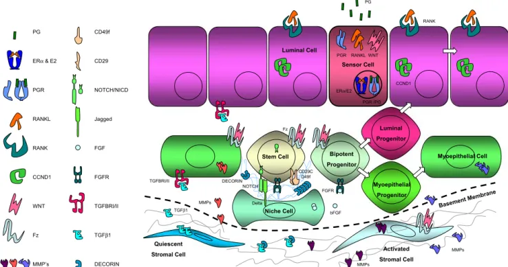
Fig. 4 Model of the adult stem cell niche that is activated in response to progesterone released by the ovaries during estrous cycles and pregnancy. The basal lamina remains intact and myoepithelial cells are recruited to fulfill niche cell function. Progesterone acting on the luminal sensor cell induces wnt-4 and RANKL. The secreted wnt-4
activates the stem cell compartment acting either directly on the stem cells and/or indirectly via the niche cells. RANKL, on the other hand induces proliferation of neighboring luminal cells through induction of cyclin D1
stroma; wt epithelium does not grow out in EGFR deficient stroma whereas EGFR deficient epithelium does reconsti-tute when placed into a wt fat pad [26]. Although these data do not preclude additional, direct effects on the epithelial cells, they strongly argue that the prime target for amphiregulin is the stroma. Infact, during ductal outgrowth a unique situation arises during mammary gland develop-ment; at the TEBs, the outer epithelial cells, the cap cells, which are considered to be stem cells, directly interact with stromal cells because the basal lamina that usually separates epithelial and stromal compartments, is degraded.
Thus, a picture emerges of a stem cell niche consisting of three different cells; the ERα positive sensor cell, the EGFR positive stromal cell and the ERα negative stem cell (Fig.3). All of them remain quiescent until they are“switched on” by estrogens. In response to estrogens, the“sensors” synthesize and secrete amphiregulin. Membrane-bound amphiregulin is subsequently activated by ADAM17, which is also of epithelial origin but not necessarily produced by the sensor cells [27]. The activated amphiregulin in turn acts on stromal cells.
The identity of the stromal factors that talk back to the epithelial components of the stem cell niche remains to be revealed. Work of Z. Werb and colleagues has unraveled several attractive candidates, partly through expression microarrays, partly by inference from in vitro culture models [28–30]. Thus, the stroma might respond by releasing and activating metalloproteinases such as MMP14 or MMP2, reported EGFR targets that are required to restructure of the ECM by degrading collagen type I and to release of other growth factors and [31]. FGF 7 and FGF 2 are particularly attractive candidates to mediate a proliferative signal from the stroma, both have been shown to induce ductal morphogenesis in a 3D matrigel assay. Interestingly, bFGF is a critical component in mammosphere medium suggest-ing that activation of FGF signalsuggest-ing is important to stem cells [32]. Whether FGF acts directly on the stem cell or indirectly via other niche cells remains to be determined.
It is also conceivable that growth inhibitory influences exerted by TGF-β signaling are relieved by a stromal derived factor. This could happen extracellularly through the secretion of the TGF-β inhibitor decorin [33] or intracellularly through induction of expression of inhibitory SMADs. Intriguingly, decorin was among the genes highly expressed in mammospheres [32]. Activation of EGFR and possibly other receptor tyrosine kinases, as well as of NF-κB or signal transducer and activator of transcription (STAT) induced by cytokine receptor signaling can result in the transcription of SMAD-6 and-7 [34].
Besides the fibroblasts, the stroma also includes adipo-cytes and endothelial cells which are not featured in our model (Fig.3). Furthermore, communication with infiltrat-ing immune cells may comprise another very important level of regulation [35].
Progesterone and the Stem Cell Niche
At the end of puberty, when the ductal system reaches the edges of the mammary fat pad and the mouse is sexually mature, 17-β-estradiol almost completely looses its mito-genic effects. Proliferation and morphogenesis are now driven by progesterone, serum levels of which rise with each estrous cycle and during pregnancy (R. Rajaram, Brisken unpublished observations).
Progesterone can elicit proliferation of both receptor positive and receptor negative luminal epithelial cells and acts, like estrogens, in a paracrine fashion [36]. At least two players are induced and have been implicated in mediating the hormone’s paracrine effects, the Receptor Activator for Nuclear Factor κB Ligand (RANKL) [37, 38] and wnt-4 [23] (Fig. 4). It has been suggested that RANKL is important in eliciting proliferation through induction of cyclin D1 in neighboring cells [37–39]. Wnt signaling, on the other hand, has been implicated in the control of stem cells [40–43] and this may account for its strong oncogenic effects.
We envision a scenario in which the sensor cell responds to progesterone stimulation by synthesizing and secreting wnt-4. Wnt-4 mobilizes stem cells either acting on them directly or indirectly through activation of niche cells. This activation results in an asymmetric cell division yielding a new stem cell and a daughter cell that give rise, possibly through further asymmetric cell divisions to transient amplifying cells. These cells in turn are induced to proliferate by RANKL.
The Ever-Changing Stem Cell Niche
The mammary stem cells need to respond to distinct factors at distinct developmental stages. The initial stages of mammary gland development, we did not discuss here, are thought to be controlled by epithelial–mesenchymal cross talk involving PTHrP [44–46], BMP [47], FGF 10 [48,49], and Wnt signaling [50, 51]. During puberty, estrogen controls the stem cell niche with amphiregulin and EGFR signaling being of central importance. In the adult mam-mary gland, progesterone is the major stimulant of stem/ progenitor cell stimulation and a prime mediator is wnt-4.
The highly specific requirement of EGFR versus wnt signaling in response to either estrogens or progestins, respectively, is underlined by the following observations:
In contrast to ERα deficient epithelia that do not proliferate throughout all developmental stages, amphire-gulin−/− mammary epithelia undergo subsequent develop-mental steps i.e. side branching and alveologenesis. This argues that estrogens specifically require amphiregulin to exert their effects whereas progesterone, which also induces amphiregulin expression [52], does not require its presence.
Different wnts, such as wnt-2 and wnt-5a [28], are expressed in the TEBs, yet, wnt signaling is specifically required to mediate progesterone function. Ectopic expres-sion of wnt-1 in the ERα deficient background by means of the MMTV-wnt-1 transgene did not lead to a rescue of the ductal elongation phenotype [53]. Ectopic wnt activation did, however, overcome the side branching defect in this mutant, furthermore it rescued the phenotype of PR deficient mammary epithelium [23]. These findings suggest that the stem cell promoting effects of wnt signaling are specific to progesterone-induced events and are not involved in the estrogen-induced activation of the stem cell niche.
We propose that the niches that promote estrogen- and progesterone-induced stem cell expansion are overlapping entities. Thus, the hormone receptor positive sensor cell and the stem cell are identical but the niche cell that is recruited in response to estrogens versus progesterone is different.
What about the sensor cells, how special are they? After all, in the adult virgin more than 30% of the luminal cells may be expressing steroid receptors; do they all induce stem cell niches or are there different subpopulations of ERα/PR positive cells, those that control stem cells and those that stimulate proliferation of transient amplifying cells? Maybe all the ERα/PR positive cells do the same job and release the same factors, but the biological outcome on the part of the responding cell is determined by the differentiation state of the responding cell. Alternatively, the impact of the steroid receptor positive cells is an indirect one only. They induce proliferation of luminal epithelial cells, which leads to depletion of progenitor cells; this depletion is sensed by the stem cell niche, which reacts by stimulating asymmetric divisions of the stem cell.
Breast Cancer: Messing Around with the Stem Cell Niche
The stem cell theory provides an attractive model that accounts for chemo resistance and tumor heterogeneity [54–57]. What is the relevance of our models to human breast cancer? Is breast cancer a disease of stem cells and their niches?
The Progesterone-Wnt Connection
Progesterone exposure, as experienced by premenopausal women during menstrual cycles and by postmenopausal women under hormone replacement therapy, increases breast cancer risk [58]. We speculate that this may be due to stimulation of stem cells.
As discussed, wnt signaling is important in mediating progesterone action [23] and evidence has accumulated that it is involved in stem cells self-renewal in different organs [41]
including the mammary gland [42,43]. Intriguingly, the wnt pathway has long been established as strongly oncogenic in the mouse mammary gland, yet, a role of wnt signaling in human breast cancer has not been forthcoming because mutations in intracellular signaling components such as β-catenin and APC were not identified [59]. More recently, however, evidence has accumulated that the secreted inhibitor of wnt signaling, secreted frizzled related protein-1 (SFRP-1), is down modulated during breast carcinogenesis suggesting that activation of the pathway may occur [60]. To assess whether deregulation of wnt signaling in human breast epithelial cells may perturb their homeostasis, we ectopically expressed Wnt-1 in them using a retroviral vector. The increased wnt signaling activity triggered a cascade of events resulting in oncogenic conversion of these cells. The breast epithelial cells ectopically expressing Wnt-1 proliferated more than the controls. However, at passage 6 to 7, the majority of the infected cells senesced like the control cultures. At the same time, a more dynamic subpopulation began to appear, which continued to proliferate and began to detach from the dish and to grow as multi cellular aggregates. The Wnt-1-HMECs could be dissociated, would briefly attach and then continue to grow in suspension. Injected into mammary glands of immuno-compromised mice, they gave rise to tumors that showed morphological and molec-ular hallmarks of a subtype of human breast carcinomas, medullary carcinoma. Consistent with the wnt-1-infected cell strains being transformed, the Rb and p53 pathways were inactivated and the cells were triploid.
What are the mechanisms underlying this wnt-1 induced transformation of primary human breast epithelial cells? The first clue is, that wnt-1 expression activated a DNA damage response. This could exert the selective pressure necessary to inactivate checkpoints. However, other onco-genes such as large T also induced the DNA damage response and did so even more effectively, yet they did not transform the infected cells. The wnt-1 transformed cells had increased Notch signaling activity when compared to the parental cells. Notch activation was required for both the in vitro phenotype and in vivo tumor growth. In the mammosphere stem cell assay Notch signaling promotes stem cell expansion [61], reviewed in [62]. The observa-tions that transformation is not quantitative but occurs only in a small subset of cells and that we observe upregulation of Notch signaling in the culture as a late event hint at the possibility that Wnt-1 targets a particular susceptible cell population possibly stem and/or progenitor cells that slowly grows out. This view is further supported by our observa-tion that the oncogenic wnt-1 cell strains express keratin 18. Usually, expression of this marker of luminal epithelial cells, is lost in the course of in vitro culture of primary human breast epithelial cells; the Wnt-1-transduced cell strains however still express keratin 18 in line with them
being derived from a rare subset of cells with luminal features. Thus, the strong oncogenic effects of ectopic wnt-1 expression in both the mouse mammary gland and human breast epithelial cells may be linked to this pathway promoting proliferation and/or survival of a rare progeni-tor/stem cell population.
In the mouse, wnt-4 is a progesterone target [23] that is induced during diestrous [63]. We speculate that in the human breast, similarly, wnt-4 or other wnts are progester-one targets and as such are induced during the luteal phase of the menstrual cycle in ERα/PR receptor positive luminal cells. The wnt activates stem cells either directly or indirectly via niche cells to generate a pool of transient amplifying cells in anticipation of a potential pregnancy. The repeated stimulation of progenitors, concomitant with an activation of a DNA damage response may predispose to malignant transformation, all the more if a given cell has already accumulated genetic changes. This may explain why menstrual cycles are a risk factor for breast cancer. Perspectives
The next challenges lie in better defining stem cells and their niches. Will we be able to identify stage-specific markers for the niche? Are there universal niche markers; maybe the specifics of the ECM composition do not change throughout development?
The fat pad reconstitution assay needs to be more standardized. Substantial variation in FACS profiles elabo-rated in different laboratories and discrepancies in the reconstitution potential of different cell population derive most likely from differences in cell preparation protocols that could readily be reduced. Eventually, the techniques need to be applied to human breast tissue samples, although the basic principle of hormonal control and paracrine interactions are likely conserved among the two species differences in the local factors that are recruited and in surface antigens are to be expected.
The further progress is likely to have important bearings on our understanding of the origins of breast cancer and may open new avenues for its treatment.
Acknowledgements The authors thank Manfred Beleut for helpful discussions and critical comments on the manuscript.
References
1. Daniel, C. W., & Silberstein, G. B. (1987). Postnatal development of the rodent mammary gland. In C. W. Daniel (Ed.), The mammary gland (pp. 3–31). New York: Plenum.
2. Sakakura, T. (1987). Developmental biology of the mammary gland. In M. C. a. D. Neville C. W. (Ed.), The mammary gland (pp. 37–63). New York: Plenum.
3. Brisken, C., Kaur, S., Chavarria, T. E., Binart, N., Sutherland, R. L., Weinberg, R. A., et al. (1999). Prolactin controls mammary gland development via direct and indirect mechanisms. Developments in Biologicals, 210(1), 96–106.
4. DeOme, K. B., Faulkin, L. J., Jr., Bern, H. A., & Blair, P. B. (1959). Development of mammary tumors from hyperplastic alveolar nodules transplanted into gland-free mammary fat pads of female C3H mice. Cancer Research, 19, 511–520.
5. Shackleton, M., Vaillant F., Simpson, K. J., Stingl, J., Smyth, G. K., Asselin-Labat, M. L., et al. (2006). Generation of a functional mammary gland from a single stem cell. Nature, 439(7072), 84–88. 6. Stingl, J., Eirew, P., Ricketson, I., Shackleton, M., Vaillant, F., Choi, D., et al. (2006). Purification and unique properties of mammary epithelial stem cells. Nature, 439(7079), 993–997. 7. Sleeman, K. E., Kendrick, H., Robertson, D., Isacke, C. M.,
Ashworth, A., & Smalley, M. J. (2007). Dissociation of estrogen receptor expression and in vivo stem cell activity in the mammary gland. Journal of Cell Biology, 176(1), 19–26.
8. Asselin-Labat, M. L., Shackleton, M., Stingl, J., Vaillant, F., Forrest, N. C., Eaves, C. J., et al. (2006). Steroid hormone receptor status of mouse mammary stem cells. Journal of the National Cancer Institute, 98(14), 1011–1014.
9. Chepko, G., Slack, R., Carbott, D., Khan, S., Steadman, L., & Dickson, R. B. (2005). Differential alteration of stem and other cell populations in ducts and lobules of TGFalpha and c-Myc transgenic mouse mammary epithelium. Tissue Cell, 37 (5), 393–412.
10. Chepko, G., & Smith, G. H. (1997). Three division-competent, structurally-distinct cell populations contribute to murine mam-mary epithelial renewal. Tissue Cell, 29(2), 239–253.
11. Chepko, G., & Smith, G. H. (1999). Mammary epithelial stem cells: Our current understanding. Journal of Mammary Gland Biology and Neoplasia, 4(1), 35–52.
12. Kordon, E. C., & Smith, G. H. (1998). An entire functional mammary gland may comprise the progeny from a single cell. Development, 125(10), 1921–1930.
13. Rizvi, A. Z., & Wong, M. H. (2005). Epithelial stem cells and their niche: There’s no place like home. Stem Cells, 23(2), 150–165. 14. Lin, H. (2002). The stem-cell niche theory: Lessons from flies.
Nature Reviews. Genetics, 3(12), 931–940.
15. Wilson, A., & Trumpp, A. (2006). Bone-marrow haematopoietic-stem-cell niches. Nature Reviews. Immunology, 6(2), 93–106. 16. Chepko, G., & Dickson, R. B. (2003). Ultrastructure of the
putative stem cell niche in rat mammary epithelium. Tissue Cell, 35(2), 83–93.
17. Boulanger, C. A., Mack, D. L., Booth, B. W., & Smith, G. H. (2007). Interaction with the mammary microenvironment redirects spermatogenic cell fate in vivo. Proceedings of the National Academy of Sciences of the United States of America, 104(10), 3871–3876.
18. Villadsen, R., Fridriksdottir, A. J., Ronnov-Jessen, L., Gudjonsson, T., Rank, F., Labarge, M. A., et al. (2007). Evidence for a stem cell hierarchy in the adult human breast. Journal of Cell Biology, 177(1), 87–101.
19. Moraes, R. C., Zhang, X., Harrington, N., Fung, J. Y., Wu, M. F., Hilsenbeck, S. G., et al. (2007). Constitutive activation of smoothened (SMO) in mammary glands of transgenic mice leads to increased proliferation, altered differentiation and ductal dysplasia. Development, 134(6), 1231–1242.
20. Daniel, C. W., Silberstein, G. B., & Strickland, P. (1987). Direct action of 17 beta-estradiol on mouse mammary ducts analyzed by sustained release implants and steroid autoradiography. Cancer Research, 47(22), 6052–6057.
21. Mallepell, S., Krust, A., Chambon, P., & Brisken, C. (2006). Paracrine signaling through the epithelial estrogen receptor alpha is required for proliferation and morphogenesis in the mammary
gland. Proceedings of the National Academy of Sciences of the United States of America, 103(7), 2196–2201.
22. Stumpf, W. E., Narbaitz, R., & Sar, M. (1980). Estrogen receptors in the fetal mouse. Journal of Steroid Biochemistry, 12, 55–64.
23. Brisken, C., Heineman, A., Chavarria, T., Elenbaas, B., Tan, J., Dey, S., et al. (2000). Essential function of Wnt-4 in mammary gland development downstream of progesterone signaling. Genes and Development, 14(6), 650–654.
24. Coleman, S., Silberstein, G. B., & Daniel, C. W. (1988). Ductal morphogenesis in the mouse mammary gland: Evidence support-ing a role for epidermal growth factor. Developments in Bio-logicals, 127(2), 304–315.
25. Ciarloni, L., Mallepell, S., & Brisken, C. (2007). Amphiregulin is an essential mediator of estrogen receptor {alpha} function in mammary gland development. Proceedings of the National Academy of Sciences of the United States of America, 104(13), 5455–5460.
26. Wiesen, J., Young, P., Werb, Z., & Cunha, G. (1999). Signaling through the stromal epidermal growth factor receptor is necessary for mammary ductal development. Development, 126(2), p335– p344.
27. Gschwind, A., Hart, S., Fischer, O. M., & Ullrich, A. (2003). TACE cleavage of proamphiregulin regulates GPCR-induced proliferation and motility of cancer cells. EMBO Journal, 22 (10), 2411–2421.
28. Kouros-Mehr, H., & Werb, Z. (2006). Candidate regulators of mammary branching morphogenesis identified by genome-wide transcript analysis. Developmental Dynamics, 235(12), 3404–3412. 29. Fata, J. E., Werb, Z., & Bissell, M. J. (2004). Regulation of mammary gland branching morphogenesis by the extracellular matrix and its remodeling enzymes. Breast Cancer Research, 6(1), 1–11. 30. Simian, M., Hirai, Y., Navre, M., Werb, Z., Lochter, A. & Bissell,
M. J. (2001). The interplay of matrix metalloproteinases, morphogens and growth factors is necessary for branching of mammary epithelial cells. Development, 128(16), 3117–3131. 31. Sternlicht, M. D., Kouros-Mehr, H., Lu, P. & Werb, Z. (2006).
Hormonal and local control of mammary branching morphogenesis. Differentiation, 74(7), 365–381.
32. Dontu, G., Abdallah, W. M., Foley, J. M., Jackson, K. W., Clarke, M. F., Kawamura, M. J., et al. (2003). In vitro propagation and transcriptional profiling of human mammary stem/progenitor cells. Genes and Development, 17(10), 1253–1270.
33. Yamaguchi, Y., Mann, D. M., & Ruoslahti, E. (1990). Negative regulation of transforming growth factor-beta by the proteoglycan decorin. Nature, 346(6281), 281–284.
34. Derynck, R., & Zhang, Y. E. (2003). dependent and Smad-independent pathways in TGF-beta family signalling. Nature, 425 (6958), 577–584.
35. Schwertfeger, K. L., Rosen, J. M., & Cohen, D. A. (2006). Mammary gland macrophages: Pleiotropic functions in mammary development. Journal of Mammary Gland Biology and Neoplasia, 11(3–4), 229–238.
36. Brisken, C., et al. (1998). A paracrine role for the epithelial progesterone receptor in mammary gland development. Proceed-ings of the National Academy of Sciences of the United States of America, 95(9), p5076–p5081.
37. Mulac-Jericevic, B., et al. (2003). Defective mammary gland morphogenesis in mice lacking the progesterone receptor B isoform. Proceedings of the National Academy of Sciences of the United States of America, 100(17), 9744–9749.
38. Brisken, C., et al. (2002). IGF-2 is a mediator of prolactin-induced morphogenesis in the breast. Developmental Cell, 3(6), 877–887. 39. Cao, Y., et al. (2001). IKKalpha provides an essential link between RANK signaling and cyclin D1 expression during mammary gland development. Cell, 107(6), 763–775.
40. Willert, K., et al. (2003). Wnt proteins are lipid-modified and can act as stem cell growth factors. Nature, 23(6938), 448–452. 41. Reya, T., & Clevers, H. (2005). Wnt signalling in stem cells and
cancer. Nature, 434(7035), 843–850.
42. Li, Y., Welm, B., Podsypanina, K., Huang, S., Chamorro, M., Zhang, X. et al. (2003). Evidence that transgenes encoding components of the Wnt signaling pathway preferentially induce mammary cancers from progenitor cells. Proceedings of the National Academy of Sciences of the United States of America, 100(26), 15853–18858.
43. Liu, B. Y., McDermott, S. P., Khwaja, S. S., & Alexander, C. M. (2004). The transforming activity of Wnt effectors correlates with their ability to induce the accumulation of mammary progenitor cells. Proceedings of the National Academy of Sciences of the United States of America, 101(12), 4158–4163.
44. Foley, J., Dann, P., Hong, J., Cosgrove, J., Dreyer, B., Rimm, D., et al. (2001). Parathyroid hormone-related protein maintains mammary epithelial fate and triggers nipple skin differentiation during embryonic breast development. Development, 128(4), 513– 525.
45. Dunbar, M. E., & Wysolmerski, J. J. (1999). Parathyroid hormone-related protein: A developmental regulatory molecule necessary for mammary gland development. Journal of Mammary Gland Biology and Neoplasia, 4(1), 21–34.
46. Wysolmerski, J. J., Philbrick, W. M., Dunbar, M. E., Lanske, B., Kronenberg, H. & Broadus, A. E. (1998). Rescue of the parathyroid hormone-related protein knockout mouse demon-strates that parathyroid hormone-related protein is essential for mammary gland development. Development, 125(7), 1285– 1294.
47. Hens, J. R., Dann, P., Zhang, J. P., Harris, S., Robinson, G. W., & Wysolmerski, J. (2007). BMP4 and PTHrP interact to stimulate ductal outgrowth during embryonic mammary development and to inhibit hair follicle induction. Development, 134(6), 1221–1230. 48. Rudland, P. S., Platt-Higgins, A. M., Wilkinson, M. C., & Fernig,
D. G. (1993). Immunocytochemical identification of basic fibroblast growth factor in the developing rat mammary gland: Variations in location are dependent on glandular structure and differentiation. Journal of Histochemistry and Cytochemistry, 41 (6), 887–898.
49. Mailleux, A. A., Spencer-Dene, B., Dillon, C., Ndiaye, D., Savona-Baron, C., Itoh, N., et al. (2002). Role of FGF10/FGFR2b signaling during mammary gland development in the mouse embryo. Development, 129(1), 53–60.
50. van Genderen, C., Okamura, R. M., Farinas, I., Quo, R. G., Parslow, T. G., Bruhn, L., et al. (1994). Development of several organs that require inductive epithelial–mesenchymal interactions is impaired in LEF-1-deficient mice. Genes and Development, 8 (22), 2691–2703.
51. Chu, E. Y., Hens, J., Andl, T., Kairo, A., Yamaguchi, T. P., Brisken, C., et al. (2004). Canonical WNT signaling promotes mammary placode development and is essential for initiation of mammary gland morphogenesis. Development, 131(19), 4819–4829.
52. Das, S. K., Chakraborty, I., Paria, B. C., Wang, X. N., Plowman, G., & Dey, S. K. (1995). Amphiregulin is an implantation-specific and progesterone-regulated gene in the mouse uterus. Molecular Endocrinology, 9(6), 691–705.
53. Bocchinfuso, W. P., Hively, W. P., Couse, J. F., Varmus, H. E., & Korach, K. S. (1999). A mouse mammary tumor virus-Wnt-1 transgene induces mammary gland hyperplasia and tumorigenesis in mice lacking estrogen receptor-alpha. Cancer Research, 59(8), 1869–1876.
54. Wang, J. C., & Dick, J. E. (2005). Cancer stem cells: Lessons from leukemia. Trends in Cell Biology, 15(9), 494–501. 55. Smalley, M., & Ashworth, A. (2003). Stem cells and breast cancer:
56. Al-Hajj, M., & Clarke, M. F. (2004). Self-renewal and solid tumor stem cells. Oncogene, 23(43), 7274–7282.
57. Dontu, G., Liu, S., & Wicha, M. S. (2005). Stem cells in mammary development and carcinogenesis: Implications for prevention and treatment. Stem Cell Reviews, 1(3), 207–213. 58. Pike, M. C., & Ross, R. K. (2000). Progestins and menopause:
Epidemiological studies of risks of endometrial and breast cancer. Steroids, 65(10–11), 659–664.
59. Brennan, K. R., & Brown, A. M. (2004). Wnt proteins in mammary development and cancer. Journal of Mammary Gland Biology and Neoplasia, 9(2), 119–131.
60. Ugolini, F., Charafe-Jauffret, E., Bardou, V. J., Geneix, J., Adelaide, J., Labat-Moleur, F., et al. (2001). WNT pathway and mammary carcinogenesis: Loss of expression of
candi-date tumor suppressor gene SFRP1 in most invasive carcino-mas except of the medullary type. Oncogene, 20(41), 5810– 5817.
61. Dontu, G., Jackson, K. W., McNicholas, E., Kawamura, M. J., Abdallah, W. M., & Wicha, M. S. (2004). Role of Notch signaling in cell-fate determination of human mammary stem/ progenitor cells. Breast Cancer Research, 6(6), R605–R615. 62. Liu, S., Dontu, G., & Wicha, M. S. (2005). Mammary stem cells,
self-renewal pathways, and carcinogenesis. Breast Cancer Re-search, 7(3), 86–95.
63. Silberstein, G. B., Van Horn, K., Hrabeta-Robinson, E., & Compton, J. (2006). Estrogen-triggered delays in mammary gland gene expression during the estrous cycle: Evidence for a novel timing system. Journal of Endocrinology, 190(2), 225–239.