HAL Id: hal-02698136
https://hal.inrae.fr/hal-02698136
Submitted on 1 Jun 2020
HAL is a multi-disciplinary open access
archive for the deposit and dissemination of
sci-entific research documents, whether they are
pub-lished or not. The documents may come from
teaching and research institutions in France or
abroad, or from public or private research centers.
L’archive ouverte pluridisciplinaire HAL, est
destinée au dépôt et à la diffusion de documents
scientifiques de niveau recherche, publiés ou non,
émanant des établissements d’enseignement et de
recherche français ou étrangers, des laboratoires
publics ou privés.
Effects of neurotransmitters, gut hormones, and
inflammatory mediators on mucus discharge in rat colon
Pascale Plaisancié, Aline Barcelo, Frederique Moro, Jean Claustre, Jean-Alain
Chayvialle, Jean-Claude Cuber
To cite this version:
Pascale Plaisancié, Aline Barcelo, Frederique Moro, Jean Claustre, Jean-Alain Chayvialle, et al..
Effects of neurotransmitters, gut hormones, and inflammatory mediators on mucus discharge in rat
colon. AJP - Gastrointestinal and Liver Physiology, American Physiological Society, 1998, 275 (5),
pp.G1073-G1084. �hal-02698136�
275:1073-1084, 1998.
Am J Physiol Gastrointest Liver Physiol
Chayvialle and Jean-Claude Cuber
You might find this additional information useful...
29 articles, 13 of which you can access free at: This article cites
http://ajpgi.physiology.org/cgi/content/full/275/5/G1073#BIBL
9 other HighWire hosted articles, the first 5 are: This article has been cited by
[PDF] [Full Text] [Abstract] , September 1, 2002; 132 (9): 2506-2513. J. Nutr.
G. S. Patten, M. Y. Abeywardena, E. J. McMurchie and A. Jahangiri Rat Ileum
Dietary Fish Oil Increases Acetylcholine- and Eicosanoid-Induced Contractility of Isolated [PDF] [Full Text] [Abstract] , September 1, 2002; 283 (3): G521-G528.
Am J Physiol Gastrointest Liver Physiol
J. Claustre, F. Toumi, A. Trompette, G. Jourdan, H. Guignard, J. A. Chayvialle and P. Plaisancie Effects of peptides derived from dietary proteins on mucus secretion in rat jejunum
[PDF] [Full Text] [Abstract] , November 1, 2005; 289 (5): G949-G959.
Am J Physiol Gastrointest Liver Physiol
R. Hokari, H. Lee, S. C. Crawley, S. C. Yang, J. R. Gum Jr, S. Miura and Y. S. Kim Vasoactive intestinal peptide upregulates MUC2 intestinal mucin via CREB/ATF1 [PDF] [Full Text] [Abstract] , April 1, 2006; 290 (4): G805-G812.
Am J Physiol Gastrointest Liver Physiol
Bado
P. Plaisancie, R. Ducroc, M. E. Homsi, A. Tsocas, S. Guilmeau, S. Zoghbi, O. Thibaudeau and A. Luminal leptin activates mucin-secreting goblet cells in the large bowel
[PDF] [Full Text] [Abstract] , April 1, 2010; 59 (2): 127-139.
J. Electron Microsc. (Tokyo)
S. P. Liu, C. Y. Chang, W. H. Huang, Y. S. Fu, D. Chao and H. T. Huang cells and plasma leakage of rat small intestine
Dimethylthiourea pretreatment inhibits endotoxin-induced compound exocytosis in goblet
on the following topics:
http://highwire.stanford.edu/lists/artbytopic.dtl
can be found at Medline items on this article's topics
Physiology .. Rats Physiology .. Colon
Physiology .. Large Intestine
Biophysics .. Calcitonin Gene-Related Peptide Endocrinology .. Bombesin
Biochemistry .. Mucus Glycoproteins
including high-resolution figures, can be found at: Updated information and services
http://ajpgi.physiology.org/cgi/content/full/275/5/G1073
can be found at:
AJP - Gastrointestinal and Liver Physiology
about Additional material and information
http://www.the-aps.org/publications/ajpgi
This information is current as of September 6, 2010 .
http://www.the-aps.org/. Society. ISSN: 0193-1857, ESSN: 1522-1547. Visit our website at
American Physiological Society, 9650 Rockville Pike, Bethesda MD 20814-3991. Copyright © 1998 by the American Physiological abnormal function of the gastrointestinal tract, hepatobiliary system, and pancreas. It is published 12 times a year (monthly) by the
publishes original articles pertaining to all aspects of research involving normal or AJP - Gastrointestinal and Liver Physiology
on September 6, 2010
ajpgi.physiology.org
Effects of neurotransmitters, gut hormones, and
inflammatory mediators on mucus discharge in rat colon
PASCALE PLAISANCIE´ ,1,2ALINE BARCELO,1 FREDERIC MORO,1 JEAN CLAUSTRE,1
JEAN-ALAIN CHAYVIALLE,1AND JEAN-CLAUDE CUBER1,2
1Institut National de la Sante´ et de la Recherche Me´dicale U-45, Hoˆpital Edouard Herriot, 69347
Lyon Cedex 03; and2Laboratoire d’Ecologie et de Physiologie du Syste`me Digestif, Institut National
de la Recherche Agronomique, Centre de Recherche de Jouy-en-Josas, 78352 Jouy-en-Josas, France Plaisancie´, Pascale, Aline Barcelo, Frederic Moro,
Jean Claustre, Jean-Alain Chayvialle, and Jean-Claude Cuber. Effects of neurotransmitters, gut hormones, and
inflammatory mediators on mucus discharge in rat colon. Am.
J. Physiol. 275 (Gastrointest. Liver Physiol. 38): G1073–
G1084, 1998.—The effect of potential mediators of mucus secretion was investigated in the isolated vascularly perfused rat colon by using a sandwich enzyme-linked immunosorbent assay for rat colonic mucin and by histochemical analysis. Bethanechol (100–200 µM), bombesin (100 nM), and vasoac-tive intestinal peptide (VIP, 100 nM) provoked a dramatic mucin discharge (maximal response at 900, 900, and 600% of control loops, respectively). VIP-stimulated mucin secretion was abolished by tetrodotoxin, whereas atropine was without effect. In contrast, both tetrodotoxin and atropine signifi-cantly decreased mucin release induced by bombesin. Isopro-terenol or calcitonin gene-related peptide was without effect. Serotonin (1–5 µM) and peptide YY (10 nM) evoked mucin discharge, whereas glucagon-like peptide-1 did not release mucin. Finally, bromolasalocid (20 µM), interleukin-1b (0.25 nM), sodium nitroprusside (1 mM), and dimethyl-PGE2(2.5
µM) induced mucus discharge. The results demonstrated a good correlation between the immunological method and histological analysis. In conclusion, these findings suggest a role for the enteric nervous system, the enteroendocrine cells, and resident immune cells in mediation of colonic mucus release.
colonic mucin release; enzyme-linked immunosorbent assay; cavitation; isolated colon
THE INTESTINAL MUCOSAis covered by a mucus gel layer
that plays important physiological roles including lubri-cation, protection against colonization by pathogenic bacteria and their toxins, protection against luminal proteases arising from bacterial and mucosal cells, and constitution of a diffusion barrier for small molecules (11). In the large bowel, mucus also represents a nutrient for the strict anaerobic bacteria. The major structural components in mucus are mucins, which are high molecular-weight glycoproteins produced by gob-let cells of the surface epithelium (11). These cells store mucin in apically located granules that are secreted at a slow baseline rate to maintain the mucus coat over the epithelium. In response to stimulation, intestinal goblet cells may dramatically accelerate their dis-charge of mucins by way of two processes (10, 32). In many mucus cells, a phenomenon of compound exocyto-sis is induced, resulting in deep apical membrane cavitation that is visible under the microscope. Some intestinal mucus cells can also respond to stimulation by another process, presumably by accelerating
single-granule exocytosis. Although mucus plays a central role in mucosal protection, factors that govern this dis-charge of mucus are not fully defined, particularly in the colon.
Colonic mucus cells are located in the vicinity of enteric nerves, enteroendocrine cells, and resident im-mune cells, which suggests a potential role of neuro-transmitters, hormonal peptides of the large bowel, and inflammatory mediators. This hypothesis is supported by the observation that cholinergic agents stimulate colonic mucus discharge both in vivo (31) and in mucosal explants (24). Experiments performed with several colonic mucus-secreting cell lines also showed that carbachol (19, 21), vasoactive intestinal peptide (VIP) (21), nitric oxide (NO) (3), interleukin (IL)-1 (18), and PGE2(26) stimulate colonic mucin discharge.
How-ever, the colonic wall contains many other candidates to influence mucus secretion.
The present study was then undertaken to investi-gate in detail the influence of various neurotransmit-ters, hormonal peptides, and inflammatory mediators on colonic mucin secretion. For this purpose, we used the preparation of isolated vascularly perfused rat colon, a new model to study mucin secretion. In this preparation, mucus cells keep their physiological orien-tation and environment and are submitted to well-defined luminal, neural, and blood-borne stimuli in a manner that eliminates influences potentially encoun-tered in vivo. A sandwich ELISA was developed to measure accurately mucin released from isolated vascu-larly perfused rat colon. Discharge of mucus granule was also evaluated by histological analysis. These experiments indicate that colonic mucus cells can re-spond to different classes of mediators produced by nerves, enteroendocrine cells, and the immune system.
MATERIALS AND METHODS
Materials
The following reagents were purchased from Sigma (Saint Quentin-Fallavier, France): guanidinium hydrochloride, phenylmethylsulfonyl fluoride (PMSF), dithiothreitol (DTT), Tris, orcinol, bovine thyroglobulin, gum arabic, proteinase K, biotin disulfide N-hydroxysuccinimide ester, polyoxyethylene-sorbitan (Tween 20), o-phenylenediamine dihydrochloride tablets (OPD), bovine albumin (BA, lyophilized powder), avidin-peroxydase conjugate, bethanechol, isoproterenol, cal-citonin gene-related peptide (CGRP) (rat CGRP-I), peptide YY (PYY), glucagon-like peptide-1-(7—36) amide, serotonin, VIP, bombesin (BBS), sodium nitroprusside (SNP), IL-1b, 16,16-dimethyl-PGE2 (dmPGE2), TTX, and atropine. EDTA
was supplied by Merck (Nogent-sur-Marne, France).
Bromola-0193-1857/98 $5.00 Copyrightr1998 the American Physiological Society G1073
on September 6, 2010
ajpgi.physiology.org
salocid was kindly provided by Roche Discovery Welwyn (Welwyn Garden City, England). Electrophoresis products were purchased from Bio-Rad (Ivry sur Seine, France). Ce-sium chloride (CsCl) and DIG Glycan detection kit were supplied by Boehringer Mannheim (Meylan, France). BSA (30% solution) was purchased from Biovalori (Cassens, France). Hyperamine 25, a mixture of amino acids, was obtained from Braun medical (Boulogne, France).
Methods
ELISA for rat colonic mucin.PURIFICATION OF RAT COLONIC
MUCIN. Male Wistar rats (Centre d’Elevage De´pre´, Saint
Doulchard, France) were killed by a lethal dose of pentobarbi-tal sodium. Colonic mucins were then isolated from mucosal scrapings, essentially as previously described by Tytgat et al. (33, 34). Briefly, scrapings were homogenized in the dark for 24 h at 4°C in homogenization buffer, pH 7.5 (6 M guanidin-ium hydrochloride, 50 mM Tris, 1 mM PMSF, 5 mM EDTA, 100 mM DTT). Sulfhydride groups were carboxymethylated by adding 250 mM iodoacetamide in the dark for another 24 h. Mucins were then purified by equilibrium centrifuga-tion on three consecutive CsCl density gradients (Centrikon T-2055 ultracentrifuge, TFT 50.38 rotor, 100,000 g, 70 h at 12°C). The resulting gradients were fractionated into 12 equal fractions. For analysis, the fractions were dialyzed extensively against distilled water (4°C, 24 h) and then assayed for hexoses using an orcinol assay at 540 nm (12). Protein content was measured by the method of Bradford (Bio-Rad), and nucleic acid detection was performed by absorbance at 260 nm. The high molecular-weight nature of purified mucin was verified by SDS-PAGE and Western blot using an immunological detection system for glycoproteins (DIG Glycan detection kit) (14).
PREPARATION OF POLYCLONAL ANTISERUM AGAINST RAT CO
-LONIC MUCIN. Antisera against rat colonic mucin (RCM),
obtained in rabbits after repeated injections of the purified mucin (250 µg), were tested for the presence of anti-rat colonic mucin antibody (anti-RCM) by performing dot-blot immunoassay. Briefly, serial dilutions (from 200 to 6.25 µg/ml) of purified mucin, BA, bovine thyroglobulin, or gum arabic were blotted on nitrocellulose. Blots were incubated with anti-RCM (1:1,000 or 1:5,000). This was followed by incubation with goat anti-rabbit IgG conjugated to horserad-ish peroxydase (1:1,000). Blots were then developed using the electrochemiluminescence method (Pierce, Rockford, IL).
The polyclonal antiserum was then studied for epitope specificity. For this purpose, purified mucin was digested with 0.1 mg/ml proteinase K in 50 mM Tris, pH 7.4 (1 h, 37°C), to test the reactivity toward protease-sensitive epitopes. The digested mucin was blotted on nitrocellulose and processed as described previously.
Determination of specific antibody titer in polyclonal anti-serum was performed by dot blot with purified mucin. The highest titer was identified by the intensity of the signal. With the use of the serum with the highest antibody titer (45C), an immunoglobulin-rich fraction was prepared using DEAE-Sephadex A50 (pH 7.2). This was used to coat ELISA plates (as described inELISA PROCEDURE) and was referred to as coating antibody 45C (c-45C). A part of the immunoglobulin-rich fraction was also used to prepare enzyme-labeled anti-body (biotinylated-45C) for the ELISA using a succinimide ester of biotin (13).
IMMUNOHISTOCHEMISTRY. Recognition of mucin by
anti-RCM was studied histologically. Sections (5 µm) of paraffin-embedded colonic tissue were incubated with diluted normal blocking serum and then with anti-RCM antiserum (1:1,000 dilution). After washing in PBS, sections were sequentially
exposed to biotinylated goat anti-rabbit antibody, avidin-biotinylated peroxydase complex, and 3,38-diaminobenzidine solution. All the reagents were purchased from Vector Labora-tories, Burlingame, CA. The sections were then counter-stained, cleared, and mounted.
ELISA PROCEDURE. Wells of a microtiter plate
(NUNC-Immuno Plate; Polylabo Paul block & cie, Strasbourg, France) were coated with 100 µl of diluted c-45C (10 µg/ml in 50 mM phosphate buffer) and incubated 1 h at 37°C and then overnight at 4°C. On the following day, the microtiter plate was washed three times with PBS containing 0.1% Tween (PBS-Tween, pH 7.2). The remaining binding sites in the wells were blocked by addition of 250 µl of PBS-Tween-BA (0.1 g albumin in 100 ml PBS-Tween) for 1 h at room temperature, and the plate was washed again. At this stage, 100 µl of sample diluted in PBS-Tween-BA were added for 1 h at room temperature. Then, after three washes, the wells were incubated with 100 µl of the biotinylated-45C diluted in PBS-Tween-BA (7.5 µg/ml) for 1 h at room temperature. After the wells were washed again, 100 µl of avidin-peroxidase conjugate were added and allowed to bind for 1 h at room temperature. The plate was washed five times. One hundred microliters of the OPD solution were then added to each well, and the color was allowed to develop in the dark for 5–10 min. The reaction was stopped by adding 25 µl of 3 M sulfuric acid to each well. The absorbance was read at 492 nm on a micro-ELISA plate reader. Purified mucin ranging in concen-tration from 0 to 500 ng/ml was treated in the same way as the test samples to obtain a mucin standard curve. All assays were performed in duplicate.
PREPARATION OF TISSUES FOR CROSS-REACTIVITY STUDIES.
Scrapings (stomach, intestine, colon) from rat, mouse, and guinea pig were treated for reduction and carboxymethyl-ation, as described in PURIFICATION OF RAT COLONIC MUCIN. Other rat tissues (lung, kidney, pancreas, liver, spleen, muscle) were first homogenized for 1 min using a homogenizer (Ultra-Turrax, Janke and Kundel) and then treated as men-tioned above. After dialysis (4°C, 24 h), homogenates of scrapings and tissues were analyzed for mucin by ELISA and for protein by Bradford assay. Tissue mucin content was expressed according to protein content.
Study of mucin secretion and measurement of mucin.
SURGICAL PREPARATION. The operative procedure to prepare an
isolated vascularly perfused rat colon was previously de-scribed (27). Briefly, male Wistar rats (250–350 g) were anesthetized with pentobarbital sodium (50 mg/kg ip). The proximal and transverse colons were freed from their visceral fixations. The ileocolic veins and arteries were tied. Both ends of the colonic loop (10 cm in length) were then equipped with Silastic tubing. A metal cannula and a Silastic one were then quickly inserted into the superior mesenteric artery and portal vein, respectively. The arterial perfusion started imme-diately at a rate of 2.5 ml/min with a Krebs-Henseleit buffer (2 mM CaCl2, 6 mM KCl, 3.18 mM NaH2PO4, 104 mM NaCl,
1 mM MgSO4, 41.6 mM NaHCO3, pH 7.4) containing 25%
washed bovine erythrocytes, 3% BSA, 5 mM glucose, and 1% hyperamine 25 (vol/vol). The pressure of perfusion, continu-ously recorded with a mercury manometer, ranged from 55 to 75 mmHg. The colonic lumen was flushed out with pre-warmed isotonic saline and then with air, and both ends of the intestinal loop were ligated. The preparation was then re-moved and transferred to a bath containing isotonic saline at 37°C.
EXPERIMENTAL PROTOCOL. The experiments consisted of a
10-min equilibration period, followed by a 30-min period of stimulation. Immediately following the equilibration period, the loops were filled by injection with 1.0 ml of prewarmed
on September 6, 2010
ajpgi.physiology.org
saline (37°C). All vascularly perfused drugs and peptides were dissolved in the Krebs-Henseleit buffer supplemented with 3% BSA. They were then delivered at a rate of 0.25 ml/min through a catheter close to the vascular inflow. The drug concentrations represented the final concentrations in the arterial inflow cannula. The control group underwent a 10-min equilibration period, followed by a 30-min period during which Krebs-Henseleit buffer supplemented with 3% BSA was administered to the vascular inflow (0.25 ml/min). The data from the controls were obtained from a separate group of animals. In some experiments, 1 µM TTX or 10 µM atropine infusion was started at the equilibration period and then lasted over the 30-min perfusion of secretagogues. Such experiments had a total duration of 40 min. Oxygen satura-tion, glucose consumption (a measure of metabolic activity), and rate of glucose conversion to lactate (an indicator of anaerobic metabolism) remained constant during the perfu-sion period (27).
At the end of the experimental period, loops were cut at both ends. Fluid content was carefully collected. To remove mucus adherent to the mucosal surface, loops were carefully emptied by manual massage, subsequently flushed with 15–20 ml air, and drained. Luminal content (fluid content1 adherent mucus gel) was weighed, sonicated, and frozen at 220°C for subsequent determination of mucinlike immunore-activity and luminal DNA content. The adherent mucous gel was taken into account because it represents a substantial part of the secreted immunoreactive material. The empty colonic loops were weighed, measured (cm), homogenized for 1 min in PBS using a homogenizer, and then stored at220°C. Tissue homogenates were then analyzed for DNA content.
SAMPLE ANALYSIS. Samples of luminal contents were
incu-bated for 24 h with 100 mM DTT for reduction and were then assayed for ELISA reactivity. All luminal assays were per-formed in three dilutions (1:4,000, 1:8,000, and 1:16,000). Colonic loop homogenates were thawed and briefly homog-enized. Aliquots were sonicated for 20 s and then analyzed for DNA content using the fluorimetric method of Hinegardner (15). The amount of mucin secreted from each loop was expressed as micrograms of mucin per milligram of tissue DNA. Samples of luminal content were also analyzed for DNA content. This analysis served as an indirect measure of tissue viability, and loops were discarded when luminal DNA con-tent represented more than 2% of the total DNA (tissue DNA plus luminal DNA). Total luminal content (fluid content 1 adherent mucus gel) recovered from the lumen was expressed as milligrams of luminal content per centimeter of colonic loop.
Histological analysis and goblet cell morphology.HISTOLOGI
-CAL TECHNIQUE. To provide an estimate of depletion of mucus
stores from goblet cells, the effect of several pharmacological agents was also monitored by histological analysis, as de-scribed previously (28). Briefly, pieces of colon (2–2.5 cm) taken at the end of the isolated colon experiment were fixed into Karnovsky’s fixative, processed in Epon, sectioned at 2 µm, stained with alcian blue (AB, pH 2.5) followed by the periodic acid-Schiff reaction (PAS), and then counterstained with hematoxylin. The AB/PAS method yielded blue color when mostly acidic mucins were present, purple when neu-tral mucins were also present, and magenta when mainly neutral mucins were present. The corresponding cells were called stained mucus cells.
QUANTITATIVE HISTOLOGY. An estimate of depletion of mucus
stores from goblet cells may be provided microscopically (29, 32). In brief, intestinal goblet cells can respond to stimulation either by a process of compound exocytosis resulting in deep apical membrane cavitation or by another mechanism that
decreases the intracellular mucus store but without apical cavitation (10, 32). When the latter mechanism leads to total depletion of mucus granules, goblet cells are no longer stained by AB/PAS and appear indistinguishable from other epithe-lial cells. Thus the extent of mucus secretion may be quanti-fied both by assessing the number of mucus cells per crypt section and the percentage of cavitated mucus cells per preparation. For this purpose, 15–30 crypts per slide were analyzed, and 500–1,000 mucus cells were counted per sample of colon. A crypt was considered when it was cut along or nearly along the length of the crypt lumen (at least two-thirds of the length of the crypt). Progressing from the crypt base to crypt opening, all stained mucus cells belonging to the epithelium were counted on both sides of the lumen. All mucus cells were evaluated for the presence of cavitation of their apical membrane. A mucus cell with clear apical inden-tation into the intracellular store of mucus granules was taken as showing signs of the cavitation that accompanies recent compound exocytotic activity. Four to seven rat colons were prepared for each set of experiments. All slides were analyzed by a single investigator who was blinded to the drug treatment.
Statistical analysis. Data are presented as mean values6
SE. Data were compared using Student’s t-test (comparison of 2 means) or an analysis of variance with a Bonferroni test (comparison of.2 means) where appropriate. P , 0.05 was considered significant.
RESULTS
Development of Immunoassay for RCM
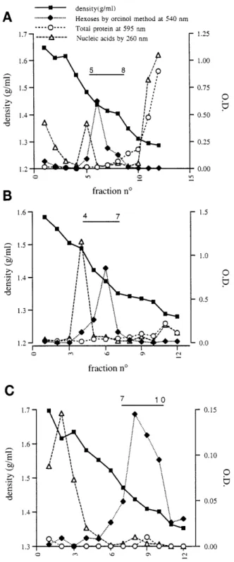
RCM, used for raising antiserum in rabbits, was isolated from colonic mucosal scrapings by applying three successive CsCl density gradients (Fig. 1). After the third gradient, mucin-containing fractions were pooled and stored lyophilized at220°C. By immunohis-tochemistry, the antiserum developed against purified RCM reacted specifically with colonic goblet cells as well as with the mucus blanket (Fig. 2). In a control experiment, purified antigen was preincubated for 30 min at 37 °C with the antiserum. Substituting antise-rum for the antigen-antiseantise-rum preincubation mix showed no reaction toward colonic mucus cells. Simi-larly, substituting anti-RCM antiserum for PBS pro-duced no reaction toward mucus cells. The antiserum was also tested for mucin specificity and epitope identi-fication by immunodetection on dot blot. Our results showed that the antiserum reacted with the purified mucin. No reaction was observed with BA, bovine thyroglobulin, and gum arabic. SDS-PAGE of the puri-fied mucins and Western blotting with our antibody revealed a single band corresponding to high molecular-weight immunoreactive material (not shown). Finally, proteolytic digestion of the purified mucin with protein-ase K resulted in a very strong decreprotein-ase in reactivity on dot blot (data not shown), thus indicating that protease-sensitive domains in mucins are crucial for binding to the antibodies.
A sandwich ELISA for RCM was then set up using the anti-RCM antiserum. A typical standard curve obtained by using increasing concentrations of purified mucin from 0 to 500 ng/ml is shown in Fig. 3A. Linearity was obtained between 8 and 500 ng mucin/ ml. The mucin specificity of the antiserum was further G1075
REGULATION OF MUCUS SECRETION IN RAT COLON
on September 6, 2010
ajpgi.physiology.org
examined by ELISA of scraping and tissue homoge-nates. The extracts of rat colon gave the highest response. Rat stomach and small intestine reacted to a lesser extent (Fig. 3B). Homogenates of lung, kidney, pancreas, liver, spleen, muscle, and serum did not react in ELISA. No cross-reactivity was detected with mouse and guinea pig gastric, intestinal, or colonic scrapings. Serial dilutions of luminal samples taken from control preparation or from colonic loop after a 30-min stimula-tion period with bethanechol or dmPGE2gave results in
ELISA that could be superimposed on the mucin
stan-dard curve, producing parallel slopes. This immunore-active material was distributed on a CsCl density gradient in a similar way to purified RCM, with a peak at a density of 1.4 g/ml (Fig. 3C), which was
characteris-Fig. 1. Isolation of mucus glycoproteins from rat colon. Distribution of material after first CsCl density gradient is shown in A. Gradient had a starting density of 1.40 g/ml and contained 4 M guanidinium hydrochloride. After centrifugation, tubes were fractionated, leading to collection of 12 fractions. Distribution of hexoses in first gradient showed peak at density of 1.43 g/ml that was characteristic of mucin. Major contaminants in mucin-containing fractions were proteins in top of gradient (fractions 8–12) and nucleic acids in bottom of gradient (fractions 1–6). Mucin-containing fractions indicated by horizontal bar were pooled and loaded on an identical gradient represented in B. Fractions 4–7 of second gradient had a mean density of 1.40 g/ml, corresponding with a peak in hexoses concentration (B). This gradient induced a complete separation of mucin from proteins but not from nucleic acids. Mucin-containing fractions were then pooled and run on third gradient to separate mucin from nucleic acids (C). This gradient had a starting density of 1.50 g/ml and contained 0.2 M guanidinium hydrochloride. In this gradient, DNA was detected in fractions 1–4 whereas hexoses were in fractions 8–10. O.D., optical density.
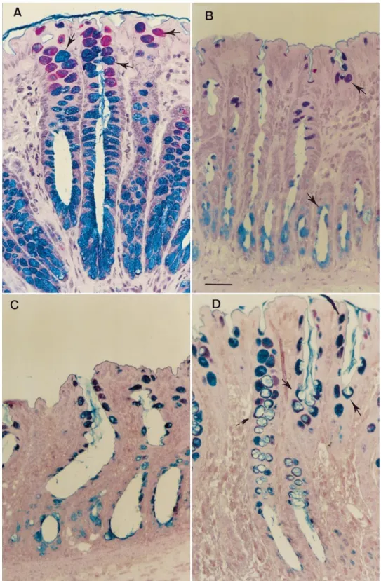
Fig. 2. Immunohistochemistry of paraffin-embedded rat colonic tis-sue after incubation with 1:1,000 anti-rat colonic mucin antiserum. The section is oriented with intestinal lumen at top. Immunolabeling is evident as a brown reaction product. Micrograph shows that only goblet cells and mucus blanket are immunoreactive. Arrows and asterisk indicate examples of immunoreactive goblet cells and mucus blanket, respectively. Bar5 10 µm.
on September 6, 2010
ajpgi.physiology.org
tic of mucins (34). No immunoreactive material was present on the top or on the bottom of the gradient.
Release of Colonic Mucus by Cholinergic and Adrenergic Agonists
Intra-arterial infusion of 200 µM bethanechol in the isolated vascularly perfused rat colon evoked a sharp increase in mucin secretion (Table 1). As shown in Fig. 4, the effect of bethanechol was concentration-depen-dent over the range 10–200 µM. The first significant response was obtained with 100 µM bethanechol. This mucin discharge was associated with a decrease in the number of stained mucus cells per crypt section as compared with the control group (Table 1, Fig. 5). Isoproterenol (1 µM) did not induce any significant increase in the release of colonic mucus (119 6 24 µg/mg DNA, n5 5, P . 0.05).
Total luminal contents in response to bethanechol and isoproterenol are shown in Table 2. Bethanechol
Fig. 3. Standard curve, cross-reactivity of prepared mucin antiserum with rat gastrointestinal scrapings, and CsCl gradient density profile of colonic luminal content. A: calibration curve of ELISA. Mucin was diluted in PBS-Tween-bovine albumin buffer to contain 0–500 ng mucin/ml. ELISA was performed as described in MATERIALS AND METHODS. Points represent mean of 2 replicate samples for each concentration of purified mucin. B: cross-reactivity of prepared mucin antiserum with rat gastrointestinal scrapings. Scrapings were ob-tained as described in MATERIALS AND METHODS and analyzed for mucin reactivity. C: CsCl gradient density profile of colonic luminal content. Samples were first homogenized in homogenization buffer and alkylated by addition of 0.25 M iodoacetamide. They were then centrifuged at 100,000 g for 70 h at 4°C, separated into fractions, and dialyzed against water before assay. Starting density of CsCl gradient was 1.40 g/ml, and gradient contained 4 M guanidinium hydrochloride.
Table 1. Quantitative histology and ELISA assay from
isolated vascularly perfused rat colon after infusion of potential mucus secretagogues
Quantitative Histology ELISA Percent of cavitated mucus cells Number of mucus cells/crypt n Mucin, µg/mg DNA n Control group 10.863.0 29.861.5 7 103614 9 Bethanechol (200 µM) 10.462.5 16.961.3* 6 9586204* 6 BBS (100 nM) 8.861.9 19.261.0* 6 8976140* 9 VIP (100 nM) 22.364.0* 25.161.4 4 574676* 10 Isoproterenol (1 µM) 7.261.0 30.761.2 4 119624 5 PYY (10 nM) 30.262.8* 29.661.5 5 264627* 6 tGLP-1 (10 nM) 9.161.0 32.461.3 4 169645 5 Serotonin (5 µM) 32.062.7* 27.461.3 5 451651* 6 dmPGE2(2.5 µM) 35.764.2* 21.461.8* 4 537683* 5 SNP (1 mM) 19.964.5 30.661.4 4 269625* 6 Values are means6 SE; n 5 no. of animals. *P , 0.05 compared with control group. Control group underwent a 10-min equilibration period followed by a 30-min period during which Krebs-Henseleit buffer supplemented with 3% BSA was administered to the vascular inflow. A reduced number of mucus cells indicated a release of mucin from colonic mucosa, and cavitation of mucus cells was the hallmark of accelerated mucus discharge by a process of compound exocytosis. BBS, bombesin; VIP, vasoactive intestinal peptide; PYY, peptide YY; tGLP, truncated glucagon-like peptide; dmPGE2, dimethyl-PGE2; SNP, sodium nitroprusside.
G1077
REGULATION OF MUCUS SECRETION IN RAT COLON
on September 6, 2010
ajpgi.physiology.org
(200 µM) led to a significant increase in luminal content recovered from the lumen, but luminal DNA content was similar to that observed in the control group (Table 2). On the contrary, isoproterenol (1 µM) did not modify the luminal content.
Neuropeptides and Mucin Release
Intra-arterial administration of 100 nM VIP induced colonic mucin discharge (Table 1). On stimulation with 10 nM VIP, the rise in mucin release was less pro-nounced (Fig. 6A). A greater proportion of cavitated goblet cells was found in loops submitted to intra-arterial infusion of VIP as compared with controls (Table 1). The effects of TTX (1 µM) and atropine (10 µM) were tested on intra-arterial infusion of 100 nM VIP. As shown in Fig. 6A, pretreatment with TTX abolished VIP-induced mucin release (P, 0.05 vs. VIP alone). In contrast, atropine did not inhibit the mucin secretory response. In control experiments (without VIP), TTX (1 µM) or atropine (10 µM) alone had no effect on mucin secretion (1106 8 µg/mg DNA, n 5 6,
P. 0.05, and 97 6 12 µg/mg DNA, n 5 6, P . 0.05,
respectively). As shown in Table 2, VIP (100 nM) induced a small increase in luminal content that was not associated with a significant rise in luminal DNA content.
BBS (100 nM) produced a sharp rise in the discharge of immunoreactive material (Table 1). Thin section of tissue demonstrated that colonic preparations exposed to 100 nM BBS for 30 min exhibited a decrease in the number of stained mucus cells compared with control preparations (Fig. 5, Table 1). In contrast, BBS infusion did not modify the percentage of cavitated mucus cells. As shown in Fig. 6B, mucin secretion induced by BBS
was completely abolished by pretreatment with 1 µM TTX. Atropine (10 µM) decreased BBS-stimulated co-lonic mucin secretion by 60% (Fig. 6B).
CGRP, at final concentrations up to 5 nM, did not elicit any release of mucin (916 10 µg/mg DNA, n 5 6,
P . 0.05), whereas a significant increase in total
luminal content was observed (Table 2).
Release of Colonic Mucin by Hormones of the Distal Gut
Intra-arterial infusion of PYY (10 nM) in the isolated vascularly perfused rat colon induced release of colonic mucin (Table 1). As shown in Fig. 7A, the responses were related to the concentration of PYY over the range 0.01–10 nM. Numerous goblet cells became deeply cavitated after 30 min of 10 nM PYY stimulation (Table 1).
In contrast, intra-arterial truncated glucagon-like peptide-1 (10 nM) did not induce a significant increase in mucin release (1696 45 µg/mg DNA, n 5 5, P . 0.05 vs. control group).
The effect of serotonin on mucin secretion is shown in Fig. 7B. The mucinlike immunoreactivity was 4516 51 µg/mg DNA, n5 6, P , 0.05, and 245 6 24 µg/mg DNA,
n 5 6, P , 0.05, after 5 and 1 µM serotonin,
respec-tively. Stained mucus cells with apical membrane cavi-tation were very numerous after administration of 5 µM serotonin (Table 1, Fig. 5). The response to 5 µM serotonin was unaffected by intra-arterial TTX (1 µM) (373 6 32 µg/mg DNA, n 5 6, P . 0.05 vs. 5 µM serotonin alone).
Release of Colonic Mucin by Mediators of the Immune System
Intra-arterial perfusion of bromolasalocid (20 µM), a degranulator of connective and mucosal mast cells, produced a modest increase in mucin release: 2546 64 µg/mg DNA (n 5 5, P , 0.05 vs. control group). In contrast, luminal administration of 20 µM bromolasa-locid failed to induce significantly mucin discharge: 1206 15 µg/mg DNA (n 5 6, P . 0.05 vs. control group). Intra-arterial administration of a stable analog of PGE2 (dmPGE2, 2.5 µM) in the isolated vascularly
perfused rat colon led to a fivefold increase in mucin secretion (Table 1, Fig. 8). By light microscopy, a rise in the percentage of cavitated mucus cells was observed. The number of stained mucus cells per crypt section was also significantly reduced (Table 1).
As can be seen from Fig. 8, IL-1b (0.25 nM) provoked a modest rise in the secretion of mucin. The mucinlike immunoreactivity release was 2006 35 µg/mg DNA(n 5 6,
P, 0.05 vs. control group) after administration of IL-1b.
SNP (1 mM), an NO generator, increased mucin secretion (Table 1, Fig. 8). By morphological analysis, SNP showed a tendency to increase the percentage of cavitated mucus cells, although this value did not achieve statistical significance (Table 1).
DISCUSSION
To measure mucus released from isolated vascularly perfused rat preparation, we developed a sandwich
Fig. 4. Effects of increasing doses of bethanechol on mucin secretion in isolated vascularly perfused rat colon. Data are means6 SE. *P , 0.05 vs. control group.
on September 6, 2010
ajpgi.physiology.org
ELISA for RCMs. For this purpose, RCMs were purified using a validated procedure (33), and these purified glycoproteins were then used to elicit a polyclonal antiserum. Because glycan structures are also present in glycolipids and in other glycoproteins, it was neces-sary to obtain a polyclonal antiserum that reacts primarily with the protein part of RCM. Our data demonstrated that anti-RCM antiserum no longer rec-ognized RCM after proteinase K digestion, thus
indicat-ing that antigenic determinants were localized to a nonglycosylated segment of colonic mucins. In agree-ment with Satchithanandam et al. (30), our study also showed that the anti-RCM antiserum is highly specific for antigenic determinants found only in scrapings of rat digestive tract. The anti-RCM antiserum detected only mucus blanket and intracellular mucin granules in goblet cells by immunohistochemistry. Furthermore, the immunoreactive material released from isolated
Fig. 5. Thin-section histology of per-fused rat colon. Bar5 10 µm. A: crypt of a control preparation. This colonic loop underwent 10-min equilibration pe-riod, followed by 30-min period during which Krebs-Henseleit buffer sup-plemented with 3% BSA was admin-istered to the vascular inflow. Goblet cells (alcian blue/periodic acid-Schiff stained) are filled with mucus. Arrows indicate examples of stained mucus cells. B: crypts of perfused rat colon after 30-min perfusion with 200 µM bethanechol. Few stained mucus cells (arrows) were observed along the length of the crypt. C: colonic mucosa after administration of 100 nM bombesin (BBS) for 30 min. Few stained mucus cells were observed along the length of the crypt. D: colonic crypt after 30-min perfusion with 5 µM serotonin. Numer-ous mucus cells with deep cavitation of apical cell surface were easily observed (arrows).
G1079
REGULATION OF MUCUS SECRETION IN RAT COLON
on September 6, 2010
ajpgi.physiology.org
vascularly perfused rat colon after administration of two well-known mucus secretagogues (bethanechol or dmPGE2) (20, 25) had the same affinity with the
antiserum as the purified mucin. Overall, these data suggest a selective recognition of epitopes belonging to the mucus fraction of intestinal contents by the present antiserum. The sandwich ELISA developed in this study is then suitable for specific and quantitative determination of colonic mucin in rats. The major advantages of this sandwich method are that the assays are accurate and are carried out in 5 h with precoated plates and that samples do not need to be purified before use. Consequently, we used it in studies of mucus secretion to measure mucins in diluted and unpurified samples. The interference of other glycopro-teins or proglycopro-teins is negligible.
Studies providing direct evidence for mucin dis-charge from the colonic epithelium by neurotransmit-ters and neuropeptides are scarce. Tumoral mucus-secreting cell lines were instead used to answer these questions. In the course of characterizing the effect of endogenous factors on colonic mucin secretion, we found that theb-adrenergic agonist isoproterenol was not a stimulant of mucin discharge in the isolated vascularly perfused rat colon. Similarly, studies per-formed in organ culture showed thata- and b-adrener-gic agonists did not result in accelerated secretion of mucin from rabbit colon (24). Taken together, adrener-gic transmitters do not appear to be major determi-nants in the regulation of colonic mucin discharge.
The possibility that gastrointestinal neuropeptides may induce colonic mucin secretion has not been clearly
explored previously. The present study demonstrated that VIP caused a sharp release of colonic mucus by compound exocytosis. In contrast, Neutra et al. (24) reported that VIP did not accelerate mucin discharge in rabbit small and large intestine in organ culture. In another study consistent with the former investigation Table 2. Total luminal content (fluid content1
adherent mucus gel) and luminal DNA content after infusion of potential mucus secretagogues in isolated vascularly perfused rat colon
Luminal Content, mg/cm Luminal DNA Content, % of total DNA n Control group 129607 0.6560.10 9 Bethanechol 200 µM 176616* 0.4460.10 6 Bethanechol 100 µM 154604 0.6060.10 5 Bethanechol 20 µM 157609 0.5760.08 5 Bethanechol 10 µM 134608 0.4060.10 6 Isoproterenol 1 µM 117623 0.4560.08 5 BBS 100 nM 150613 0.8060.20 9 BBS 10 nM 146607 0.6260.12 5 VIP 100 nM 167611* 0.6860.06 10 VIP 10 nM 151603 0.5660.10 6 CGRP 5 nM 163609* 0.4860.07 6 PYY 10 nM 113613 0.7060.13 6 PYY 1 nM 130608 0.6460.05 6 PYY 0.1 nM 134601 0.7660.10 4 tGLP-1 10 nM 125618 0.5060.15 5 Serotonin 5 µM 153610 0.4360.10 6 Serotonin 1 µM 121610 0.4160.10 6 dmPGE22.5 µM 166610* 0.5660.14 5 SNP 1 mM 197604* 0.8360.14 6 SNP 0.1 mM 142616 0.5560.15 5 IL-1b 151608 0.4060.07 6 Bromolasalocid 20 µM 164611* 0.4560.10 5 Values are means6 SE. CGRP, calcitonin gene-related peptide; IL, interleukin. * P, 0.05 compared with control group.
Fig. 6. Effect of 2 neuropeptides [vasoactive intestinal peptide (VIP) and BBS] on mucin secretion in rat colonic preparations. A: dose-related effect of VIP on mucin discharge and effect of TTX (1 µM) or atropine (10 µM) on secretory response induced by 100 nM VIP. Colonic preparations were pretreated for 10 min with TTX or atropine before being challenged for 30 min with 100 nM VIP. Values are means6 SE. *P , 0.05 vs. control group. †P , 0.05 vs. 100 nM VIP alone. B: dose-related effect of BBS on mucin secretion and effect of TTX or atropine on secretory response induced by 100 nM BBS. Data are means6 SE. *P , 0.05 vs. control group. †P , 0.05 vs. 100 nM BBS alone.
on September 6, 2010
ajpgi.physiology.org
(19), VIP (1 µM) did not alter basal mucin secretion in the HT-29-CL.16E colonic cell line, despite the presence of functional VIP-receptors. An attractive hypothesis that could explain the difference between these results is that VIP stimulates mucin release indirectly through the activation of the enteric nervous system, which is left intact in the isolated vascularly perfused rat prepa-ration. This hypothesis is supported by the observation that pretreatment with the neurotoxin TTX completely
abolished VIP-evoked mucin secretion from rat colon but had no effect on baseline mucin discharge. This result is in agreement with early observations indicat-ing that enteric nerves may be involved in VIP-stimulated intestinal secretion (16). Indeed, it has been pointed out that the VIP effect on bicarbonate secretion is mediated by the enteric nervous system in rabbit proximal duodenum (16). In our study, the possibility that muscarinic receptors participate in the VIP-evoked mucin secretion may be excluded, because
Fig. 7. Effects of peptide YY (PYY) and serotonin on mucin release.
A: luminal mucin secretion in response to PYY. Values are means6
SE. * P , 0.05 vs. control group. B: luminal mucin secretion in response to serotonin, the major product of the enterochromaffin cells. Values are means6 SE. *P , 0.05 vs. control group.
Fig. 8. Effects of bromolasalocid, dimethyl-PGE2(dmPGE2), interleu-kin (IL)-1b, (A) and sodium nitroprusside (B) on mucin secretion in isolated vascularly perfused rat colon. * P, 0.05 vs. control group.
G1081
REGULATION OF MUCUS SECRETION IN RAT COLON
on September 6, 2010
ajpgi.physiology.org
atropine did not alter the secretagogue-induced effect on mucin discharge.
The neuropeptide gastrin-releasing peptide (GRP)/ BBS is another neuropeptide found in mammalian gastrointestinal tract. GRP/BBS is thought to stimu-late secretion from endocrine cells in the stomach and intestine (7). The present study showed for the first time that BBS markedly increased the discharge of colonic mucin. Similarly, Hootman and De Ondarza (17) were able to show that, in isolated pancreatic duct, BBS stimulated goblet cell degranulation. Together, these results suggest another secretory function for BBS in the digestive tract. Our morphometric analysis additionally indicated that BBS induced mucus secre-tion by a mechanism that did not result in the cavita-tion of apical membrane. This histological picture resembles the aspect of colonic crypts after 30-min perfusion with bethanechol. To gain an insight in the mechanism underlying the BBS-evoked mucus secre-tion in the isolated vascularly perfused rat colon, TTX or atropine was administered intra-arterially before BBS infusion. Mucin secretion elicited by BBS was completely inhibited by TTX. Interestingly, atropine reduced by ,60% the BBS-induced mucin response, suggesting a partial cholinergic mediation. Together, these data indicate that BBS evokes mucin discharge through the activation of enteric nerves, part of which involves the participation of muscarinic receptors.
CGRP-containing nerves are found in the submuco-sal layer of the gut wall. The present study showed that CGRP did not induce mucin output from the isolated vascularly perfused rat colon. Another interesting fact is that luminal DNA content was not modified, al-though CGRP significantly increased fluid accumula-tion. Overall, it is likely that the moderate colonic fluid distension induced by fluid accumulation in our model was not sufficient by itself to induce mucosal damage and mucin discharge. This hypothesis is strengthened by the observation that the most potent fluid secreta-gogues administered in this report (bethanechol, VIP, SNP) did not significantly modify luminal DNA con-tent. Furthermore, histological analysis of the stimu-lated preparations did not reveal any damage of the epithelium, suggesting that mucin secretion obtained after fluid secretagogues was not induced by nonspe-cific breakdown of the epithelial barrier and mucosal damage. The conclusion to be drawn from these results is that mucin release from our model is the result of a true secretory response induced by secretagogues and was not the consequence of colonic loop distension and pressure necrosis.
Besides a neural mechanism, the secretion of colonic mucin may also implicate hormonal or local paracrine pathways. Supporting this hypothesis, El-Sahli et al. (8) were able to observe that enteroendocrine L cells in human colon emit long cytoplasmic processes to the neighboring goblet cells, suggesting a paracrine action on mucus secretion. This hypothesis was therefore tested in the present study. L cells produce and secrete PYY, a 36-amino-acid polypeptide hormone that has a variety of biological effects on the gastrointestinal
tract, including inhibition of gastric and pancreatic secretion and of gastric emptying and intestinal motil-ity (35). The present study showed that intra-arterial infusion of PYY (10 nM) produced a threefold increase in mucin release. This result is in agreement with our previous finding that PYY (10 nM) induced cavitation of the apical granule mass of goblet cells in the isolated vascularly perfused rat colon (28). In the present study, intra-arterial infusion of PYY mimicking a physiologi-cal concentration of the circulating peptide (0.1 nM) (1, 2) induced a modest rise in mucin secretion, thus excluding a major hormonal action of this peptide. However, the possibility that PYY exerted a local paracrine action on mucin release cannot be ruled out. Consequently, it is conceivable that PYY, released by intraluminal stimuli, could act locally on mucus cells to protect the colonic mucosa.
The other prominent enteroendocrine cell in the colon is the enterochromaffin cell, which synthesizes the biogenic amine serotonin. Serotonin stimulated mucin secretion from the dog gastric antrum (22). Moore et al. (23) have also demonstrated that serotonin (3 and 5 µM) induces the release of mucin in an isolated loop of rat small intestine. Used at the same concentra-tions, serotonin also induced a clear rise in colonic mucin secretion in our model. The present study also showed for the first time that this mucus discharge was mediated by compound exocytosis. In contrast, seroto-nin was without effect on mucin release in rabbit small and large intestine in organ culture (24). It is likely that these discrepancies are related to the different models used.
The accelerated discharge of mucus may also involve resident immune cells of the normal colon, which consist of lymphocytes, macrophages, and mast cells. Several studies performed with isolated gastric mucous cells and the human goblet cell line HT-29-CL.16E showed that mucin was released by several mediators secreted by immune cells (3, 4, 18). Similarly, the present study demonstrates that several immunoregu-latory mediators induce a rise in mucus discharge from colonic goblet cells in rats. Two different populations of mast cells have been identified in the colon: connective tissue mast cells and mucosa mast cells that are found in large number. The present study demonstrated that the degranulator compound bromolasalocid, which acts on both types of mast cells (9), induced a small increase in mucin secretion. Furthermore, luminal administra-tion of bromolasalocid did not induce mucin discharge, thus suggesting that this drug did not directly degranu-late goblet cells.
NO is a mediator released by mast cells, but also by macrophages, neutrophils, and enteric nerves. With a model of isolated rat gastric mucous cells, Brown et al. (4) showed that NO donors induced mucin secretion. Subsequently, a specific effect of NO donors to stimu-late mucus secretion was described in the goblet cell line HT-29-CL.16E (3). Similarly, the present study demonstrated that NO liberated from SNP caused a concentration-dependent increase in colonic mucin re-lease. Taken together, these results suggest that NO
on September 6, 2010
ajpgi.physiology.org
may be implicated in the control of gastrointestinal mucin release.
The immunoregulatory cytokine IL-1 is more specifi-cally secreted by macrophages (6). IL-1 has been demon-strated to induce a rise in the release of mucin in explant culture of mouse duodenum (5) and in the goblet cell line HT-29-CL.16E (18). Our observation that IL and SNP elicited an increase in mucin release into the colonic lumen must be tempered because the magnitude of the response was relatively low. It repre-sented at most a doubling of basal secretion calculated from data obtained in a different animal group. Be-cause baseline responses may vary among animal groups, the significance of the results is questionable when rather weak stimulants are tested.
In conclusion, we have provided evidence that the sandwich ELISA is useful to measure accurately mucin secretion in the isolated vascularly perfused rat colon. This approach already led to the demonstration that colonic mucin secretion may be induced by several factors, such as a classical neurotransmitter, two gut neuropeptides, an hormonal peptide of the distal gut, a biogenic amine, and several mediators of the immune system. Additionally, the morphological method en-abled us to show that accelerated secretion of mucus is accomplished by two different mechanisms according to the nature of secretagogues. Several factors, including VIP, PYY, and serotonin, induced a phenomenon of compound exocytosis, resulting in deep cavitation of apical membrane surface. On the contrary, bethanechol and BBS provoked mucus secretion by another process, presumably by accelerating single-granule exocytosis.
The kind advice of Dr. C. Vincent is gratefully acknowledged. The authors also thank G. Burlet and A. Desvignes for skillful technical assistance.
Address for reprint requests: P. Plaisancie´, Institut National de la Sante´ et de la Recherche Me´dicale U-45, Pavillon Hbis, Hoˆpital Ed. Herriot, 69347 Lyon Cedex 03, France.
Received 3 December 1997; accepted in final form 10 July 1998.
REFERENCES
1. Adrian, T. E., A. J. Bacarese-Hamilton, H. A. Smith, P.
Chohan, K. J. Manolas, and S. R. Bloom. Distribution and
postprandial release of porcine peptide YY. J. Endocrinol. 113: 11–14, 1987.
2. Bilchik, A. J., O. J. Hines, T. E. Adrian, D. W. McFadden,
J. J. Berger, M. J. Zinner, and S. W. Asley. Peptide YY is a
physiological regulator of water and electrolyte absorption in the canine small bowel in vivo. Gastroenterology 105: 1441–1448, 1993.
3. Branka, J. E., G. Vallette, A. Jarry, and C. L. Laboisse. Stimulation of mucin exocytosis from human epithelial cells by nitric oxide: evidence for a dependent and cGMP-independent pathway. Biochem. J. 323: 521–524, 1997.
4. Brown, J. F., A. C. Keates, P. J. Hanson, and B. J. R. Whittle. Nitric oxide generators and cGMP stimulate mucus secretion by rat gastric mucosal cells. Am. J. Physiol. 265 (Gastrointest. Liver
Physiol. 28): G418–G422, 1993.
5. Cohan, V. L., A. L. Scott, C. A. Dinarello, and R. A.
Prender-gast. Interleukin-1 is a mucus secretagogue. Cell. Immunol. 136:
425–434, 1991.
6. Dinarello, C. A. Interleukin-1 and interleukin-1 antogonism.
Blood 77: 1627–1652, 1991.
7. Dockray, G. J. Physiology of enteric neuropeptides. In:
Physiol-ogy of the Gastrointestinal Tract (3rd ed.), edited by L. R.
Johnson. New York: Raven, 1994, p. 169–209.
8. El-Sahli, M., L. Grimelius, E. Wilander, B. Ryberg, L.
Terenius, J. M. Lundberg, and K. Tatemoto.
Immunocyto-chemical identification of polypeptide YY (PYY) cells in the human gastrointestinal tract. Histochemistry 77: 15–23, 1983. 9. Fargeas, M. J., V. Theodourou, J. Fioramonti, and L.
Bueno. Relationship between mast cell degranulation and
jeju-nal myoelectric alterations in intestijeju-nal anaphylaxis in rats.
Gastroenterology 102: 157–162, 1992.
10. Forstner, G. G. Signal transduction, packaging and secretion of mucins. Annu. Rev. Physiol. 57: 585–605, 1995.
11. Forstner, J. F., and G. G. Forstner. Gastrointestinal mucus. In: Physiology of the Gastrointestinal Tract (3rd ed.), edited by L. R. Johnson. New York: Raven, 1994, p. 1255–1283.
12. Franc¸ois, C., R. D. Marshall, and A. Neuberger. Carbohy-drates in protein. The determination of mannose in hen’s egg albumin by radioisotope dilution. Biochem. J. 83: 335–341, 1962. 13. Guesdon, J. L., T. Ternynck, and S. Avrameas. The use of avidin-biotin interaction in immunoenzymatic techniques. J.
Histochem. Cytochem. 27: 1131–1139, 1979.
14. Haselbeck, A., and W. Ho¨ sel. Description and application of an
immunological detection system for analyzing glycoproteins on blots. Glycoconj. J. 7: 63–74, 1990.
15. Hinegardner, T. T. An improved fluorimetric assay for DNA.
Anal. Biochem. 39: 197–201, 1971.
16. Hogan, D. L., B. Yao, J. H. Steinbach, and J. I. Isenberg. The enteric nervous system modulates mammalian duodenal muco-sal bicarbonate secretion. Gastroenterology 105: 410–417, 1993. 17. Hootman, S. R., and J. De Ondarza. Regulation of goblet cell degranulation in isolated pancreatic ducts. Am. J. Physiol. 268 (Gastrointest. Liver Physiol. 31): G24–G32, 1995.
18. Jarry, A., G. Vallette, J. E. Branka, and C. Laboisse. Direct secretory effect of interleukin-1 via type I receptors in human colonic mucous epithelial cells (HT29-CL.16E). Gut 38: 240–242, 1996.
19. Laburthe, M., C. Augeron, C. Rouyer-Fessard, I.
Roumag-nac, J. J. Maoret, E. Grasset, and C. Laboisse. Functional
VIP receptors in the human mucus-secreting colonic epithelial cell line CL.16E. Am. J. Physiol. 256 (Gastrointest. Liver Physiol. 19): G443–G450, 1989.
20. Loeschke, K., T. Schmid, and U. M. Farack. Inhibition by loperamide of mucus secretion in the rat colon in vivo. Eur. J.
Pharmacol. 170: 41–46, 1989.
21. McCool, D. J., M. A. Marcon, J. F. Forstner, and G. G.
Forstner. The T84 human colonic adenocarcinoma cell line
produces mucin in culture and releases it in response to various secretagogues. Biochem. J. 267: 491–500, 1990.
22. Menguy, R. Regulation of secretion of mucus from the gastric antrum. Ann. NY Acad. Sci. 140: 797–803, 1967.
23. Moore, B. A., K. A. Sharkey, and M. Mantle. Role of 5-HT in cholera toxin-induced mucin secretion in the rat small intestine.
Am. J. Physiol. 270 (Gastrointest. Liver Physiol. 33): G1001–
G1009, 1996.
24. Neutra, M. R., L. J. O’Malley, and R. D. Specian. Regulation of intestinal goblet cell secretion. II. A survey of potential secretagogues. Am. J. Physiol. 242 (Gastrointest. Liver Physiol. 5): G380–G387, 1982.
25. Phillips, T. E. Both crypt and villus intestinal goblet cells secrete mucin in response to cholinergic stimulation. Am. J.
Physiol. 262 (Gastrointest. Liver Physiol. 25): G327–G331, 1992.
26. Phillips, T. E., C. M. Stanley, and J. Wilson. The effect of 16,16-dimethylprostaglandin E2on proliferation of an intestinal goblet cell line and its synthesis and secretion of mucin glycopro-teins. Prostaglandins Leukot. Essent. Fatty Acids 48: 423–428, 1993.
27. Plaisancie´, P., C. Bernard, J. A. Chayvialle, and J. C.
Cuber. Regulation of glucagon-like peptide-1-(7—36) amide
secretion by intestinal neurotransmitters and hormones in the isolated vascularly perfused rat colon. Endocrinology 135: 2398– 2403, 1994.
28. Plaisancie´, P., A. Bosshard, J. C. Meslin, and J. C. Cuber. Colonic mucin discharge by a cholinergic agonist, prostaglandins
G1083
REGULATION OF MUCUS SECRETION IN RAT COLON
on September 6, 2010
ajpgi.physiology.org
and peptide YY in the isolated vascularly perfused rat colon.
Digestion 58: 168–175, 1997.
29. Sakata, T., and W. V. Engelhardt. Influence of short chain fatty acids and osmolality on mucus release in rat colon. Cell Tissue
Res. 219: 371–377, 1981.
30. Satchithanandam, S., M. M. Cassidy, A. T. Kharroubi, R. J.
Calvert, A. R. Leeds, and G. V. Vahouny. Alterations in rat
intestinal mucin patterns following luminal infusion of acetylsali-cylic acid and prostaglandins derivatives. Dig. Dis. Sci. 35: 1518–1527, 1990.
31. Specian, R. D., and M. R. Neutra. Regulation of intestinal goblet cell secretion. I. Role of parasympathetic stimulation.
Am. J. Physiol. 242 (Gastrointest. Liver Physiol. 5): G370–G379,
1982.
32. Specian, R. D., and M. G. Oliver. Functional biology of intestinal goblet cells. Am. J. Physiol. 260 (Cell Physiol. 29): C183–C193, 1991.
33. Tytgat, K. M. A. J., F. J. Bovelander, F. J. M. Opdam, A. W. C.
Einerhand, H. A. Bu¨ ller, and J. Dekker. Biosynthesis of rat
MUC2 in colon and its analogy with human MUC2. Biochem. J. 309: 221–229, 1995.
34. Tytgat, K. M. A. J., H. A. Bu¨ ller, F. J. M. Opdam, Y. C. Kim, A. W. C. Einerhand, and J. Dekker. Biosynthesis of human
colonic mucin: MUC2 is the prominent secretory mucin.
Gastro-enterology 107: 1352–1363, 1994.
35. Walsh, J. H. Gastrointestinal hormones. In: Physiology of the
Gastrointestinal Tract (3rd ed.), edited by L. R. Johnson. New
York: Raven, 1994, p. 2–128.
on September 6, 2010
ajpgi.physiology.org


![Fig. 6. Effect of 2 neuropeptides [vasoactive intestinal peptide (VIP) and BBS] on mucin secretion in rat colonic preparations](https://thumb-eu.123doks.com/thumbv2/123doknet/14554736.537375/10.918.79.447.165.520/effect-neuropeptides-vasoactive-intestinal-peptide-secretion-colonic-preparations.webp)

