ORIGINAL ARTICLE
Preliminary results on the postmortem measurement
of 3-beta-hydroxybutyrate in liver homogenates
Cristian Palmiere&Patrice Mangin&Dominique Werner
Received: 18 December 2012 / Accepted: 6 May 2013 / Published online: 31 May 2013 # Springer-Verlag Berlin Heidelberg 2013
Abstract The concentrations of 3-beta-hydroxybutyrate (3HB) in blood and two liver samples were retrospectively examined in a series of medicolegal autopsies. These cases included diabetic ketoacidosis, nondiabetic individuals presenting moderate to severe decompositional changes and nondiabetic medicolegal cases privy of decompositional changes. 3HB concentrations in liver sample homogenates correlate well with blood values in all examined groups. Additionally, decompositional changes were not associated with increases in blood and liver 3HB levels. These results suggest that 3HB can be reliably measured in liver homog-enates when blood is not available at autopsy. Furthermore, they suggest that metabolic disturbances potentially leading or contributing to death may be objectified through liver 3HB determination even in decomposed bodies.
Keywords Postmortem biochemistry . 3-Beta-hydroxybutyrate . Liver . Diabetes . Ketoacidosis
Introduction
Specific molecules may prove much more difficult to iden-tify in biological samples collected during autopsy com-pared to clinically derived specimens. Indeed, the presence of compounds generated by putrefaction and the often-altered (decomposed) nature of the samples limit the direct applicability of clinically validated assays in a postmortem
setting [1]. While peripheral blood, urine, and gastric contents represent the ideal biological fluids for toxicological purposes, they may be partly or completely unavailable in infant autop-sies and severely decomposed corpses. Furthermore, should body damage in cases such as traffic accidents, fire fatalities, and explosions be extensive, the collection of sufficient amounts of blood, urine, and other samples for toxicology and biochemistry would be consequently limited.
Significant effort has therefore been directed towards identifying alternative, liquid or solid, biological specimens for toxicological analyses. Alternatives considered include liver, kidney, skeletal muscle, brain, lung, adipose tissue, and bone marrow samples as well as possible larvae of insects feeding on the host [1,2].
Postmortem biochemical analyses are routinely performed on vitreous humor (glucose and electrolytes), whole blood (glycated hemoglobin and ketones) and postmortem serum obtained from peripheral or cardiac blood (markers of hepatic, cardiac and renal functions, various hormones, tryptase, markers of inflammation, and sepsis). Urine, pericardial, ce-rebrospinal, and synovial fluids are occasionally used for chemical and biochemical investigations. However, blood and urine samples collected during autopsy are usually re-served for toxicological analyses. Vitreous humor, pericardial, synovial, and cerebrospinal fluids are not systematically sam-pled. Additionally, their use is rather episodic and usually limited to situations in which blood and urine are unavailable. As far as toxicology is concerned, the nature of the biolog-ical fluids collected from putrefied bodies may significantly reduce their application possibilities, rendering the direct ap-plication of clinically validated methods to decomposed sam-ples for biochemical purposes impracticable.
Over the last 20 years, there has been a growing interest in the development of methods for qualitative and quantita-tive analysis of several compounds in postmortem matrices alternative to blood, despite the existence of objective limits related to sample handling and processing.
C. Palmiere (*)
:
P. ManginUniversity Center of Legal Medicine, University of Lausanne, Rue du Bugnon 21, 1011 Lausanne, Switzerland
e-mail: [email protected] D. Werner
Laboratory of Clinical Chemistry, Lausanne University Hospital, 1011 Lausanne, Switzerland
These difficulties are mainly due to decompositional changes, postmortem autolysis, bacterial contamination, tis-sue homogenization, challenges in obtaining representative samples, time consuming techniques, as well as analytical and chromatographic obstacles. The analytical methods must therefore be able to minimize the role of potentially interfering substances such as lipids, proteins, and numerous other molecules which are usually present in high concen-trations in such matrices [3–6].
Tissue samples obtained from the liver and kidney are often used in postmortem toxicology analysis, espe-cially when blood is unavailable. Kidney and liver tis-sues are suitable to prepare homogenates; yet, as already mentioned, they also contain high concentrations of lipids, which may interfere with the analytical proce-dures’ performance.
As a specimen, liver has the advantage of being relatively unaffected by postmortem redistribution com-pared to blood, although drug concentrations in its left side, which is proximal to the stomach and small intes-tine, may be affected by the postmortem diffusion of some compounds. Since most drugs are metabolized in the liver, both the parent substance and its metabolites may be present in high concentrations in the hepatic tissue. Additionally, one of the main problems in using liver samples for forensic purposes is the lack of a database concerning the hepatic concentrations of most analyzed molecules. The results obtained from liver analysis are therefore more complicated to interpret [7]. In the realm of postmortem biochemistry, C-reactive protein (CRP) has mainly been determined in postmortem serum from femoral blood. It has, however, been experi-mentally measured in other biological matrices, such as pericardial fluid, vitreous humor, cerebrospinal fluid, and liver homogenates, at times with promising results [8]. Astrup and Thomsen [9] investigated CRP levels in 50 forensic autopsy cases in different specimens, including liver samples, and concluded that liver CRP levels correlate well with postmortem serum concentrations, suggesting the possibility of using liver samples for CRP determination should blood or postmortem serum prove unavailable. Apart from this example, liver homogenates have not been used further for postmortem biochemical purposes.
The aim of this study was to investigate 3-beta-hydroxybutyrate (3HB) levels in blood and liver sam-ples in a series of medicolegal autopsies that included cases of diabetic ketoacidosis and bodies presenting decompositional changes. The intention was to charac-terize 3HB concentrations in liver homogenates in comparison to blood levels as well as evaluate the usefulness of 3HB determination in liver samples in order to assess the metabolic disturbances potentially leading or contributing to death.
Material and methods Forensic autopsy cases
A total of 48 subjects (28 males and 20 females) were included in this study and retrospectively selected from 2008 to 2012. Subject age range was between 16 and 78 years old, with a mean age of 54.6 years. All cases included in the study underwent complete autopsies preceded by unenhanced CT scans. Autopsies were or-dered by the public prosecutor due to unclear circum-stances of death and the bodies were transferred to the medicolegal center. Toxicology and histology were performed in all cases included in this study despite the presence of decompositional changes in most organs in a portion of the selected cases. Medical records, social histories of the deceased when available, as well as police reports were reviewed in all cases before conclusions were made.
The availability of both blood (peripheral or cardiac) and liver samples at autopsy was the inclusion criterion for the cases chosen.
The selected study population consisted of 7 diabetic ketoacidosis cases without decompositional changes, 1 insulin-dependent diabetic individual presenting severe decompositional changes, 20 nondiabetic cases (according to medical records) presenting moderate to severe decompositional changes, and 20 nondiabetic cases (according to medical records) without decompositional changes.
Vitreous glucose concentrations were measured in all cases where vitreous humor was collected during autopsy.
According to the literature [10], diabetic ketoacidosis was determined to be the cause of death when vitreous glucose concentrations were above 10 mmol/l (corre-sponding to 104 mg/ml) and blood 3HB levels were higher than 2.5 mmol/l (corresponding to 26 mg/ml), as well as when other causes of death were excluded based on all postmortem investigations. The insulin-dependent diabetic individual with severe decompositional changes showed a cardiac blood 3HB concentration consistent with the hypoth-esis of ketoacidosis as the cause of death (9.6 mmol/l, corre-sponding to 100 mg/ml). Vitreous humor was unavailable in this case.
The causes of death in the 20 individuals presenting moderate to severe decompositional changes were hangings (two cases), gunshot wounds (two cases), drug intoxication (three cases), drowning (two cases), natural deaths (six cases), and unknown (five cases).
The causes of death in the 20 individuals without decompositional changes were determined to be drug intoxication (four cases), hangings (six cases), coronary thrombosis (three cases), and ischemic heart disease (seven cases).
Blood samples
Peripheral blood samples were collected by aspiration with a sterile needle and a syringe from the left or right femoral vein(s) during autopsy. Blood samples were drawn after clamping the vein(s) at the proximal end and lifting the lower limb(s) for several minutes. Peripheral blood was not available in 11 out of 20 cases with moderate to severe decompositional changes as well as the insulin-dependent diabetic individual presenting a high degree of decomposi-tion. In these cases, blood was sampled from the right and left cardiac cavities. Both peripheral and cardiac blood samples were stored in tubes containing sodium fluoride and immediately frozen at−20 °C.
Vitreous samples
Undiluted vitreous humor samples were obtained by aspira-tion using a sterile needle and syringe as soon as possible upon arrival of the bodies at the morgue. Right and left vitreous samples were collected through a scleral puncture at the lateral canthus, aspirated from the center of each eye, pooled in the same syringe, and mixed together. After col-lection, vitreous samples were immediately centrifuged at 3,000×g for 15 min. The separated supernatant was collect-ed and storcollect-ed in preservative-free tubes. Vitreous samples were transferred to the laboratory immediately after having performed the autopsies. When glucose determination was delayed, vitreous samples were stored at−20 °C.
Liver samples
Two samples (A sample and B sample) were collected in each case from the central part of the right lobe of the liver during autopsy. The first set of samples (A samples) was immediately homogenized using a laboratory blender. The tissue specimens were cut into small pieces with a dispos-able scalpel and 4 g was weighed out. Four milliliters of sterile, sodium chloride solution 0.9 % was added to the tissue (dilution 1:1). The second set of samples (B samples) was stored in preservative-free tubes and frozen at−20 °C. After 30 days, samples were thawed overnight at 4 ° C and homogenized using the same protocol as was used with the A samples. Complete homogenization of the tissue samples was not always possible due to specimen quality. However, approximately 90 % tissue homogenization was achieved by using the laboratory blender for 4 min.
Laboratory assays
3HB concentrations were determined on a Cobas Mira Plus (Roche Diagnostics, Switzerland) by an enzymatic photo-metric method adapted in house from the technique
described by Ruell and Gass [11]. Frozen femoral and cardiac blood samples were thawed overnight at 4 ° C and deproteinized with perchloric acid. Supernatant was used for analysis. Similarly, liver homogenates (samples A and B) were deproteinized with perchloric acid and supernatant was used for analysis. 3HB concentrations were expressed in millimole per liter.
Glucose was determined on vitreous samples by enzy-matic assays on a Dimension® Xpand® Plus Integrated Chemistry System (Siemens Healthcare Diagnostics Inc., Deerfield, IL, USA). Glucose concentrations were expressed in millimole per liter.
Statistical analysis
In diabetic ketoacidosis cases, in nondiabetic cases presenting moderate to severe decompositional changes and in nondiabetic cases privy of decompositional changes, 3HB concentrations in blood and liver samples were com-pared by matched pair test. The comparisons between pa-tient groups were carried out by using the Wilcoxon test.
The linear relationship between 3HB in blood and liver samples was explored by Pearson’s product–moment test. Correlation coefficient value≥0.8 and between 0.7 and 0.79 was considered to define a very strong and strong correla-tion between variables, respectively, while coefficient rang-ing between 0.69 and 0.5 and 0.49 to 0.3 was considered to define moderate and low correlation, respectively.
Ethical aspects
Ethical aspects were discussed with the local ethics commit-tee. Since the medicolegal autopsies had been ordered by the judicial authorities, analyses were performed as part of the investigations and no further ethical permission was required.
Results
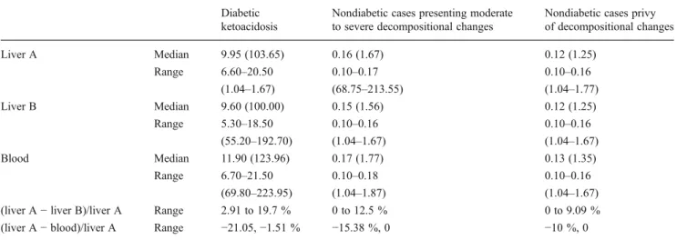
3HB concentrations in blood and liver samples as well as the relative differences between values observed in A sam-ple versus B samsam-ple and A samsam-ple versus blood in the three studied groups are summarized in Table1.
In cases of diabetic ketoacidosis, 3HB levels ranged from 6.70 to 21.50 mmol/l (corresponding to 69.80 and 223.95 mg/ml, respectively), from 6.60 to 20.50 mmol/l (corresponding to 68.75 and 213.55 mg/ml, respectively), and from 5.30 to 18.50 mmol/l (corresponding to 55.20 and 192.70 mg/ml, respectively) in blood, A samples and B samples, respectively. Concentrations were statistically higher in blood than A samples (mean difference 1.46; 95 % CI 0.84, 2.09; p value <0.001) and in A samples than B samples (mean difference 0.78; 95 % CI−0.27, 1.28; p
value=0.008). The relative differences between 3HB levels in A samples versus B samples and A samples versus blood
were ≤21 % of A sample concentration in all cases of
diabetic ketoacidosis.
Similarly, in nondiabetic cases presenting moderate to severe decompositional changes and in nondiabetic cases privy of decompositional changes, 3HB concentrations were statistically higher in blood than A samples (mean differ-ence 0.01; 95 % CI 0.008, 0.01; p value <0.001 and mean difference 0.0045; 95 % CI 0.0021, 0.0089; p value <0.001, respectively) and in A samples than in B samples (mean difference 0.009; 95 % CI 0.005, 0.012; p value <0.001 and mean difference 0.002; 95 % CI 0.0001, 0.0039; p value= 0.042, respectively) (Table1).
The relative differences between 3HB levels in A samples
versus B samples and A samples versus blood were ≤15
and≤10 % of A sample concentration in all the nondiabetic cases presenting decompositional changes and in all the nondiabetic cases without decompositional changes, respectively.
3HB concentrations were significantly higher in cases of diabetic ketoacidosis than in the other studied groups in both blood and liver samples (p<0.001 for every comparison). 3HB values were higher in nondiabetic cases presenting moderate to severe decompositional changes than in nondiabetic cases privy of decompositional changes in both blood and liver samples (p<0.001 for every comparisons). However, in all tested samples of both groups, 3HB con-centrations were lower than the blood clinical reference value (0.17 mmol/l, corresponding to 1.77 mg/ml).
Linear dependence between the three variables was shown by Pearson’s product–moment coefficient in the three groups (Table 2). Pearson’s product–moment
coeffi-cient was≥0.9, p value<0.001, and R-square ≥0.84 in every correlation between variables and in every group.
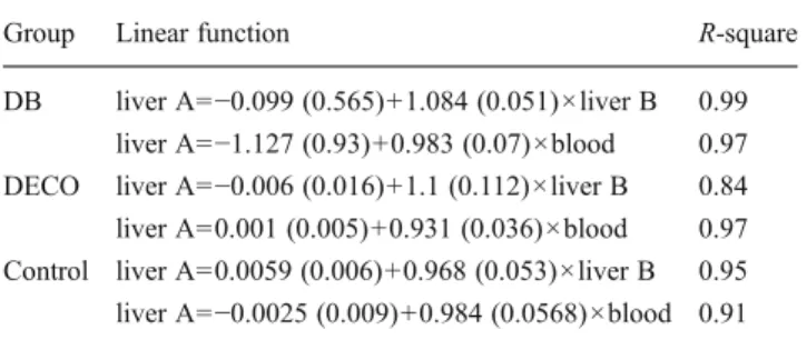
The linear function of 3HB levels in A samples versus B samples and in A samples versus blood is reported in Table3. Slopes and intercept of the regression line equations were very close to 1 and 0, respectively, and their values ±2 standard error included 1 and 0 for every correlation (Table3).
Table 1 3-Beta-hydroxybutyrate concentrations in blood and liver samples and relative difference between values observed in A samples versus B samples and A samples versus blood in the three studied groups
Diabetic ketoacidosis
Nondiabetic cases presenting moderate to severe decompositional changes
Nondiabetic cases privy of decompositional changes Liver A Median 9.95 (103.65) 0.16 (1.67) 0.12 (1.25) Range 6.60–20.50 0.10–0.17 0.10–0.16 (1.04–1.67) (68.75–213.55) (1.04–1.77) Liver B Median 9.60 (100.00) 0.15 (1.56) 0.12 (1.25) Range 5.30–18.50 0.10–0.16 0.10–0.16 (55.20–192.70) (1.04–1.67) (1.04–1.67) Blood Median 11.90 (123.96) 0.17 (1.77) 0.13 (1.35) Range 6.70–21.50 0.10–0.18 0.10–0.16 (69.80–223.95) (1.04–1.87) (1.04–1.67) (liver A− liver B)/liver A Range 2.91 to 19.7 % 0 to 12.5 % 0 to 9.09 % (liver A− blood)/liver A Range −21.05, −1.51 % −15.38 %, 0 −10 %, 0 3HB concentrations are expressed in millimole per liter and (milligram per deciliter)
Table 2 Correlation between 3HB in blood and liver samples in the three studied groups assessed by Pearson’s product– moment coefficient test
DB diabetic ketoacidosis, DECO nondiabetic cases presenting moderate to severe
decompositional changes, Con-trol nondiabetic cases without decompositional changes
Group Variable by variable Pearson’s product–moment coefficient test Correlation p value
DB Liver A vs blood 0.99 <0.001
Liver B vs blood 1 <0.001
Liver B vs liver A 0.99 <0.001
DECO Liver A vs blood 0.99 <0.001
Liver B vs blood 0.9 <0.001
Liver B vs liver A 0.92 <0.001
Control Liver A vs blood 0.96 <0.001
Liver B vs blood 0.96 <0.001
Equivalency emerged from the bivariate fit of 3HB levels in A samples versus blood/B samples in diabetic
ketoacidosis cases is shown in Fig. 1a. Equivalency
emerged from the bivariate fit of 3HB levels in A samples versus blood/B samples in nondiabetic cases presenting decompositional changes (DECO) and in nondiabetic cases without decompositional changes (control) is shown in Fig.1b. The regression line for every correlation described angle bisector between the axes.
Discussion
The postmortem diagnosis of diabetes mellitus with fatal complications, particularly ketoacidosis, requires the de-termination of specific biochemical markers. Vitreous glucose has been proposed by several authors as the most appropriate laboratory parameter to estimate ante-mortem blood glucose levels [12–16]. Other analyses involving the determination of the glycosylation level of some blood proteins [17–22] and ketone measure-ment in body fluids [23–31] have also been indicated to corroborate the hypotheses of constant antemortem hyperglycemia and increased blood ketone concentra-tions at the time of death, respectively.
Determination of ketone bodies in blood or alternative biological fluids, along with measurements of other biochem-ical parameters including vitreous glucose, urine adrenaline, and postmortem serum hormones, proteins, and markers of chronic alcohol consumption, has also been proposed in diag-nosing alcoholic and starvation ketoacidosis as well as in supporting the hypothesis of hypothermia as the cause of death [10,32–41].
The possibilities of establishing reliable diagnoses of diabetic ketoacidosis by using biochemical investigations can be extremely compromised when vitreous and blood are unavailable during autopsy or advanced putrefactive changes are present [42].
These limitations, however, may be drastically re-duced if ketone measurements in the postmortem setting can be performed in biological fluids other than blood with equally reliable results [25, 26, 32, 35, 38, 39]. The need to identify alternative biological samples is therefore paramount. These should be collectable even when autopsies are performed on bodies presenting ad-vanced decompositional changes and, above all, the measured compounds should normally be present and not increase with the onset of putrefaction.
Decompositional phenomena may markedly influence toxicological and biochemical results because they induce modifications in drug concentrations and may be responsi-ble for molecule degradation. Additionally, putrefaction may lead to the production of numerous lower molecular weight compounds, drugs, or new substances by way of artefact, which may affect the way in which case results are interpreted [3,43–46].
Iten and Meier [10] analyzed the relationship between blood 3HB concentrations and the interval after death in a series of medicolegal autopsies and did not observe any statistical increase in postmortem blood 3HB levels. These findings led to the conclusion that decompositional changes were not associated with 3HB production and that blood 3HB levels in decomposed bodies could be considered an appropriate biochemical parameter in the estimation of 3HB concentrations at the time of death. Similar results were obtained in two former studies, in which we investigated
Table 3 The linear function of 3HB levels in A samples versus B samples and A samples versus blood in the three studied groups (in parentheses standard error of intercept and slope)
Group Linear function R-square
DB liver A=−0.099 (0.565)+1.084 (0.051)×liver B 0.99 liver A=−1.127 (0.93)+0.983 (0.07)×blood 0.97 DECO liver A=−0.006 (0.016)+1.1 (0.112)×liver B 0.84 liver A=0.001 (0.005)+0.931 (0.036)×blood 0.97 Control liver A=0.0059 (0.006)+0.968 (0.053)×liver B 0.95 liver A=−0.0025 (0.009)+0.984 (0.0568)×blood 0.91 DB diabetic ketoacidosis, DECO nondiabetic cases presenting mode-rate to severe decompositional changes, Control nondiabetic cases without decompositional changes
+and , ood-bl d DB +; and , ver -li B DB X and- -, blood-DECO; and , blood-Control, X and - -, liver B-DECO; and , liver B-Control
Fig. 1 Bivariate fit of 3HB levels in A samples versus blood/B samples in diabetic ketoacidosis cases (DB) (a) and in nondiabetic cases presenting decompositional changes (DECO) and in nondiabetic cases without decompositional changes (control) (b)
blood 3HB levels in a series of medicolegal autopsies that included bodies with decompositional changes [40, 47]. Kadiš et al. [39] had already postulated that 3HB does not increase after death but, at most, may decrease due to spontaneous molecule degradation.
Besides decompositional changes, other situations fre-quently encountered in the forensic setting may force the pathologist to deal with biological fluid insufficiency or unavailability during autopsy or to collect biological sam-ples of poor quality for toxicological and biochemical pur-poses. Indeed, only small amounts of blood may be sampled during infant autopsy and specimens are often missing completely in severely damaged victims [48]. In these situ-ations, the collection of alternative biological samples that can be reliably analyzed and whose results can be faithfully exploited to produce pertinent data is of utmost importance. This is especially crucial when faced with pathological processes, such as hypothermia, diabetic, alcoholic, and starvation ketoacidosis, which may be characterized exclu-sively by biochemical changes and limited (or absent) mor-phological findings [49].
The liver has been deemed the primary solid tissue of use in postmortem toxicology with the xenobiotic analysis resulting from this tissue often complementing blood toxi-cology data. Furthermore, the liver has the advantage of being the main metabolic organ, allowing sufficient quanti-ties of tissue to be collected during autopsy for analyses. Liver samples are therefore the specimen of choice should blood prove unavailable due to massive blood loss, fire victims, or advanced decompositional changes. Liver sam-ples can additionally be readily homogenized. Though the liver is relatively unaffected by postmortem redistribution or postmortem diffusion compared with blood, xenobiotic con-centrations in the lobe proximal to the stomach and small intestine may artificially increase due to postmortem diffu-sion in cases of oral drug overdose. Therefore, use of tissue from the deep part of the right lobe is preferred for toxico-logical purposes [50–52].
In this study, we aimed to evaluate 3HB levels in liver samples obtained from medicolegal autopsies including cases of diabetic ketoacidosis fatalities and bodies presenting decompositional changes. Our goal was to char-acterize 3HB concentrations in liver homogenates and com-pare them with peripheral (or cardiac) blood 3HB levels in order to evaluate the usefulness of 3HB determination in liver samples to objectify the metabolic disturbances that can potentially cause or contribute to death.
The results of our study indicate that 3HB concentrations in liver homogenates correlate well with blood values in all exam-ined groups, thus allowing diabetic ketoacidosis to be diagnosed. Additionally, in nondiabetic decomposed bodies and nondiabetic individuals without decompositional changes, 3HB concentrations in both blood and liver samples were
within clinical blood reference values, suggesting that decompositional changes are not associated with increased blood 3HB levels. This observation concurs with the conclu-sions of two former studies that focused on the interpretation of postmortem blood 3HB levels in decomposed bodies [10,39]. Lastly, according to the results of our analysis, decompositional changes do not seem to be associated with increases in 3HB liver concentrations.
Though further studies are required to confirm these pre-liminary observations, our data suggest that 3HB can be reliably measured in liver homogenates when blood is unavailable during autopsy or when priority in using limited amounts of blood must be given to toxicological investiga-tions. Liver homogenates can be considered appropriate, al-ternative, biological samples for 3HB determination when blood, vitreous, urine, pericardial, and cerebrospinal fluids cannot be collected during autopsy and biochemical investi-gations could prove useful in evaluating the metabolic distur-bances that may have potentially led or contributed to death.
Acknowledgments The authors are grateful to the anonymous re-viewers, whose constructive and useful comments improved the qual-ity of the article.
Conflict of interest The authors have no conflict of interest to declare.
References
1. Drummer OH, Gerostamoulos J (2002) Postmortem drug analysis: analytical and toxicological aspects. Ther Drug Monit 24(2):199– 209
2. Morley SR, Bolton J (2012) Variation in postmortem liver sam-pling: implications for postmortem toxicology interpretation. J Clin Pathol 65(12):1136–1137
3. Butzbach DM (2010) The influence of putrefaction and sample storage on postmortem toxicology results. Forensic Sci Med Pathol 6(1):35–45
4. Skopp G (2010) Postmortem toxicology. Forensic Sci Med Pathol 6(4):314–325
5. Drummer OH (2004) Postmortem toxicology of drugs of abuse. Forensic Sci Int 142(2–3):101–113
6. Drummer OH (2007) Requirements for bioanalytical procedures in postmortem toxicology. Anal Bioanal Chem 388(7):1495–1503 7. Margalho C, Franco J, Corte-Real F, Vieira DN (2011) Illicit drugs
in alternative biological specimens: a case report. J Forensic Leg Med 18(3):132–135
8. Augsburger M, Iglesias K, Bardy D, Mangin P, Palmiere C (2012) Diagnostic value of lipopolysaccharide-binding protein and procalcitonin for sepsis diagnosis in forensic pathology. Int J Leg Med. doi:10.1007/s00414-012-0780-9
9. Astrup BS, Thomsen JL (2007) The routine use of C-reactive protein in forensic investigations. Forensic Sci Int 172(1):49–55 10. Iten PX, Meier M (2000) Beta-hydroxybutyric acid—an indicator
for an alcoholic ketoacidosis as cause of death in deceased alcohol abusers. J Forensic Sci 45(3):624–632
11. Ruell PA, Gass GC (1991) Enzymatic measurement of 3-hydroxybutyrate in extracts of blood without neutralization. Clin Ann Biomed 28(Pt 2):183–184
12. Karlovsek MZ (1995) Postmortem diagnosis of diabetes mellitus and diabetic coma: a comparison of HbA1, glucose, lactate and combined glucose and lactate values in vitreous humor and in cerebrospinal fluid. In: Jacob B, Bonte W (eds) Advances in Forensic Sciences: Forensic Criminalistic 2, Vol. 4. Verlag Dr Köstner, Berlin, pp 38–48
13. Karlovsek MZ (2004) Diagnostic values of combined glucose and lactate values in cerebrospinal fluid and vitreous humor—our experiences. Forensic Sci Int 146(Suppl146):s19–s23
14. Zilg B, Alkass K, Berg S, Druid H (2009) Postmortem identifica-tion of hyperglycemia. Forensic Sci Int 185(1–3):89–95
15. Boulagnon C, Garnotel R, Fornes P, Gillery P (2011) Postmortem biochemistry of vitreous humor and glucose metabolism: an up-date. Clin Chem Lab Med 49(8):1265–1270
16. Palmiere C, Sporkert F, Vaucher P, Werner D, Bardy D, Rey F, Lardi C, Brunel C, Augsburger M, Mangin P (2012) Is the formula of Traub still up to date in antemortem blood glucose level esti-mation? Int J Leg Med 126(3):407–413
17. Chen C, Glagov S, Mako M, Rochman H, Rubenstein AH (1983) Postmortem glycosylated hemoglobin (HbA1c): evidence for a history of diabetes mellitus. Ann Clin Lab Sci 13(5):407–410 18. Hindle EJ, Rostron GM, Gatt JA (1985) The diagnostic value of
glycated haemoglobin levels in postmortem blood. Ann Clin Biochem 22(Pt. 2):144–147
19. Valenzuela A (1988) Postmortem diagnosis of diabetes mellitus. Quantitation of fructosamine and glycated hemoglobin. Forensic Sci Int 38(3–4):203–208
20. Ritz S, Mehlan G, Martz W (1996) Postmortem diagnosis of diabetic metabolic derangement: elevated alpha 1-antitrypsin and haptoglobin glycosylation levels as an index of antemortem hy-perglycemia. J Forensic Sci 41(1):94–100
21. Goullé JP, Lacroix C, Bouige D (2002) Glycated hemoglobin: a useful postmortem reference marker in determining diabetes. Forensic Sci Int 128(1–2):44–49
22. Wineker RE, Hammett-Stabler CA, Chapman JF, Ropero-Miller JD (2002) HbA1c as a postmortem tool to identify glycemic control. J Forensic Sci 47(6):1373–1379
23. Osuna E, Vivero G, Conejero J, Abenza JM, Martínez P, Luna A, Pérez-Cárceles MD (2005) Postmortem vitreous humor beta-hydroxybutyrate: its utility for the postmortem interpretation of diabetes mellitus. Forensic Sci Int 153(2–3):189–195
24. Kanetake J, Kanawaku Y, Mimasaka S, Sakai J, Hashiyada M, Nata M, Funayama M (2005) The relationship of a high level of serum beta-hydroxybutyrate to cause of death. Leg Med (Tokyo) 7(3):169–174
25. Felby S, Nielsen E, Thomsen JL (2008) The postmortem distribu-tion of ketone bodies between blood, vitreous humor, spinal fluid and urine. Forensic Sci Med Pathol 4(2):100–107
26. Elliott S, Smith C, Cassidy D (2010) The postmortem relationship between beta-hydroxybutyrate (BHB), acetone and ethanol in ketoacidosis. Forensic Sci Int 198(1–3):53–57
27. Hess C, Musshoff F, Madea B (2011) Disorders of glucose metabolism—post mortem analyses in forensic cases: part I. Int J Leg Med 125(2):163–170
28. Musshoff F, Hess C, Madea B (2011) Disorders of glucose metabolism—post mortem analyses in forensic cases—part II. Int J Leg Med 125(2):171–180
29. Palmiere C, Mangin P (2012) Postmortem chemistry update part I. Int J Leg Med 126(2):187–198
30. Hockenhull J, Dhillo W, Andrews R, Paterson S (2012) Investigation of markers to indicate and distinguish death due to alcoholic ketoacidosis, diabetic ketoacidosis and hyperosmolar
hyperglycemic state using postmortem samples. Forensic Sci Int 214(1–3):142–147
31. Heninger M (2012) Postmortem vitreous beta-hydroxybutyrate: interpretation in a forensic setting. J Forensic Sci 57(5):1234–1240 32. Teresiński G, Buszewicz G, Mądro R (2002) The influence of ethanol on the level of ketone bodies in hypothermia. Forensic Sci Int 127(1–2):88–96
33. Teresiński G, Buszewicz G, Mądro R (2005) Biochemical back-ground of ethanol-induced cold susceptibility. Leg Med (Tokyo) 7(1):15–23
34. Teresiński G, Buszewicz G, Mądro R (2009) Acetonaemia as an initial criterion of evaluation of a probable cause of sudden death. Leg Med (Tokyo) 11(1):18–24
35. Denmark LN (1993) The investigation of beta-hydroxybutyrate as a marker for sudden death due to hypoglycemia in alcoholics. Forensic Sci Int 62(3):225–232
36. Thomsen JL, Felby S, Theilade P, Nielsen E (1995) Alcoholic ketoacidosis as a cause of death in forensic cases. Forensic Sci Int 75(2–3):163–171
37. Brinkmann B, Fechner G, Karger B, DuChesne A (1998) Ketoacidosis and lactic acidosis—frequent causes of death in chronic alcoholics. Int J Leg Med 111(3):115–119
38. Pounder DJ, Stevenson RJ, Taylor KK (1998) Alcoholic ketoacidosis at autopsy. J Forensic Sci 43(4):812–816
39. Kadiš P, Balažic J, Ferlan-Marolt V (1999) Alcoholic ketoacidiosis: a cause of sudden death of chronic alcoholics. Forensic Sci Int 103(Suppl):s53–s59
40. Palmiere C, Sporkert F, Werner D, Bardy D, Augsburger M, Mangin P (2012) Blood, urine and vitreous isopropyl alcohol as biochemical markers in forensic investigations. Leg Med (Tokyo) 14(1):17–20 41. Petersen TH, Williams T, Nuwayhid N, Harruff R (2012)
Postmortem detection of isopropanol in ketoacidosis. J Forensic Sci 57(3):674–678
42. Smialek JE, Levine B (1998) Diabetes and decomposition: a case of diabetic ketoacidosis with advanced postmortem changes. Am J Forensic Med Pathol 19(1):98–101
43. Gerostamoulos D, Beyer J, Staikos V, Tayler P, Woodford N, Drummer OH (2012) The effect of the postmortem interval on the redistribution of drugs: a comparison of mortuary admission and autopsy blood specimens. Forensic Sci Med Pathol 8(4):373–379 44. Drummer OH (2007) Postmortem toxicology. Forensic Sci Int
165(2–3):199–203
45. Statheropoulos M, Spiliopoulou C, Agapiou A (2005) A study of volatile organic compounds evolved from the decaying human body. Forensic Sci Int 153(2–3):147–155
46. Statheropoulos M, Agapiou A, Spiliopoulou C, Pallis GC, Sianos E (2007) Environmental aspects of VOCs evolved in the early stages of human decomposition. Sci Total Environ 385(1– 3):221–227
47. Palmiere C, MdM L, Sabatasso S, Mangin P, Augsburger M, Sporkert F (2012) Usefulness of postmortem biochemistry in forensic patholo-gy: illustrative case reports. Leg Med (Tokyo) 14(1):27–35 48. Maeda H, Ishikawa T, Michiue T (2011) Forensic biochemistry for
functional investigation of death: concept and practical application. Leg Med (Tokyo) 13(2):55–67
49. Luna A (2009) Is postmortem biochemistry really useful? Why is it not widely used in forensic pathology? Leg Med (Tokyo) Suppl:s27–s30
50. Dinis-Oliveira RJ, Carvalho F, Duarte JA, Remião F, Marques A, Santos A, Magalhães T (2010) Collection of biological samples in forensic toxicology. Toxicol Mech Methods 20(7):363–414 51. Flanagan RJ, Connally G, Evans JM (2005) Analytical toxicology:
guidelines for sample collection postmortem. Toxicol Rev 24(1):63–71 52. Skopp G (2004) Preanalytical aspects in postmortem toxicology.