Gli3 Controls Corpus Callosum Formation by Positioning Midline Guideposts During
Telencephalic Patterning
Dario Magnani1, Kerstin Hasenpusch-Theil1, Carine Benadiba2, Tian Yu3, M. Albert Basson3, David J. Price1, Cécile Lebrand2 and Thomas Theil1
1
Centre for Integrative Physiology, University of Edinburgh, Edinburgh, UK2Department of Cellular Biology and Morphology, University of Lausanne, Lausanne, Switzerland and3Department of Craniofacial Development, King’s College London, London, UK
Address correspondence to Thomas Theil. Email: [email protected]
The corpus callosum (CC) represents the major forebrain commis-sure connecting the 2 cerebral hemispheres. Midline crossing of callosal axons is controlled by several glial and neuronal guideposts specifically located along the callosal path, but it remains unknown how these cells acquire their position. Here, we show that the Gli3 hypomorphic mouse mutant Polydactyly Nagoya (Pdn) displays agenesis of the CC and mislocation of the glial and neuronal guide-post cells. Using transplantation experiments, we demonstrate that agenesis of the CC is primarily caused by midline defects. These defects originate during telencephalic patterning and involve an up-regulation of Slit2 expression and altered Fgf and Wnt/β-catenin signaling. Mutations in sprouty1/2 which mimic the changes in these signaling pathways cause a disorganization of midline guide-posts and CC agenesis. Moreover, a partial recovery of midline ab-normalities in Pdn/Pdn;Slit2−/− embryos mutants confirms the functional importance of correct Slit2 expression levels for callosal development. Hence, Gli3 controlled restriction of Fgf and Wnt/ β-catenin signaling and of Slit2 expression is crucial for positioning midline guideposts and callosal development.
Keywords: corpus callosum, Fgf8, Gli3, Pdn, Slit2
Introduction
The corpus callosum (CC) connects neurons of the 2 cerebral hemispheres and coordinates information between the left and right cortex. CC malformations have been associated with mental retardation involving a wide range of cognitive, behav-ioral, and neurological consequences (Richards et al. 2004;
Paul et al. 2007) and have been identified in over 50 human congenital syndromes (Richards et al. 2004). During CC for-mation, several guidance events control midline crossing of callosal axons. The midline zipper glia (MZG) have been suggested to initiate the fusion of the dorsal midline produ-cing the substrate on which callosal axons navigate (Silver et al. 1993). Moreover, several guidepost cells are located along the path of callosal axons including the midline glial cell populations composed of the indusium griseum glia (IGG) and the glial wedge (GW) (Richards et al. 2004), and GABAergic and glutamatergic neurons that transiently popu-late the CC (Niquille et al. 2009). Finally, axons from the cin-gulate cortex pioneer the CC and function as scaffolds for neocortical axons (Koester and O’Leary 1994; Rash and Ri-chards 2001; Piper, Plachez et al. 2009). Several axon-guidance molecules, including Slit2, that are produced by midline glial cells and by the glutamatergic neurons have es-sential roles in callosal development (Bagri et al. 2002; Ni-quille et al. 2009). While these studies reveal complex
interactions between callosal axons and their environment, it remains largely unknown how guidepost cells acquire their correct positions and how the expression of essential gui-dance molecules is regulated.
Gli3 encodes a zinc-finger transcription factor with crucial roles in early patterning of the dorsal telencephalon (Theil et al. 1999;Tole et al. 2000;Kuschel et al. 2003;Fotaki et al. 2006) acting both cell autonomously (Quinn et al. 2009) and cell nonautonomously by controlling the expression of signal-ing molecules essential for telencephalic development (Grove et al. 1998; Theil et al. 1999; Tole et al. 2000; Aoto et al. 2002). Moreover, Gli3 functions in axon pathfinding in the forebrain. The Gli3 hypomorphic mouse mutant Polydactyly Nagoya (Pdn) shows defects in the corticothalamic and thala-mocortical tracts (Magnani et al. 2010) and lacks the CC (Naruse et al. 1990) though for unknown reasons. Using transplantation experiments, we here demonstrate that midline abnormalities are primarily responsible for agenesis of the corpus callosum (ACC). We show that Pdn mutants display mislocated glial and neuronal guidepost cells. The Pdn cingulate cortex contains ectopic glial cells transecting the path of callosal axons. These midline abnormalities are associated with an up-regulation and down-regulation of Fgf and Wnt/β-catenin signaling, respectively. These changes in these signaling pathways are mimicked in Sprouty1/2 double mutants, which display a mislocation of midline guideposts and ACC. Pdn mutants also show an up-regulation of Slit2 expression, and positioning of the neuronal guideposts is largely rescued in Pdn/Pdn;Slit2−/− double mutants suggesting that maintaining correct Slit2 expression levels is crucial for callosal development. Collectively, these analyses reveal a novel role for Gli3 in controlling the positioning of midline guideposts by regulating Fgf and Wnt/β-catenin sig-naling and Slit2 expression levels and provide new insights into the mechanisms underlying CC pathogenesis.
Materials and Methods Mice
The mutant mouse lines Pdn,τGFP, Slit2, Sprouty1, and Sprouty2 and mating strategies have been described previously (Naruse et al. 1990;
Pratt et al. 2000;Plump et al. 2002; Basson et al. 2005;Shim et al. 2005;Simrick et al. 2011). All experimental procedures involving mice were performed in accordance with local guidelines. In analyses of Pdn mutant phenotypes, heterozygous and wild-type embryos did not show qualitative differences and both were used as control embryos. For quantitative analyses, wildt-ype and Pdn/Pdn embryos were com-pared to avoid the possible risk of Pdn/+ embryos having subtle defects. For each marker and each stage, 3–5 embryos were analyzed. Cerebral Cortex January 2014;24:186–198
doi:10.1093/cercor/bhs303
In Situ Hybridization and Immunohistochemistry
Antisense RNA probes for Bmp7 (Furuta et al. 1997), Msx1 (Hill et al. 1989), Sema3C (Bagnard et al. 2000), Slit2 (Erskine et al. 2000), Fabp7 (Genepaint. RNA probe 653), Fgf8 (Crossley and Martin 1995), Sprouty2 (Minowada et al. 1999), Axin2 (Lustig et al. 2002), Wnt7b (Parr et al. 1993), Wnt8b (Richardson et al. 1999), Nf1b (IMAGE: 4038233), Nf1x (IMAGE: 3491917), Emx1 (Simeone et al. 1992), and Six3 (Oliver et al. 1995) were labeled with digoxigenin. In situ hybrid-ization on 12-μm serial paraffin sections of mouse brains were per-formed as described (Theil 2005).
Immunohistochemical analysis was performed as described pre-viously (Theil 2005) using antibodies against the following molecules: β-III-tubulin (Tuj1 antibody; 1:1000, Sigma); brain lipid-binding protein (Blbp; 1:500, CHEMICON); calbindin (CB; 1:1000, Swant); cal-retinin (CR; 1:1000, CHEMICON); Glast (1:5000, CHEMICON); glia fi-brillary acidic protein (GFAP; 1:1000, DakoCytomation); green fluorescent protein (GFP; 1:1000, Abcam); Nf1a (1:1000, Active Motif ); neural cell adhesion molecule L1 (1:1000, CHEMICON); Neu-rofilament (2H3; 1:5, DSHB); Neuropilin-1 (Npn-1; 1:1000, R&D Systems); Satb2 (1:50, Abcam); Tbr1 (1:2500, CHEMICON). Primary antibodies for immunohistochemistry were detected with Alexa- or Cy2/3-conjugated fluorescent secondary antibodies. For non-fluorescent detection, we used biotinylated goat antimouse antibodies (Dako) followed by avidin-HRP and DAB detection (Vector Labs).
CB+neurons in the indusium griseum of E16.5 and E18.5 embryos were quantified by determining total CB+cell numbers in this region. For quantifying CR+neurons, a box with constant area (170μm2for
E16.5 and 297μm2for E18.5 embryos) was placed in the cingulate
cortex immediately dorsal to the CC, and the numbers of CR+neurons were counted within this box. Numerical values are given as a proportion of CR+cells perμm2. For statistical analyses, an analysis
of variance test was used followed by a Bonferroni’s multiple com-parison test.
Explant Culture
Organotypic slice cultures of rostral levels of the embryonic mouse telencephalon were prepared as previously described (Magnani et al. 2010). Brain slices were cultured on polycarbonate culture mem-branes (8-μm pore size; Corning Costar) in organ tissue dishes con-taining 1 mL of medium (Neurobasal/B-27 [Gibco] supplemented with glutamine, glucose, penicillin, and streptomycin). For transplan-tation experiments, slices were cultured for 72 h,fixed with 4% PFA, and processed for antiGFP immunofluorescence as described above. For Fgf blocking experiments, slices were cultured in the presence of either DMSO or of 25 or 100μM SU5402 (Calbiochem) for 48 h , fixed with 4% PFA, and processed for in situ hybridization or Blbp immunofluorescence as described above.
Quantitative Reverse Transcription PCR
Total RNA was prepared from the E14.5 rostromedial telencephalon of wild-type or Pdn/Pdn embryos. Quantitative reverse transcription polymerase chain reaction (qRT-PCR) was performed using a TaqMan® Gene Expression Assay (Applied Biosystems) for Slit2
(Mm00662153.m1, probe dye FAM-MGB) with ACTB (#4352933, probe dye FAM-MGB) and GAPDH (#4352932, probe dye FAM-MGB) as endogenous controls and a 7000 Sequence Detection System. The abundance of each transcript in the original RNA sample was extrapo-lated from PCR reaction kinetics using sequence detection software (SDS) version 1.2.3 running an absolute quantification protocol in-cluding background calibrations.
Results
CC Midline GuidePost Cells are Severely Disorganized in Pdn/Pdn Brains
Neurofilament, Tuj1 and L1 immunhistochemical stainings, and cortical DiI labeling confirmed a previous description of CC malformation in Pdn mutants (Naruse et al. 1990),
showing that the path of callosal axons is disrupted at several positions in the cingulate cortex and that those axons which approach the midline fail to cross it, forming Probst bundles instead (Fig. 1 and Supplementary Fig. 1). To gain insights into the origins of these defects, we analyzed the navigation of the cingulate pioneer axons and the formation of glial and neuronal guideposts that are essential for callosal develop-ment (Paul et al. 2007). In P0 control animals, the cingulate pioneer axons are immunopositive for Npn-1 occupying the dorsal-most part of the CC (Fig. 1A,B). In Pdn mutants, Npn-1+axons fail to project to the contralateral hemisphere, but form dense bundles ipsilaterally (Fig.1C,D). Glutamater-gic guidepost neurons express Tbr1, CR, or CB (Niquille et al. 2009). In control embryos, CR+ and CB+ neurons are both located in the IG region, and CR+ neurons are also found within the CC where they delineate its ventral and dorsal parts (Niquille et al. 2009; Fig. 1A,B,E,F). In Pdn mutants, CR+neurons are dramatically disorganized, but maintain their spatial association with callosal axons, with clusters of CR+ neurons surrounding the Probst bundles (Fig. 1C,D). CB+ neurons remain concentrated in the medial cortex although they are more diffusely distributed and clusters of CB+ neurons intermingle abnormally with callosal axons (Fig.1G, H). Finally, GFAP immunostaining labels the GW, the IGG, and the MZG in control embryos (Fig.1I,J). In Pdn brains, several GFAP+fascicles are formed ectopically in the cingulate cortex (Fig. 1K,L). Some fascicles span the whole cortical width and transect the path of callosal axons. The IGG could not be identified and the MZG expands into more ventral regions of the septum. Taken together, these data show a dra-matic disorganization of glial and neuronal guidepost cells.
Given the severity of this disorganization, we started to study the origins of these midline defects by investigating the formation of the midline guideposts and of the cingulate pioneer neurons at earlier stages. In E16.5 control embryos, cingulate pioneer axons approach the midline and start to cross it (Supplementary Fig. 2A,B). In Pdn mutants, few axons have reached the corticoseptal boundary (CSB) and many have abnormally formed clusters in the cingulate cortex (Supplementary Fig. 2C,D). Tbr1+, CR+, and CB+ glutamater-gic guidepost neurons form a well organized band of neurons at the CSB of control embryos, but their organization is se-verely disturbed in Pdn embryos with less CB+neurons in the IG region (Supplementary Figs 2A–L and 7P). In the cingulate cortex, the cortical plate is disrupted in several positions where callosal axons stop their navigation (Supplementary Fig. 2G,H). Finally, radial glial cells (RGCs) at the CSB, which co-express GFAP and the RGC marker Glast, have started to differentiate into GW cells, to translocate to the pial surface, and to form the IGG in control embryos (Supplementary Fig. 2M,N). In Pdn mutants, GFAP+;Glast+cells are present ec-topically in the cingulate cortex and extend projections from the ventricular to the pial surface (Supplementary Fig. 2O,P). Taken together, thesefindings suggest that midline guidance cues are already disorganized in Pdn mutants when callosal axons approach the CSB.
Agenesis of the CC in Pdn Mutants is Caused by CC Midline Defects
Since Gli3 is widely expressed in progenitor cells that give rise to callosal neurons and to guidepost cells, agenesis of the
CC in Pdn mutants could either be caused by the disorganiz-ation of midline guideposts or by a primary failure of callosal axons to navigate in the midline region leading to the for-mation of Probst bundles and to a secondary redistribution of guideposts. Previous marker and BrdU birthdating analyses in Pdn/Pdn mutants failed tofind major defects in cortical lami-nation (Magnani et al. 2010). Moreover, Satb2 upper layer cal-losal projection neurons are borne at E15.5 (Supplementary Fig. 3), suggesting that these neurons are specified correctly. To test directly whether Pdn mutant callosal axons are capable of following midline guidance cues we performed in vitro transplantation experiments using mice ubiquitously ex-pressing aτGFP fusion protein (Pratt et al. 2000). Homotopi-cal transplantation of frontal cortex of E17.5 GFP+ embryos into cortical sections of age-matched GFP−embryos resulted in growth of axons into the host tissue and in midline cross-ing of callosal axons (n = 8 of 9; Fig.2A). After transplantation of Pdn/Pdn;GFP+ cortex into control cortex, Pdn/Pdn axons also migrated across the midline (n = 7 of 8; Fig. 2B). However, control;GFP+callosal axons did not grow into Pdn/ Pdn;GFP−dorsomedial cortex (n = 0 of 7; Fig.2C) and only a
few axons projected along the surface of the mutant host tissue (n = 4 of 7; Fig. 2C). In contrast, corticofugal axons project into the lateral cortex and striatum under these con-ditions (Magnani et al. 2010). These results show that normal levels of Gli3 are not required to generate callosal neurons with the ability to project their axons across the midline, but indicate a requirement for Gli3 in the generation of the midline guideposts.
The Pdn Mutation Affects the Patterning in the Rostromedial Telencephalon
Next, we became interested in identifying causes underlying these midline defects. Our previous analyses showed that Pdn mutants display patterning defects during early telencephalic development (Kuschel et al. 2003). We therefore hypoth-esized that these defects might cause the defective positioning of the midline guidance cues. To test this idea, we started to analyze the development of the E12.5 corticoseptal region where callosal axons later cross the midline. We showed pre-viously that expression of the Emx1 homobox gene is lost in Figure 1. Disorganization of midline structures in P0 Pdn/Pdn brains. (A–D) Npn-1 stains the pioneer axons of the cingulate cortex (CiC) in control and Pdn/Pdn mutants. Pdn/ Pdn Npn-1+axons fail to reach the contralateral hemisphere, instead forming Probst bundles (C and D). (A and B) CR labels glutamatergic guidepost neurons. (C and D) In Pdn/
Pdn brains, CR+neurons are disorganized and clusters of CR+neurons are associated with Probst bundles (arrowhead in D). (E and F) CB labels guidepost neurons located in
the IG. (G and H) CB+neurons abnormally cluster with Tuj1+callosal axons in the Pdn/Pdn cortex (arrowheads in H). (I–J) GFAP immunofluorescence labels the GW, the IGG, and the MZG in control brains. (K and L) In Pdn/Pdn brains, GFAP+fascicles are ectopically formed within the CiC (arrowhead in L) and transect the path of callosal axons. The
Pdn mutants (Kuschel et al. 2003). Moreover, Emx1 mutants display ACC (Qiu et al. 1996;Yoshida et al. 1997) and Emx1 has recently been shown to belong to a group of transcription factors including Six3 and Nfia whose expression domains delineate the regions where the CC, the hippocampal, and anterior commissures cross the midline at E16.5 (Moldrich et al. 2010). As these genes have important roles in forebrain and/or callosal development (Qiu et al. 1996;das Neves et al. 1999; Lagutin et al. 2003; Shu et al. 2003; Campbell et al. 2008;Plachez et al. 2008;Piper, Moldrich et al. 2009), we in-vestigated their expression at the E12.5 CSB where callosal axons later cross the midline. In control embryos, Six3 is ex-pressed in the septum, but Six3 expression expands dorsally in Pdn mutants (Supplementary Fig. 4A,E). Nfia, Nfib, and Nfix, are expressed at high levels in the cortex and at lower levels in the dorsalmost septum (Shu et al. 2003; Plachez et al. 2008; Supplementary Fig. 4B–D). In Pdn mutants, their cortical expression domains are lost, while low-level septal expression remains except for Nfia which is strongly ex-pressed in the septum (Supplementary Fig. 4F–H). Taken to-gether, these data indicate that the expression of several transcription factors with important roles in callosal develop-ment is altered in Pdn mutants, suggesting that the CSB is poorly defined.
Previous analyses had also shown a requirement of Gli3 for the correct expression of several signaling molecules in the telencephalon, including Shh, Bmp/Wnt genes, and Fgf8 (Grove et al. 1998;Theil et al. 1999; Tole et al. 2000;Aoto et al. 2002; Kuschel et al. 2003; Magnani et al. 2010). We
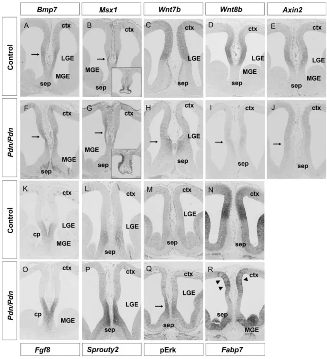
therefore analyzed the expression of these signaling mol-ecules specifically at the E12.5 CSB. This analysis revealed a slight extension of Shh expression into the ventral-most part of the septum in Pdn/Pdn embryos, but Shh signaling as judged by Ptc1 expression remains confined to the septum and does not reach the CSB (Supplementary Fig. 5). More-over, Bmp7, which is essential for callosal development (Sanchez-Camacho et al. 2011), and its target gene Msx1 are expressed on the cortical side of the CSB though only at caudal levels with no obvious difference between control and Pdn/Pdn embryos (Fig. 3A,B,F,G). In contrast, we observed severe changes in the Wnt7b/8b expression patterns. In control embryos, Wnt7b and Wnt8b expression are confined to the dorsomedial telencephalon with a sharp expression boundary at the CSB (Fig. 3C,D), while Wnt7b and Wnt8b expression is nearly absent from the Pdn dorsomedial tele-ncephalon, and Wnt7b transcription is increased in the septum (Fig. 3H,I). Consistent with reduced Wnt/β-catenin signaling, expression of the Wnt target gene Axin2 is severely reduced in Pdn mutants (Fig.3E,J).
Since telencephalic patterning is controlled by a balance between Bmp/ Wnt/β-catenin and Fgf signaling (Theil et al. 1999; Kuschel et al. 2003; Shimogori et al. 2004) and since Fgf8 is required for callosal development (Huffman et al. 2004; Moldrich et al. 2010), we also investigated Fgf8 expression in Pdn mutants. In control embryos, Fgf8 tran-scripts are confined to the commissural plate, but expand further dorsally in the E12.5 Pdn corticoseptal region (Fig. 3K,O) consistent with our previous whole-mount Figure 2. Pdn/Pdn callosal axons are able to cross the cortical midline in a control environment. (A) Transplantation of E17.5 control GFP+frontal cortex leads to the migration of GFP+callosal axons across the midline (n = 8 of 9). (B) Pdn/Pdn;GFP+cortex transplantation into control cortex also shows crossing callosal axons (n = 7 of 8). (C) After
transplantation of control; GFP+cortical tissue into Pdn/Pdn; GFP-frontal cortex callosal axons do not project into the intermediate zone (n = 0 of 7), only few axons project along the surface of the mutant tissue (arrowhead; n = 4 of 7). (D) Experimental procedure illustrating transplant experiments: Tissue from the frontal cortex of E17.5 GFP+
embryos was homotopically transplanted into cortical sections of age-matched GFP− embryos and the migration pattern of callosal axons was monitored using GFP immunofluorescence after 72 h of culture.
expression analysis (Kuschel et al. 2003). Expression of and phospho-Erk ( pErk), targets of Fgf signaling, also extends dorsally into the cortex (Fig. 3L,M,P,Q). A similar expansion of Fgf8 and sprouty2 expression were already observed in E11.5 Pdn embryos (data not shown), indicating that Fgf signaling is ectopically activated during patterning. We also analyzed Fabp7 expression which in control embryos marks neurogenic RGC on the cortical side of the CSB (Fig. 3N) and which is increased upon up-regulation of Fgf
signaling in the rostromedial telencephalon (Faedo et al. 2010). Interestingly, the Pdn dorsomedial cortex lacks this high-level Fabp7 expression domain, but shows clusters of RGCs with high levels of Fabp7 expression next to cells having little Fabp7 transcripts (Fig. 3R) reminiscent of the ectopic Glast+ fibers which we observed at E16.5. Taken together, these analyses indicate severe changes in Fgf and Wnt/β-catenin signaling in the rostromedial telencephalon of Pdn mutants.
Figure 3. Altered Wnt/β-catenin and Fgf signaling in the E12.5 Pdn rostromedial telencephalon. (A, B, F, and G) Bmp7 expression and that of its target gene Msx1 are detected on the cortical side of the CSB at caudal levels in both control and Pdn/Pdn mutants. Arrows in (A) and (F) demarcate the Bmp7 expression domain. The arrows in (B) and (G) point at the Msx1 expression domain and the insets show Msx1 expression in the telencephalic roofplate. (C, D, H, and I) Wnt7b and Wnt8b are expressed in the dorsomedial telencephalon with a sharp expression boundary at the CSB. In Pdn mutants, cortical Wnt7b and Wnt8b expression are strongly reduced (arrows in H and I), and Wnt7b expression is shifted ventrally into the septum. (E and J) Axin2 expression is severely reduced in the Pdn dorsomedial cortex (arrow in J). (K–M and O–Q) Fgf8, Sprouty2, and pErk expression are normally confined to the commissural plate (cp) and septum, respectively, but are shifted dorsally and expressed at higher levels at the Pdn/Pdn corticoseptal boundary. (N and R) Fabp7 is expressed at high levels in the dorsomedial cortex of control embryos with a sharp expression boundary at the CSB. The Pdn dorsomedial cortex lacks this Fabp7 high-level expression domain, but shows clusters of cells expressing high levels of Fabp7 (arrowheads in R).
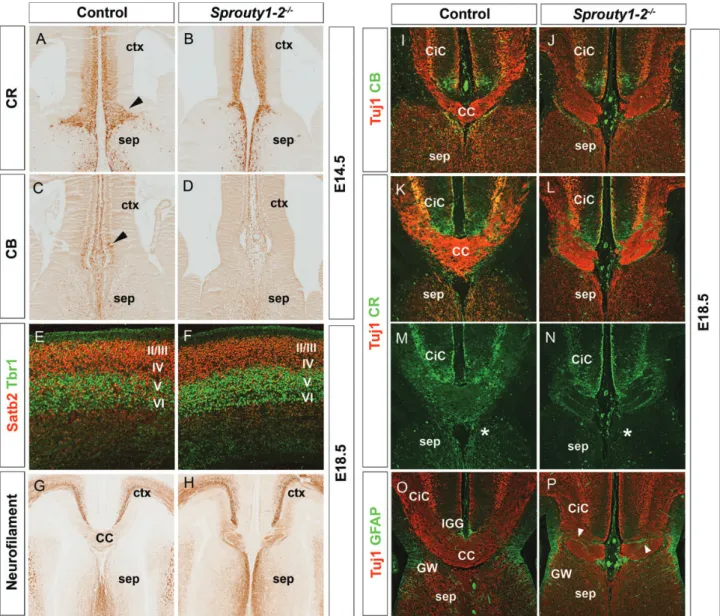
Sprouty1/2 Double Mutants Show Agenesis of the CC To investigate the importance of these changes in Fgf and Wnt/β-catenin signaling for callosal development, we made use of Sprouty1/2 double mutants. Sprouty1 and Sprouty2 encode negative feedback regulators of Fgf signaling (Kim and Bar-Sagi 2004). In the E12.5 rostromedial telencephalon of Sprouty1/2 double mutants, Fgf signaling is up-regulated which in turn leads to a down-regulation of Wnt/β-catenin sig-naling (Faedo et al. 2010) similar to the situation in Pdn mutants. Wefirst determined the effects of these alterations to the signaling pathways on the development of guidepost neurons. At E14.5, prior to the arrival of callosal axons, the CR+guidepost neurons accumulate at the CSB forming a well organized band of neurons which, however, is largely missing in Sprouty1/2 double mutants (Fig. 4A,B). The mutants also lack CB+neurons that can already be detected in the midline region of control embryos (Fig.4C,D), suggesting that the development of guidepost neurons is disturbed in these mutants before callosal axons approach the CSB. Next, we analyzed CC formation in E18.5 embryos. While the for-mation of Satb2+callosal projection neurons and their posi-tioning in the upper cortical layers is not affected (Fig.4E,F). Neurofilament and Tuj1 staining revealed agenesis of the CC in Sprouty1/2 mutants (Fig.4G–L,O,P). Callosal fibers project toward the midline, but fail to cross and form ectopic axon bundles. The analysis of the midline guideposts showed no dramatic differences in the distribution of CB+neurons, but CR+ neurons formed abnormal fibers in the ectopic axon bundles (Fig. 4I–N). Several GFAP+ glia fibers abnormally cluster at the CSB, transecting the path of callosal axons, while the IGG could not be identified (Fig.4O,P). Taken to-gether, these data show that up-regulation of Fgf signaling is sufficient to induce callosal malformation.
Fgf Signaling is Reduced in the E16.5 Pdn Cingulate Cortex
A recent analysis had shown that Fgf signaling is required between E15.5 and E17.5 for the translocation of glial cells toward the indusium griseum (Smith et al. 2006). Since inter-fering with Fgf signaling at this stage leads to glial transloca-tion defects very similar to those in E18.5 Pdn mutants, we investigated Fgf8 expression and that of its target gene sprouty2 in E16.5 Pdn embryos. In the rostral cortex of control embryos, both genes are expressed in the IGG and in the GW and sprouty2 expression expands into the cingulate cortex (Supplementary Fig. 6A,B). At more caudal levels, Fgf8 and sprouty2 transcripts were detected in the septum and in the stria medullaris thalami (Supplementary Fig. 6C,D). In contrast, Fgf8 expression is absent from the IG region and from the GW of Pdn embryos and is confined to the caudal septum (Supplementary Fig. 6E,G). At this caudal level, septum and cingulate cortex are only connected by a thin bridge of tissue. This abnormal morphology and the absence of Fgf8 expression in the GW and IG region suggests that Fgf8 might not signal to the cingulate cortex. Consistent with this idea, sprouty2 is only expressed in the septum but not in the cingulate cortex of Pdn mutants (Supplementary Fig. 6F, H). Taken together with the results of our E12.5 analysis, these data strongly suggest an early phase when Fgf signaling is up-regulated in the E12.5 rostromedial Pdn telencephalon causing patterning defects and a clustering of RGCs followed
by a later phase with a down-regulation of Fgf signaling in the E16.5 cingulate cortex due to an abnormal morphology of the Pdn rostral midline tissue. This down-regulation coincides with the glial translocation defect in Pdn mutants.
Positioning of Midline Guidance Cues is Rescued in Pdn/Pdn;Slit2−/−Embryos
Thefindings described above indicate that altered Fgf signal-ing plays an important part in the development of the Pdn callosal phenotype. However, the callosal phenotype of sprouty1/2 embryos appears relatively mild compared with that of Pdn mutants, suggesting additional abnormalities in Gli3 mutants. We therefore started to analyze the expression of axon guidance molecules in Pdn mutants. In E16.5 control embryos, Sema3c is expressed in glutamatergic guidepost and cingulate neurons thereby attracting callosal axons toward the midline (Niquille et al. 2009;Piper, Plachez et al. 2009), but its expression is only slightly reduced in Pdn mutants (Sup-plementary Fig. 7). Slit2 normally prevents callosal axons from projecting into the septum (Bagri et al. 2002) and is already expressed in the commissural plate of E9.5 embryos (Yuan et al. 1999) and in the septum of E12.5 control embryos (Fig. 5A). Interestingly, our in situ hybridization showed a slight expansion of Slit2 expression into the cortical region of E12.5 Pdn embryos (Fig. 5E). This expansion became more prominent by E14.5 when strong Slit2 expression is confined to the septum of control embryos with a graded but weaker expression in cortical midline progeni-tors. In contrast, Slit2 expression is up-regulated in the rostro-medial Pdn cortex and Slit2 transcripts were ectopically detected in the septal midline (Fig.5B,C,F,G). To confirm this potential increase in Slit2 expression, we used qRT-PCR on rostromedial telencephic tissue to show a significant increase in Slit2 mRNA expression levels (Fig.5I). Moreover, expanded Slit2 expression is maintained in the E16.5 cingulate cortex (Fig. 5D,H). Thus, Pdn mutants show an expansion of Slit2 expression in the rostromedial cortex from patterning stages until time points when callosal axons approach the CSB.
To test for a role of this expanded Slit2 expression, we ana-lyzed CC development in Pdn/Slit2 double mutants. Initially, we determined the positioning of guidepost cells in E16.5 embryos. This analysis showed that the organization of the cortical midline is much improved in Pdn/Pdn;Slit2+/−and in Pdn/Pdn;Slit2−/− embryos. The positioning of the CB+ and CR+ guidepost neurons is largely rescued (Supplementary Fig. 8B–D,G–I). The numbers of CB+neurons are increased in double mutants, though not to wild-type levels, while CR+ neurons are present in normal numbers in the double mutants (Supplementary Fig. 8P,Q). The formation of GFAP+ GW cells is restricted to the CSB, although the GFAP staining appears more irregular with a few isolated GFAP+ fascicles (Supplementary Fig. 8L–N). Moreover, in contrast to Pdn/Pdn embryos, L1+ callosal axons progress through the cingulate cortex without disruption in double-mutant embryos (Sup-plementary Fig. 8B–D;G–I;L–N). We also analyzed the posi-tioning of guidepost cells in Slit2−/−embryos (Supplementary Fig. 8E,J,O). While the CB+and many CR+guidepost neurons acquire their correct position in the prospective IG region of Slit2−/−mutants, some CB+and CR+neurons intermingle ec-topically with callosal axons in the septum, where callosal axons are misdirected. In addition, there is a dramatic
increase in the number of callosal axons reaching the midline region in Slit2−/−embryos as reported previously (Bagri et al. 2002).
Finally, we analyzed CC formation in E18.5 Pdn/Slit2 double mutants. This analysis confirmed our findings on the much improved organization of midline guidepost neurons, but callosal axons do not cross the midline (Fig.6C,D,H,I,M, N). In the Pdn cingulate cortex, the intermediate zone is dis-rupted by several, large Probst bundles (Fig.6B,G,L). In Pdn/ Pdn;Slit2+/−and in Pdn/Pdn;Slit2−/−embryos, callosal axons migrate uninterrupted through the cingulate cortex without forming Probst bundles (Fig.6C,D,H,I,M,N). CB+neurons are located in the IG region similar to control embryos, but are scattered in the Pdn cortex (Fig.6A–D). CR+neurons occupy positions in the dorsomedial cortex of the double mutants, while these cells are mostly associated with the Probst
bundles in Pdn mutants (Fig. 6F–I). In addition to their correct position, normal numbers of CB+and CR+neurons are present in the midline region of double mutants (Fig.6P,Q). In contrast, the midline glia develops abnormally in Pdn/Slit2 double mutants (Fig. 6K–N). The IGG is missing and ectopic glial fascicles are still formed at the CSB but only in the ven-tralmost part of the cortex (Fig. 6M,N). Interestingly, the guidepost neurons are also severely affected in Slit2−/− mutants. Few CR+ neurons occupy their normal position in the IG, while large clusters of CR+neurons were detected ven-trally to the callosal axons crossing the midline (Fig. 6E,J,O). In addition, 2 large ectopic bundles offibers were also found at either side of the CC as described previously (Bagri et al. 2002). Taken together, these analyses show a remarkable recovery of midline morphology in Pdn/Slit2 double mutants.
Figure 4. Sprouty1–2 double mutants lack the CC. (A and B) In E14.5 control embryos, CR+guidepost neurons form a well organized band of neurons at the CSB, which is largely missing in Sprouty1/2 double mutants. (C and D) Unlike control embryos, CB+guidepost neurons were not detected at the CSB of Sprouty1/2 double mutants at E14.5.
(E and F) At E18.5, Satb2+callosal neurons are normally positioned in the upper cortical layers II/III and IV above the Tbr1+ neurons in layer V and VI. (G–L, O, and P) Neurofilament and Tuj1 staining reveal agenesis of the CC in E18.5 sprouty1-2 mutants. Callosal fibers fail to cross the midline and form ectopic axon bundles. (I–J) No obvious differences in the distribution of CB+guidepost neurons are detected in the dorsomedial cortex of Sprouty1-2 double mutants. (K–N) CR+guidepost neurons form abnormal fiber bundles within the Probst bundles (O and P) GFAP immunofluorescence reveals abnormally formed midline glia populations. Several GFAP+gliafibers abnormally cluster at
Up-Regulation of Fgf Signaling Controls Slit2 Expression and is Required for RGC Clustering in Pdn Mutants Taken together, our analyses demonstrate roles for Fgf signal-ing and Slit2 in positionsignal-ing callosal guidance cues raissignal-ing the possibility that both pathways are interconnected. To test for this, we employed an ex vivo explant assay in which we pre-pared coronal sections of E13.5 control and Pdn/Pdn rostral telencephalon, including the commissural plate as the Fgf8 signaling centre, and maintained these sections in culture for 48 h in the presence of DMSO or various concentrations of SU5402, which selectively inhibits Fgf signaling. Wefirst de-termined the effects of these treatments on the expression of sprouty2. Under control conditions, sprouty2 expression is de-tected in the septum on sections of control and Pdn/Pdn embryos (Fig.7A,B). While the addition of 100μM SU5402 se-verely disrupted tissue morphology (data not shown), sprouty2 expression was abolished in the presence of 25μM SU5402 (Fig. 7C), indicating that this concentration is suf fi-cient to block Fgf signaling in this ex vivo explant culture assay. Next, we analyzed the expression of Slit2 after SU5402 treatment. In the presence of DMSO, Slit2 transcripts are con-fined to the septum of control embryos (Fig. 7D), but Slit2 expression expands into the cortex and into the ventral-most septum on Pdn/Pdn sections (Fig. 7E). SU5402 treatment of Pdn mutant sections resulted in a loss of Slit2 expression in this latter tissue and in reduced expression in the cortex (Fig. 7F), suggesting that up-regulated Fgf signaling in Pdn mutants plays at least a partial role in controlling Slit2 expression. Finally, we used the same assay to determine a role for Fgf signaling in the formation of the ectopic RGC clusters. Immunofluorescence for the Blbp antigen which is encoded by Fabp7 revealed RGCs in the cortex dorsally to the CSB on control sections and widespread RGC clusters on Pdn mutant sections (Fig. 7G,H) similar to our in vivo findings (compare with Fig. 3N,R). In contrast, addition of 25μM SU5402 nearly completely abolished the formation of RGC
clusters on Pdn/Pdn sections (Fig. 7I) strongly suggesting that their formation depends on up-regulated Fgf signaling.
Discussion
Several glial and neuronal guidepost cells are organized in strategic positions at the CSB and play crucial roles in the midline crossing of callosal axons, but it remains largely unknown how the guideposts acquire their correct position. The Gli3 hypomorphic mutant Pdn provides an interesting model to address this as the normal distribution of callosal guideposts is severely affected in this mutant. The cortical midline region contains ectopic glialfibers that transect the path of callosal axons and shows an up-regulation of the Slit2 guidance molecule. Several lines of evidence strongly suggest that the ACC in Pdn mutants is caused by these midline defects rather than by defects in callosal axons. Cortical layer-ing, the expression of the callosal determinant Satb2 (Alcamo et al. 2008;Britanova et al. 2008) and the birthdate of upper layer callosal neurons are not affected in Pdn embryos (Magnani et al. 2010). Moreover, Pdn mutant callosal axons are capable of midline crossing in a wild-type environment. Finally, molecular changes in the cortical midline relevant to the callosal malformation occur as early as E12.5. As these al-terations occur well before callosal axons arrive at the midline, our findings strongly suggest that Gli3-controlled early patterning events are crucial for setting up the spatial organization of midline guideposts and hence for callosal development.
Pdn mutants showing a very severe callosal phenotype present an interesting tool to identify pathways controlling patterning of the CSB. In fact, our analyses led to the identi fi-cation of altered activities in key signaling pathways and of changed expression patterns of several transcription factors emphasizing this link between patterning and callosal devel-opment. First, several transcription factors with important Figure 5. Slit2 expression expands into the rostromedial cortex of Pdn/Pdn embryos. (A and E) In E12.5 control embryos, Slit2 expression is confined to the septum, but expands into cortical regions of Pdn mutants (arrowhead in E). Note the decreased size of the LGE in Pdn mutants as described previously (Magnani et al. 2010). (B–D) In E14.5 and E16.5 control embryos, Slit2 transcripts are detected in the septum and in the prospective cingulate cortex in a graded fashion. (F–H) In Pdn/Pdn embryos, Slit2 expression is increased in the future cingulate cortex (arrows). There is also ectopic Slit2 expression in the midlineof the septum, which is enlarged in E14.5 Pdn/Pdn embryos (arrowheads in F and G). (I) Quantification of Slit2 expression levels in the rostromedial telencephalon of E14.5 wild-type and control embryos. Asterisk (*) denotes statistically significant changes with P≤ 0.05 (Mann–Whitney test).
functions in early forebrain and callosal development have altered expression patterns in the corticoseptal region of E12.5 Pdn embryos. Mutations of the human and mouse SIX3 genes lead to holoprosencephaly (Wallis et al. 1999) and to severe truncations of the prosencephalon (Lagutin et al. 2003), respectively, but the severity of these phenotypes might obscure potential role(s) in callosal formation. In con-trast, Emx1 mutants show ACC due to a lack of the indusium griseum (Qiu et al. 1996;Yoshida et al. 1997). Furthermore, Nfia, Nfib, and Nfix have high expression level domains dor-sally to the CSB (Shu et al. 2003; Campbell et al. 2008;
Plachez et al. 2008) overlapping with the domains of Wnt7b/ 8b expression, suggesting regulatory relationships between these genes. Mutations in Nfia and Nfib lead to callosal defects due to malformations in the midline glial cell popu-lations and to defective development of the cingulate pioneer neurons (Shu et al. 2003; Steele-Perkins et al. 2005; Piper, Moldrich et al. 2009). Our data suggest that these factors have an earlier patterning role that might be obscured by redun-dancy between these factors.
Secondly, we identified altered Fgf signaling and Wnt/ β-catenin signaling at the CSB in E12.5 Pdn mutants as Figure 6. CC development in E18.5 Pdn/Slit2 double mutants. (A, F, and K) Immunostaining on control brain sections revealing L1+callosal axons, CR+and CB+guidepost
neurons and GFAP+midline glia cells. (B, G, and L) In the Pdn cingulate cortex, the path of L1+ axons is interrupted at several positions (arrowheads), large L1+Probst bundles are formed and midline glia and neuronal populations are disorganized. (C, D, H, I, M, and N) In Pdn/Pdn;Slit2+/−and in Pdn/Pdn;Slit2−/−embryos, callosal axons reach the CSB without forming Probst bundles, but do not cross the midline. Also, organization and positioning of midline guideposts is partially rescued in the Pdn/Slit2 double mutants. (C and D) In Pdn/Pdn;Slit2+/−and in Pdn/Pdn;Slit2−/−embryos, CB+neurons are normally located in the IG region similar to control embryos. (H and I) Pdn/Pdn;Slit2+/−and in Pdn/
Pdn;Slit2−/−CR+, sling neurons are normally localized in the cingulate cortex. (M and N) In Pdn/Slit2 double mutants, the IGG is absent and ectopic glial fascicles are formed at the CSB. (E, J, and O) Formation of guidepost neurons in Slit2−/−mutants. CR+neurons form large ectopic clusters adjacent to large Probst bundles (arrowhead), but are largely
missing from their normal position in the IG (J). Note the presence of glialfibers intermingling with the callosal axons crossing the midline (O). (P and Q) Quantification of CB+ (P) and CR+(Q) neurons in the IG region. *P < 0.05, **P < 0.01, and ***P < 0.001 (Bonferroni
important regulators of callosal development. In fact, Sprouty1/2 double mutants, in which increased Fgf signaling down-regulates Wnt/β-catenin signaling in the rostromedial telencephalon (Faedo et al. 2010), display agenesis of the CC. Interestingly, these mutants already show defective develop-ment of CR+ and CB+guidepost neurons at the E14.5 CSB. Although we cannot exclude the possibility that elevated Fgf signaling after E14.5 might further disrupt callosal formation, this altered development of guidepost neurons prior to the arrival of callosal axons strongly suggest that increased levels of Fgf signaling at patterning stages already interfere with guidepost and hence callosal development. This idea is sup-ported by recent findings on callosal development in Rfx3 mutant mice in which a mild up-regulation of Fgf signaling underlies a mislocalization of glutaminergic guidepost neurons (Benadiba et al. 2012). Consistent with recent find-ings on a regulatory role of Fgf signaling in RGC development (Kang et al. 2009;Sahara and O’Leary 2009), we also show here that up-regulating Fgf signaling is required for the for-mation of RGC clusters in the rostromedial telencephalon of Pdn mutants. Our Blbp/GFAP double staining further indi-cates that these RGC clusters give rise to the ectopic glial cells in the E16.5 Pdn cingulate cortex, which due to morphologi-cal alterations lacks Fgf signaling at this state. This lack is likely to result in a failure of ectopic glial cells to translocate (Smith et al. 2006). Taken together, thesefindings indicate 2 phases for Fgf signaling in callosal development. During a newly identified, early patterning phase Fgf signaling sets the CSB and positions glial and neuronal guidepost cells. In a
second phase, Fgf signaling is required for glial cell transloca-tion (Smith et al. 2006). These data also demonstrate that a reduction and an increase in Fgf signaling can cause ACC, strongly suggesting that regulating Fgf8 expression levels is crucial for callosal development. This regulation might involve a positive feedback loop with Shh (Ohkubo et al. 2002) and/or an interaction with Wnt7b and Wnt8b which have complementary expression patterns to Fgf8 at the CSB. Previous analyses have implicated Wnt5a and Ryk-mediated Wnt/Ca2+signaling in promoting the escape of callosal axons from the midline into the contralateral hemisphere (Keeble et al. 2006;Hutchins et al. 2011). Moreover, the meninges and neurons of the cingulate cortex use a cascade of signals in-cluding Wnt3 to regulate midline crossing of cingulate pioneer axons (Choe et al. 2012). In contrast, Wnt8b mutant mice show normal callosal development probably due to re-dundancy with other Wnt molecules (Fotaki et al. 2010). However, Wnt7b/8b expression is already down-regulated before the onset of ectopic Fgf8 expression in the E9.0 Pdn telencephalon (Ueta et al. 2008). This and the reduced Wnt/ β-catenin signaling in the sprouty1/2 double mutants (Faedo et al. 2010) suggest an antagonistic interaction between Fgf and Wnt/β-catenin signaling to control Fgf8 expression levels in the commissural plate, thereby regulating patterning of the CSB and positioning of midline guideposts (Fig.8).
Finally, the up-regulation of Slit2 expression represents a major cause of the Pdn callosal phenotype. Pdn/Slit2 double mutants show a dramatic improvement in the growth of corti-cal axons toward the midline and in midline organization Figure 7. Effects of blocking Fgf signaling on midline development. (A–C) Sprouty2 expression is detected in the septum of DMSO treated control and Pdn/Pdn sections (arrows in A and B) but completely abolished after treatment with 25μM SU5402. (D–E) Under control conditions, Slit2 expression is confined to the septum (arrows in D), but expands into the cortex (arrowheads in E) and into the ventralmost septum (asterisks in E) of Pdn/Pdn mutant sections. (F) Treatment of Pdn/Pdn sections with 25μM SU5402 resulted in reduced Slit2 expression in the cortex (arrowhead) and to a loss of expression in the ventralmost septum. (G–I) Up-regulation of Fgf signaling is required for RGC cluster formation. (G) Blbp marks RGCs in the cortex dorsal to the CSB (arrow). (H and I) In Pdn/Pdn, sections treated with DMSO, Blbp+ cells form widespread cluster (asterisks in H), while their formation is nearly completely abolished after treatment with 25μM SU5402 (I).
suggesting 2, mutually non-exclusive roles for Slit2 in callosal development. First, Slit2 could control the permissiveness of the cingulate cortex for the growth of callosal axons. Indeed, callosal axons approach the CSB without forming Probst bundles in Pdn/Slit2 double mutants, and many callosal axons approach the midline but miss-project into the septum in Slit2−/−mutants (Bagri et al. 2002). This idea is also con-sistent with the temporal expression profile of Slit2, which becomes down-regulated in the control cingulate cortex after E14.5 (Fig.5). Alternatively, Slit2 could regulate the migration of guidepost neurons into the cortical midline (Niquille et al., 2009) similar to its effect on the migration of LGE guidepost cells (Bielle et al. 2011). The positioning and the numbers of guidepost neurons are largely rescued in the Pdn/Slit2 double mutants, while CR+ neurons form ectopic clusters in Slit2−/−embryos. Taken together, thesefindings raise the in-teresting possibility that a major role of Slit2 in callosal devel-opment is to coordinate the migration of callosal axons with that of the guidepost neurons.
Interestingly, Slit2 expression is already expanded in E12.5 Pdn embryos suggesting that early patterning regulates its expression. Slit2 could be a downstream target of Fgf signaling given its coexpression with sprouty2 (Yuan et al. 1999) and its down-regulation in the septum of Fgfr1 mutant mice (Tole et al. 2006) and after blocking Fgf signaling on rostromedial tissue sections (Fig. 7). Alternatively, Gli3 or transcription factors downstream of Gli3, such as Emx1 or the Nfi transcrip-tional regulators, could repress Slit2 expression in the
rostrodorsal telencephalon. Irrespective of the exact mechan-ism, the up-regulation of Slit2 provides a link between early patterning and the coordination of midline development.
In summary, our analyses provide insights into how early patterning of the cortical midline controls the organization of midline guideposts and the formation of a permissive environment allowing callosal axons to approach the CSB. In this process, Gli3 takes centre stage by controlling Fgf and Wnt/β-catenin signaling at the rostral midline and the expression of several transcription factors and of the Slit2 axon guidance molecule. Interestingly, the human GLI3 gene is mutated in Acrocallosal syndrome patients who lack the CC (Elson et al. 2002). CC malformations are also a frequent hall-mark of ciliopathies in which the function of the primary cilium and hence Gli3 processing is affected (Tobin and Beales 2009). Therefore, our findings provide a framework for understanding the defective processes underlying the ACC in Acrocallosal syndrome and in ciliopathies.
Supplementary Material
Supplementary material can be found at: http://www.cercor. oxfordjournals.org/.
Funding
This work was supported by grants from the Deutsche For-schungsgemeinschaft (TH770/6-1) and from the Medical Figure 8. Schematic representation of the major changes during CSB formation and midline crossing of callosal axons in Pdn/Pdn mutants. (A) During patterning stages, Gli3 restricts the expression of Fgf8 and Slit2 to the commissural plate and is required for Wnt7b/8b expression in the rostromedial cortex of control embryos thereby establishing the CSB and controlling a balance between Fgf and Wnt/β-catenin signaling. Arrows and lines indicate genetic interactions except for the direct regulation of Gli3 expression by Wnt/ β-catenin signaling (Hasenpusch-Theil et al. forthcoming 2012). Reducing Gli3 expression levels in Pdn/Pdn mutants leads to a down-regulation of Wnt7b/8b expression and a concomitant up-regulation of Slit2 expression and Fgf signaling, which in turns results in RGC clustering. (B) Schematic drawing representing the organization of glial cell populations (green) CR+( pink) and CB+(red) guidepost neurons in control embryos to channel callosal axons (blue) across the midline. (C) Disorganization of guideposts results in the formation of Probst bundles (Pb) in Pdn/Pdn embryos. Ectopic glial fascicles span the width of the cortex and provide a barrier to the migration of callosal axons. Callosal axons may migrate in and out of the image plane to circumvent the ectopic glial fascicles as indicated by the dotted lines. Guidepost neurons associate with the Probst bundles.
Research Council (G0801359) (TT) and the Wellcome Trust (091475) (A.B.).
Notes
We thank Drs Thomas Becker, Christopher D. Conway, Bénédicte Durand, John Mason, and Tom Pratt for critical comments on the manuscript. We are grateful to Trudi Gillespie for help with confocal imaging and Gail Martin for the Sprouty2 mouse line. We thank Neil Campbell who worked on this project as part of his Honours re-search. Conflict of Interest: None declared.
References
Alcamo EA, Chirivella L, Dautzenberg M, Dobreva G, Farinas I, Grosschedl R, McConnell SK. 2008. Satb2 regulates callosal projec-tion neuron identity in the developing cerebral cortex. Neuron. 57:364–377.
Aoto K, Nishimura T, Eto K, Motoyama J. 2002. Mouse GLI3 regulates Fgf8 expression and apoptosis in the developing neural tube, face, and limb bud. Dev Biol. 251:320–332.
Bagnard D, Thomasset N, Lohrum M, Puschel AW, Bolz J. 2000. Spatial distributions of guidance molecules regulate chemorepulsion and chemoattraction of growth cones. J Neurosci. 20:1030–1035. Bagri A, Marin O, Plump AS, Mak J, Pleasure SJ, Rubenstein JL,
Tessier-Lavigne M. 2002. Slit proteins prevent midline crossing and determine the dorsoventral position of major axonal pathways in the mammalian forebrain. Neuron. 33:233–248.
Basson MA, Akbulut S, Watson-Johnson J, Simon R, Carroll TJ, Shakya R, Gross I, Martin GR, Lufkin T, McMahon AP et al. 2005. Sprouty1 is a critical regulator of GDNF/RET-mediated kidney induction. Dev Cell. 8:229–239.
Benadiba C, Magnani D, Niquille M, Morle L, Valloton D, Nawabi H, Ait-Lounis A, Otsmane B, Reith W, Theil T et al. 2012. The cilio-genic transcription factor RFX3 regulates early midline distribution of guidepost neurons required for corpus callosum development. PLoS Genet. 8:e1002606.
Bielle F, Marcos-Mondejar P, Keita M, Mailhes C, Verney C, Nguyen Ba-Charvet K, Tessier-Lavigne M, Lopez-Bendito G, Garel S. 2011. Slit2 activity in the migration of guidepost neurons shapes thal-amic projections during development and evolution. Neuron. 69:1085–1098.
Britanova O, de Juan Romero C, Cheung A, Kwan KY, Schwark M, Gyorgy A, Vogel T, Akopov S, Mitkovski M, Agoston D et al. 2008. Satb2 is a postmitotic determinant for upper-layer neuron speci fi-cation in the neocortex. Neuron. 57:378–392.
Campbell CE, Piper M, Plachez C, Yeh YT, Baizer JS, Osinski JM, Litwack ED, Richards LJ, Gronostajski RM. 2008. The transcription factor Nfix is essential for normal brain development. BMC Dev Biol. 8:52.
Choe Y, Siegenthaler JA, Pleasure SJ. 2012. A cascade of morphogenic signaling initiated by the meninges controls corpus callosum for-mation. Neuron. 73:698–712.
Crossley PH, Martin GR. 1995. The mouse Fgf8 gene encodes a family of polypeptides and is expressed in regions that direct outgrowth and patterning in the developing embryo. Development. 121:439–451.
das Neves L, Duchala CS, Tolentino-Silva F, Haxhiu MA, Colmenares C, Macklin WB, Campbell CE, Butz KG, Gronostajski RM. 1999. Disruption of the murine nuclear factor I-A gene (Nfia. results in perinatal lethality, hydrocephalus, and agenesis of the corpus cal-losum. Proc Natl Acad Sci USA. 96:11946–11951.
Elson E, Perveen R, Donnai D, Wall S, Black GC. 2002. De novo GLI3 mutation in acrocallosal syndrome: broadening the phenotypic spectrum of GLI3 defects and overlap with murine models. J Med Genet. 39:804–806.
Erskine L, Williams SE, Brose K, Kidd T, Rachel RA, Goodman CS, Tessier-Lavigne M, Mason CA. 2000. Retinal ganglion cell axon
guidance in the mouse optic chiasm: expression and function of robos and slits. J Neurosci. 20:4975–4982.
Faedo A, Borello U, Rubenstein JL. 2010. Repression of Fgf signaling by sprouty1–2 regulates cortical patterning in two distinct regions and times. J Neurosci. 30:4015–4023.
Fotaki V, Larralde O, Zeng S, McLaughlin D, Nichols J, Price DJ, Theil T, Mason JO. 2010. Loss of Wnt8b has no overt effect on hippo-campus development but leads to altered Wnt gene expression levels in dorsomedial telencephalon. Dev Dyn. 239:284–296. Fotaki V, Yu T, Zaki PA, Mason JO, Price DJ. 2006. Abnormal
posi-tioning of diencephalic cell types in neocortical tissue in the dorsal telencephalon of mice lacking functional Gli3. J Neurosci. 26:9282–9292.
Furuta Y, Piston DW, Hogan BL. 1997. Bone morphogenetic proteins (BMPs) as regulators of dorsal forebrain development. Develop-ment. 124:2203–2212.
Grove EA, Tole S, Limon J, Yip L, Ragsdale CW. 1998. The hem of the embryonic cerebral cortex is defined by the expression of multiple Wnt genes and is compromised in Gli3-deficient mice. Develop-ment. 125:2315–2325.
Hasenpusch-Theil K, Magnani D, Amaniti EM, Han L, Armstrong D, Theil T. Forthcoming 2012. Transcriptional analysis of Gli3 mutants identifies Wnt target genes in the developing hippo-campus. Cereb Cortex. [Epub ahead of print].
Hill RE, Jones PF, Rees AR, Sime CM, Justice MJ, Copeland NG, Jenkins NA, Graham E, Davidson DR. 1989. A new family of mouse homeo box-containing genes: molecular structure, chromo-somal location, and developmental expression of Hox-7.1. Genes Dev. 3:26–37.
Huffman KJ, Garel S, Rubenstein JL. 2004. Fgf8 regulates the develop-ment of intra-neocortical projections. J Neurosci. 24:8917–8923. Hutchins BI, Li L, Kalil K. 2011. Wnt/calcium signaling mediates axon
growth and guidance in the developing corpus callosum. Dev Neurobiol. 71:269–283.
Kang W, Wong LC, Shi SH, Hebert JM. 2009. The transition from radial glial to intermediate progenitor cell is inhibited by FGF sig-naling during corticogenesis. J Neurosci. 29:14571–14580. Keeble TR, Halford MM, Seaman C, Kee N, Macheda M, Anderson RB,
Stacker SA, Cooper HM. 2006. The Wnt receptor Ryk is required for Wnt5a-mediated axon guidance on the contralateral side of the corpus callosum. J Neurosci. 26:5840–5848.
Kim HJ, Bar-Sagi D. 2004. Modulation of signalling by Sprouty: a de-veloping story. Nat Rev Mol Cell Biol. 5:441–450.
Koester SE, O’Leary DD. 1994. Axons of early generated neurons in cingulate cortex pioneer the corpus callosum. J Neurosci. 14:6608–6620.
Kuschel S, Rüther U, Theil T. 2003. A disrupted balance between Bmp/Wnt and Fgf signaling underlies the ventralization of the Gli3 mutant telencephalon. Dev Biol. 260:484–495.
Lagutin OV, Zhu CC, Kobayashi D, Topczewski J, Shimamura K, Puelles L, Russell HR, McKinnon PJ, Solnica-Krezel L, Oliver G. 2003. Six3 repression of Wnt signaling in the anterior neuroecto-derm is essential for vertebrate forebrain development. Genes Dev. 17:368–379.
Lustig B, Jerchow B, Sachs M, Weiler S, Pietsch T, Karsten U, van de Wetering M, Clevers H, Schlag PM, Birchmeier W et al.. 2002. Negative feedback loop of Wnt signaling through upregulation of conductin/Axin2 in colorectal and liver tumors. Mol Cell Biol. 22:1184–1193.
Magnani D, Hasenpusch-Theil K, Jacobs EC, Campagnoni AT, Price DJ, Theil T. 2010. The Gli3 hypomorphic mutation Pdn causes se-lective impairment in the growth, patterning, and axon guidance capability of the lateral ganglionic eminence. J Neurosci. 30:13883–13894.
Minowada G, Jarvis LA, Chi CL, Neubuser A, Sun X, Hacohen N, Krasnow MA, Martin GR. 1999. Vertebrate Sprouty genes are induced by FGF signaling and can cause chondrodysplasia when overexpressed. Development. 126:4465–4475.
Moldrich RX, Gobius I, Pollak T, Zhang J, Ren T, Brown L, Mori S, De Juan Romero C, Britanova O, Tarabykin V et al.. 2010. Molecular
regulation of the developing commissural plate. J Comp Neurol. 518:3645–3661.
Naruse I, Kato K, Asano T, Suzuki F, Kameyama Y. 1990. Develop-mental brain abnormalities accompanied with the retarded pro-duction of S-100 beta protein in genetic polydactyly mice. Brain Res Dev Brain Res. 51:253–258.
Niquille M, Garel S, Mann F, Hornung JP, Otsmane B, Chevalley S, Parras C, Guillemot F, Gaspar P, Yanagawa Y et al.. 2009. Transi-ent neuronal populations are required to guide callosal axons: a role for semaphorin 3C. PLoS Biol. 7:e1000230.
Ohkubo Y, Chiang C, Rubenstein JL. 2002. Coordinate regulation and synergistic actions of BMP4, SHH and FGF8 in the rostral prosen-cephalon regulate morphogenesis of the telencephalic and optic vesicles. Neuroscience. 111:1–17.
Oliver G, Mailhos A, Wehr R, Copeland NG, Jenkins NA, Gruss P. 1995. Six3, a murine homologue of the sine oculis gene, demarcates the most anterior border of the developing neural plate and is ex-pressed during eye development. Development. 121:4045–4055. Parr BA, Shea MJ, Vassileva G, McMahon AP. 1993. Mouse Wnt genes
exhibit discrete domains of expression in the early embryonic CNS and limb buds. Development. 119:247–261.
Paul LK, Brown WS, Adolphs R, Tyszka JM, Richards LJ, Mukherjee P, Sherr EH. 2007. Agenesis of the corpus callosum: genetic, devel-opmental and functional aspects of connectivity. Nat Rev Neurosci. 8:287–299.
Piper M, Moldrich RX, Lindwall C, Little E, Barry G, Mason S, Sunn N, Kurniawan ND, Gronostajski RM, Richards LJ. 2009. Multiple non-cell-autonomous defects underlie neocortical callosal dysgen-esis in Nfib-deficient mice. Neural Dev. 4:43.
Piper M, Plachez C, Zalucki O, Fothergill T, Goudreau G, Erzurumlu R, Gu C, Richards LJ. 2009. Neuropilin 1-Sema signaling regulates crossing of cingulate pioneering axons during development of the corpus callosum. Cereb Cortex. 19(Suppl 1):i11–21.
Plachez C, Lindwall C, Sunn N, Piper M, Moldrich RX, Campbell CE, Osinski JM, Gronostajski RM, Richards LJ. 2008. Nuclear factor I gene expression in the developing forebrain. J Comp Neurol. 508:385–401.
Plump AS, Erskine L, Sabatier C, Brose K, Epstein CJ, Goodman CS, Mason CA, Tessier-Lavigne M. 2002. Slit1 and Slit2 cooperate to prevent premature midline crossing of retinal axons in the mouse visual system. Neuron. 33:219–232.
Pratt T, Sharp L, Nichols J, Price DJ, Mason JO. 2000. Embryonic stem cells and transgenic mice ubiquitously expressing a tau-tagged greenfluorescent protein. Dev Biol. 228:19–28.
Qiu M, Anderson S, Chen S, Meneses JJ, Hevner R, Kuwana E, Peder-sen RA, Rubenstein JL. 1996. Mutation of the Emx-1 homeobox gene disrupts the corpus callosum. Dev Biol. 178:174–178. Quinn JC, Molinek M, Mason JO, Price DJ. 2009. Gli3 is required
autono-mously for dorsal telencephalic cells to adopt appropriate fates during embryonic forebrain development. Dev Biol. 327:204–215. Rash BG, Richards LJ. 2001. A role for cingulate pioneering axons in
the development of the corpus callosum. J Comp Neurol. 434:147–157.
Richards LJ, Plachez C, Ren T. 2004. Mechanisms regulating the devel-opment of the corpus callosum and its agenesis in mouse and human. Clin Genet. 66:276–289.
Richardson M, Redmond D, Watson CJ, Mason JO. 1999. Mouse Wnt8B is expressed in the developing forebrain and maps to chromosome 19. Mamm Genome. 10:923–925.
Sahara S, O’Leary DD. 2009. Fgf10 regulates transition period of corti-cal stem cell differentiation to radial glia controlling generation of neurons and basal progenitors. Neuron. 63:48–62.
Sanchez-Camacho C, Ortega JA, Ocana I, Alcantara S, Bovolenta P. 2011. Appropriate Bmp7 levels are required for the differentiation of midline guidepost cells involved in corpus callosum formation. Dev Neurobiol. 71:337–350.
Shim K, Minowada G, Coling DE, Martin GR. 2005. Sprouty2, a mouse deafness gene, regulates cell fate decisions in the auditory sensory epithelium by antagonizing FGF signaling. Dev Cell. 8:553–564.
Shimogori T, Banuchi V, Ng HY, Strauss JB, Grove EA. 2004. Embryo-nic signaling centers expressing BMP, WNT and FGF proteins in-teract to pattern the cerebral cortex. Development. 131:5639–5647.
Shu T, Butz KG, Plachez C, Gronostajski RM, Richards LJ. 2003. Ab-normal development of forebrain midline glia and commissural projections in Nfia knock-out mice. J Neurosci. 23:203–212. Silver J, Edwards MA, Levitt P. 1993. Immunocytochemical
demon-stration of early appearing astroglial structures that form bound-aries and pathways along axon tracts in the fetal brain. J Comp Neurol. 328:415–436.
Simeone A, Gulisano M, Acampora D, Stornaiuolo A, Rambaldi M, Boncinelli E. 1992. Two vertebrate homeobox genes related to the Drosophila empty spiracles gene are expressed in the embryonic cerebral cortex. EMBO J. 11:2541–2550.
Simrick S, Lickert H, Basson MA. 2011. Sprouty genes are essential for the normal development of epibranchial ganglia in the mouse embryo. Dev Biol. 358:147–155.
Smith KM, Ohkubo Y, Maragnoli ME, Rasin MR, Schwartz ML, Sestan N, Vaccarino FM. 2006. Midline radial glia translocation and corpus callosum formation require FGF signaling. Nat Neurosci. 9:787–797.
Steele-Perkins G, Plachez C, Butz KG, Yang G, Bachurski CJ, Kinsman SL, Litwack ED, Richards LJ, Gronostajski RM. 2005. The transcrip-tion factor gene Nfib is essential for both lung maturation and brain development. Mol Cell Biol. 25:685–698.
Theil T. 2005. Gli3 is required for the specification and differentiation of preplate neurons. Dev Biol. 286:559–571.
Theil T, Alvarez-Bolado G, Walter A, Rüther U. 1999. Gli3 is required for Emx gene expression during dorsal telencephalon develop-ment. Developdevelop-ment. 126:3561–3571.
Tobin JL, Beales PL. 2009. The nonmotile ciliopathies. Genet Med. 11:386–402.
Tole S, Gutin G, Bhatnagar L, Remedios R, Hebert JM. 2006. Develop-ment of midline cell types and commissural axon tracts requires Fgfr1 in the cerebrum. Dev Biol. 289:141–151.
Tole S, Ragsdale CW, Grove EA. 2000. Dorsoventral patterning of the telencephalon is disrupted in the mouse mutant extra-toes(J). Dev Biol. 217:254–265.
Ueta E, Kurome M, Teshima Y, Kodama M, Otsuka Y, Naruse I. 2008. Altered signaling pathway in the dysmorphogenesis of telencepha-lon in the Gli3 depressed mouse embryo, Pdn/Pdn. Congenit Anom (Kyoto). 48:74–80.
Wallis DE, Roessler E, Hehr U, Nanni L, Wiltshire T, Richieri-Costa A, Gillessen-Kaesbach G, Zackai EH, Rommens J, Muenke M. 1999. Mutations in the homeodomain of the human SIX3 gene cause holoprosencephaly. Nat Genet. 22:196–198.
Yoshida M, Suda Y, Matsuo I, Miyamoto N, Takeda N, Kuratani S, Aizawa S. 1997. Emx1 and Emx2 functions in development of dorsal telencephalon. Development. 124:101–111.
Yuan W, Zhou L, Chen JH, Wu JY, Rao Y, Ornitz DM. 1999. The mouse SLIT family: secreted ligands for ROBO expressed in pat-terns that suggest a role in morphogenesis and axon guidance. Dev Biol. 212:290–306.