HAL Id: hal-03081947
https://hal.archives-ouvertes.fr/hal-03081947
Preprint submitted on 18 Dec 2020
HAL is a multi-disciplinary open access
archive for the deposit and dissemination of sci-entific research documents, whether they are pub-lished or not. The documents may come from teaching and research institutions in France or abroad, or from public or private research centers.
L’archive ouverte pluridisciplinaire HAL, est destinée au dépôt et à la diffusion de documents scientifiques de niveau recherche, publiés ou non, émanant des établissements d’enseignement et de recherche français ou étrangers, des laboratoires publics ou privés.
Olga Rosspopoff, Christophe Huret, A. Collier, Miguel Casanova, Peter J.
Rugg-Gunn, J.-F. Ouimette, C. Rougeulle
To cite this version:
Olga Rosspopoff, Christophe Huret, A. Collier, Miguel Casanova, Peter J. Rugg-Gunn, et al.. Mech-anistic diversification of XIST regulatory network in mammals. 2020. �hal-03081947�
Mechanistic diversification of XIST regulatory network in mammals
Olga Rosspopoff1, Christophe Huret1, Amanda J. Collier2,3, Miguel Casanova1, Peter J. Rugg-Gunn2,3, Jean-François Ouimette1* and Claire Rougeulle1*.
5
1 Université de Paris, Epigenetics and Cell Fate, CNRS, F-75013 Paris.
2 Epigenetics Programme, The Babraham Institute, Cambridge CB22 3AT, UK.
3Wellcome Trust – Medical Research Council Cambridge Stem Cell Institute, University of Cambridge, Cambridge CB2 1QR, UK.
*[email protected]; [email protected]
10
SUMMARY
X chromosome inactivation (XCI) is a developmental regulatory process that initiates with remarkable diversity in various mammalian species. Here we addressed the contribution of XCI 15
regulators, most of which are lncRNA genes characterized in the mouse, to this mechanistic diversity. By combining analysis of single-cell RNA-seq data from early human embryogenesis with various functional assays in naïve and primed pluripotent stem cells and in differentiated cells, we demonstrate that JPX is a major regulator of XIST expression in human and in mouse. However, the underlying mechanisms differ radically between species and require Jpx RNA in the mouse and the 20
act of transcription of JPX locus in the human. Moreover, biogenesis of XIST is affected at different regulatory steps between these species. This study illustrates how diversification of LRGs modes of action during evolution provide opportunities for innovations within constrained gene regulatory networks.
25
KEYWORDS
X chromosome inactivation, XIST, JPX, lncRNA, pluripotency, single-cell RNA-seq, human embryogenesis, evolution, gene regulatory networks
Graphical abstract
Xist RNA Ftx Jpx lncRNA Post-transcriptional effect JPX transcription XIST transcription FTX RNAPII recruitment Ht1 Mouse Human RNAPII CTCF mature RNAIntroduction
X chromosome inactivation (XCI) is a fundamental epigenetic process that ensures dosage compensation for X-linked genes expression between male and female mammals. X chromosome silencing is triggered early in development by the accumulation of the long non-coding RNA (lncRNA) XIST, which acts as a scaffold for multiple protein complexes, involved amongst others in 5
chromatin remodeling, nuclear organization and RNA modification (Furlan and Rougeulle, 2016). The concerted action of these ribonucleoprotein factors results in the conversion of one of the two X chromosomes in females into a compact and transcriptionally silent structure. XIST expression has to be tightly controlled in order to ensure female-restricted inactivation of a single X chromosome in a timely manner. However, it remains intriguing that such an essential process follows species-specific 10
routes in which the dynamics of XIST expression in early developmental stages differs markedly between mouse and human. While in the mouse Xist is restricted to females and to a single X, XIST expression in human pre-implantation development transiently initiates in a manner that is independent of the sex and of the number of X-chromosomes. XIST accumulation precedes the establishment of proper XCI in human pre-implantation embryos resulting in active XIST-coated X-15
chromosomes (Okamoto et al., 2011). However, the situation eventually homogenizes and Xist/XIST
RNA coating becomes restricted to a single Xi in both mouse and human post-inactivation (post-XCI)
cells (Vallot et al., 2016). These observations raise questions regarding the functional conservation of
XIST regulatory network across species. We previously identified the transcription factor YY1 as a
potent activator of XIST in mouse and human (Makhlouf et al., 2014), but the activity of the regulatory 20
elements from the genomic region surrounding XIST, known as the X-inactivation center (XIC), has never been addressed in species other than the mouse.
Chromosomal rearrangements in mouse and human have allowed the determination of the physical boundaries of the XIC, defined as necessary and sufficient to trigger XCI. In addition to Xist, other 25
genes were mapped to the mouse Xic, including several protein-coding genes (Slc16a2, Cnbp2, Chic1 and Rnf12) and four additional lncRNA genes (Linx, Tsix, Jpx, and Ftx). While the order and orientation of Xic-linked genes are globally preserved between mouse and human (Chureau et al., 2002; Duret et al., 2006), the human XIC underwent a dramatic expansion and is about three times larger
compared to its mouse counterpart. Particularly relevant are the lncRNA genes hosted within the Xic,
30
which have been linked to Xist regulation in the mouse: Tsix and Linx acts as major repressor of Xist expression while Jpx and Ftx acts as positive regulators. The mechanistic dissection of these Xic-linked and other lncRNA loci highlighted that their molecular function is not only mediated by the RNA molecule itself, but may also involve various entities such the act of transcription or key regulatory elements embedded within their locus (Cho et al., 2018; Engreitz et al., 2016; Furlan et 35
al., 2018; Paralkar et al., 2016). For instance, the antisense transcription of the Tsix gene over the Xist locus contributes to the monoallelic repression of Xist (Navarro et al., 2005); Tsix transcription is itself controlled by the upstream lncRNA gene, Linx (Giorgetti et al., 2014; Nora et al., 2012). Ftx transcription has been shown to be essential for Xist expression cis (Furlan et al., 2018) while Jpx was proposed to act through its RNA molecule by binding and titrating away the CTCF protein from 5
the Xist promoter (Sun et al., 2013). Therefore, it appears more appropriate to define loci producing lncRNAs as lncRNA genes (LRGs) to better emphasize their mechanistic versatility. The antagonistic action of the Xic-linked LRGs is likely facilitated by the spatial segregation of the Xist- and Tsix-associated regulators in two adjacent and oppositely regulated topologically Tsix-associated domains (TADs) (Nora et al., 2012). These TADs also delimit internal long-range interactions to ensure 10
contacts between regulatory elements and their target genes; intra-TAD interactions have been described between the Xist and Ftx LRGs and between Tsix promoter and Linx LRG (Furlan et al., 2018; Nora et al., 2012).
The molecular players controlling XCI have been largely characterized in the mouse, in part due to 15
the lack of cellular models recapitulating the early stage of XIST activation in human. While the differentiation of female mouse embryonic stem cells (ESCs) recapitulates Xist upregulation and XCI, no human ex-vivo model faithfully reproduces the biallelic upregulation of XIST observed during in
vivo human embryogenesis. Conventional hESCs displays the hallmarks of primed pluripotency
including an inactive X-chromosome coated by XIST (Vallot et al., 2016). In addition, loss of XIST 20
expression may occur spontaneously upon prolonged culture of primed hESCs through a process identified as “XCI erosion”, that involves the ectopic reactivation of a subset of genes from the Xi (Mekhoubad et al., 2012; Vallot et al., 2015). Nevertheless, the post-XCI context in primed hESCs differ from that of differentiated cells and their use as an XCI model has been proven effective in the identification human-specific XCI regulators such as the lncRNA XACT (Vallot et al., 2013). It is 25
only recently that methods were developed to reset primed hESCs into the naïve state of pluripotency that matches several features of human pre-implantation embryos, including active X-chromosomes coated by XIST (Guo et al., 2017; Sahakyan et al., 2017; Theunissen et al., 2016; Vallot et al., 2016). Indeed, resetting of eroded primed hESCs triggers XIST upregulation concomitantly to X-chromosome reactivation (XCR), although XIST upregulation is often mono-allelic and restricted to 30
the former Xi (Sahakyan et al., 2017; Vallot et al., 2016). While the resetting of these cells is currently the only method to trigger XIST upregulation in human, it has never been used to functionally characterize regulators of XIST induction. Altogether, experimental systems are now available to probe the XIST regulatory network in human.
From an evolutionary standpoint, one major challenge is that both LRG functionality, if any, and their mechanism of action are hardly predictable based on the DNA sequence alone. The sequence conservation pattern of LRG evolving under functional constraints is therefore difficult to predict. For instance, it is known that syntenic LRG often display strong primary sequence turnover during evolution, even among closely related species (Hezroni et al., 2015; Necsulea et al., 2014; Ulitsky et 5
al., 2011; Washietl et al., 2014), whose impact on LRGs functional conservation is still poorly understood. In rare studies where the functional conservation of lncRNA molecules has been addressed, orthologues display short patches of conserved sequence that are necessary, but not sufficient, for their function (Lin et al., 2014; Ulitsky et al., 2011). While this sharp contrast with the evolutionary stability of protein-coding genes raised controversies on LRGs functionality, 10
experimental investigations have been too limited to provide a definitive understanding of the rules underlying LRGs functional conservation. X-chromosome inactivation provides an interesting experimental paradigm to test this since LRG orthologs are found in the human XIC (Romito and Rougeulle, 2011).
15
In this study, we investigated the regulatory network involved in the initial steps of XIST expression in human during early embryogenesis. The analysis of single-cell RNA-seq data from early human embryos designated JPX as a potent candidate for XIST regulation in human. Using a panel of functional approaches to target various modules of JPX LRG, we could show that, while human JPX transcripts are dispensable for this process, transcription of the JPX locus is essential to sustain XIST 20
transcription in post-XCI cells. This process is fostered within a sub-TAD domain that involves RNA polymerase II-mediated 3D interactions. By resetting primed hESCs carrying various deletions of
JPX promoter region, we demonstrate that this system is suitable to investigate regulators of XIST
upregulation in human and identify the JPX LRG as a major cis-regulator of XIST transcriptional activation. We also re-addressed the role of Jpx RNA, matching cellular models and functional 25
approaches between human and mouse. We could identify that Jpx RNA acts as a positive regulator of Xist in mouse post-XCI cells and our findings suggest that Jpx regulates Xist accumulation in a post-transcriptional manner. In addition to identifying of a novel regulator of XIST expression in human, these findings provide a striking demonstration of the mechanistic diversification of orthologous LRGs, which sheds new light on the importance of these noncoding elements in defining 30
species-specific regulatory mechanisms within constrained gene regulatory networks.
RESULTS
Identification of candidate regulators of XIST during early human embryonic development. To examine whether human XIC-linked genes are involved in the initial upregulation of XIST in vivo, we investigated their expression kinetics during early embryogenesis (Figure 1A), using single-cell RNA-seq datasets obtained from human pre-implantation embryos (Petropoulos et al., 2016; Yan et 5
al., 2013). XIST expression initiates between the four- and eight-cell stages (Figure S1A), corresponding to embryonic day 4 (E4, Figure 1B), and increases hereafter, more predominantly in females than in males. While most of XIC-linked genes remained lowly expressed throughout pre-implantation development, RLIM and JPX show the highest levels at the early embryonic days, although they display different expression trajectories (Figure 1C). The expression of the protein-10
coding gene RLIM is the highest at E3 in both male and female embryos and rapidly decrease in the following days, prior to XIST induction (Figure S1B). This pattern likely reflects strong maternal inheritance of RLIM transcripts, which is consistent with previous observations made in the mouse (Shin et al., 2010). In contrast, low levels of JPX could be detected at the 2-4 cells stage, followed by a major burst of expression at the 8-cell stage (Figure S1A) or E4, coinciding with XIST initial 15
induction (Figure 1D). Except at the E3 stage, JPX was broadly expressed, independently from the sex of the embryos, although JPX levels were almost twice in females compared to male embryos, suggesting an early transcription from the two active X-chromosomes (Figure 1D). Using an RPKM threshold to define XIST and JPX expressing cells in female embryos (Figure 1E), we found that the majority of the cells were expressing either JPX or JPX and XIST concomitantly, with very few XIST-20
only expressing cells at the early embryonic days (E3 and E4, Figure 1F). This pattern suggests that
JPX activation may shortly precede XIST induction and that the two genes become eventually
co-expressed in a vast proportion of cells as development progresses. JPX and XIST expression levels were weakly correlated at early stages of embryogenesis (Figure 1G) and within embryonic lineages (Figure 1H), indicating that JPX transcriptional activation, but not the level of its RNA products, may 25
be a prerequisite for its function. These results point toward the JPX LRG as a candidate for the regulation of the initial induction of XIST expression during pre-implantation development.
JPX RNA is dispensable for XIST expression in human
The JPX LRG derived from the pseudogenization of the protein-coding gene USPL after the 30
divergence of eutherians and marsupials, and evolved concomitantly to XIST (Elisaphenko et al., 2008; Hezroni et al., 2017), although the two genes display distinct evolutionary trajectories (Figure 2A). While XIST present strong signs of positive selection in both intronic and exonic regions, the
JPX LRG evolved through a quasi-neutral selection, as illustrated by a conservation score close to
zero along the entire locus (Figure 2B). This strong sequence turnover is essentially due to species-35
Figure 1. Identification of candidate regulators of XIST during early human embryonic development (A) Single cell RNA-seq data from E3 to E7 pre-implantation embryos (Petropoulos et al., 2016) were used to probe for XIST regulators. Also shown is the timing of XIST induction along with observed XCI
dynamics.
(B) XIST expression is upregulated in male and female embryos from E4. RPKM: Reads Per Kilobase Million
(C) Analysis of single cell expression of XIC-linked genes from E3 to E6 reveals that JPX induction precedes that of XIST. (See also Figures S1A and S1B).
(D) JPX is expressed with comparable kinetics in male and female embryos.
(E) The plots represent the distribution of the log2 RPKM values for JPX and XIST, in female cells from E3 to E6 stages. Red dashed line represents the cutoff used to define JPX and XIST expressing cells, on which panel (F) is based.
(F) Combined analysis of JPX and XIST expression in single cells showed that the proportion of cells expressing JPX alone decreased during development, while the percentage of cells co-expressing the two genes increased (Chi-square test). n.s., not significant, ***p<0.001; ****p<0.0001.
(G-H) JPX and XIST expression levels were weakly correlated in early stages of embryogenesis and in the different lineages. Figure 1 RPKM 0 20 40 60 E3 E4 E5 E6 CHIC1 TSIX XIST JPX FTX ZCCHC13 SLC16A2 RLIM C 0 30 60 90 120 Female Male E3 E4 E5 E6 E7 XIST A
Frequency (E3 to E6)
E log2 (RPKM JPX) log2 RPKM=1 log2 RPKM=-2 0.0 0.1 0.2 0.3 0.4 -10 -5 0 5 0.00 0.05 0.10 0.15 -10 -5 0 5 % of cells 0 25 50 75 100 E3 E4 E5 E6 Both expressed None expressed Only JPX Only XIST (30) (89) (166) (230) **** **** n.s. **** D log2 (RPKM XIST) RPKM 0 20 40 60 JPX Female Male E3 E4 E5 E6 E7 RPKM G log2 (RPKM XIST) E3 (30) E4 (89) E5 (166) -10 -5 0 5 -10 -5 0 5 log2 (RPKM JPX )
Early female cells
R =0.30 p-val. =3.3 E-7 H EPI (35) ICM (31) PE (16) TE (520) -10 -5 0 5 log2 (RPKM XIST) -10 -5 0 5 log2 (RPKM JPX ) R=0.079 p-val.=0.0511 B F Embryonic lineages E6 E4 E3 E5 E7 XIST induction XCI scRNA-seq pre-implantation embryos XIST regulators ?
Frequency (E3 to E6)
CHIC1 TSIX XIST JPX FTX ZCCHC13 SLC16A2 RLIM
Figure S1: Identification of candidate regulators of XIST during early human embryonic development, related to Figure 1
(A) JPX reads can be detected from the 2-4 cell stages, possibly linked to limited maternal contribution (Yan et al., 2013), but a burst of JPX expression could be seen from the 8-cell stage. At later stages, JPX expression can be detected in the three compartments of the embryos – epiblast (EPI), primitive endoderm (PE) and trophectoderm (TE).
(B) Expression of the protein-coding gene RLIM is the highest at E3 in both male and female embryos and decreases afterwards, suggesting maternal contribution.
Figure supplementary 1 TSIX XIST JPX FTX Oocyte 700 Zygote 2-cell 4-cell 8-cell Morulae ChrX 73,100,000 73,200,000 73,300,000 Refseq genes EPI PE TE 0 A B 0 30 60 90 E3 E4 E5 E6 E7 Female Male RPKM RLIM
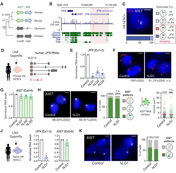
specific integration of transposable elements in this region (Chureau et al., 2002; Kolesnikov, 2010), resulting in poor multiple alignment of the homologous region of five eutherian species (Figure 2B). As observed for numerous LRGs (Hezroni et al., 2017; Hezroni et al., 2015; Washietl et al., 2014), signs of purifying selection on the JPX gene are concentrated toward the promoter region, including the first exon that contains two highly conserved region of ~20 nucleotides embedded within the 5
mouse and human transcripts. As the human and mouse genes bear limited sequence identity (Chureau et al., 2002), we examined several features of JPX in human, such as its expression pattern and inactivation status in multiple human cell lines. We found that JPX expression is not restricted to pre-implantation development but is ubiquitous across a wide range of human tissues (Figure S2A). Similarly to the pre-XCI state, JPX transcripts levels appeared consistently higher in females 10
compared to males, suggesting expression from both active and inactive X and, thus, escaping from XCI. To confirm this hypothesis, we performed RNA fluorescence in situ hybridization (FISH) to detect simultaneously sites of JPX active transcription and of XIST RNA accumulation. In both pluripotent and differentiated cellular contexts, JPX was expressed in every cell and a pinpoint of transcription was associated in cis to XIST RNA cloud in about ~80 to 85% of cells (Figure 2C and 15
S2B), confirming a strong tendency for JPX to escape XCI.
We next analyzed the function of JPX transcripts as they were described as the major functional component in the mouse (Tian et al., 2010). We used an LNA-GapmeR (LGs)-mediated knockdown (KD) strategy (Figure 2D) that have been previously used for the functional analysis of several 20
lncRNAs (Furlan et al., 2018; Leucci et al., 2016; Luo et al., 2016; Tripathi et al., 2010). To address
JPX function in an embryonic context, we carried out this analysis in non-eroded primed female
hESCs, with a percentage of XIST-expressing cells above 90% (Vallot et al., 2015). Robust depletion of JPX mature transcripts could be achieved in both H9 and WIBR2 lines (Figure 2E and S2C) using three distinct hLGs targeting JPX second exon, which is common to all of JPX RNA isoforms. As 25
previous studies reported that oligonucleotides containing LNA bases could result in transcriptional inhibition (Beane et al., 2007), we verified that hLGs were not affecting JPX nascent transcription by performing JPX RNA-FISH (Figure 2F), allowing us to address unambiguously the function of the mature transcripts. In both hESCs lines, XIST RNA levels and accumulation within the nuclei remained unaffected by JPX KD, as monitored by RT-qPCR (Figure 2G and S2C) and RNA-FISH 30
(Figure 2H-I and S2D). Similar results were obtained in differentiated cells such as fetal fibroblasts (Figure S2E-F), indicating that JPX mature RNA is dispensable for XIST expression once XCI is established.
Figure 2: JPX RNA is dispensable for XIST expression in human
(A) Scheme of JPX evolution in vertebrates. Colored arrows represent LRGs and grey arrows their protein-coding ancestors.
(B) JPX genomic sequence is weakly conserved, as illustrated by the conservation score in 100 vertebrates and alignment of the human genomic region to five other mammalian species.
(C) JPX escapes XCI in primed H9 hESCs, as assessed by JPX/XIST double RNA-FISH (see also Figure S2A and S2B).
(D) Scheme of LGs lipofection strategy in primed H9 hESCs and of JPX RNA isoforms (red lines: LGs-targeted exons).
(E) JPX RNA levels are reduced following LG transfection, RT-qPCR, n=3 (see also Figure S2C). (F) LGs targeting human JPX RNA did not impact on the number of cells transcribing JPX in primed H9 hESCs, RNA-FISH. Percentages indicate JPX positive cells, Fischer’s exact test.
(G) XIST RNA levels are unaffected following JPX LG-depletion in primed H9 and WIBR2 hESC (see Figure S2C), RT-qPCR, n=3.
(H-I) JPX KD did not affect the number of cells expressing XIST (Chi-square test) or the volume of XIST RNA territory, RNA-FISH, Mann-Whitney test (see also Figure S2D-S2F).
(J) In naïve hESCs, JPX was efficiently downregulated 48h post-lipofection (unpaired one-tailed t-test, n=2). JPX KD did not result in changes in XIST RNA levels, RT-qPCR. Expression of pluripotency markers was not affected in these conditions (see Figure S2G), neither was the activity status of the X chromosomes (see Figure S2H).
(K) JPX KD did not impact on the number of cells expressing XIST, RNA-FISH.
Scale bars are 5 µm. Error bars represent standard deviation; n.s., not significant; *p<0.05, **p<0.01 and ***p<0.001. Number of counted cells is in brackets.
human JPX RNAs 5’ 3’ hLG1-3 Figure 2 JPX on Xa no JPX JPX on Xi JPX Xa+Xi escaping Outcomes (%) G human JPX RNAs J C H9 hESCs JPX XIST 0 50 100 LNA K J
Normalized RNA levels
JPX (Ex1-2) 0.0 0.2 0.4 0.6 0.8 1.0 1.2 ControlhLG1 0.0 0.2 0.4 0.6 0.8 1.0 1.2 XIST (Ex5-6) ControlhLG1 XIST Control hLG1 (135)(125) 0 50 100 % of cells ControlhLG1 * LNA GapmRs Naïve H9 hESCs 0.0 0.5 1.0 * * * Normaliz ed RN A levels ControlhLG1hLG2hLG3 % of cells 0 50 100 (252) (254) n.s. V olume (μm 3) XIST RNA territory D XIST (Ex5-6) E
ControlhLG1hLG2hLG3 Control hLG1 Control hLG1
0.0 0.5 1.0 0 5 10 15 20 25 (107) (98) n.s. LNA GapmRs Primed H9 hESCs H XIST Control hLG1 96,03%(252) 92,91%(254) JPX (Ex1-2) JPX Control hLG1 100%(252) 91,73%(254), n.s. Normalized RN A levels G I F XIST patterns XIST patterns A TSIX XIST JPX FTX 73,900,000 74,100,000 hg38, chrX Chimp Rhesus Mouse Rat Dog Cons. 100 vert. 5 -5 Human Opossum Mouse Chicken 91 MYA 147.7 180 XIST JPX Lnx2b Uspl B
Figure S2: JPX RNA is dispensable for XIST expression in human, related to Figure 2
(A) JPX is ubiquitously expressed across human tissues (Transcripts per Million from the GTEx Project), with ~2-fold higher expression in female tissues compared to male, in agreement with JPX escaping XCI; the box plots shown as median and 25th and 75th percentiles over the violon plots.
(B) JPX escapes XCI in female fetal fibroblasts, as assessed by JPX/XIST double RNA-FISH.
(C-D) KD of human JPX RNA did not impact XIST expression and accumulation in primed WIBR2 hESCs as assessed by RT-qPCR (n=3) and RNA-FISH (Chi square test).
(E-F) XIST RNA levels were not affected by JPX KD in female fetal fibroblasts as assessed by RT-qPCR (n=3) and RNA-FISH (Chi square test).
(G) JPX KD did not affect the pluripotency status of the cells as assessed by the quantification of naïve (KLF4) and primed (HERVH) markers, RT-qPCR, n=3.
(H) JPX KD did not affect X-chromosome activity, as determined by ATRX RNA-FISH (active X marker). Scale bars are 5 µm. Error bars represent standard deviation; n.s., not significant. Number of counted cells is in brackets. A XIST hLG1 Control % of cells E Figure supplementary 2 C XIST hLG1 Control 46,9% (262) 47,6% (267); n.s. D F 0 50 100 (116) (118) n.s. ControlhLG1 LNA GapmRs WIBR2 hESCs LNA GapmRs IMR90 Fibroblasts 0.0 0.2 0.4 0.6 0.8 1.0 1.2 hLG1hLG2hLG3 hLG1hLG2hLG3 JPX XIST
Normalized to control WIBR2 hESCs
0 10 20 30 40 (78) (95)n.s. V olume (μm 3) Control hLG1 Normalized to control 0.0 0.2 0.4 0.6 0.8 1.0 1.2 JPX XIST hLG1hLG2hLG3 hLG1hLG2hLG3 IMR90 ATRX Control hLG1 H G
Log2 Fold change(Naive/WTprimed)
ATRX patterns no ATRX mono. XaXi bi. XaXa KLF4 HERVH 0 50 100 (135) (125) % of cells ControlhLG1 -6 -4 -2 0 2 4 6 Control hLG1
Whole BloodSkeletal MuscleHeart Brain - CortexTestisKidneyLiver Lung Spinal cordMammary Tissue
ColonBladderSpleenProstateVaginaUterusOvaryPituitary 10 20 30 40 XIST patterns XIST RNA territory LNA GapmRs Naïve NK2 hESCs JPX on Xa no JPX JPX on Xi JPX Xa+Xi escaping Outcomes (%) Fibroblasts JPX XIST 0 50 100 Female tissue Male tissue
JPX expression in human tissues
T
ranscripts Per Millions
Considering that JPX is broadly expressed during human pre-implantation development, we hypothesized that JPX RNA could function specifically in a pre-XCI context. To test this, we performed JPX KD in naïve hESCs that were reset from primed hESCs using recently published methods (Sahakyan et al., 2017; Vallot et al., 2017). These cells display XIST-expressing active X-chromosomes and represent the closest in vitro model of pre-XCI as observed in pre-implantation 5
human embryos (Guo et al., 2017; Sahakyan et al., 2017; Theunissen et al., 2016; Vallot et al., 2017).
JPX was efficiently knocked-down in naïve hESCs, without impacting on the expression of
naïve-specific markers (Figure S2G). In these conditions, neither XIST expression (Figure 2J) nor its pattern of accumulation (Figure 2K) were affected by JPX RNA depletion. We also monitored the activity status of the X-chromosomes by monitoring the ATRX expression by RNA-FISH as its expression 10
only restricted to fully active X-chromosome (Vallot et al., 2015); ATRX transcription remained biallelic in a vast majority of cells suggesting that JPX is not involved in XCI per se (Figure S2H). Taken together, these results exclude a function of the JPX RNA in the expression of XIST and XCI in both pre- and post-XCI cells.
15
XIST expression requires JPX transcription.
Considering that JPX is the closest gene in 5’ to XIST (Johnston et al., 2002), we wondered whether
JPX could be part of the XIST cis-regulatory landscape, independently from its RNA transcripts. We
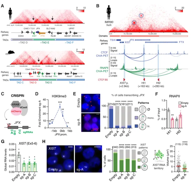
characterized long-range interactions surrounding XIST promoter region using published Hi-C (Rao et al., 2014) and ChIA-PET datasets (Ji et al., 2016) generated from female cell lines. This revealed 20
a partitioning of the human XIC into discrete spatial domains that resembled the organization into topological associated domains (TADs) of the mouse syntenic region (Nora et al., 2012) (Figure 3A). Notably, the boundaries of the XIST-associated TAD (~TAD E) fall within XIST locus and upstream of the RLIM gene in both species (Figure 3A). Closer inspection of the XIST-associated TAD in human (Dowen et al., 2014; Hnisz et al., 2016; Ji et al., 2016) revealed that CTCF-CTCF loops formed 25
an insulated chromatin neighborhood hosting preferential contacts between XIST promoter region and the JPX gene (Figure 3B). The CTCF loops anchor a region in the vicinity of XIST TSS (XISTp, +2,9 kb) to two upstream intragenic regions, or hotspots, located within the JPX (Ht1, +163 kb) and
FTX (Ht2, +283 kb) genes. All anchors were co-occupied by CTCF and the cohesin complex, but
were not enriched in chromatin marks or proteins associated with active enhancers (Figure S3A). 30
Although a similar organization of the XIST-associated TAD was observed on the sole active X chromosome of male fibroblasts (Figure S3B), we found that XIST/JPX long-range interactions were associated with RNA polymerase II (RNAPII) only in female datasets (Li et al., 2012), while no peak was reported in that of male (Figure 3B, data not shown). This suggests a female-specific transcriptional association of XIST and JPX that likely occurs on the Xi (Figure 3B). This hypothesis 35
is further supported by the high number of cells (>80%) co-transcribing XIST and JPX from the Xi as assessed by RNA-FISH in several cell lines (Figure 2A). These long-range interactions were also detected in CTCF and SMC1 ChIA-PET datasets obtained from female naïve hESCs (Ji et al., 2016), suggesting that these loops occur independently from the XCI status of the cells (Figure S3C). 5
As insulated chromatin neighborhoods have been shown to favor the communication between genes and their regulatory elements (Ji et al., 2016; Sun et al., 2019), we hypothesized that XIST/JPX long-range interactions could provide a structural framework for JPX transcription to regulate XIST expression. To test this hypothesis, we used a CRISPR inhibition strategy (CRISPRi) (Gilbert et al., 2013) in female primed H9 hESCs, where three guide RNAs were used independently to recruit a 10
catalytically inactive Cas9 fused to a KRAB co-repressor to the JPX 5’ region, in order to prevent its transcription (Figure 3C). This system efficiently triggered the local deposition of the H3K9me3 repressive mark on a restricted region surrounding JPX transcription start site (TSS) (Figure 3D). As a result, RNAPII recruitment to JPX TSS was compromised (Figure S3D) and transcription at the locus was severely impaired; JPX remained transcribed in less than ~15% of the cells as assessed by 15
RNA-FISH (Figure 3E) and JPX RNA levels were reduced by 90% (Figure S3E). RNAPII occupancy was also decreased at the Ht1 (Figure 3F), indicating that this strategy efficiently reduced RNAPII processing along the 70 kb of the JPX gene. In these conditions, XIST steady-state RNA levels were significantly reduced (Figure 3G), as were the percentage of cells with XIST accumulation (Figure 3H) and focal enrichment of the H3K27me3 repressive mark (Figure S3F). In addition, the cells that 20
retained XIST expression displayed smaller XIST RNA cloud compared to the control condition (Figure 3H), indicating that all cells in the population were affected by JPX inhibition. As XIST repression may result from XCI erosion, we tested whether inhibition of JPX transcription could favor this process. For this, we monitored the accumulation of the XACT lncRNA and the transcription of the POLA1 gene from the Xi by RNA-FISH, as the expression of these genes from the Xi are early 25
markers of XCI erosion and precedes the loss of XIST expression in H9 hESCs (Vallot et al., 2015). Inhibition of JPX transcription did not trigger either XACT or POLA1 transcription from the XIST-coated Xi (Figure S3G) and the two genes remained solely expressed from the active X. Moreover, we could not link the reduction of XIST expression to perturbation of YY1 binding on XIST promoter (Figure S3H), a known regulator of XIST expression in human cells (Makhlouf et al., 2014), 30
suggesting that JPX acts through YY1-independent mechanisms. We also verified that reduced XIST expression did not result from an ectopic deposition of H3K9me3 at XIST promoter due to the CRISPRi strategy (Figure S3I). Altogether, our results, which were reproduced in another primed hESCs line (WIBR2, Figure S3J-K), demonstrate that JPX transcription is required for proper XIST expression.
Figure 3: XIST expression requires JPX transcription.
(A) The human XIC is partitioned in discrete topological domains (TAD C to F) that are syntenic to that of the mouse Xic (TAD D to F); Xist/XIST TAD E boundaries are highlighted by black arrows. HiC data from human fetal fibroblasts and mouse ESC (Bonev et al., 2017; Rao et al., 2014).
(B) XIST and JPX interact (Hi-C, IMR90) through CTCF- and RNAPII-mediated loops (ChIA-PET K562). Called loops are highlighted by blue circles. (See also Figure S3A-C).
(C-D) Scheme of CRISPRi strategy to inhibit JPX transcription in primed hESCs. In this condition, a strong and local enrichment of H3K9me3 could be observed at JPX promoter, ChIP-qPCR, n=4. (See also Figure S3D).
(E) The number of cells expressing JPX was strongly reduced in CRISPRi conditions, with both JPX alleles being efficiently silenced one week after lentiviral infection with the guides. Left: Representative images. Right: scoring of JPX RNA-FISH signals (Chi-square test). (See also Figure S3E).
(F) Inhibition of JPX transcription reduced RNAPII availability at the Ht1, ChIP-qPCR, n=3.
(G) XIST steady state RNA levels were reduced upon inhibition of JPX transcription (RT-qPCR, n=4). (See also Figure S3F).
(H) JPX CRISPRi resulted in a decrease in the number of cells expressing XIST (Chi-square test) and on the volume of XIST RNA cloud (Mann-Whitney test), RNA-FISH. (See also Figures S3H-K).
Error bars represent standard deviation; n.s., not significant; *p<0.05; **p<0.01; ***p<0.001; ****p<0.0001. Unpaired two-tailed t-tests to the empty condition unless stated otherwise. Number of counted cells is in brackets.
0.00 0.01 0.02 0.03 0.04 0.05 Figure 3 B XIST p. (+2.9kb) Ht1 (+163 kb) Ht2 (+283 kb) TSIX XISTJPX FTX chrX 73,100,000 73,300,000 73,500,000 Domains Refseq genes IMR90 CTCF BS [ 0-30] Loops Signal [ 2-15] CTCF ChIA-PET [ 0-30] Loops Signal [ 2-15] RNAPII ChIA-PET XaXi Refseq genes 103,000,000 Chic1 Slc16a2 Abcb7 Uprt Zdhhc15 Tsix Xist Jpx Ftx Cdx4 Rlim Linx Dmrt1c/a
TAD D TAD E TAD F
TAD C 103,400,000 103,800,000 104,200,000 104,400,000 chrX, mm10 0 15 A TADs 73,000,000 72,500,000 73,500,000 74,000,000 74,500,000 CDX4 CHIC1 SLC16A2 RLIM ABCB7 UPRT ZDHHC15 TSIX XIST JPX FTX 0 10
~TAD D ~TAD E ~TAD F
chrX, hg19 Refseq genes TADs * * * G XIST sg-A Empty % of cells Volume (μm 3) (342) (345) (264) (313)**** 0 50 100 **** **** (115) (116)**** 0 5 10 15 20 25 H XIST (Ex5-6) Global RNA levels
Emptysg-A sg-Bsg-C Emptysg-A sg-B sg-C Emptysg-A
XIST patterns JPX sgRNAs A B C dCas9 KRAB mCherry C JPX sg-A Empty **** (214)(217)**** ****(161)(211) % of cells transcribing JPX 0 50 100 sg-A sg-B sg-C Empty
Fold Enrichment sg-A
H3K9me3 **** *** ** n.s. JPX prom. -1kb 0kb 1kb 0 10 20 30 40 D CRISPRi ** Ht1 Ht2 RNAPII sg-A Empty 0.0 0.5 1.0 1.5 % of input F Patterns Ø mono. bi. E XIST RNA territory 2 20
Figure S3: XIST expression requires JPX transcription, related to Figure 3
(A) Within the XIST-associated TAD, CTCF binding sites are co-occupied by the cohesin complex (RAD21 and SMC3). The hotspots (Ht1/Ht2) are not enriched with enhancer-associated chromatin marks (H3K27Ac) or protein (CEBPB) in female fetal fibroblasts; active promoters in this region are highlighted by the H3K4me3 ChIP-seq track.
(B) Structural organization of XIST-associated TAD in male fibroblasts (HFF-c6; (Dekker et al., 2017). Called loops are highlighted by blue circles.
(C) In naïve hESCs from (Ji et al., 2016), JPX and XIST are interacting through SMC1-mediated loops, which coincide with CTCF-mediated loops in Fig.2a. The heatmap represents raw SMC1 interactions and arcs represents high confidence loops.
Figure supplementary 3 Normalized RNA levels XIST sg-A Empty 72%(550)**** 90%(469) **** V olume (μm 3) (166) (111) XIST RNA territory Empty sg-A 0 20 10 15 5 J
XIST H3K27me3 Merge
sg-A Empty F K % of input 0kb 1kb 2kb 0 1 2 3 RNAPII** * * sg-A Empty D JPX prom. E Global RNA levels 0.00 0.01 0.02 0.03 0.04 ** ** ** JPX (Ex1-2) Emptysg-A sg-Bsg-C 0 50 100 Emptysg-A sg-B sg-C % of cells XIST+ H3K27me3 (220)****(216)****(202)****(118) Ø CRISPRi JPX WIBR2 hESCs 0.0 0.5 1.0 JPX XIST * * * * ** sg-Asg-Bsg-C sg-Asg-Bsg-C XACT XIST Empty sg-A (145) (99) no XACT XACT on Xa XACT biallelic n.s. G POLA1 XIST Empty sg-A 0 50 100 % of cells no POLA1 POLA1 on Xa POLA1 biallelic Emptysg-A 0 50 100 Xi-reactivation XACT XIST JPX ChrX POLA1 % of cells Patterns H3K9me3 -2kb 0kb 2kb 0 10 20 30 XIST promoter % of input sg-A Empty sg-A Empty YY1 0kb 1kb 2kb 0 1 2 3 4 5 % of input Xi-reactivation (145) (99) H I * B ChrX, hg3873,800,000 74,000,000 74,200,000 74,400,000 TSIX XISTJPX FTX Refseq genes Ht 1 Ht 2 0 5 Male Fibroblast TSIX XIST JPX FTX hg19, chrX 73,100,000 73,300,000 73,500,000 [0-100] [0-100] [0-20] [0-20] [0-30] [0-20] CTCF RAD21 SMC3 H3K4me3 CEBPB H3K27Ac (0kb) XIST TSS (+163 kb)Ht 1 (+283 kb)Ht 2 (+91kb) (+442kb) CTCF ori. A ChrX, hg19 73,100,000 73,300,000 73,500,000 CTCF (+ peaks) SMC1 Loops [0-4] [5-30] Refseq genes 0 2 Naive hESCs (pre-XCI) C TSIX XISTJPX FTX
(D-E) JPX CRISPRi prevented RNAPII recruitment at JPX promoter (ChIP-qPCR, n=3) and resulted in a strong decrease of JPX RNA levels, RT-qPCR, n=4.
(F) JPX CRISPRi led to the simultaneous loss of XIST RNA clouds (RNA-FISH) and H3K27me3 foci (IF); right panel represents the fraction of double positive cells for XIST and H3K27me3 cells, Fischer’s exact test.
(G) In contrast to XCI erosion, JPX CRISPRi did not trigger XACT reactivation from the XIST-coated Xi, as assessed by double XIST and XACT RNA-FISH (Chi-square test). Similarly, no Xi-reactivation of
POLA1 transcription could be identified by double XIST and POLA1 RNA-FISH (Fischer’s exact test).
(H) JPX CRISPRi did not affect YY1 binding at XIST promoter, ChIP-qPCR, n=3.
(I) JPX CRISPRi did not result in ectopic H3K9me3 enrichment at XIST promoter, ChIP-qPCR, n=4. (J-K) Inhibition of JPX transcription in WIBR2 hESC induced a decrease in XIST RNA levels (RT-qPCR, unpaired two-tailed t-test, n=3), in the number of cells expressing XIST (Fischer’s exact test) and in the volume of XIST RNA cloud (Mann-Whitney test) – red bars: median.
Error bars represent standard deviation; n.s., not significant; *p<0.05; **p<0.01; ***p<0.001; ****p<0.0001. Unpaired two-tailed t-tests to the empty condition unless stated otherwise. Number of counted cells is in brackets.
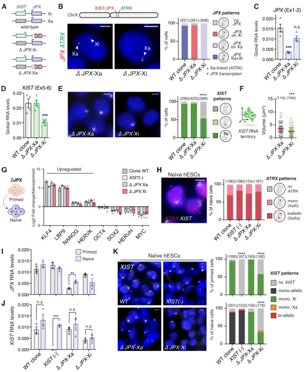
XIST expression requires a functional JPX allele in cis.
To further explore the contribution of JPX transcription to XIST regulation, we generated deletions of JPX promoter region in primed hESCs using the CRISPR-Cas9 technology. Based on JPX promoter features and its expression in H9 cells, we designed guides RNAs to delete a ~7kb region 5
encompassing the three first exons of JPX (Figure S4A). We developed a strategy where hESCs co-transfected with two sgRNAs, each targeting a region upstream and downstream of JPX TSS, could be selected based on the expression of a fluorescent gene, GFP and mCherry, respectively (Figure S4B). Therefore, FACS-sorting of double positive cells maximizes the probability to obtain clones with a direct deletion of JPX promoter. This approach allowed us to interrogate the contribution of 10
JPX transcription to XIST regulation in an allele-specific manner, and tease apart cis- from
trans-effects of the LRG (Figure 4A). However, as no SNP could be identified in H9 cells within the deleted region, we systematically performed simultaneous JPX and ATRX RNA-FISH to determine on which of the two X-chromosomes (Xa or Xi) JPX was still transcribed. Using this strategy, we selected two clones carrying heterozygous deletions of JPX promoter region for further investigation, in which the 15
deletion occurred either on the active X-chromosome (∆JPX-Xa) or on the inactive X (∆JPX-Xi) (Figure 4B and S4C-D). Interestingly, the two clones displayed different JPX RNA levels depending of the deleted allele, with the expression level in ∆JPX-Xa clone reaching only ~15% of that of the WT clone, and ~80% in the ∆JPX-Xi clone (Figure 4C). This is in agreement with JPX being predominantly expressed from the Xa in WT cells, as found for most genes that escape XCI (Carrel 20
et al., 1999). Remarkably, we found that XIST expression was perturbed exclusively in the ∆JPX-Xi clone with RNA; XIST RNA levels were reduced by half compared to the WT and ∆JPX-Xa clone (Figure 4D) with only ~55% of the cells displaying XIST RNA accumulation (Figure 4E) and H3K27me3 foci (Figure S4E). Moreover, XIST RNA territory in the remaining XIST-positive cells were significantly smaller in the ∆JPX-Xi clone (Figure 4F), indicating that XIST expression was also 25
impacted in those cells. Altogether these results show that transcription originating from the JPX promoter region is required to sustain XIST expression in cis in human post-XCI cells. Moreover, the fact that JPX RNA levels were the least perturbed in the ∆JPX-Xi clone (Figure 4C) confirmed that
JPX RNA does not control XIST expression in human.
30
As JPX induction seems to precede XIST upregulation during human pre-implantation development, we investigated whether its transcription could be important for the de novo induction of XIST expression. To do so, we proceeded to the chemical resetting of the primed ∆JPX and WT lines into the naïve state of pluripotency (Guo et al., 2017). As a control, we also converted fully XIST-negative eroded primed hESCs to ensure that the resetting process could efficiently trigger XIST upregulation 35
and X-chromosome reactivation (XCR) as observed with this method (Guo et al., 2017) and others (Sahakyan et al., 2017; Theunissen et al., 2016; Vallot et al., 2017). After 7 passages (~45 days) in naïve culture medium, all cell lines displayed dome-shaped colonies (Figure S4F) and proper induction of key naïve-specific pluripotency markers (Figure 4G) such as the transcription factors KLF4 and LBP9 (Takashima et al., 2014), indicating an efficient transition to the naïve-like state. 5
XCR was observed to same extent in all cell lines, as inferred from the bi-allelic transcription of
ATRX (~75-85%, Figure 4H), which confirmed that XCR occurred independently from the
XIST-expressing status of the parental primed hESCs (Sahakyan et al., 2017). XCR could also be detected at the level of XIC-linked genes, notably by the biallelic transcription of the Xa-specific FTX gene (Figure S4G) and for JPX, which is more expressed in naïve ∆JPX-Xa cells compared to primed 10
(Figure 4I). We found that XIST was strongly reactivated upon the resetting of the eroded cells (Figure 4J), with the proportion of naïve XIST-expressing cells reaching about 95%, as assessed by RNA-FISH (Figure 4K). In agreement with previous studies, XIST accumulation remained mostly monoallelic and likely confined to the former Xi, with less than 3% of XaXa cells displaying biallelic
XIST RNA clouds. Resetting of XIST-expressing cells (WT and ∆JPX-Xa clones) did not increase the
15
proportion of biallelic XIST RNA clouds and XIST RNA clouds remained associated to the former Xi (Sahakyan et al., 2017). By contrast, XIST reactivation could not be observed in cells where JPX is deleted on the Xi allele, in which XIST expression remained low and restricted to an even lower percentage of cells, compared to primed conditions (38% naïve vs. 58% primed, Figure 4K). Deletion of the JPX promoter region thus prevents de novo XIST upregulation in cis during conversion of 20
primed to naïve human ESCs. These analyses altogether suggest that JPX transcription is required for proper XIST upregulation in cis during early human pre-implantation development.
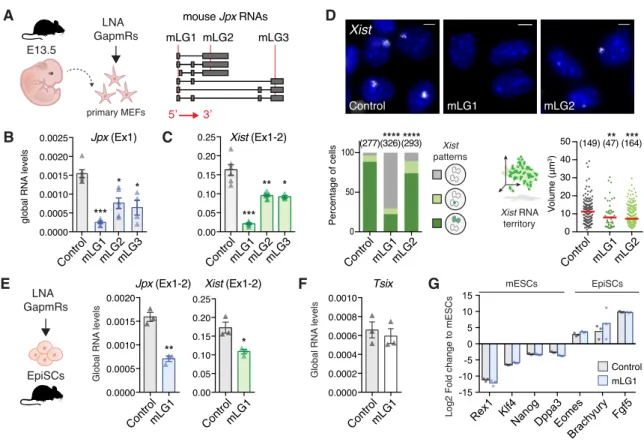
Jpx RNA regulates XIST expression in mouse post-XCI cells.
Previous studies have shown that the deletion of a single allele of the Jpx gene or shRNA-mediated 25
knockdown of Jpx is sufficient to prevent Xist upregulation (Carmona et al., 2018; Tian et al., 2010), with Jpx acting through RNA-based mechanisms, both in cis and in trans. Considering the discrepancy with the results we obtained in human, we decided to revisit the function of Jpx RNA in the mouse, by rigorously matching both our experimental approaches and the cellular models. We performed LGs-mediated KD experiment in mouse embryonic fibroblasts (pMEFs) and murine 30
epiblast-derived stem cells (EpiSCs). Murine EpiSCs share similarities with primed hESCs in terms of transcriptional signatures, signaling pathways and XCI status (Brons et al., 2007; Tesar et al., 2007), while pMEFs parallel the primary fibroblasts of fetal origin used in this study (Figure 2C-H and S2F-G). Both cell types display one inactive X-chromosome coated by Xist and express Jpx. First, we investigated mouse Jpx RNA function in primary mouse embryonic fibroblasts (pMEFs) 35
Figure 4: XIST expression requires a functional JPX allele in cis.
(A) Schematic representation of WT and JPX-deleted hESC clones. (See also Figure S4A and S4B) (B) Determination of the JPX-targeted allele by simultaneous JPX/ATRX RNA-FISH. ATRX is transcribed exclusively from the Xa in primed hESCs. (See also Figure S4C and S4D).
(C) ∆JPX-Xa cells displayed a ~80% reduction of JPX RNA levels compared to WT, while a moderate decrease was observed in ∆JPX-Xi cells (~20%), suggesting an asymmetric expression of JPX from the two X chromosomes, RT-qPCR, n=4.
(D) XIST steady states RNA levels were reduced when JPX promoter was deleted in cis (∆JPX-Xi), but not in trans (∆JPX-Xa), RT-qPCR, n=4.
(E-F) In ∆JPX-Xi cells, both the number of cells expressing XIST (Chi-square test) and the volume of XIST RNA cloud (Mann-Whitney test) were reduced, RNA-FISH. (See also Figure S4E).
(G) Average log2 fold-change of transcript levels between the naïve and the parental primed hESC clones for a selection of markers, RT-qPCR, n=3. (See also Figure S4F).
(H) The Xi was properly reactivated in more than 70% of cells in the different cell lines based on biallelic expression of ATRX, RNA-FISH. (See also Figure S4G).
(154) (196) (190) (181) 0 50 100 Figure 4 G -5 0 5 10
Log2 Fold change(Naive/primed)
KLF4 LBP9
NANOG HERVK OCT4 SOX2HERVH MYC Clone WT XIST(-) ∆ JPX Xa ∆ JPX Xi Upregulated H K 0.000 0.005 0.010 0.015 0.020 WT clone ∆ JPX -Xa XIST (-) ∆ JPX -Xi *** n.s n.s n.s % of primed cells 0 50 100 (166)(167)****(169)(182) XIST patterns mono-allelic no XIST bi-allelic mono. Xi mono. Xa % of naive cells 0 50 100 WT clone ∆ JPX -Xa XIST (-) ∆ JPX -Xi **** (156) (153) (201) (176) **** WT XIST XIST(-) ∆ JPX-Xa ∆ JPX-Xi JPX RN A levels 0.000 0.005 0.010 0.015 0.020 Primed Naive ** XIST RN A levels I J Naïve hESCs % of naive cells A *** 0.00 0.01 0.02 0.03 XIST (Ex5-6) D 0 5 10 15 V olume (μm 3) % of cells Global RN A levels E (114)(104)*** **** WT clone∆ JPX-Xa ∆ JPX-Xi 0 50 100 (290)(423)(399) ∆ JPX-Xa ∆ JPX-Xi WT clone∆ JPX-Xa ∆ JPX-Xi ATRX patterns no ATRX mono. (XaXi) biallellic (XaXa) Naïve Primed ∆JPX Naïve hESCs ATRX XIST WT clone ∆ JPX -Xa XIST (-) ∆ JPX -Xi ∆ JPX-Xi ∆ JPX-Xa Xa-linked (ATRX) JPX transcription B no JPX JPX on Xi JPX on Xa JPX Xa+Xi WT clone∆ JPX-Xa ∆ JPX-Xi % of cells (251)(261) (306) X PJ X R T A 0 50 100 ∆ JPX-Xi ∆ JPX-Xa Xa Xa Xi ATRX XIST/JPX ChrX ∆ JPX-Xi ∆ JPX-Xa wild-type Xa Xi XIST JPX *** 0.000 0.005 0.010 0.015 0.020 C Global RN A levels n.s. JPX (Ex1-2) WT clone∆ JPX-Xa ∆ JPX-Xi XIST RNA territory XIST patterns JPX patterns F
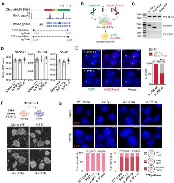
Figure S4: XIST expression requires a functional JPX allele in cis, related to Figure 4 (A) Map of the JPX 5’ region, with various promoter features indicated, such as the chromatin
(chromHMM) and transcriptional states (H9 RNA-seq, H3K4me3). The bottom tracks show the guides position (in green, guides coupled to a Cas9-GFP; in red guides coupled to a Cas9-mCherry) and sequence alignments spanning the deleted region to the reference genome in the two clones.
(B) Scheme of the strategy to produce genomic deletion using the CRISPR-Cas9 system coupled to FACs-sorting in hESCs.
(C) PCR-genotyping of the two heterozygote clones obtained.
(D) JPX deletion did not impact on the steady state RNA levels of key pluripotency markers, RT-qPCR, error bars represent standard deviation, n=4.
(E) Deletion of JPX promoter on the Xi, but not the Xa, led to a decrease in the percentage of cells with
XIST RNA clouds and H3K27me3 foci (Immunofluorescence) (Fischer’s exact test). P-value: <0.0001
(****).
(F) Resetting primed hESCs to the naïve state results in similar colonies with dome-shaped appearance, independently of the genotype.
(G) Analysis of FTX gene expression by RNA-FISH showed robust biallelic expression following resetting of primed into naïve hESCs.
Scale bars are 5 µm. Number of counted cells is in brackets. Figure supplementary 4 0.00 0.01 0.02 0.03 0.04 0.00 0.02 0.04 0.06 0.00 0.01 0.02 0.03 0.04 0.05 ∆JPX Xa
NANOG OCT3/4 SOX2
∆JPX Xi Clone WT ∆JPX Xa ∆JPX Xi Clone WT ∆JPX Xa ∆JPX Xi Clone WT D Global RNA levels
XIST H3K27me3 Merge
∆ JPX Xi ∆ JPX Xa 0 50 100 % of cells **** ∆ JPX Xa ∆ JPX Xi (274)(243) E Clone WT XIST(-) ∆JPX Xa ∆JPX Xi ∆JPX Xa ∆JPX Xi Naive Primed 0 50 100 (203)(156)(156)(176) 0 50 100 (161)(167)(186)(178) WT clone ∆ JPX -Xa XIST (-) ∆ JPX -Xi % of naive cells % of primed cells FTX patterns no FTX mono. XaXi biallellic XaXa WT clone FTX XIST(-) FTX B Cas9-GFP Cas9-mCherry Primed hESCs GFP+ mCherry+ colonies picking A 1 kb ∆JPX Xi deletion sgRNAs JU3 JD3 JPX ∆JPX Xa deletion JU1 JD1 sgRNAs Refseq genes RNA-seq H9 0 200 ChromHMM K562 C 1000 500 1000 500 500 250 WT allele deletion inversion ∆ JPX Xi ∆ JPX Xa Clone WT XIST + H3K27me3 Ø F Naïve Primed ∆JPX clones + XIST(-) WT clone ∆ JPX -Xa XIST (-) ∆ JPX -Xi Naïve Cult G
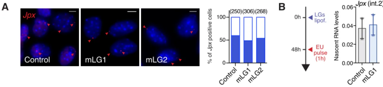
derived from 13.5 days post-coitum mouse embryos using three distinct LNA Gapmers targeting different exons, to minimize the probability of random off-target effects (Stojic et al., 2018) (Figure 5A). This approach was efficient in depleting Jpx RNA (Figure 5B), and the strongest effect was obtained with the mLG1, which is expected to target all Jpx RNA isoforms. The three LGs induced a decrease in spliced Xist RNA levels (Figure 5C) that correlated with the extent of Jpx RNA depletion 5
(Pearson correlation = 0.96, p-val. = 0.035), suggesting a dose-dependent effect of Jpx RNA. RNA-FISH analyses revealed that most of the cells were affected by Jpx KD, as both the percentage of Xist positive cells and the volume of the remaining Xist RNA clouds were reduced (Figure 5D). As previously, we verified that the observed effects of Jpx mLGs were not due to transcriptional inhibition through quantification of Jpx nascent transcripts by RNA-FISH and after ethynyl uridine 10
(EU) incorporation followed by pull-down (Figure S5A-B). We could thus conclude that the observed
Xist downregulation can be attributed to the depletion of Jpx mature transcripts only, and not to
alterations of Jpx ongoing transcription. Similarly to pMEFs, depletion of Jpx RNA in EpiSCs led to a decrease in Xist RNA levels (Figure 5E), without ectopic expression of Xist negative regulators in the mouse, namely its antisense Tsix (Figure 5F) or the pluripotency factors REX1 and KLF4 (Figure 15
5G) (Navarro et al., 2010). These data demonstrate the contribution of mouse Jpx RNA to the maintenance of Xist expression in post-XCI cells and confirm the role of Jpx as a potent regulator of
Xist.
Mechanisms of XIST regulation by JPX have diversified during evolution 20
To further decipher the mechanisms underlying the different molecular function of JPX, we investigated which step of Xist/XIST biogenesis was under the control of the Jpx/JPX LRG in mouse and human. Since previous work reported that Jpx RNA could activate Xist by evicting the CTCF protein from its TSS (Sun et al., 2013), we investigated CTCF binding profile across the Xist promoter upon Jpx
KD in pMEFs. Binding of CTCF to a position ~1kb upstream of Xist TSS was significantly increased
25
upon Jpx KD (Figure 6A), an effect we also observed on the imprinting control region of H19 (Figure
S6A). Nevertheless, we probed the impact of this change of CTCF binding on Xist transcription by
measuring the level of Xist premature transcripts. Quite unexpectedly, we could not detect changes in Xist premature transcript, suggesting that transcription was unaffected by Jpx KD (Figure 6B). To confirm this, we performed nascent RNAs pulldown (Figure 6C) and single-cell level by RNA-FISH 30
using stranded oligo-probes detecting Xist first intron (Figure 6D). While these approaches were suitable to detect Xist transcriptional changes upon Yy1 KD, a known regulator of Xist transcription (Figure 6C-D and Figure S6B-E) (Makhlouf et al., 2014), we could not detect any transcriptional deregulation upon Jpx KD. Altogether, these data demonstrate that Jpx RNA acts downstream of Xist transcription and is required for proper Xist RNA accumulation.
Figure 5: Jpx RNA regulates XIST expression in mouse post-XCI cells.
(A) Schematic representation of LNA GapmeRs (LG) lipofection in primary MEFs; LGs-targeted regions (red lines) are indicated on Jpx RNA isoforms.
(B-C) LG-transfected pMEFs showed reduced Jpx and Xist RNA levels, RT-qPCR; n=4.
(D) Jpx KD reduced the number of Xist expressing cells (Chi-square test) and the volume of Xist RNA cloud (Mann-Whitney test), RNA-FISH. Red bars: median.
(E) In EpiSCs, Jpx KD led to a decrease in Xist RNA levels, RT-qPCR, n=3. (F) Tsix is not re-expressed in EpiSC transfected with Jpx-targeting LGs.
(G) Log2 expression fold change for a selection of markers (Brons et al., 2007; Tesar et al., 2007) in EpiSC transfected with control or Jpx-targeting LG, normalized to expression in mESC, RT-qPCR, n=3.
Error bars represent standard deviation; n.s., not significant; *p<0.05; **p<0.01; ***p<0.001; ****p<0.0001. Unpaired two-tailed t-tests to the control LG unless stated otherwise.
See also Figure S5. Figure 5 **** *** 0.00 0.05 0.10 0.15 0.20 0.25 0.0000 0.0005 0.0010 0.0015 0.0020 0.0025 (277)(326)(293) global RNA levels Jpx (Ex1) Xist mLG1 mLG2 Control A D mLG1 mLG2 mLG3 mouse Jpx RNAs Percentage of cells B ControlmLG1mLG2mLG3 Xist (Ex1-2) ControlmLG1mLG2mLG3 *** ** * V olume (μm 3) 0 10 20 30 40 50 (149)(47)**(164)*** Xist RNA territory LNA GapmRs primary MEFs 5’ 3’ Control mLG1mLG2 Control mLG1mLG2 Xist patterns E13.5 C * * 0 50 100 **** 0.0000 0.0002 0.0004 0.0006 0.0008 0.0010 Rex1 Klf4NanogDppa3Eomes Brachyury Fgf5 Control mLG1 mESCs EpiSCs
Log2 Fold change to mESCs
G F Global RNA levels Tsix ** 0.00 0.05 0.10 0.15 0.20 0.25 0.0000 0.0005 0.0010 0.0015 0.0020 Global RNA levels ControlmLG1 Jpx (Ex1-2) Xist (Ex1-2)
* ControlmLG1 ControlmLG1 -15 -10 -5 0 5 10 15 LNA GapmRs EpiSCs E
Figure S5: Jpx RNA regulates XIST expression in mouse post-XCI cells, related to Figure 5
(A) RNA-FISH of Jpx performed 48h after LGs lipofection in pMEFs. Left panel: representative images. Right panel: quantification of Jpx positive cells (blue fill). Number of counted cells is in brackets. (B) Nascent RNA pulldown of EU-labelled nascent transcripts experimental scheme and Jpx RNA quantification, RT-qPCR, n=2. Error bars represent standard deviation.
Figure supplementary 5 0 50 100 0.00 0.02 0.04 0.06 B 0h 48h EU pulse (1h) LGs lipof. Nascent RNA levels Jpx (int.2) Jpx mLG1 mLG2 Control (250)(306)(268) % of Jpx positive cells A Control mLG1mLG2 ControlmLG1
In striking contrast, XIST unspliced RNA levels were strongly decreased following inhibition of human JPX transcription, independently of CTCF binding changes (Figure 6E-F). This was further supported by the observed reduction of phospho-Ser5 RNAPII recruitment at XIST promoter (Figure 5c). Severe impairment of XIST ongoing transcription following JPX transcriptional inhibition was also evident at the single-cell level, when stranded oligo-FISH probes were used to detect human 5
XIST intronic regions (Figure 6H). This suggests a transcriptional crosstalk between JPX and XIST in
human, where ongoing transcription across the JPX locus would favor the recruitment of the transcription machinery at XIST, possibly through local 3D interaction and chromosomal looping. Our results therefore demonstrate that not only the functional module of Jpx/JPX differs between human and mouse, but also their mode of action to regulate Xist/XIST.
10
DISCUSSION
Here, we interrogated the XIST regulatory network in human early development, and the extent to which regulatory networks essentially based on LRGs operate similarly in different species. Through unbiased analysis of expression dynamics of XIC-linked genes, we identified JPX as the best 15
candidate for promoting XIST activation, and through functional investigation in various human contexts, we demonstrated a major and ubiquitous role for JPX in XIST expression. Doing so, we provided the first evidence that resetting human primed to naïve pluripotent stem cell may constitute a system of choice to study the regulatory network at stake for post-fertilization XIST activation in human. We further determined that transcription across JPX is important in this process and that JPX 20
RNA is not involved in XIST regulation in human cells. Furthermore, JPX acts in cis to promote XIST transcription, while we show that Jpx RNA acts downstream of Xist transcription on Xist metabolism in the mouse. The choice of cellular models was critical for the comparative analysis of JPX/Jpx mode of action in human and mouse. Indeed the early steps of XCI differs markedly in these species, with initial XIST up-regulation being uncoupled from XCI in human, and, more importantly, not 25
currently recapitulated in any ex vivo model. We therefore chose to focus most of our investigation on the maintenance of XIST expression, for which comparable cellular systems were available in the two species studied. The reproducibility of the results in two post-XCI contexts, primed pluripotent stem cells and fetal fibroblasts, strengthens our conclusions and provide the first evidence that an LRG from the XIC plays similar function in different mammalian species but operates via different 30
mechanisms. Jpx/JPX therefore stands as a key component of the Xist/XIST regulatory network in mouse and human.
Figure 6: Mechanisms of XIST regulation by JPX have diversified during evolution.
(A) In the mouse, CTCF binding to Xist proximal promoter region is increased upon Jpx KD in pMEF, ChIP-qPCR, n=3. (see also Figure S6A)
(B) KD of Jpx RNA did not impact on Xist premature transcript levels, intronic RT-qPCR, n=3 (See also Figure S6B-D).
(C) Xist transcription was affected by Yy1 KD, but not by Jpx KD, in pMEFs when quantified after pulldown of EU-labelled nascent transcripts, RT-qPCR, n=3.
(D) Jpx KD did not affect Xist ongoing transcription (intron 1 stranded oligo-FISH probes) but only on its accumulation (p510 probe) (Fisher's exact test). Yy1 KD affects both transcription and accumulation of
Xist. (See also Figure S6E)
(E) In human, JPX CRISPRi did not affect CTCF enrichment at XIST promoter or at both interaction hotspots, ChIP-qPCR, n=3.
(F) XIST premature transcripts levels are reduced following JPX CRISPRi, intronic RT-qPCR, n=3. (G) Inhibition of JPX transcription prevented RNAPII (CTD-phospho-Serine5) recruitment at XIST promoter, ChIP-qPCR, n=4.
(H) The number of cells with XIST RNA accumulation (G1A probe) and transcription (intronic stranded oligo-FISH probes) is reduced following JPX CRISPRi.
Error bars represent standard deviation; *p<0.05; **p<0.01; ***p<0.001; ****p<0.0001. Unpaired two-tailed t-tests to control condition unless stated otherwise.
Figure 6 E Human % of input 0.0 0.1 0.2 0.3 CTCF sg-A Empty Ht1 Ht2 XISTp. neg. XIST (int1) ** ** ** Global RNA levels Emptysg-A sg-B sg-C 0.0000 0.0005 0.0010 0.0015 0.0020 F
XIST G1A XIST int. Merge Empty sg-A 0 50 100 sg-A Empty (305)(300)**** sg-A Empty (305)(300)**** 0 50 100 Patterns other XIST cloud Patterns no signal XIST int.1-2 -1kb 0kb 1kb 0 20 40 60 80 100 XIST TSS RNAPII-Ser5P sg-A Empty Fold Enrichment * * *
XIST accumulation XIST transcription
% of cells % of cells H G 0 50 100 0 50 100 C D % of cells
Xist p510 Xist intron 1 Merge Control mLG1 (0h) (48h) EU LGs B Global RNA levels Xist (int1) Mouse A 0.00 0.02 0.04 0.06 0.08 0.10 mLG1 Control * * 0kb +2kb -2kb From Xist P1 TSS P1 P2 % of input CTCF 0.0000 0.0005 0.0010 0.0015 0.0020 ControlhLG1hLG2hLG3 0.00 0.02 0.04 0.06 si-YY1 si-CTL ControlmLG1 nascent RNA levels si-YY1 si-CTL
ControlmLG1 ControlmLG1si-CTLsi-YY1
Xist accumulation *** (196)(243)(210)(157)*** (196)(243)* (210)(157)*** Xist transcription % of cells Xist Patterns no signal Xist intron 1 Patterns other Xist cloud *
Figure S6: Mechanisms of XIST regulation by JPX have diversified during evolution, related to Figure 6
(A) CTCF binding at different control positions upon LNA GapmeR transfection. Rs14c and NanS represent respectively positive and negative positions for CTCF binding, ChIP-qPCR, n=3.
(B) Expression of Yy1 mRNA and protein levels following siRNA transfection as in (Makhlouf et al., 2014).
(C-D) Expression levels of Xist mature and premature RNA following siRNA transfection.
(E) Representative images of the effect of Yy1 depletion on Xist accumulation and transcription, RNA-FISH using a probe covering Xist locus or intronic probes. Scale bars: 5 µm.
Error bars represent standard deviation; unpaired two-tailed t-test, *p<0.05; **p<0.01. Figure supplementary 6 NanS Rs14c ICR H19 Control mLG1 * Percentage of input CTCF 0.00 0.05 0.10 0.15 B 0.00 0.02 0.04 0.06 Yy1 si-YY1 si-CTL YY1 VINC. si-YY1 si-CTL Global RNA levels si-RNA YY1 MEFs 0.0000 0.0005 0.0010 0.0015 0.0020 0.0025 0.00 0.05 0.10 0.15 0.20 0.25 ** Xist (int3) Xist (Ex1-2) si-YY1 si-CTL ** Global RNA levels C si-YY1 si-CTL D A si-YY1 si-CTL
Xist p510 Xist intron 1 Merge
Addressing the functional conservation of LRGs is a challenge given their fast evolutionary rate. Overexpression of human JPX RNA in trans was recently shown to complement heterozygous deletion of mouse Jpx during the establishment of XCI, suggesting that the human RNA might be functional in an ectopic context (Karner et al., 2019). Such rescue experiments using orthologous LRGs, as opposed to our strategy to tackle the role of mouse and human Jpx/JPX in the respective 5
species, reveal the effect of the environment on LRG mode of action but do not interrogate LRGs’ function in their endogenous contexts. The lack of XIST deregulation upon JPX RNA depletion in all human cellular contexts that we tested, together with the fact that deleting JPX impacts on XIST only in cis strongly argues against a major role for JPX RNA in trans during human XCI.
10
The mechanistic diversification we observed between mouse and human might be a consequence of the changes within the chromatin neighborhood encompassing the JPX locus in human. For instance, our previous work suggested that transcriptional activity of Xist in the mouse is, at least partially, regulated in cis by transcription of the neighboring Ftx LRG, independently of the Ftx RNA products (Furlan et al., 2018). Interestingly, Ftx is located 141 kb upstream of Xist, which is comparable to the 15
distance bridging XIST promoter to the interaction hotspot Ht1 (~163 kb) within the human JPX, and interacts through CTCF-mediated loops with Xist (Furlan et al., 2018). It is within this insulated chromatin neighborhood, which has been reshaped between mouse and human, that constraints on the JPX locus might have favored diversification of JPX mode of action on XIST. One compelling hypothesis from this model is that XIST transcriptional cis-regulators in eutherian species could have 20
been co-opted based on features such as linear distance from XIST promoter, local 3D organization and XCI escaping profile. Importantly, this scenario is reminiscent of what has been observed for enhancer evolution (Villar et al., 2015), and is thus likely not restricted to JPX evolution but may apply to other orthologous LRGs.
25
Finally, what our study provides is a proof of concept that orthologues may act differently in various species, thus epitomizing the mechanistic plasticity of LRGs through evolution. Diversification of LRGs across evolution could confer molecular drift to developmental processes, contribute to species adaptability and fitness: their strong turnover offers a plausible mechanism for generating phenotypic diversity in the control of gene expression across evolution. One major challenge is the systematic 30
identification of versatile LRGs. Indeed, both LRGs functionality, if any, and their mechanism of action are hardly predictable based on the DNA sequence alone. Like for JPX, syntenic LRGs often display strong primary sequence turnover during evolution, even among closely related species (Hezroni et al., 2015; Necsulea et al., 2014; Ulitsky et al., 2011; Washietl et al., 2014). The impact of such turnover on LRGs functional conservation is still poorly understood. In rare studies where 35
the functional conservation of lncRNA molecules has been addressed, orthologues display short patches of conserved sequence that are necessary, but not sufficient, for their function (Lin et al., 2014; Ulitsky et al., 2011). This contrasts sharply with the evolutionary stability of protein-coding genes and has often raised controversies about LRGs functionality. Our study pave the way for systematic experimental investigations of LRGs functional conservation with the aim to provide a 5
definitive understanding of underlying rules. Whether mechanistic or functional, this plasticity appears to be an essential parameter to take into consideration in the context of animal modelling of human diseases involving LRGs.