Doctoral Dissertation submitted to the
Faculty of Informatics of the Università della Svizzera Italiana in partial fulfillment of the requirements for the degree of
Doctor of Philosophy
presented by
under the supervision of
Prof. Dr. Rolf Krause and Dr. Santiago Fernandez Gonzalez
Rolf Krause ICS, USI - Switzerland Santiago Fernandez Gonzalez IRB, USI - Switzerland
Olaf Schenk ICS, USI - Switzerland
Luca Gambardella IDSIA, USI-SUPSI - Switzerland Marcus Thelen IRB, USI - Switzerland
Matteo Matteucci DEIB, Politecnico di Milano - Italy
Dissertation accepted on 03 March 2020
Research Advisor Co-Advisor
Prof. Dr. Rolf Krause Dr. Santiago Fernandez Gonzalez
PhD Program Director
I certify that except where due acknowledgement has been given, the work presented in this thesis is that of the author alone; the work has not been submit-ted previously, in whole or in part, to qualify for any other academic award; and the content of the thesis is the result of work which has been carried out since the official commencement date of the approved research program.
Diego Ulisse Pizzagalli Lugano, 03 March 2020
To my beloved
I am the vine, you are the branches The Bible, John 15:5
The immune system has a critical role in diseases of primary importance such as infec-tions and cancer. Hence, it represents a target for novel therapeutic strategies.
However, the immune system relies on a complex network of cell-to-cell interactions which remains largely unknown, or difficult to be interpreted.
The combination of experimental data with computational methods is of paramount im-portance to analyze these interactions. Indeed, recently established 2-photon intravital microscopes (2P-IVM), can capture videos of cells while interacting in organs of living an-imals. These interactions are often associated with specific movement patterns. Hence, computer vision methods have the potential to extract knowledge from these videos by analyzing the movement of cells. Unfortunately, common analysis methods poorly apply to 2P-IVM videos capturing the cells of the immune system. This is mainly due to the complex appearance and biomechanical properties of these cells, as well as challenges introduced by in vivo imaging. Additionally, a lack of publicly available 2P-IVM datasets hampers the development of novel analysis methods along with data-driven studies of the immune system. Finally, common measures of cell motility, poorly describe the dy-namic behavior of immune cells.
In this thesis, we address these limitations by
• Making available the first database of 2P-IVM videos and tracks of immune cells. • Modeling as graph the content of 2P-IVM videos, from pixels to biological processes. • Developing, refining, and applying a variety of computational methods to extract
knowledge from this graph.
• Shifting the analysis of cell motility towards the recognition of cell actions, which does not necessarily require cell tracking.
This combination of microscopy data, graph-based methods, and action-based models allowed us to quantify the complex movement patterns of neutrophils, revealing different phases of the immune response to influenza vaccination.
Supervisors and colleagues. I am thankful to my supervisors Prof. Rolf Krause,
Dr. Santiago Fernandez Gonzalez, Prof. Marcus Thelen and colleagues Dr. Niko-laos Chatziandreou, Dr. Yagmur Farsakoglu, Dr. Miguel Palomino-Segura who
shaped the evolution of the project from its primordial-soup. Tommaso Virgilio,
Irene Latino, Daniel Molina-Romero, Dr. Juliana Falivene, Silvia Zanaga, Dr. Jordi Sintes, Nina Germic, Dr. Mauro Di Pilato, Sabrina Casella, Paola Antonello, Ilaria Pierangeli who provided biomedical data, insights and experience. Benedikt The-len, Gabriele Rovi, Concetta Piazza, Rocco D’Antuono, Diego Morone, Dr. Davide Ey-nard, Dr. Ganna Marchenko, Dr. Alessandro Giusti, Prof. Giuseppe Pozzi, for
tech-nical support. Tutored students. I am thankful to Alain Pulfer who inherits the project in its evolved yet chaotic form, and for sharing passion on systems biology.
Radu Theodorescu, Elisa Palladino for help in LTDB. Ilaria Arini who accidentally
named "Dijkstra algorithm" in 2016, going to be a main ingredient of the thesis.
Liudmila Karagyaur for discussions on physics and appreciation of the Hough
Transform. USIMakers. Community of makers which I co-founded at USI with
Dr. Ivan Elhart and Prof. Marc Langheinrich. It provided the sufficiently needed
amount of scientific distraction. Italian Red Cross - Gruppo Clown Tiramisu. This group of people provided energy and skills for stress management, team building, communication and teaching. Beloved. I am thankful to my family for having taught appreciating the beauty in the little things and for having sup-ported my career decisions, Madalina Mahrus for having improved each-other, having prepared all the days my lunch-box, and transformed by weaknesses into opportunities. Funding. I am thankful to SystemsX.ch - the Swiss Initiative in Systems Biology - for funding (iPhD project 2013/124) and training.
Contents xi
List of Figures xiii
List of Tables xv
1 Introduction 1
1.1 The immune system and its actors . . . 2
1.2 Actions of immune cells, an ontology . . . 9
1.3 Open research questions . . . 19
2 State of the art methods to study immune cell behavior in vivo 21 2.1 Intravital imaging pipeline . . . 21
2.1.1 Image acquisition . . . 23
2.1.2 Cell staining . . . 25
2.1.3 Organs and surgical models . . . 26
2.1.4 Cell detection . . . 27
2.1.5 Cell tracking . . . 31
2.2 Quantification of immune cell migration and interaction . . . 34
2.2.1 Measures to quantify cell migration and interaction . . . . 35
2.2.2 Quantification of cell actions . . . 37
2.3 Relevant computer vision methods . . . 40
2.3.1 Superpixels . . . 40
2.3.3 Action recognition . . . 45
2.4 Clustering algorithms for data-driven research . . . 47
3 Objectives of the thesis 51 4 LTDB, A database of videos and tracks of immune cells from intravital microscopy 53 5 Graph-based algorithms 73 5.1 A trainable, graph-based clustering algorithm . . . 73
5.2 Optimized implementation of the Dijkstra algorithm on images . . 97
5.3 Outlook . . . 107
6 Application of graph-based methods for analyzing immune cell mi-gration and interaction 113 6.1 Modeling 2P-IVM data as a hierarchical graph . . . 113
6.2 Grouping pixels in space and color - superpixels . . . 116
6.3 Grouping pixels in time . . . 120
6.4 HoT-POF: an interpretable descriptor of cell motility . . . 133
6.5 Detection and quantification of neutrophil response to apoptosis . 140 6.6 Semi-supervised colocalization . . . 144
6.7 De-bounced kiss and run analysis . . . 153
6.8 Swarm detection . . . 156
7 Characterization of the dynamic behavior of neutrophils following influenza vaccination 157 8 Conclusion, and future perspectives 189 8.1 Workflow and implementation . . . 190
8.2 IMMUNEMAP, making 2-photon microscopy data FAIR . . . 192
8.3 Experimental definition of imaging protocols . . . 195
8.4 Tracking with CARE . . . 196
8.5 Detection and discovery of additional cell actions . . . 197
8.6 Relevance of actions . . . 198
1.1 Different layers of defense of the immune system . . . 4
1.2 Structure of a lymph node . . . 6
1.3 Characteristics and examples of patrolling leukocytes . . . 11
1.4 Characteristics and examples of directed leukocytes . . . 12
1.5 Characteristics and examples of arrested leukocytes . . . 14
1.6 Characteristics and examples of contact formation . . . 15
1.7 Characteristics and examples of swarm formation . . . 18
2.1 Image acquisition and analysis pipeline. . . 22
2.2 Two-photon point-wise excitation . . . 23
2.3 Surgical models for intravital imaging . . . 27
2.4 Measures to quantify cell migration and interaction . . . 34
3.1 Interdisciplinary of the thesis. . . 51
4.1 LTDB - Data generation workflow . . . 62
4.2 LTDB - Challenges of cell tracking . . . 63
4.3 LTDB - Data organization and format . . . 64
4.4 LTDB - Use cases . . . 65
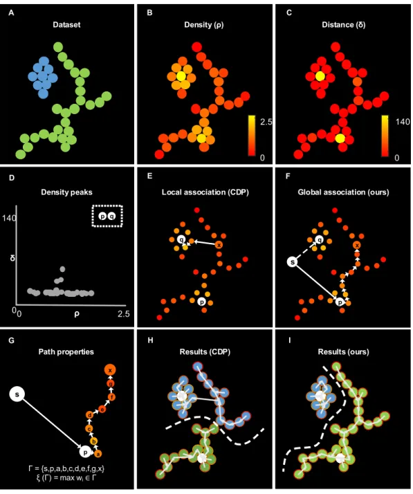
5.1 Clustering rules on a simplified example. . . 87
5.2 Evaluation on a synthetic dataset with heterogeneous structures . 88 5.3 Robustness to density peak detection. . . 89
5.4 Robustness to background noise. . . 90
5.6 Recognition of different heart rhythms. . . 92
5.7 Graph structure. . . 93
5.8 Evaluation on the 12 synthetic datasets from ClustEval . . . 94
5.9 Performance degradation assay with respect to number of training paths. . . 95
5.10 Benchmark on high dimensional synthetic datasets. . . 96
5.11 Results on the bonemarrow leukemia dataset. . . 96
5.12 Iterative implementation of the Dijkstra algorithm . . . 100
5.13 Iterative Dijkstra algorithm - benchmark . . . 102
5.14 Iterative Dijkstra - Shortest paths found at each iteration . . . 105
5.15 Iterative Dijkstra - Results on 2D lattices . . . 106
5.16 Clustering via Hough Transform - simplified example . . . 110
5.17 Analogy with the Hough Transform . . . 110
5.18 Give and receive votes . . . 111
6.1 Hierarchical graph from pixels to biological knowledge . . . 115
6.2 Clustering of non-convex groups of pixels in color and space . . . 117
6.3 Qualitative results on 2P-IVM data . . . 118
6.4 Quantitative results on BSDS . . . 119
6.5 Tracking vs. Optical Flow . . . 120
6.6 Optical Flow errors on 2P-IVM data . . . 121
6.7 Shortest path properties as descriptor . . . 124
6.8 Graph-based optical flow - proposed structure . . . 125
6.9 Optical Flow - Qualitative results on a squeezing neutrophil . . . . 126
6.10 Optical Flow - Results on synthetic example . . . 126
6.11 Optical Flow - Registration error quantification . . . 127
6.12 Optical Flow as tracking predictor - accuracy (1) . . . 128
6.13 Optical Flow as tracking predictor - accuracy (2) . . . 129
6.14 Effect of unstable centroids . . . 129
6.15 Optical Flow Ground Truth generation . . . 131
6.16 Downsampling of Optical Flow . . . 131
6.17 HoT-POF arechitecture . . . 134
6.18 HoT-POF example: neutrophils 2PIVM . . . 136
6.19 HoT-POF example: B cell colture . . . 137
6.20 Apoptosis morphodynamics . . . 142
6.21 Effect of an apoptotic cell on the neighborhood . . . 142
6.22 Apoptosis detection via HoT-POF . . . 143
6.23 Apoptosis detection performances . . . 143
6.25 Improving tracking of poorly visible cells . . . 147
6.26 Improving tracking of non-specifically stained cells . . . 149
6.27 Usage of path-based features to classify superpixels . . . 152
6.28 Example of contact formation (Neutrophil-Macrophage) . . . 154
6.29 Contact detection with hystheresis . . . 155
6.30 De-bounced contact detection with brightness fluctuations . . . 155
6.31 Swarm detection using clustering . . . 156
7.1 Neutrophil recruitment and distribution into the popliteal lymph node (PLN) after influenza vaccine administration . . . 178
7.2 Production of CXCL1 and IL-1↵ induce recruitment of neutrophils to the PLN in response to vaccination . . . 179
7.3 Neutrophils phagocyte and transport influenza virus . . . 180
7.4 Time-course of neutrophil motility . . . 181
7.5 Neutrophils exhibit different actions in the early response to vac-cination . . . 182
7.6 Characterization of neutrophils swarm formation in the PLN after influenza vaccination . . . 183
7.7 Neutrophils - Supplementary 1 . . . 184
7.8 Neutrophils - Supplementary 2 . . . 185
8.1 Workflow of the PhD project . . . 191
8.2 IMMUNEMAP architecture . . . 193
8.3 IMMUNEMAP partners . . . 194
8.4 Effect of time-step on measured cell speed . . . 195
1.1 Main types of immune cells and functions . . . 5
2.1 Two-photon magnifications . . . 24
2.2 State of the art methods for foreground segmentation . . . 30
2.3 Track based measures to quantify cell migration and interaction . 36 2.4 Tools to detect and quantify cell actions . . . 37
4.1 LTDB - Biomechanical and technical problematics . . . 66
4.2 LTDB - Dataset overview . . . 67
4.3 LTDB - Channel specification . . . 68
4.4 LTDB - Experimental settings . . . 69
4.5 LTDB - Microscopy platforms . . . 70
4.6 LTDB - Comparison of tracking operators . . . 71
4.7 LTDB - File format . . . 72
7.1 Gates for track-based activity recognition . . . 176
Systems biology is a recently established research paradigm which combines ex-perimental data with computational methods to observe, model and interpret complex biological processes [78].
In this thesis, we focused on the combination of microscopy data with graph-based algorithms to investigate the complexity of the immune system in vivo. Hence, we contribute towards the reverse engineering of biology [54].
In this chapter, we introduce the immune system describing it as a set of ac-tors performing certain actions.
The first section "The immune system and its actors" provides a brief introduction to the cellular and molecular mechanisms involved in immunity, describing the main types of immune cells as "actors" and the dynamism of the immune system which involves cell migration.
In the second section "Actions of immune cells" we review the main actions that the immune cells can perform, both in response to an immune challenge (e.g. infection) and in homeostasis.
Lastly, in the third section, we describe the main open research questions which are partially addressed within this thesis.
The actor-action point-of-view to describe the immune system is one of the main contributions of the thesis, resulting in a novel way of analysis.
Immunity is a word that derives from the latin expression "immunus" which meant the exemption from public duties. Later, (approx. 400 years B.C), the term immunity started to be used as protection from disease.
Although immunity has been historically associated to infective diseases, recently it became evident that the mechanisms involved in immunity, play a central role in the pathogenesis of neoplastic, autoimmune, neurodegenerative and many other diseases.
These diseases of primary importance worldwide, posing enormous health and economic challenges. Therefore, immunological research is essential for improv-ing human health.
Indeed, while evolution conferred to humans the capabilities of defending from otherwise deadly microbes and parasites, the co-evolution of microbes and the increased possibilities of transmission still pose major risks of infection, resulting in the major cause of death in low-economy countries.
Moreover, the recently increased life span of people in high-income countries, pose the new challenge of protection from age-related diseases (WHO).
Immunity involves a complex network of interactions between cells and molecules which, together, represent the immune system. Such a network is distributed among different organs and changes over time to allow an exchange of informa-tion between its components. This dynamism is essential to promptly mount an immune response in the entire body and returning to homeostasis once an infec-tion is resolved. Moreover, similarly to brain circuits, the network of interacinfec-tions between immune cells can be remodeled to learn responding to new pathogens. Nowadays it is clear that a functional network is essential to protect the host from pathogens, while a dysfunctional network eventually leads to disease. The human immune system is composed of stratified lines of defense which are summarized in (Figure 1.1).
Physical barriers. The first layer of defense of the immune system is repre-sented by the physical barriers. These involve both specific organs and tissues (i.e. skin) that avoid external pathogens to enter in the body, and chemicals that create a hostile environment for their replication (i.e. acid pH of mucosae). If a
new pathogen breaches these barriers, an immediate, but non-specific immune response can be triggered.
Blood proteins. When a pathogen is present within the body, proteins circulat-ing in the blood can bind to its surface and rapidly neutralize its pathogenicity, potentially up to nanoseconds. These proteins include the complement system and antibodies.
Cells. The immune system involves specialized cells. These are called immune cells, also referred to as leukocytes, and represent the main actors of the im-mune system. These cells respond up to several days following an imim-mune stim-ulus, both by interacting between them, with other cells of the host and with the pathogen.
More in detail, the earlier responders to a pathogenic challenge are typically innate immune cells. These cells have the capacity to contain and eliminate microbes in a generic way involving phagocytosis, and secretion of toxic com-pounds. However, certain types of infection might require a more advanced type of response to be eliminated. Indeed, organisms which were more able to tailor the immune response towards a specific pathogen had more chances to survive. This gave rise to organisms in which a second layer of protection, named adap-tive immune system, exists. This second layer involves the constant selection and expansion of specific cells, and the production of proteins that specifically bind to microbial antigens (antibodies). Although this process may require up to several days to complete, it can confer protection to the host for an extended amount of time.
Vaccination artificially induce the adaptive immune response towards certain pathogens, aiming at conferring long-lasting protection to otherwise deadly in-fections.
NK B T BARRIER Microbe DC Complement Low affinity antibody High affinity antibodies Time
0 hours 12 hours … days
INNATE ADAPTIVE
Neutrophil
DC
Type of immune cells. Immune cells are classically grouped according to their function in infectious diseases. These are summarized in Table 1.1
However, increasing evidence suggests that immune cells can execute a broad range of non-classical functions. For instance, neutrophils which are classically described as cells involved in tissue clearing and microbial containment were recently found important in shaping the adaptive immune response [145]. Additionally, the behavior of immune cells is dynamic, varying over time and in response to other changes in the tissues. To this end, in the next section, we review the literature describing the different functions associated with different immune cells in several experimental conditions.
Type Main cells Description
Phagocytic cells
Neutrophils, Monocytes, Macrophages
Phagocytic cells primarily internalize microbes and contrast their dissemination. This might involve the creation of inflammatory conditions, tissue damage and phagocytosis of cell particles. Additionally, phagocytic cells can digest the internalized microbes and present microbial proteins (antigens) to other specialized cells.
While neutrophils and monocytes typically circulate within the blood stream after being recruited from the bone marrow, macrophages are typically tissue resident cells which differentiate from monocytes.
Apart from phagocytosis these cells have roles in the initiation of the immune response by secreting important molecules for the activation and recruitment of other cells to the site of infection.
Granulocytes Neutrophils,
Mast cells, Basophils, Eosinophils
Granulocytes are characterized by the presence of granules in the cytoplasm containing chemicals with inflammatory and/or antimicrobial activity. These cells are involved in both the innate and adaptive immune response. Additionally, they are essential in the control of parasitic infections and they are involved in several allergic diseases. While neutrophils and eosinophils are found in blood, Mast cells are found in mucosae and in the skin. Basophils can be found both in tissues and circulating in the blood.
Antigen Presenting Cells (APCs) Dendritic cells, Macrophages, B cells, others
APCs have the capacity of capturing antigens and presenting these to specialized cells of adaptive immunity. For this reason, APCs are considered the bridge between the innate and the adaptive immune response. Several cells can serve as APCs. Amongst these dendritic cells have the capacity to capture microbes in peripheral tissues and transport them to the lymph node. In the lymph node, other resident APCs regulate the selection of B cells.
Lymphocytes B cells,
T cells, NK cells
Lymphocytes are the main cells present in the lymph. While B and T cells are the main players for adaptive immune response, NK cells are innate-like.
B cells are responsible for the humoral immune response which involves the generation and secretion of proteins that specifically bind an antigen (antibodies). B cells are also involved in antigen presentation and in memory. T cells are responsible for cellular immunity. They can be divided in Cytotoxic T cells that can induce the death of stressed/infected/damaged cells, Helper T cells that produce cytokines to regulate the adaptive immune response. NK cells are involved in the lysis (via the secretion of toxic granules) of infected or tumor cells displaying an altered set of proteins on their surface.
Organs. Lastly, the aforementioned immune response can happen at different locations, including both the site of infection and specialized lymphoid organs. Amongst these, we focus on the lymph node (LN), as the organ in which the im-mune system encounters antigens and adaptive imim-mune response take place. LNs have a compartmentalized structure that is associated with their function. Three main compartments, namely the cortex, paracortex, and medulla are gen-erated by specific types of hematopoietic stromal cell population, which give rise to their anatomical structure [191, 237] Different populations of lymphocytes are specifically localized in the compartments: B cells occupy the cortical folli-cles, T cells are recruited and confined in the paracortical region, macrophages are present in the superficial cortex and the medullary regions, and dendritic cells (DC), are associated with T cells in the paracortex and the medullary cords [267, 237]. Importantly, the existence of a prominent network of lymphatic ves-sels facilitates the transport of lymph-borne antigen and pathogens towards the LN [200, 237], while a network of blood vessels facilitates the recruitment of other cells from the bloodstream an their circulation through the LN.
Although monocytes and neutrophils are promptly recruited to the lymph node following inflammation, tumor cells can use similar mechanisms to migrate to the lymph nodes. Hence, lymph nodes represent important sites where tumors can spread to other organs, or where tumor-antigens can trigger the development of an immune response towards the tumors.
Figure 1.2. Structure of a LN. Schematic representation of the major compartments of the LN
and the major immune cells populations, DC, SSM and MM stands for dendritic cells, subcapsular sinus macrophages and medullary macrophages, respectively
Cell migration. Cell migration is a topic of central biomedical significance. It is a process that involves the continuous relocation of cells in different organs, anatomical compartments, and tissues. This is involved in many pathologies in-cluding developmental and inflammatory diseases and tumor metastasis.
Amongst the multitude of cells that migrate, immune cells are amongst the most dynamic. Indeed, they continuously migrate to scan the environment, share in-formation and interact with potential pathogens, for instance.
Immune cells engage both long-range migration and short-range migration which are essential for immunity [36]. Long-range migration includes the movement of cells from one organ to another via blood or lymph. By contrast, short-range migration happens inside specific organs or tissues.
In the presence of inflammatory or other recruiting stimuli, endothelial cells of blood and lymphatic vessels can express specific adhesion molecules to allow the extravasation of circulating immune cells. As a consequence, circulating immune cells enhance the interaction with the blood endothelium, progressively decreas-ing their speed. This makes the switch from a passive cell transportation to an active migration on the vessel walls, finally allowing immune cells to leave the vessel and migrating to other tissues.
From a biomechanical point of view, a force that accelerates the cell is required for migration. Immune cells actively generate this force by remodeling their cytoskeleton and adhering to the surrounding cells or structures. This process involves both intracellular and extracellular mechanisms. The former includes the continuous destruction and reconstruction of the actin filaments forming the cytoskeleton. The latter include the response to extracellular signals such as gra-dients of chemical compounds that can attract immune cells (chemoattractants). Chemoattractants include chemokines, cytokines, pathogen debris, lipids and de-graded components [84]. Chemoattractants can polarize a cell, conferring to it an efficient shape to migrate. For instance, the exposure of chemokine gradients rapidly break the symmetry of blood-borne cells by promoting the formation of an anterior-posterior axis [244].
Attraction, remodeling are the main ingredients that confer to immune cells the capacity to migrate towards a specific direction. Indeed, migration is achieved by the cyclic expansion of the leading front (pseudopod) towards the direction of the chemoattractant and the retraction of the opposite front (uropod), re-sembling the migration modes of amoeba and therefore inheriting the name of
amoeboid migration" [141].
Another external mechanism that facilitates cell migration is adhesion. This volves the binding of specific molecules expressed by the migratory cells (i.e. in-tegrins) with other molecules expressed on the surface of the surrounding cells or extracellular structures. These bindings provide anchorage and support to mi-gration [269].
The migration of immune cells can be classified based on the direction and the type of adhesion. Chemotaxis and Chemokinesis refer respectively to cells moving towards the source of a chemotactic gradient and without a specific target (i.e. when a gradient is not present or multiple gradients exist). Similarly, Haptotaxis
and Haptokinesis refer to migration modes involving high or null adhesion with
the substrate.
Finally, cell migration is regulated by internal programs [101]. For instance, neu-trophils regularly stop, sense the presence of chemoattractant gradients, change the direction of movement, and move towards that direction. This cyclic process involves both directional memory and refractory periods of time [101]. Addi-tionally, this process changes according to with the cell type. For instance, while monocytes can rapidly invert their polarization axis, other leukocytes such as neutrophils are more likely to invert their direction by turning [256].
Although cell migration has been extensively described in vitro in chemotaxis assays, recent studies showed that cell migration in vivo can exhibit different patterns [172]. Therefore, it is important to investigate the behavior of immune cells under physiological conditions.
Additionally, cell migration has been extensively studied by quantifying the motil-ity of individual cells. However, the migration patterns of a collectivmotil-ity of im-mune cells can be difficult to interpret. To this end, Mayor and colleagues argue that the migration of immune cells could be better explained by considering cells as part of supracellular entities [232], allowing to detect and quantify cell mi-gration at a higher scale [233].
Immune cells perform a broad range of biological functions which are associated with migratory and interaction patterns.
Indeed, an increasing number of studies identified recurrent movement patterns of immune cells and associated these to specific biological functions. This is in contrast to previous studies in vitro where the movement of immune cells was classically modeled as particles following either a directional or random motion. In this section, we report these patterns from the literature, and we refer to them as actions considering immune cells as active and dynamic objects able to perform different actions over time [211].
This point of view has some advantages with respect to the standard way of analysis. Firstly, it facilitates the interpretation of cell movement by identifying meaningful and previously described patterns. Secondly, it bridges immunology with the computer vision field, where actions are routinely used to quantify the movement of humans.
An action can be defined as the modification of the environment performed by an actor. Actions are typical of animals and humans which can sense the envi-ronment and modify it according to their needs.
Similarly, cells respond to chemical stimuli and perform actions at tissutal level. For instance, thanks to their migratory capacity, they can relocate within tissues and organs and give rise to relevant immunological reactions [265].
Definition. Patrolling is an activity of motile immune cells associated with exten-sive monitoring of tissues [18]. Patrolling cells exhibit long tracks in a confined area which results in low directionality. Moreover, the direction of patrolling cells is not correlated to flow direction (i.e. blood) or other external forces that could transport the cells [35]. The speed of patrolling cells instead varies according to the cell type, conditions, and anatomical site. For instance, patrolling monocytes exhibited a speed of 36um/min in the endothelium of carotid arteries and 9 um/min in the mesenteric venules [35] while patrolling B cells exhibit a speed of 6 um/min in the lymph node follicles.
Biological relevance and cases.
Continuous screening of pathogens. The endothelium of blood vessels is
contin-uously monitored by patrolling monocytes [35, 259, 18, 17]. These cells can recognize infection and damage signals via Toll-Like Receptors (TLR), which are cellular receptors binding to proteins expressed by several pathogens. Indeed, TLR triggers a rapid response of monocytes, with tissue invasion and differen-tiation into macrophages [18]. Additionally, patrolling monocytes promote the recruitment and activation of neutrophils in acute inflammation [79] and initi-ate a local neutrophil response via a TLR7-dependent paracrine secretion of pro-inflammatory cytokines, such as IL-1 , CXCL1, TNF, CCL3, or IL-6 [35, 39]. Neu-trophils patrolling in the blood can adhere to the endothelial walls [133, 201], and get promptly recruited in the inflammation site within a few hours.
Tumor immunosurveillance. Within the tumor microenvironment, patrolling
mono-cytes were also associated to immunosurveillance, rapidly detecting tumor ma-terial via CX3CR1 [104], establishing interactions with metastasizing cells, and promoting recruitment and activation of NK cells in an animal model of lung carcinoma [104].
Similarly, within the LN, NK cells exhibit a patrolling pattern while searching for cognate targets and transformed cells [87]. By contrast to other cells, while patrolling NK cells form transient cell to cell contacts for the clearance of MHC-mismatched targets [87].
Maximize antigen encountering. In the lymph node, patrolling B cells
continu-ously survey subcapsular macrophages and follicular dendritic cells for surface-displayed antigen or soluble antigens in their environment [56]. Moreover, within the germinal centers (GC), patrolling B cells exhibited a probing, dendritic mor-phology that gives them a larger surface area and greater opportunity for antigen encountering [56]. Similarly, Beuneu and colleagues [26] report that NK cells maintain a patrolling behavior during priming, suggesting that the patrolling pattern is an efficient strategy for sensing and integrating cytokine signals in the vicinity of multiple DCs.
Also within lymphatic capillaries, DCs [113], CD4 + effector and memory T cells [241] were associated with patrolling behavior. Considering that lymphatic cap-illaries transport antigens to the lymph node, this further suggests a role of the patrolling pattern for antigen encountering.
T NK T NK B Mo Mo 10um 10um
• Long, confined tracks
• Mid speed
• Low directionality
• Long, confined tracks
• Mid speed
• Low directionality
Endothelium screening
Maximize antigen
encountering Tumor immuno-surveillance
Neu (CFP) M (cd169 rfp) Blood (FITC) SHG A B C D E M M DC Ag
Figure 1.3. Characteristics and examples of patrolling leukocytes. A. Sketch showing the
characteristic long tracks confined in an area, which are associated to a mid speed and low direc-tionality (high confinement). B. 2P-IVM micrograph showing a patrolling neutrophil (light blue) migrating between macrophages (red) in the subcapsular sinus of a lymph node following infec-tion. C. Sketch showing a monocyte (Mo) patrolling the endothelium of blood vessels. D. Sketch showing a B cell patrolling in the lymph node to survey antigen presenting cells (M: macrophages, DC: dendritic cells). E. Sketch showing a natural killer (NK) patrolling for immuno-surveillance in tissue with a tumor cell (T).
Definition. Directional migration is associated with the movement of cells to-wards a target. Such a target must exist for a sufficient amount of time to bias the migration. This happens when a chemical cue is located within a delimited spatial region, driving the migration toward the source. Directed migration is opposed to undirected migration, which reflects the movement toward a non-specific, or rapidly changing, target. Cells undergoing directed migration exhibit high directionality and possibly high speeds.
Biological relevance and case.
Response to chemotactic gradients. As leukocytes sense a biased signal, they move
toward its source to mediate a plethora of functions, such as tissue repair [60], microbial detection [60], cell priming, and other cell-to-cell interactions [84]. Neutrophils exhibit a directed migration pattern while migrating toward an flammation site such as epithelial injuries [198], inflammation [122] and in-fection [230]. Although studied with less frequency, other leukocytes migrate accordingly to a directional pattern. Macrophages perform directed migration in interstitial tissue in response to bacterial infection or tissue injury [275].
Influence of anatomical structures. The architecture of organs and tissues can
influence cell movements, conferring properties of directed migration. Within the lymph node, recruited B cells and T cells migrate following directed trajecto-ries to relocate precisely in their respective areas [84]. B cells can also migrate
from the follicle area to the interface with the T cell zone to increase the proba-bility of contact with the T cells [84]. Dendritic cells, even mostly known to be sessile, do migrate from the dermal interstitial space to the afferent lymphatics [151].
In a model of ovarian carcinoma, CD8+ T cells exhibited a directed migration pattern along collagen fibers as if they were migrating in a conduit [30]. By con-trast, in the paracortical area of the LN, T cells exhibited a highly non-directed migration, which is probably an efficient way of movement to avoid obstacles in complicated microenvironments [127]. Neu Neu Response to chemotactic gradients Inflamation Neu Response to chemotactic gradients Inflamation T Migration along structures (fibers) T T T Tc T Migration along structures (fibers) T T T Tc Neu (CFP) M (cd169 rfp)
Blood (FITC) SHG 40um • Straight tracks
• Possibly high speed
• High directionality
• Straight tracks
• Possibly high speed
• High directionality
• Straight tracks
• Possibly high speed
• High directionality
A B C D
Figure 1.4. Characteristics and examples of directed leukocytes. A. Sketch showing the
characteristic straight tracks associated to a high directionality and possibly high speed. B. 2P-IVM micrograph showing a neutrophil (light blue) exhibiting directed migration towards the subcapsular sinus area of a lymph node following infection. C. Sketch showing a neutrophil (Neu) directed towards the source of a chemotactic gradient. D. Sketch showing a T cell (Tc) moving with directed migration while following collagen fibers (blue structures) between tumor cells (T).
Definition. An immune cell is said to arrest when it does not move for a suf-ficiently large amount of time. Indeed, the migration of immune cells involves cyclic stop-and-go periods [101]. Hence, to define a cell as arrested, an observa-tion time larger than the period of the stop and go cycle is required.
Additionally, considering that a cell can keep a certain motility, a threshold on the speed or displacement is required for the definition of arrested cells. Friedl and Weigelin [84] reviewed the interstitial migration patterns of immune cells and described arrested cells with a speed less than a threshold (i.e. 1um / min).
Biological relevance and cases. Arresting is an action of immune cells associ-ated with both adhesive interactions and to specific internal states of cells.
Adhesive interactions during recruitment. Friedl and colleagues reviewed the
mi-gratory patterns of several types of leukocytes, describing that during recruit-ment, integrin-dependent adhesive interactions with stromal cells are associated with the arresting of leukocytes. This in line with the more recent studies by [113] where arrested T cells were observed in the lymphatics.
Arresting during activation. In neutrophils, arresting was associated with the
oxidative burst [134], which is a state in which reactive oxygen species are gen-erated. This occurs both during phagocytosis and in response to soluble antigens [46]. Beuneu and colleagues [26] report that NK cells do not arrest while be-ing activated by dendritic cells. However, NK cells were reported arrested in the medullary part of the lymph-node [75] following influenza vaccination. Al-though these arrested NK cells were forming stable contacts with macrophages, this behavior was not associated with NK-mediated lysis. Therefore we consider that it might represent a different activation pattern.
Arresting during intracellular communication and signaling. Effective intracellular
communication requires arresting. This is supported by Germain and colleagues [89] describing that the regulation of speed by T cells is essential during priming to adhere to DCs and forming swarms of T cell around individual DCs. Similarly, Qi et al.[218] report that B cells arrest on DCs inducing intracellular calcium sig-naling.
Arresting during killing. The formation of stable contacts with a target cell is
one of the most described reasons for a cell to arrest. For instance, CD8+ T cells arrest during the formation of the cytotoxic synapses with target tumor cells and resume migrating after killing the target [128, 29].
Definition. Cellular contacts are defined as the absence of a space (empty pixels) between cells [190].
Biological relevance and cases. Being the distance between cells in contact sufficiently small, a precise exchange of chemical stimuli is possible. Therefore, contact formation is a form of precise cell-to-cell communication that allows, for instance, the encountering of protein clusters on the cell surface [186, 68], initi-ate the inniniti-ate immune response [75], and guide the adaptive immune response
ROS . O2 -O.2 -O2 -. O2 -O.2 -O2 -Activation D ROS . O2 -. O2 -Activation D 5um Adhesion C 5um Adhesion C • Confined tracks • Low speed • Low directionality A • Confined tracks • Low speed • Low directionality A Neu (CFP) M (cd169 rfp) Blood (FITC) SHG B Neu (CFP) M (cd169 rfp) Blood (FITC) SHG B
Figure 1.5. Characteristics and examples of arrested leukocytes. A. Sketch showing the
track of an arrested cell, associated to a low speed and high confinement. B. 2P-IVM micrograph showing a neutrophil (light blue) arresting in proximity of a macrophage (red) in the subcapsular sinus area of a lymph node following infection. C. Sketch showing a neutrophil (Neu) arresting during an adhesive interaction with an epithelial cell layer. D. Sketch showing a neutrophil arresting during the production of reactive oxigen species.
[270].
Immunological synapses. One of the most well-studied cases of contact formation
is the immune synapse occurring between DCs and T Cells in the lymph node. DCs play a crucial role in initiating the immune cell response [32], as they scan the surrounding environment in search for antigens to capture and present to naive T cells [32]. At first, T cells would engage many short-lived contacts with the surrounding DCs, reducing their pace due to the multiple interactions [174]. Upon successful encounter between T cells and antigen-presenting DCs, long-term and stable contacts occur and T cell arrest. This leads to the activation of T cells, which finally recover motility and proliferate.
In an OT-I model, a comparison between antigen-specific CD8+ T Cells and poly-clonal CD8+ T revealed that already 18 h after immunization, antigen-specific cells significantly decreased their speed from 14 um/min to 3 um/min, followed by the formation of stable interactions with DCs [132]. By contrast, polyclonal CD8+ T cells maintained the same initial speed [132].
This is in agreement with the study by Thorsten and colleagues where different phases of TCs-DCs interaction were associated with different contact duration [172]. Additionally, contacts between T cells and DCs can happen in other or-gans and compartments such as the lymphatic capillaries of the ear skin [113].
Integration of multiple signals. NKs, form short-term contacts with DCs by
recog-nizing IL-15 (expressed on the surface of DCs) in addition to soluble signals. By contrast to in vitro observations, where long-lasting contacts between NKs and DCs were observed, in vivo acquisitions in the LN revealed that NK cells main-tain a motile behavior during their activation [26]. This yields to the formation
of brief contacts with a duration of 1-3 min, suggesting an efficient strategy to sense and integrate cytokine signals in the vicinity of multiple DCs.
Cytotoxic synapses and lysis. Cytotoxic T leucocytes (CTL) can establish cytotoxic
synapses with other cells which eventually yield to cell death [69]. Cytotoxic synapses formed by CD8+ T cells, also known as "death kiss", relies on a shared molecular mechanism with CD4+ T cell immunological synapses [69]. However, CD8+ T cell synapses seem to be more stable and efficient in killing the target [22]. Two known killing mechanisms involve binding of FasL to Fas, resulting in apoptotic death by caspase activation [247]. The second mechanism involves Ca2+ dependent release of perforin and granzymes, yielding to activation of different apoptotic pathways [247] The latter mechanism is faster and more dif-fused since it does not require specific receptors [69]. Common targets of CTLs are virus-infected cells or transformed cells. However, CTLs killing efficiency was reported to be affected by the affinity for the ligand, possibly resulting in slow killing [69]. In vivo, CTLs were observed to kill tumoral B cells at a rate of 2.4 cells per hour [173].
NK cells form contacts to lyse target cells through degranulation. In the context of tumor cell lysis, NK-mediated lysis can happen either via contacts of long du-ration with a single NK or via multiple short contacts with several NK cells which are associated with increased NK activation [26].
• Low distance • Colocalization • Possible arrest • Low distance • Colocalization • Possible arrest • Low distance • Colocalization • Possible arrest A • Low distance • Colocalization • Possible arrest A C Precise communication Dc Tc Protein clusters Precise communication C Dc Tc Protein clusters Cytotoxic synapses T D Tc Cytotoxic granules Cytotoxic synapses T D Tc Cytotoxic granules M (cd169 rfp) Neu (GFP) SHG 10um B M (cd169 rfp) Neu (GFP) SHG 10um B
Figure 1.6. Characteristics and examples of contact formation. A. Sketch showing the
mor-pohodynamics of contact formations, characterized by a low distance between two cells, and the possible overlapping of colors. B. 2P-IVM micrograph showing a neutrophil (green) establishing a contact with a macrophage (violed). 3D reconstructions are shown to highlight the shape of the cells during the formation of the contacts. C. Sketch showing a T cell (Tc) forming an im-munological synapse with a Dendritic cell (Dc), with a cluster of proteins in the contact area. D. Sketch showing a T cell (Tc) accumulating cytotoxic granules in contact with a tumor cell (T).
Definition. Swarming is an activity that involves a collectivity of immune cells clustering in a defined space or moving towards a common target in a coordi-nated manner [49].
Biological relevance and cases. The swarming process was extensively de-scribed for neutrophils, observed to form aggregates in inflamed tissues in a way reminiscent of the swarming intelligence exhibited by social insects [49]. Kienle and colleagues attribute two main functions to the swarming process: Host pro-tection and tissue disruption.
Host protection. Swarm formation was reported in infection models as a strategy
to contain pathogens and protecting the host. Indeed, the presence of pathogens induces the release of signals that can trigger the formation of swarms in neu-trophils [230]. These swarms yielded to the confinement of pathogens in isles where microbicidal compounds concentrate [130]. In the case of sterile photo burning [122] and needle damage [198], neutrophils formed abrupt and long-lasting clusters of large dimensions, suggesting a role of these cells in the remod-eling and repair of the tissue.
Altering organ architecture and shaping the immune response The formation of
swarms can alter the architecture of immune organs. For instance, the swarms formed by neutrophils were reported to disrupt the network of resident macrophages in the subcapsular sinus (SCS) area of the lymph node in parasitic infection mod-els [53, 130, 49]. Considering that these macrophages are important to contain [153] or promoting [189] the spread of pathogens, and to activate adaptive im-munity [44, 153], the alteration of this layer by swarms might influence the immune response.
Cell death is known to induce chemotaxis of phagocytic cells [221, 95] which was reported to induce the formation of neutrophil swarms with a size propor-tional to the number of death cells [140]. In viral infection models, macrophages resident in the SCS area of the lymph node were reported to undergo cell death followed by the release of cytokine storms and formation of neutrophil swarms [214]. This suggests an association of swarming with cell death and tissue re-modeling. However, remains to be better investigated if neutrophil swarms are the cause or the consequence of the disruption of the macrophage network. Interestingly, NKs were also observed to form swarms in the SCS area of the lymph node and interact with resident cd11b+ cells. However, the accumulation
of NK cells in the SCS area was linked to another function, which is the promo-tion of their activapromo-tion by specific antigen-presenting cells (APCs) [103].
Other cell types such as T cells were reported to from swarms around APCs fol-lowing immunization. Since most of the interactions in the swarms were main-tained over time, the authors highlighted that stable swarms may keep newly arrived T cells at the boundaries of the swarm, limiting their interaction with DCs [32].
From the above-listed swarm triggers, it emerges that swarms are linked with specific biological functions during inflammation. Neutrophil swarms have tis-sue cleaning and remodeling functions, they are involved in wound repair, and are believed to prevent pathogen spread to nearby tissues [130]. Other cell types, as seen for the NKs, may resort to swarming mechanisms to efficiently promote the encounter with APCs in a fast and directed fashion.
Swarming formation in neutrophil. One of the most characterized cases of swarm formation involves neutrophils. This was reported to follow five distinct phases in infected lymph nodes [130]. In the first phase, a self-contained number of patrolling neutrophils detect tissue injury and chemotactically migrate toward the injury site. In the second phase, recruitment is amplified through secondary cell death as supported in [140] . In this phase, neutrophils are characterized by increased speed and directionality. In the third stage, further amplification is mediated by leukotriene B4 (LTB4) released by neutrophils, promoting long-distance chemotaxis up to a 300 um [140]. In this stage, swarm size increases in a seemingly feed-forward manner [130], leading to the accumulation of a large cell number. In the fourth stage, neutrophils slow down to form stable clusters: increased local interactions, as well as integrin based displacement, stabilize the aggregate. Neutrophils remodel and seal the damaged tissue and finally, in the fifth phase, they disperse leading to the resolution of the previously established swarms. Swarms have been classified according to their size and persistence [49]. Transient swarms have less than 150 cells and they are reported to last up to 40 minutes, while larger swarms counting up to 300 cells can last even hours [49]. Duration and swarm size were linked to the severity of the damage, with extended lesions recruiting thousands of neutrophils involved in swarms lasting for days [130].
• Common target • High volume • Common target • High volume A • Common target • High volume A Neu (CFP) M (cd169 rfp)
Blood (FITC) SHG 20um B
Neu (CFP) M (cd169 rfp)
Blood (FITC) SHG 20um
B C Host protection Microbicidal compound Pathogen Isle Neu Host protection C Microbicidal compound Pathogen Isle Neu Shaping immune response D Keep out zone Dc Tc Tc Shaping immune response D Keep out zone Dc Tc Tc
Figure 1.7. Characteristics and examples of swarm formation. A. Sketch showing the tracks
cells towards a common target, resulting in the accumulation of cells in a confined area (high density). B. 2P-IVM micrograph showing a neutrophil swarm (light-blue) following infection int he subcapsular area of a lymph node. C. Sketch showing a swarm of neutrophils (Neu) to contain pathogens in an isle enriched with microbicidal compounds. D. Sketch showing a swarm of T cell (Tc) accumulating around an antigen presenting dendritic cell (Dc) and preventing other Tc to interact with the Dc.
As described in the previous sections, the immune system involves a complex network of cell-to-cell interaction patterns which remains largely unexplored. Regarding cell actions, although a list was provided, several remain unknown. This is especially relevant for innate immune cells. Indeed, while the majority of the studies focuses on adaptive immunity, composed by cells highly effective and highly specialized, the role of generic and non-specialized immune cells re-mained secondary until recent studies highlighted their capacity to shape the adaptive component.
One promising investigation method is given by 2-photon intravital microscopy (2P-IVM). This technique is described in Chapter 2 and allows to observe the be-havior of immune cells in organs of living animals.
The main scientific questions to be solved from a biomedical point of view are • Which migration and interaction patterns occur under defined experimental
conditions (i.e. infection, vaccination, inflammation, tumor, and autoimmu-nity)?
• Which of these patterns are associated with an effective or ineffective immune response?
• How immune cells migrate and interact in different organs and tissues? In principle, these questions can be solved thanks to the availability of efficient methods for mining biomedical data machine learning [268]. Unfortunately, there is both a lack of data and methods focused on the migration and inter-action of immune cells in 2P-IVM videos.
In this chapter, we give an overview of the state-of-the-art methods to study the migration and interaction of immune cells in organs living organisms. We ini-tially focus on intravital microscopy as the main tool to visualize cell behavior and acquire data, Then, we focus on the methods to analyze the acquired data, discuss their pitfalls and peculiarities.
Additionally, we discuss the application of methods from computer vision and pattern recognition which are relevant for the analysis of intravital microscopy data but are not specifically designed for this task.
The standard imaging pipeline (see Figure 2.1) includes the acquisition of im-ages, using a specific microscope, in a surgically exposed organ of an anesthetized animal. This process is repeated over time to acquire videos. Subsequently, cells are detected and tracked within the videos and their movement quantified. Unfortunately, within the pipeline noise in the input data can be amplified, lead-ing to inaccurate or even non-reproducible results.
Indeed, imaging artifacts and the complex biomechanical properties of immune cells hamper automatic cell detection and tracking. Errors in detection and track-ing, significantly affect the measures used for quantifying cell migration and
in-teraction. Although average measure can be computed to smooth errors, biolog-ically relevant information can be lost during the averaging process.
As a consequence, manual curation of results is required. This task is time-consuming and introduces user bias, with a possible negative impact on repro-ducibility and usability of imaging software [40].
Therefore, robust computational and data analysis methods are required to foster image-driven immunological research.
A. 4D time-lapse image acquisition
t0 t1 t3 t0 t1 t3 B. Detection C. Tracking centroid track D. Movement quantification Channel 1 Channel 2
length displacement mean speed directionality
Two-photon intravital video microscopy (2P-IVM) is the gold standard to acquire time-lapse images of immune cells in organs of living organisms [107, 237]. 2P-IVM uses photons to excite fluorescence in the sample. Then, a scanner cap-tures the emitted fluorescence and converts it into a digital image. While other fluorescence microscopy techniques stimulate the sample using a single high-energy photon, emitted by a laser in the visible spectrum, 2P-IVM uses two low-energy photons, emitted by an infrared laser. Due to the increased wavelength, infrared photons penetrate the sample at remarkable depth and reduced scat-tering. This allows 2P-IVM to obtain a point-like excitation of the sample. This point-wise excitation and emitted fluorescence acquisition are repeated for all the points in a plane, then the plane is moved to a different depth. A visual example is provided in Figure 2.2. The fluorescence emitted by the sample is captured by a photo-detector, which is located outside the sample. Therefore emitted fluo-rescence undergoes diffraction and diffusion throughout the sample.
By repeating the process over time, 2P-IVM acquires 4D data (3D volumes over time, hereafter videos).
Figure 2.2. Point-wise excitation reduces photoburning and scattering.
Magnification. 2P-IVM platforms can acquire images with different magnifica-tion by using different objectives. A suitable magnificamagnifica-tion is chosen according with the type of study to be performed. We empirically identified the magnifi-cations reported in Table 2.1 to be useful to study different immune processes.
Spatial resolution. The resolution of 2P-IVM is different according to the plat-form used. On average images are discretized with a pixel size from 0.1 to 1 µm in a matrix of 512x512 pixels. These can be low magnification (1mm x 1mm)
Magnification Useful to study Limitations 10x
~1x1 mm
Overall positioning of cells throughout an organ Differences in regions / compartments
Recruitment of cells and biodistribution of molecules
Inaccurate cell morphology Too many cells for single-cell tracking Presence of non-translational deformations 25x
~500x500 um
Collective and individual cell motility Cell-to-cell interactions
Cell migration associated with anatomical structures
Might require a previous low-magnification, or different attempts to identify an appropriate area to be imaged. 40x
~300x300 um
Motility of single cells to a few number of cells. Morphodynamics
Animal movement affects the majority of field of view Possibly too few cells for statistically-driven results Table 2.1. Magnifications used to investigate different immunological processes.
images to see the overall dynamics in an organ with an increased number of cells, or high magnifications (200µm x 200µm) to see the spatiotemporal interactions of few cells.
The distance between planes (z dimension) is one order higher, from 1 to 10 µm. Typically a volume of 50um, with 10 planes is acquired.
Temporal resolution and video duration. 2P-IVM is typically configured to ac-quire a z-stack every 30 seconds when capturing videos of immune cells. This setup varies according to the experimental conditions. To image fast cells, the time step between z-stack acquisitions can be lowered up to 10 seconds. For slow processes, time-step can be increased to several minutes.
General limits exist on the temporal resolution. While in principle fast acquisi-tions can be achieved, this will induce phototoxicity in the sample with an un-predictable effect on the experiment. Therefore, this is not typically done. This temporal resolution is just sufficient to capture fast migrating immune cells such as neutrophils. However, it does not guarantee an overlap between objects in two subsequent time points. Moreover, aliasing can be introduced by faster movements, such as the flow of cells in blood vessels and the movement of the animal. To this end, it is essential that the animal is anesthetized and immobi-lized appropriately [237].
Acquisitions time can last from a few minutes to several hours. Typically, videos of one hour are recorded for better file management.
Due to the possible drift of the organ over time, real-time stabilization techniques [254] play a critical role for the successful imaging over long periods.
Bit depth. Although state-of-the-art microscopes can use a bit depth of 14bits, the signal excursion is typically limited by amplification noise. In a collection of videos from different laboratories, the Signal to Noise Ratio (S.N.R.) is assessed to 17 ± 15, exhibiting high variability from experiment to experiment [212].
2P-IVM requires fluorescent samples. Although certain molecules of living organ-isms are capable of spontaneously emitting fluorescence (i.e. auto-fluorescence), like, for instance, the collagen fibers, the great majority of the cells and tissues need to be labeled with fluorescent markers to be visible in 2P-IVM acquisitions. The most common labeling methods are
• Genetically modified (GM) animals • In vitro labelling with fluorescent dies • Administration of fluorescent antibodies
GM animals, which are predominately murine models, have specific cells express-ing a fluorescent protein. They are obtained by genetically encodexpress-ing a probe into the sequence of a cell structure, like a protein. For instance, ubiquitin C, which is a protein constitutively expressed throughout all the cells and tissues, can be made fluorescent by inserting in its gene the sequence of the reporter Green Flu-orescent Protein (GFP). As a result, all the cells of these mice, which are called UBC-GFP, emit fluorescence in the spectrum of the GFP [226].
The sequence of fluorescent proteins such as GFP usually was identified from bioluminescent organisms such as corals and jellyfishes [135].
Although it is possible to perform imaging in a reporter GM animal, some chal-lenges could make it not practical, like the relatively limited availability and high cost of commercial reporter strains. Therefore, a common alternative approach is to inject into a recipient wild type mouse (i.e. not fluorescent) fluorescent cells, previously labeled in vitro using commercial dies. Then, imaging is done into the recipient animal. According to the experimental settings, a sufficient amount of time is required prior do imaging for the cells to distribute throughout the tis-sues. This technique has the advantage that it reduces the costs and limitations of GM animal models. Additionally, it allows distinguishing different cell types, by injecting at the same time a combination of cells labeled in different colors or isolated from different transgenic reporter mice. Although the injection of la-beled cells in the bloodstream reflects the normal recruitment path of leukocytes from blood, additional effects must be taken into consideration during the
anal-ysis of results. These include the limited persistence of cells in the bloodstream (non-continuous recruitment), the possible stress and activation of cells due to the injection.
Non-fluorescent cells can be also tagged in vivo using conjugated antibodies. An-tibodies are very efficient in binding cells or structures and after injection in a living organism, they can specifically attach to their target. However, the ef-ficiency of this labeling method depends on the kinetics of the distribution of every single antibody throughout the tissues. A disadvantage of dies and anti-bodies methods is that the fluorescence signal can be altered by a not uniform distribution of the fluorophores in the substrate, resulting in entire cells or parts of them which are more intense than others. Such a problem is overcome using genetic models, in which fluorophores are homogeneously distributed.
From our observations of immune cells, we noticed that phagocytic immune cells (i.e. neutrophils) labeled with fluorescent dies or antibodies present non-uniform fluorescence, with possible internalization and high concentration into the lyso-somes.
2P-IVM can be performed into organs and tissues of living animals with great depth (500 µm to 1 mm) [237].
Immune cells have been observed in both immune-related organs such as the lymph nodes, bone marrow, spleen [237], vessels such as blood capillaries, venules and arterioles [243], lymphatics [119], and other internal organs such as the cen-tral nervous system [205], liver [86, 97], pancreas [144], gastro-intestinal tract [123] and skin [8] amongst others.
2P-IVM requires an appropriate surgical strategy to expose and access the or-gan of interest, guaranteeing at the same time immobility, low tissue damage and minimization of the inflammation associated with surgery. Indeed, insuf-ficient immobility of the organ of interest may irreversibly alter the acquisition and make the analysis not possible. Thus, very efficient organ exposure and im-mobilization are required. On the other hand, too invasive surgery could cause damage of blood or lymphatic vessels, which impairs the quality of fluorescent signals, cells recruitment, and reliability of the data.
Different organ features, like anatomical localization and movements, affect the probability of success of the surgery and subsequent acquisition. For instance,
imaging of intrathoracic organs remains challenging, mainly because of their anatomic inaccessibility, the large tissue displacement during breathing cycles, and of the loss of negative pressure in the thorax once opened that makes the organs collapsing [237]. Thus, it is clear that the establishment of advanced surgical protocols is critical for successful 2P-IVM imaging. For example, we recently proposed a surgical model to perform 2P-IVM imaging in the mouse tra-chea [207].
Figure 2.3. Surgical models adapted to perform intravital imaging.
Automatic cell detection includes two different tasks: Detecting which pixels are of a cell and which not. Associating these pixels to different cells.
These two tasks are typically achieved by: • Separating foreground from background. • Reconstructing the shape of cells.
• Separating, merging or excluding objects with size-based constraints.
The goal of these methods is to separate cells of interest from the background or other cells. Usually, another image in which only the cells of interest are visible is generated. This image can be either a binary mask, or it can have the original bit-depth of the 2P-IVM acquisition having each pixel a brightness equal to the original image for the foreground and 0 for the background, or a value proportional to the likelihood of the pixel to be foreground.
Manual thresholding. The most widely used method to separate the ground from the foreground is manual thresholding. This assumes that the back-ground has a lower brightness than the foreback-ground. Classical bioimaging soft-ware such as Imaris (Bitplane) asks the user as the first step to set a brightness threshold to exclude the background from the cells of interest. This threshold is typically global in space and time. Although a global threshold cannot deal with brightness variations in space and time, nor exclude a background with high brightness, it can be easily tuned. Indeed, users can immediately see the gener-ated images and tune this parameter intuitively until visual satisfactory results are obtained. Unfortunately, this threshold has to be modified from experiment to experiment, requiring an expert user that knows how cells should visually look. The high dependence of this approach on individually defined parameters (i.e. the threshold) lowers reproducibility and introduces bias.
Otsu thresholding. Otsu thresholding [203] is a method typically applied to automatically suggest an initial threshold. Let I :Nd ! N, I(~x) = v be a digital image that maps a pixel at coordinates ~x to a value v. Let t be a threshold. We define two classes a, b for pixels whose value is below or above the threshold
t. Then we define 2
class as the variance of the values in a class. Otsu method
iterates through all the possible threshold values t and selects t⇤that minimizes the intra-class variance. Otsu demonstrates that t⇤ also maximizes the inter-class variance
t⇤= arg min t Wa
2
a(t) + Wb 2b(t)
where Wa and Wbare weight factors depending on the number of pixels in each class.
Despite its wide use, in our evaluation, the threshold computed using the Otsu method required further manual adjustment for 2P-IVM images where brightness is not uniform.
Supervised pixel classification. Supervised machine learning is applied to clas-sify pixels in background and foreground, based on examples provided by the user.
This task is either done pixel-by-pixel by classical machine learning methods such as Support Vector Machines and Random Forest classifiers, or end-to-end, which is transforming the input image to an output image. This is typically achieved by deep neuronal networks with convolutional layers.
Amongst the available software, Ilastik [236] pioneered pixel classification by brushing. The Software allows the user to draw lines on 3D images in xy, xz and


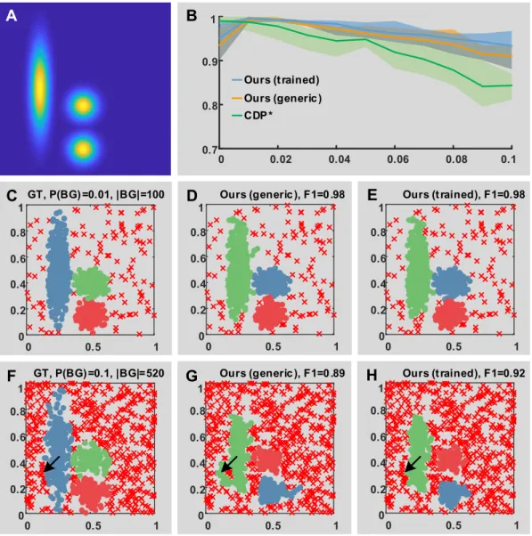
![Figure 5.2. Evaluation on a synthetic dataset with heterogeneous structures. A-B. The per- per-formances of the proposed method are evaluated on the synthetic dataset 01_chang_pathbased provided in ClustEval [ 268 ]](https://thumb-eu.123doks.com/thumbv2/123doknet/14315058.495926/108.892.149.749.246.771/evaluation-synthetic-heterogeneous-structures-formances-evaluated-synthetic-clusteval.webp)