Publisher’s version / Version de l'éditeur:
Vous avez des questions? Nous pouvons vous aider. Pour communiquer directement avec un auteur, consultez la première page de la revue dans laquelle son article a été publié afin de trouver ses coordonnées. Si vous n’arrivez pas à les repérer, communiquez avec nous à PublicationsArchive-ArchivesPublications@nrc-cnrc.gc.ca.
Questions? Contact the NRC Publications Archive team at
PublicationsArchive-ArchivesPublications@nrc-cnrc.gc.ca. If you wish to email the authors directly, please see the first page of the publication for their contact information.
https://publications-cnrc.canada.ca/fra/droits
L’accès à ce site Web et l’utilisation de son contenu sont assujettis aux conditions présentées dans le site LISEZ CES CONDITIONS ATTENTIVEMENT AVANT D’UTILISER CE SITE WEB.
Biomacromolecules, 21, 6, pp. 2236-2245, 2020-03-30
READ THESE TERMS AND CONDITIONS CAREFULLY BEFORE USING THIS WEBSITE. https://nrc-publications.canada.ca/eng/copyright
NRC Publications Archive Record / Notice des Archives des publications du CNRC :
https://nrc-publications.canada.ca/eng/view/object/?id=2f585924-8a91-46ed-9d75-f8ad3603a165 https://publications-cnrc.canada.ca/fra/voir/objet/?id=2f585924-8a91-46ed-9d75-f8ad3603a165
NRC Publications Archive
Archives des publications du CNRC
This publication could be one of several versions: author’s original, accepted manuscript or the publisher’s version. / La version de cette publication peut être l’une des suivantes : la version prépublication de l’auteur, la version acceptée du manuscrit ou la version de l’éditeur.
For the publisher’s version, please access the DOI link below./ Pour consulter la version de l’éditeur, utilisez le lien DOI ci-dessous.
https://doi.org/10.1021/acs.biomac.0c00201
Access and use of this website and the material on it are subject to the Terms and Conditions set forth at
Carboxylated chitosan nanocrystals: a synthetic route and application
as superior support for gold-catalyzed reactions
Jin, Tony; Kurdyla, Davis; Hrapovic, Sabahudin; Leung, Alfred C. W.;
Régnier, Sophie; Liu, Yali; Moores, Audrey; Lam, Edmond
Carboxylated Chitosan Nanocrystals: A Synthetic Route and
Application as Superior Support for Gold-Catalyzed Reactions
Tony Jin, Davis Kurdyla, Sabahudin Hrapovic, Alfred C. W. Leung, Sophie Régnier, Yali Liu,
Audrey Moores,
*
and Edmond Lam
*
Cite This:Biomacromolecules 2020, 21, 2236−2245 Read Online
ACCESS
Metrics & More Article Recommendations*
sı Supporting InformationABSTRACT: In this study, we demonstrate for the first time the fabrication of carboxylated chitosan nanocrystals (ChsNC) with high degree of deacetylation (DDA) at >80% and narrow size distribution. We also studied its application as a sustainable support material for metal-based catalysts. Carboxylated chitin nanocrystals (ChNCs) were initially prepared through partial cleavage of glycosidic bonds in chitin by ammonium persulfate, with concurrent oxidation of chitin C6 primary alcohols to produce carboxylate groups on the surface of the ChNCs. ChsNCs were subsequently prepared using an alkaline deacetylation procedure in the presence of NaBH4to preserve the nanorod structure of the biomaterial. The
resulting nanocrystals feature both carboxyl and amino functional groups. Transmission electron microscopy (TEM), X-ray diffraction
(XRD), and Fourier-transform infrared (FTIR) spectroscopy were used to determine the morphology and composition of these carboxylated ChNCs and ChsNCs. Subsequently, we tested the ability of the as-made ChsNCs as a biomass-based catalyst support for Au nanoparticles (NPs) using the 4-nitrophenol reduction and the aldehyde−amine−alkyne (A3) coupling reactions to
demonstrate its capabilities in regard to the ones of cellulose nanocrystals (CNCs). In particular, Au NPs over ChsNCs featured the highest turnover frequency (TOF) value for the 4-nitrophenol reduction reported for all Au-based catalysts supported on carbon-based systems. Spectroscopic and imaging techniques confirmed the importance of precisely controlling the redox state of Au as it is being deposited to afford a highly disperse active site on the bionano-support.
■
INTRODUCTIONResearch into biobased nanomaterials has seen major advance-ments in the past decade for a multitude of disciplines ranging from energy, electronics, medicine, sustainable packaging, environmental remediation, and coatings.1−3 This effort was pioneered by research on cellulose nanocrystals (CNCs), a material readily accessible via cellulose acid hydrolysis. CNCs are nontoxic materials with large surface area, high mechanical strength, and tunable colloidal and self-assembly behavior in aqueous media, making them exciting for a number of downstream applications in nanomedicinal drug delivery, food packaging, or papermaking industry, to name only a few.4−7 Their small size and the presence of coordinating groups on their surface, such as hydroxyls, sulfate half esters, and carboxylates, enable the facile deposition of metal species, which have been further utilized in catalysis.8−14In particular, it was shown that CNCs were excellent stabilizers for Cu complexes for oxidation reactions,15 Au nanoparticles (NPs) applied to nitrophenol reduction,16 Ru NPs for arene hydrogenation,17 Ag NPs for carbonyl hydrogenation,18Pd NPs for phenol hydrogenation19 and even for highly effective chirality transfer from the CNC surface in a Pd-catalyzed carbonyl hydrogenation reaction.9
While CNCs were undergoing these exciting developments, chitin, on the other hand, had been scarcely explored in this form, despite the presence of nitrogen-containing functionalities as a handle for further manipulations.20Chitin serves as a core structural material in crustaceans, fungi, and certain insects. With shrimp and crab shells as primary sources, the annual availability of chitin is in the range of 1 to 100 billion tons.21 Currently in the seafood industry, shell waste is often discarded back into the sea or in landfills, causing disposal costs as well as environmental concerns.22 Therefore, it is critical to develop valuable downstream applications for this extremely abundant material and enable the concept of the “shell biorefinery”.23 Chitin has limited solubility in aqueous and organic media,24−28 contrary to its deacetylated form, chitosan.26 While many methods exist for the conversion of chitin into chitosan, green
Received: February 11, 2020
Revised: March 25, 2020
Published: March 30, 2020
Article
pubs.acs.org/Biomac
© 2020 American Chemical Society
2236
https://dx.doi.org/10.1021/acs.biomac.0c00201
Biomacromolecules 2020, 21, 2236−2245
Downloaded via NATL RESEARCH COUNCIL CANADA on June 23, 2020 at 10:59:08 (UTC).
alternatives are still on demand.26,28,29 Both bulk chitin and chitosan have been reported as effective supports for metal NPs and applied in catalysis.30−33Interestingly, the rich literature of CNC applications as catalyst supports reveal that they possess distinct properties to bulk cellulose, enabling for instance chiral induction9or mild reaction conditions by stabilizing ultrasmall
metal NPs.17 Thus, exploring chitin and chitosan in the nanoscale for catalysis application is highly promising.
The production of chitin nanocrystals (ChNCs) and nanofibrils has been investigated before.34−36Typically, strong mineral acids are used to hydrolyze the amorphous regions of chitin to yield highly crystalline nanochitin materials. TEMPO-mediated oxidation may also produce carboxylated chitin Scheme 1. Schematic Depicting the Two Steps of Fabrication from Bulk Chitin to Carboxylated Chitosan Nanocrystals
Figure 1.LV-TEM micrographs taken at 5600× magnification of (a) ChNCs and (b) ChsNCs. (c) FTIR spectra of bulk chitin (blue), ChNCs (orange), and ChsNCs (black). (d) XRD spectra of bulk chitin, ChNCs, and ChsNCs, with Miller indices indicating the common reflection planes of α-chitin.52
nanofibrils.34However, these methods rely on the use of harsh and/or corrosive chemicals. Our group (Lam) has recently patented the procedure for producing carboxylated ChNCs from chitin using ammonium persulfate as a mild oxidant,37and other groups have followed suit and used similar methods.38−40 From ChNCs, a major goal is the formation of chitosan nanocrystals (ChsNCs) by deacetylation, since the amine functionalities will impart surface charges to the nanocrystals and thus improve dispersibility in polar solvents. We (Lam group) reported that the use of concentrated NaOH to directly deacetylate ChNCs does lead to the formation of ChsNCs.37 However, the process had severe limitations as attempts to further deacetylate ChNCs above 70% degree of deacetylation (DDA) leads to uncontrolled depolymerization of the chitin structure, resulting in the production of spherical NPs with nonuniform size distribution. Indeed, a method to fabricate ChsNCs has yet to be developed with retention of its nanorod dimensions and dispersion. Aside from the development of clean ChNC and ChsNC syntheses, hybridization of ChNCs and ChsNCs with metal NPs also remains largely unexplored.
Herein, we first report a facile and scalable procedure for fabricating carboxylated ChNCs and ChsNCs from chitin. We performed a complete physicochemical characterization and studied the colloidal behavior of the resulting NPs. Then, two methods were used to immobilize Au onto both carboxylated ChNCs and ChsNCs. ChsNC-supported Au was tested as catalysts for the reduction of 4-nitrophenol and for the aldehyde-amine-alkyne (A3) coupling reaction. For the reduction of
4-nitrophenol, we observed the highest turnover frequencies (TOFs) ever reported for carbon-based material supported Au NPs and suggest that this high activity is caused by the uniqueness in using ChsNCs as a support, outperforming CNC-based catalysts by an order of magnitude. For the A3coupling
reaction, it was found that high activity was linked to the ability of ChsNCs to effectively stabilize highly disperse Au(I) species. We hope to provide an initial framework for the design and synthesis of carboxylated ChsNCs, as well as demonstrate the applicability that this biomass material has in stabilizing active metal species than any other biomass-based support.
■
RESULTS AND DISCUSSIONSynthesis of ChNCs and ChsNCs. The synthesis of
nanopolysaccharides (CNCs and ChNCs) typically use high concentrations of strong acids (H2SO4 or HCl), where
hydrolysis occurs through protonation of the glycosidic oxygen units in the biopolymeric chain, yielding fragments of shorter chain biopolymers while preserving the glucopyranosic back-bone.41,42 It is accepted that amorphous regions in native cellulose or chitin are more accessible to acidic hydrolysis attack, leaving the crystalline regions intact. We have demonstrated that low concentrations of ammonium persulfate (APS) can hydrolyze both cellulose or chitin as efficiently as concentrated mineral acids such as sulfuric or hydrochloric acid. APS is considered a safer reagent to handle than mineral acids, which are very corrosive.37,43,44Specifically, in acidic media, persulfate radicals along with hydrogen peroxide are released and act in concert to degrade the amorphous regions of the biopolymer chain via free-radical propagation and oxidation of the glycosidic bond. The oxidative environment also explains the oxidation of surface primary alcohol functionalities into carboxylic acids.43
Bulk chitin from shrimp shells were treated with APS at 60 °C for 16 h to afford carboxylated ChNCs, as shown inScheme 1. The ChNC morphology was analyzed by low voltage TEM
(LV-TEM,Figure 1a). The average size of the nanorod ChNCs is 239 ±7 nm in length and 4.60 ± 0.06 nm in width (n = 400). AFM images confirmed these results (Figure S1a). FTIR analysis of ChNCs (Figure 1c) was used to identify the chemical transformations caused by APS treatment (full assignment in
Table S1). Overall, FTIR spectra of bulk chitin (blue line) and synthesized ChNCs (orange line) are similar, with the exception for a peak centered at 1743 cm−1 present only in ChNCs,
attributed to the CO stretching band. It confirmed that chitin C6 alcohols were oxidized into carboxylic acids in ChNCs (Figures 1c andS2). Previous studies have shown that FTIR spectra can conveniently be used to estimate the degree of oxidation (DO) of surface carboxyl groups in cellulose nanomaterials as the ratio of the intensities of the absorbance bands at 1726 cm−1(ν
(C=O)in the acid form) and at 1033 cm−1
(polysaccharide backbone).45,46We concluded that the DO was hardly affected by deacetylation, since the DOs for carboxylated ChNC and ChsNC were 0.051 and 0.052, respectively. Bulk chitin and ChNCs were characterized by X-ray diffraction (XRD, Figure 1d). Characteristic diffraction peaks located at 9.6°, 19.6°, 21.1°, and 23.7° are consistent with α-chitin polymorph for ChNCs.47−49The crystallinity index (CRI) of ChNCs was estimated to be 75.9%, similar to bulk α-chitin of 82.2%.50This result is comparable to other crystalline chitin nanomaterials, such as an 86% literature value reported for chitin nanowhiskers.51
In order to deacetylate ChNCs into ChsNCs, ChNCs were suspended into a 40% (w/w) aqueous solution of NaOH at 117 °C for 18 h. Novel to this report is the addition of 10% (w/w) NaBH4 as a crucial reagent to prevent depolymerization, as
previously reported in other contexts.37This method has been developed to control the similar “end-peeling” phenomenon of cellulosic materials in alkaline environments.53 Specifically,
NaBH4selectively reduces the terminal alditols of the polymer
chains.54LV-TEM analysis of ChsNCs confirmed the desired retention of nanorod structure across the sample (Figure 1b), where the average size of the ChsNC is 182 ± 2 nm in length and 2.68 ± 0.02 nm in width (n = 1048), with further evidence in the AFM imaging (Figure S1b). Retention of the carboxyl COOH stretch is apparent in FTIR (Figure 1c), which validates that NaBH4 does not reduce the carboxyl functionality in the
process. It is noted though that spectral features within the peaks are lost in the FTIR spectrum of ChsNCs in comparison to both ChNCs and bulk chitin, notably in the O−H and N−H stretching peaks in the 3500−3000 cm−1 region (Table S1).
This phenomenon is attributed to a loss of crystallinity within the ChsNCs. The N−H bend found at 1560 cm−1, and the C−O
stretch from the chitin backbone at 1030 cm−1can be used to
determine the DDA using a previously reported method.34,55In all cases, the DDA was measured to be higher than 80% for ChsNCs. In contrast, in the absence of methods to control end-peeling, the direct deacetylation of ChNCs to ChsNCs led to the formation of variably sized, spherical chitosan NPs, which can be seen in both AFM (Figure S1c) and TEM (Figure S3).
XRD analysis confirmed ChsNCs (Figure 1d) were
amorphized during deacetylation treatment, in agreement with the FTIR data (Figure 1c). The CRI value for ChsNCs was estimated to be 23.6%, a major decrease from 75.9%, measured for ChNCs. This crystallinity decrease was accompanied by a small shift toward higher angles and a large intensity decrease of the 020 reflection at 9.6° (Figure 1d).52A similar, yet much more intense, shift of this peak all the way to 11° had been previously reported by Chirachanchai and was associated with
Biomacromolecules pubs.acs.org/Biomac Article
https://dx.doi.org/10.1021/acs.biomac.0c00201
Biomacromolecules 2020, 21, 2236−2245
the formation of a webbed scaffold, distinct from the discrete crystals we observed for ChsNC.56 Also, previous studies reported that the decrease in intensity of the 020 reflection correlated linearly with DDA value.57 The peak at 12.5° is attributed to the 021 reflection and remained fairly intense as chitin was converted from ChNCs to ChsNCs, although a direct comparison between peak intensities is misleading because of the large difference in signal-to-noise ratios in the various XRD spectra. Interestingly for the ChsNCs XRD spectrum, a high degree of convolution is seen in the reflections that envelope the chitin peaks found at 19.6°, 21.1°, and 23.7°, which are the 110, 120, and 101 reflections, respectively, as a consequence of amorphization. Similar amorphization effects were observed in many other chitin to chitosan deacetylation procedures.57,58
DLS and ζ-potential measurements were also conducted for both ChNCs and ChsNCs and presented in Table 1. The
negative ζ-potential value of −36.9 ± 3.1 mV for ChNCs in water is consistent with the presence of negatively charged carboxylate groups at the nanorod surface. Conversely, a positive value of 47.3 ± 1.0 mV for the ζ-potential is indicative of the positive surface charge on ChsNCs from the protonated amino groups dispersed in water at pH ∼ 6.5.
As both amines and carboxylates are pH active functional groups, the pH-responsive behavior of ChsNCs was investigated by using DLS and ζ-potential measurements and compared with bulk chitosan (Figure S4).59 Different concentrations of ChsNCs in acetic acid were prepared in the range 1−0.1% (w/w) and the pH was adjusted from 1 to 12 by adding either HCl or NaOH solutions. At pH 2, ChsNC amino groups are protonated, resulting in dispersed ChsNC solutions with positive ζ-potentials beyond 50 mV. As the pH was decreased to under pH 2, a decrease in the absolute value of the ζ-potential was reported, likely because of the high ionic charge resulting from the strong HCl concentrations in these conditions, resulting in surface charge shielding. As the pH of the ChsNC solutions was increased from 2 to 7, the absolute value of the ζ-potential decreased steadily to <20 mV, consistent with the gradual deprotonation of the quaternary ammonium groups. As the solutions became very basic (pH > 10), the ζ-potential dropped below 0 mV to a few units mV in the negative scale, revealing the negative charges of the carboxylate groups. The apparent particle size was measured by DLS. Under acidic conditions and up to pH 6.5, the apparent particle size of ChsNCs remained constant around 120 nm, a value consistent with AFM and TEM measurements. Beyond 6.5 though, the particle size increased rapidly as a function of pH as a result of the drop in surface charge. This justifies that acidic conditions were kept while exploring the properties of ChsNCs for catalysis in the following.
The physicochemical characterization of the carboxylated ChsNCs confirmed that the developed process afforded crystalline, nanorod-shaped ChsNCs with DDA consistently above 80%. The scalability of the process presented inScheme 1
has been demonstrated in which ChNCs have been produced at
the 200 L batch scale, while ChsNCs have been produced at the 5 L batch scale, as shown in theSupporting Information.
Deposition of Au onto ChNCs and ChsNCs. We then
explored the use of these nanoscale biomaterials as supports for Au NPs, owing to their unique chemical functionalities and higher overall surface area compared to their bulk counterparts. In order to pave the way as initial frameworks for metal NP deposition onto ChsNC domains, two separate strategies of Au NP immobilization were investigated as outlined inScheme 2.
First, we investigated the “layer-by-layer” (LBL) method, a two-step process previously developed by Lam et. al for CNC functionalization.16This method enables the immobilization of prefabricated, carbonate-stabilized Au NPs of small size (<5 nm) onto CNCs. The positively charged polymer polydiallyldime-thylammonium chloride (PDDA) was coated onto ChsNCs, before Au NPs were immobilized over this hybrid, resulting in the formation of a light purple solid, Au@ChsNC-LBL. TEM imaging (Figure 2a) showed Au NPs deposited onto ChsNCs with an average size of 3.7 ± 0.8 nm (Figure S5) with many of them aggregated in discrete locations along the nanorods. UV− vis absorption corroborated this aggregation (Figure S6). A red shift of the localized surface plasmon resonance (LSPR) peak of the ready-made 3 nm Au NPs at 510 to 525 nm for Au@ChsNC-LBL was observed, which is a well-known effect of aggregation.60,61
This aggregation is consistent with the fact that ChsNCs feature a strongly positive surface charge, repulsing the positively charge PDDA-covered Au NPs. This is contrasted by the uniform distribution of Au NPs on CNCs using the same LBL method, since the CNCs in this case featured carboxylate anionic functionalities on their surface.16
Alternatively, we used a one-pot deposition-precipitation technique, via hydrogen reduction (HR), in which the Au salt precursor was reduced by hydrogen gas in situ within a suspension of ChsNCs, inspired by work done to access Pd@ CNCs.19As seen inFigures 2b andS7, while the HR method created larger Au NPs (6.6 ± 1.8 nm), they were far less aggregated for Au@ChsNC-HR than the immobilized Au NPs found using the LBL method. Moreover, all Au NPs were located on the surface of ChsNCs, with no “free” Au NPs unattached to a ChsNC. We hypothesized that the carboxylate functionalities on the ChsNCs could promote a suitable Table 1. DLS and ζ-Potential Measurements of ChNCs and
ChsNCs at pH 6.5
sample apparent particle size(nm) polydispersity index(PDI) ζ-potential(mV) ChNC 99.0 ± 1.9 0.189 ± 0.016 −36.9 ± 3.1 ChsNC 149.8 ± 0.3 0.187 ± 0.004 +47.3 ± 1.0
Scheme 2. Synthetic Routes to Deposit Au NPs onto the ChsNCsa
aThe two fabrication routes presented are the layer-by-layer method
coordination environment for Au salts, which were then subsequently reduced in situ into NPs, favoring good dispersity and interaction with the support.62
Along with the contrast in morphology created between the two fabrication procedures, the resulting Au species immobi-lized on ChsNCs from the two processes were also chemically dissimilar. X-ray photoelectron spectroscopy (XPS) was used to
analyze the oxidation state of Au from both LBL and HR methods, as found inFigure 2c. The two peaks in the normalized Au 4f high-resolution XPS spectra are the Au 4f5/2and Au 4f7/2
spin−orbit split peaks. For the Au 4f7/2binding energies of the
Au@ChsNC-LBL seen inFigure 2c, the peak was at 83.1 eV, which is a lower binding energy than the theoretical binding energy of metallic Au of 84.0 eV, marked by a dotted line in Figure 2.Brightfield TEM micrographs at 25000× magnification for (a) Au@ChsNC-LBL and (b) Au@ChsNC-HR, stained with 1 mM uranyl acetate. The insets for both (a) and (b) depict photographs of the two nanocomposites as dried powders. (c) Normalized Au 4f XPS spectra comparing the compositional profiles of nanocomposites fabricated in various conditions: (black line) Au@ChsNC-HR and (blue line) Au@ChsNC-LBL, with the theoretical binding energy of metallic Au at 84.0 eV shown as the dotted line. The inset depicts the deconvolution of the Au@ChsNC-HR Au 4f XPS spectrum. (d) The deconvolution of the Au@ChsNC-HR Au 4f high resolution XPS spectrum found in (c), with the legend indicating the experimental (gray), fitted (black), Au(I) (red), and Au(0) (yellow) lines.
Figure 3.Representative time-dependent UV−vis absorption spectra for the reduction of 4-nitrophenol to 4-aminophenol from 0 to 10 min using Au@ChsNC-LBL. The insets depict the reaction equation (top right) along with the plot of ln (A/A0) vs time (bottom right) depicting the pseudo-first order kinetics of the reaction
Biomacromolecules pubs.acs.org/Biomac Article
https://dx.doi.org/10.1021/acs.biomac.0c00201
Biomacromolecules 2020, 21, 2236−2245
Figure 2c. This could be attributed to the lower cluster size of the Au being in a NP form, which has been reported by other groups, as well as an electron transfer from the stabilizing carbonate ligand to the Au NP itself.63,64
As for the Au 4f spectrum of Au@ChsNC-HR (black line,
Figure 2c), the Au 4f7/2peak was at a higher binding energy than
84.0 eV. Deconvolution of this peak (Figure 2d) revealed it enveloped two subpeaks at 83.0 and 84.9 eV, attributed to Au(0) and Au(I), respectively.65,66XRD spectra further corroborated this observation of the partial reduction of Au using the HR method, contrary to the LBL method (Figure S8). No reflection planes for metallic Au were found in the Au@ChsNC-HR, while broad reflections at 38.1° and 44.3° in the Au@ChsNC-LBL material were indicative of the presence of elemental Au.67,68 This further validated the incomplete reduction of Au within the Au@ChsNC-HR material. The influence of pH during the HR reduction of Au on ChsNCs was investigated and is provided in theSupporting Information (SI) section below Figure S9. Low pH conditions favored the formation of Au(0), while at higher pH the formation of Au(I) was evident.
Catalytic Reduction of 4-Nitrophenol. The catalytic performance of Au@ChsNC-LBL and Au@ChsNC-HR for the 4-nitrophenol reduction were evaluated by UV−vis spectrosco-py (Figure 3).69−71
After addition of the catalyst to the reaction mixture, a decrease in the intensity at 400 nm from the 4-nitrophenolate anion was accompanied by a slight increase in absorption at 295 nm, corresponding to the absorption of 4-aminophenolate anion. The reaction with either catalyst system reached 99% completion within the first 10 min. All 4-nitrophenol reduction reactions were done at room temperature (298 K) unless otherwise specified. The activation energies for Au@ChsNC-LBL and Au@ChsNC-HR were 28.4 and 36.7 kJ/mol, respectively, as calculated from the Arrhenius plots shown in
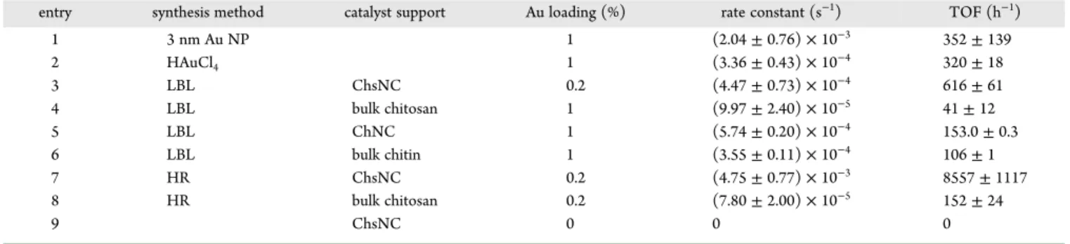
Figure S10. Table 2shows the calculated rate constants and turnover frequencies (TOFs) for various catalysts made in this work.
First, free 3 nm carbonate-stabilized Au NPs, the one used in the LBL method, proved to be active in the catalytic reaction, serving as a positive control for the reaction conditions (entry 1). The ability for in situ reduction of Au by NaBH4during the
catalyst experiment was established by testing HAuCl4directly
as a catalyst (entry 2). The observation of some catalytic activity with this system confirmed the formation of Au(0) during the reaction.72−74 The comparison between the performance of Au@ChsNC-LBL and Au NPs supported on bulk chitosan revealed the importance of bringing chitosan down to the nanoscale. Au@ChsNC-LBL featured a TOF value over an order of magnitude higher than Au NPs supported by chitosan
(entries 3 and 4). This is also seen for chitin as well, in which Au supported on ChNCs has a higher TOF value (entry 5) than Au supported on bulk chitin (entry 6). It is also important to note that the ChsNCs support for Au NPs (entry 3) outperformed the ChNCs support (entry 5). A possible explanation lies in the fact that ChsNCs being positive charges overall, may interact favorably with borohydride anions and accelerate the reaction accordingly. Furthermore, by comparing the two methods for producing catalysts on ChsNCs, the HR method (entry 7) was superior to the LBL method, with a TOF value (8557 h−1) an
order of magnitude greater than the LBL method (entry 3, 616 h−1). From prior characterization of the material, the HR
method yielded a catalyst with a significant portion of the metal in the +1 redox state, likely stabilized by carboxylate functionalities on the surface of ChsNCs. These are presumably reduced in situ under catalytic conditions to yield highly dispersed active sites. On the other hand, the LBL method results in aggregation of Au NPs present on the ChsNCs. This may explain the superior activity observed with the HR made nanocatalysts. We attempted XPS analysis postcatalysis, unfortunately too much noise inhibited a valid understanding of the Au oxidation state. A blank experiment was performed whereby we reacted 4-nitrophenol with the support alone ChsNC (entry 9). No conversion was observed in this case, indicating that the support does not act as an adsorbent for nitrophenolate anions, unlike other carbon-based supports.75 This rules out any contribution of adsorption in the 4-nitrophenol degradation activity of Au@ChsNC catalysts.
Au-ChsNC nanocatalysts exhibited superior catalytic activity compared to literature values for other Au-based catalysts immobilized on carbon-based supports (Table S2). Other metal-supported catalysts are also presented for comparison. This assessment clearly depicts that through the combined characteristics of nanoscale dimensions and unique amine and carboxylate functionalities, ChsNCs are a viable, biomass-based support capable of outperforming the current CNC-based supports popularly utilized.
By using the 4-nitrophenol model reaction as a framework for demonstrating the catalytic ability of these nanomaterials, we have concluded the efficacy of using nanoscale chitin and chitosan over their bulk counterparts from the TOF values, as depicted inTable 2. From this we also establish the ability of chitosan as a much better support for Au NPs in lieu of chitin, as well as confirm the viability of the one-pot synthesis to immobilize Au onto the ChsNC structure with control of the Au oxidation state. From this framework, we studied the activity of these nanocatalysts for another important chemical trans-formation.
Table 2. Rate Constants and Turnover Frequency Values for the Reduction of 4-Nitrophenol to 4-Aminophenol for Various Catalysts and Supports
entry synthesis method catalyst support Au loading (%) rate constant (s−1) TOF (h−1)
1 3 nm Au NP 1 (2.04 ± 0.76) × 10−3 352 ± 139 2 HAuCl4 1 (3.36 ± 0.43) × 10−4 320 ± 18 3 LBL ChsNC 0.2 (4.47 ± 0.73) × 10−4 616 ± 61 4 LBL bulk chitosan 1 (9.97 ± 2.40) × 10−5 41 ± 12 5 LBL ChNC 1 (5.74 ± 0.20) × 10−4 153.0 ± 0.3 6 LBL bulk chitin 1 (3.55 ± 0.11) × 10−4 106 ± 1 7 HR ChsNC 0.2 (4.75 ± 0.77) × 10−3 8557 ± 1117 8 HR bulk chitosan 0.2 (7.80 ± 2.00) × 10−5 152 ± 24 9 ChsNC 0 0 0
A3 Coupling Catalysis. The A3 coupling reaction is an
atom-economical, multicomponent synthesis of propargyl-amines.76 It serves as an alternative to the less sustainable reaction of using stoichiometric quantities of organometallic reagents such as butyllithium to activate the highly acidic terminal hydrogen of an alkynyl to form a metal acetylide, before subsequent addition to an imine.77,78 Much work has been performed to understand its mechanism, along with developing more effective catalysts for this reaction in terms of TOF, stability, and recyclability.79More specifically, the usage of Au as a transition metal catalyst has been studied extensively within the field.80Both metallic Au NPs79as well as Au salts in the form of Au(I)/Au(III) have the ability to catalyze this reaction.76,81,82 For example, Li and co-workers have shown that Au(I) salts are an effective catalyst for the synthesis of propargylamines in water.83Work has also been done on using biomaterials such as CNCs to support Au catalysts for A3coupling, albeit with lower
activity than their homogeneous counterparts.84Spurred by the heightened catalytic ability of Au@ChsNC-HR, where the highly dispersed gold exists as Au(0) and Au(I), we explored the ability of this nanomaterial to catalyze the A3coupling reaction.
In a standard procedure, we used model substrates, benzaldehyde, piperidine, and phenylacetylene, keeping the benzaldehyde as the limiting reagent in order to favor the formation of the imine intermediate. FromTable 3, it can be
seen that by using the free carbonate-stabilized Au NPs, negligible amounts of product were formed, evidencing that metallic Au may not play a major role as the active catalyst in the A3coupling reaction (entry 9). In contrast to this, using HAuCl
4
salts is seen to have full conversion to the proparglymine product in 24 h (entry 10). The inability for Au(0) to catalyze the reaction is further seen by using Au@ChsNC-LBL (8%, entry 11), which was confirmed previously to feature pure Au(0). In contrast, substantial conversion and yield can be seen using Au@ChsNC-HR with only 0.1 mol % Au loading (91%, entry 12). By increasing the Au loading to 0.5 mol % Au, full conversion was reported (entry 13). Lower yields of 69% and 40% were reported at 6 h for 0.5 mol % (entry 14) and 0.1 mol % (entry 15) Au loading, respectively. The effects of temperature were examined as well in which >90% yield was achieved at 50
°C (entry 16). However, when the temperature is increased to 100 °C, a substantial decrease in yield is seen (56%, entry 17). Since full conversion of the starting reactants was observed, this poor yield was likely caused by potential product degradation at elevated temperatures.
■
CONCLUSIONSIn this work, we present the first method to produce carboxylated ChsNCs from bulk chitin. Using ammonium persulfate as a mild oxidant, cleavage of the chitin amorphous regions with concurrent oxidation of the C6 alcohol group leads to the formation of carboxylated ChNCs. Moreover, through a facile procedure using NaBH4 in alkaline conditions to limit
uncontrolled depolymerization, deacetylation of ChNCs occurs to produce ChsNCs with high DDA, retention of the nanorod structure, and the presence of both surface carboxyl and amine functionalities. Two different methods were used to create Au-immobilized ChsNC catalysts in which the composition and topography of these nanomaterials can be directly altered through the reaction conditions. The catalytic properties of the nanomaterial were then investigated in the reduction of 4-nitrophenol and the A3coupling reaction. We have shown that
nanoscale chitin and chitosan have a clear advantage in activity over bulk chitin and chitosan as support materials for heterogeneous catalysis. A highly dispersed dual Au(I)/Au(0) nanocatalyst (Au@ChsNC-HR) fabricated by direct hydrogen reduction of HAuCl4on ChsNC showed significant activity for
both model catalyst reactions. To the best of our knowledge, the Au@ChsNC-HR nanocatalyst exhibits the highest reported TOF for the Au-catalyzed 4-nitrophenol reduction reaction on carbon-based supports. From this work, we hope to show the potential of deriving high value products from chitinous waste streams obtained from the seafood industry, as well as exhibit the physicochemical properties of ChsNCs conferred by the different functional groups and its nanorod structure. This work provides a prospective of ChsNCs as a new bionanoma-terial that can compete with and possibly overcome CNCs in terms of applicability and efficacy.
■
ASSOCIATED CONTENT*
sı Supporting InformationThe Supporting Information is available free of charge at
https://pubs.acs.org/doi/10.1021/acs.biomac.0c00201. Experimental procedures depicting the syntheses of ChNC and ChsNC and their scale-up, fabrication methods for Au@ChsNC, standard catalytic reaction protocols, and additional characterization information, including AFM, FTIR peak assignments, UV−vis spectra, DLS, TEM, XRD, and XPS, as well as a table comparing the rate constant and turnover frequency of the 4-nitrophenol reaction obtained in this study with other works (PDF)
■
AUTHOR INFORMATIONCorresponding Authors
Audrey Moores − Department of Chemistry and Department of Materials Engineering, McGill University, Montreal, Quebec H3A 0B8, Canada; orcid.org/0000-0003-1259-913X;
Email:audrey.moores@mcgill.ca
Edmond Lam − Aquatic and Crop Resource Development Research Centre, National Research Council of Canada, Table 3. A3Coupling Reaction Tablea
entry synthesismethod supportcatalyst Au loading(%) temp(°C) time(h) yield(%) 9b 3 nm Au NP 1 80 24 2 10b HAuCl 4 1 80 24 >99 11 LBL ChsNC 0.1 70 24 8 12 HR ChsNC 0.1 70 24 91 13 HR ChsNC 0.5 70 24 >99 14 HR ChsNC 0.5 70 6 69 15 HR ChsNC 0.1 70 6 40 16 HR ChsNC 0.5 50 24 92 17 HR ChsNC 0.5 100 24 56
aAll reactions listed are done using 1 mmol benzaldehyde, 1.2 mmol
piperidine, and 1.5 mmol phenylacetylene, with no solvent. Yield was determined through 1H NMR.bReaction done using water as the
solvent.
Biomacromolecules pubs.acs.org/Biomac Article
https://dx.doi.org/10.1021/acs.biomac.0c00201
Biomacromolecules 2020, 21, 2236−2245
Montreal, Quebec H4P 2R2, Canada; orcid.org/0000-0003-4343-4469; Email:edmond.lam@cnrc-nrc.gc.ca
Authors
Tony Jin − Department of Chemistry, McGill University, Montreal, Quebec H3A 0B8, Canada
Davis Kurdyla − Department of Chemistry, McGill University, Montreal, Quebec H3A 0B8, Canada; Aquatic and Crop Resource Development Research Centre, National Research Council of Canada, Montreal, Quebec H4P 2R2, Canada Sabahudin Hrapovic − Aquatic and Crop Resource Development
Research Centre, National Research Council of Canada, Montreal, Quebec H4P 2R2, Canada
Alfred C. W. Leung − Aquatic and Crop Resource Development Research Centre, National Research Council of Canada, Montreal, Quebec H4P 2R2, Canada
Sophie Régnier − Aquatic and Crop Resource Development Research Centre, National Research Council of Canada, Montreal, Quebec H4P 2R2, Canada
Yali Liu − Aquatic and Crop Resource Development Research Centre, National Research Council of Canada, Montreal, Quebec H4P 2R2, Canada
Complete contact information is available at:
https://pubs.acs.org/10.1021/acs.biomac.0c00201
Author Contributions
All authors have given final approval to the final version of the manuscript.
Notes
The authors declare no competing financial interest.
■
ACKNOWLEDGMENTSWe thank the Natural Science and Engineering Research Council of Canada (NSERC) Discovery Grant and Discovery Accelerator Supplement, the Canada Foundation for Innovation (CFI), the National Research Council (NRC) New Beginnings Initiative Ideation fund, the Fonds de Research du Québec Nature et Technologie (FRQNT) - Centre du Chimie Verte et Catalyse (CCVC), the National Research Council Canada (NRC), and McGill University for their financial support. We thank the Facility for Electron Microscopy Research of McGill University for help in data collection. We thank the MC2facility
at McGill University for help in acquiring the FTIR and UV−vis spectra. Specifically, we thank Dr. Hatem Titi from the MC2
facility for help in acquiring PXRD spectra along with fruitful scientific discussion.
■
REFERENCES(1) Dufresne, A. Nanocellulose: a new ageless bionanomaterial. Mater.
Today 2013, 16, 220−227.
(2) Andresen, M.; Stenstad, P.; Møretrø, T.; Langsrud, S.; Syverud, K.; Johansson, L.-S.; Stenius, P. Nonleaching Antimicrobial Films Prepared from Surface-Modified Microfibrillated Cellulose. Biomacromolecules 2007, 8, 2149−2155.
(3) Thomas, B.; Raj, M. C.; B, A. K.; H, R. M.; Joy, J.; Moores, A.; Drisko, G. L.; Sanchez, C. Nanocellulose, a Versatile Green Platform: From Biosources to Materials and Their Applications. Chem. Rev. 2018,
118, 11575−11625.
(4) Xu, Q.; Gao, Y.; Qin, M.; Wu, K.; Fu, Y.; Zhao, J. Nanocrystalline cellulose from aspen kraft pulp and its application in deinked pulp. Int. J.
Biol. Macromol. 2013, 60, 241−247.
(5) Shojaeiarani, J.; Bajwa, D.; Shirzadifar, A. A review on cellulose nanocrystals as promising biocompounds for the synthesis of nanocomposite hydrogels. Carbohydr. Polym. 2019, 216, 247−259.
(6) Cunha, A. G.; Mougel, J.-B.; Cathala, B.; Berglund, L. A.; Capron, I. Preparation of Double Pickering Emulsions Stabilized by Chemically Tailored Nanocelluloses. Langmuir 2014, 30, 9327−9335.
(7) Jackson, J. K.; Letchford, K.; Wasserman, B. Z.; Ye, L.; Hamad, W. Y.; Burt, H. M. The use of nanocrystalline cellulose for the binding and controlled release of drugs. Int. J. Nanomed. 2011, 6, 321−330.
(8) Zhou, Z.; Lu, C.; Wu, X.; Zhang, X. Cellulose nanocrystals as a novel support for CuO nanoparticles catalysts: facile synthesis and their application to 4-nitrophenol reduction. RSC Adv. 2013, 3, 26066− 26073.
(9) Kaushik, M.; Basu, K.; Benoit, C.; Cirtiu, C. M.; Vali, H.; Moores, A. Cellulose Nanocrystals as Chiral Inducers: Enantioselective Catalysis and Transmission Electron Microscopy 3D Characterization. J. Am.
Chem. Soc. 2015, 137, 6124−6127.
(10) Tang, J.; Shi, Z.; Berry, R. M.; Tam, K. C. Mussel-Inspired Green Metallization of Silver Nanoparticles on Cellulose Nanocrystals and Their Enhanced Catalytic Reduction of 4-Nitrophenol in the Presence of β-Cyclodextrin. Ind. Eng. Chem. Res. 2015, 54, 3299−3308.
(11) Yan, W.; Chen, C.; Wang, L.; Zhang, D.; Li, A.-J.; Yao, Z.; Shi, L.-Y. Facile and green synthesis of cellulose nanocrystal-supported gold nanoparticles with superior catalytic activity. Carbohydr. Polym. 2016,
140, 66−73.
(12) George, J.; Sabapathi, S. N. Cellulose nanocrystals: synthesis, functional properties, and applications. Nanotechnol., Sci. Appl. 2015, 8, 45−54.
(13) Rezayat, M.; Blundell, R. K.; Camp, J. E.; Walsh, D. A.; Thielemans, W. Green One-Step Synthesis of Catalytically Active Palladium Nanoparticles Supported on Cellulose Nanocrystals. ACS
Sustainable Chem. Eng. 2014, 2, 1241−1250.
(14) Kaushik, M.; Moores, A. Review: nanocelluloses as versatile supports for metal nanoparticles and their applications in catalysis.
Green Chem. 2016, 18, 622−637.
(15) Chauhan, P.; Yan, N. Nanocrystalline cellulose grafted phthalocyanine: a heterogeneous catalyst for selective aerobic oxidation of alcohols and alkyl arenes at room temperature in a green solvent. RSC
Adv. 2015, 5, 37517−37520.
(16) Lam, E.; Hrapovic, S.; Majid, E.; Chong, J. H.; Luong, J. H. T. Catalysis using gold nanoparticles decorated on nanocrystalline cellulose. Nanoscale 2012, 4, 997−1002.
(17) Kaushik, M.; Friedman, H. M.; Bateman, M.; Moores, A. Cellulose nanocrystals as non-innocent supports for the synthesis of ruthenium nanoparticles and their application to arene hydrogenation.
RSC Adv. 2015, 5, 53207−53210.
(18) Kaushik, M.; Li, A. Y.; Hudson, R.; Masnadi, M.; Li, C.-J.; Moores, A. Reversing aggregation: direct synthesis of nanocatalysts from bulk metal. Cellulose nanocrystals as active support to access efficient hydrogenation silver nanocatalysts. Green Chem. 2016, 18, 129−133.
(19) Cirtiu, C. M.; Dunlop-Brière, A. F.; Moores, A. Cellulose nanocrystallites as an efficient support for nanoparticles of palladium: application for catalytic hydrogenation and Heck coupling under mild conditions. Green Chem. 2011, 13, 288−291.
(20) Kim, S.-K. Chitin, Chitosan, Oligosaccharides and Their
Derivatives: Biological Activities and Applications; CRC Press, 2010.
(21) Gopalan Nair, K.; Dufresne, A. Crab Shell Chitin Whisker Reinforced Natural Rubber Nanocomposites. 1. Processing and Swelling Behavior. Biomacromolecules 2003, 4, 657−665.
(22) MacLeod, J. A.; Kuo, S.; Gallant, T. L.; Grimmett, M. Seafood processing wastes as nutrient sources for crop production. Can. J. Soil
Sci. 2006, 86, 631−640.
(23) Chen, X.; Yang, H.; Yan, N. Shell Biorefinery: Dream or Reality?
Chem. - Eur. J. 2016, 22, 13402−13421.
(24) Shahidi, F.; Arachchi, J. K. V.; Jeon, Y.-J. Food applications of chitin and chitosans. Trends Food Sci. Technol. 1999, 10, 37−51.
(25) Park, B. K.; Kim, M.-M. Applications of chitin and its derivatives in biological medicine. Int. J. Mol. Sci. 2010, 11, 5152−5164.
(26) Jayakumar, R.; Menon, D.; Manzoor, K.; Nair, S. V.; Tamura, H. Biomedical applications of chitin and chitosan based nanomaterialsA short review. Carbohydr. Polym. 2010, 82, 227−232.
(27) Margoutidis, G.; Parsons, V. H.; Bottaro, C. S.; Yan, N.; Kerton, F. M. Mechanochemical Amorphization of α-Chitin and Conversion into Oligomers of N-Acetyl-d-glucosamine. ACS Sustainable Chem. Eng. 2018, 6, 1662−1669.
(28) Di Nardo, T.; Hadad, C.; Nguyen Van Nhien, A.; Moores, A. Synthesis of high molecular weight chitosan from chitin by mechanochemistry and aging. Green Chem. 2019, 21, 3276−3285.
(29) Chen, X.; Yang, H.; Zhong, Z.; Yan, N. Base-catalysed, one-step mechanochemical conversion of chitin and shrimp shells into low molecular weight chitosan. Green Chem. 2017, 19, 2783−2792.
(30) Zhou, J.; Dong, Z.; Yang, H.; Shi, Z.; Zhou, X.; Li, R. Pd immobilized on magnetic chitosan as a heterogeneous catalyst for acetalization and hydrogenation reactions. Appl. Surf. Sci. 2013, 279, 360−366.
(31) Qiu, Y.; Ma, Z.; Hu, P. Environmentally benign magnetic chitosan/Fe3O4 composites as reductant and stabilizer for anchoring Au NPs and their catalytic reduction of 4-nitrophenol. J. Mater. Chem. A 2014, 2, 13471−13478.
(32) Wei, D.; Ye, Y.; Jia, X.; Yuan, C.; Qian, W. Chitosan as an active support for assembly of metal nanoparticles and application of the resultant bioconjugates in catalysis. Carbohydr. Res. 2010, 345, 74−81. (33) Primo, A.; Quignard, F. Chitosan as efficient porous support for dispersion of highly active gold nanoparticles: design of hybrid catalyst for carbon-carbon bond formation. Chem. Commun. 2010, 46, 5593− 5595.
(34) Fan, Y.; Saito, T.; Isogai, A. Chitin Nanocrystals Prepared by TEMPO-Mediated Oxidation of α-Chitin. Biomacromolecules 2008, 9, 192−198.
(35) Goodrich, J. D.; Winter, W. T. α-Chitin Nanocrystals Prepared from Shrimp Shells and Their Specific Surface Area Measurement.
Biomacromolecules 2007, 8, 252−257.
(36) Fan, Y.; Saito, T.; Isogai, A. Preparation of Chitin Nanofibers from Squid Pen β-Chitin by Simple Mechanical Treatment under Acid Conditions. Biomacromolecules 2008, 9, 1919−1923.
(37) Luong, J. H.; Lam, E.; Leung, C. W.; Hrapovic, S.; Male, K. B., Chitin Nanocrystals and Process for Preparation Thereof. U.S. Patent US20160272731A1, 2016.
(38) Ma, Q.; Pang, K.; Wang, K.; Huang, S.; Ding, B.; Duan, Y.; Zhang, J. Ultrafine and carboxylated β-chitin nanofibers prepared from squid pen and its transparent hydrogels. Carbohydr. Polym. 2019, 211, 118− 123.
(39) Oun, A. A.; Rhim, J.-W. Effect of oxidized chitin nanocrystals isolated by ammonium persulfate method on the properties of carboxymethyl cellulose-based films. Carbohydr. Polym. 2017, 175, 712−720.
(40) Oun, A. A.; Rhim, J.-W. Effect of isolation methods of chitin nanocrystals on the properties of chitin-silver hybrid nanoparticles.
Carbohydr. Polym. 2018, 197, 349−358.
(41) Lu, P.; Hsieh, Y.-L. Preparation and characterization of cellulose nanocrystals from rice straw. Carbohydr. Polym. 2012, 87, 564−573.
(42) Hon, D. N.-S.; Shiraishi, N. Wood and Cellulosic Chemistry,
Revised, and Expanded; CRC Press, 2000.
(43) Leung, A. C. W.; Hrapovic, S.; Lam, E.; Liu, Y.; Male, K. B.; Mahmoud, K. A.; Luong, J. H. T. Characteristics and Properties of Carboxylated Cellulose Nanocrystals Prepared from a Novel One-Step Procedure. Small 2011, 7, 302−305.
(44) Final Report on the Safety Assessment of Ammonium, Potassium, and Sodium Persulfate. Int. J. Toxicol. 2001, 20, 7−21.
(45) Habibi, Y.; Chanzy, H.; Vignon, M. R. TEMPO-mediated surface oxidation of cellulose whiskers. Cellulose 2006, 13, 679−687.
(46) Lam, E.; Leung, A. C. W.; Liu, Y.; Majid, E.; Hrapovic, S.; Male, K. B.; Luong, J. H. T. Green Strategy Guided by Raman Spectroscopy for the Synthesis of Ammonium Carboxylated Nanocrystalline Cellulose and the Recovery of Byproducts. ACS Sustainable Chem.
Eng. 2013, 1, 278−283.
(47) Wada, M.; Saito, Y. Lateral thermal expansion of chitin crystals. J.
Polym. Sci., Part B: Polym. Phys. 2001, 39, 168−174.
(48) Feng, F.; Liu, Y.; Hu, K. Influence of alkali-freezing treatment on the solid state structure of chitin. Carbohydr. Res. 2004, 339, 2321− 2324.
(49) Li, J.; Revol, J. F.; Marchessault, R. H. Effect of degree of deacetylation of chitin on the properties of chitin crystallites. J. Appl.
Polym. Sci. 1997, 65, 373−380.
(50) Lavall, R. L.; Assis, O. B. G.; Campana-Filho, S. P. β-Chitin from the pens of Loligo sp.: Extraction and characterization. Bioresour.
Technol. 2007, 98, 2465−2472.
(51) Heath, L.; Zhu, L.; Thielemans, W. Chitin Nanowhisker Aerogels. ChemSusChem 2013, 6, 537−544.
(52) Cárdenas, G.; Cabrera, G.; Taboada, E.; Miranda, S. P. Chitin characterization by SEM, FTIR, XRD, and 13C cross polarization/mass angle spinning NMR. J. Appl. Polym. Sci. 2004, 93, 1876−1885.
(53) Sjostrom, E. Wood Chemistry: Fundamentals and Applications; Gulf Professional Publishing, 1993.
(54) Batista, I.; Roberts, G. A. F. A novel, facile technique for deacetylating chitin. Makromol. Chem. 1990, 191, 429−434.
(55) Shigemasa, Y.; Matsuura, H.; Sashiwa, H.; Saimoto, H. Evaluation of different absorbance ratios from infrared spectroscopy for analyzing the degree of deacetylation in chitin. Int. J. Biol. Macromol. 1996, 18, 237−242.
(56) Lertwattanaseri, T.; Ichikawa, N.; Mizoguchi, T.; Tanaka, Y.; Chirachanchai, S. Microwave technique for efficient deacetylation of chitin nanowhiskers to a chitosan nanoscaffold. Carbohydr. Res. 2009,
344, 331−335.
(57) Zhang, Y.; Xue, C.; Xue, Y.; Gao, R.; Zhang, X. Determination of the degree of deacetylation of chitin and chitosan by X-ray powder diffraction. Carbohydr. Res. 2005, 340, 1914−1917.
(58) Clark, G. L.; Smith, A. F. X-ray Diffraction Studies of Chitin, Chitosan, and Derivatives. J. Phys. Chem. 1936, 40, 863−879.
(59) Liu, H.; Wang, C.; Zou, S.; Wei, Z.; Tong, Z. Simple, Reversible Emulsion System Switched by pH on the Basis of Chitosan without Any Hydrophobic Modification. Langmuir 2012, 28, 11017−11024.
(60) Liz-Marzán, L. M. Tailoring Surface Plasmons through the Morphology and Assembly of Metal Nanoparticles. Langmuir 2006, 22, 32−41.
(61) Moores, A.; Goettmann, F. The plasmon band in noble metal nanoparticles: an introduction to theory and applications. New J. Chem. 2006, 30, 1121−1132.
(62) Zhao, P.; Li, N.; Astruc, D. State of the art in gold nanoparticle synthesis. Coord. Chem. Rev. 2013, 257, 638−665.
(63) Konova, P.; Naydenov, A.; Venkov, C.; Mehandjiev, D.; Andreeva, D.; Tabakova, T. Activity and deactivation of Au/TiO2 catalyst in CO oxidation. J. Mol. Catal. A: Chem. 2004, 213, 235−240. (64) Arrii, S.; Morfin, F.; Renouprez, A. J.; Rousset, J. L. Oxidation of CO on Gold Supported Catalysts Prepared by Laser Vaporization: Direct Evidence of Support Contribution. J. Am. Chem. Soc. 2004, 126, 1199−1205.
(65) Molnár, Á. The use of chitosan-based metal catalysts in organic transformations. Coord. Chem. Rev. 2019, 388, 126−171.
(66) Sun, C.; Qu, R.; Chen, H.; Ji, C.; Wang, C.; Sun, Y.; Wang, B. Degradation behavior of chitosan chains in the ‘green’ synthesis of gold nanoparticles. Carbohydr. Res. 2008, 343, 2595−2599.
(67) Tsunoyama, H.; Sakurai, H.; Ichikuni, N.; Negishi, Y.; Tsukuda, T. Colloidal Gold Nanoparticles as Catalyst for Carbon-Carbon Bond Formation: Application to Aerobic Homocoupling of Phenylboronic Acid in Water. Langmuir 2004, 20, 11293−11296.
(68) Chen, Y.; Gu, X.; Nie, C.-G.; Jiang, Z.-Y.; Xie, Z.-X.; Lin, C.-J. Shape controlled growth of gold nanoparticles by a solution synthesis.
Chem. Commun. 2005, 4181−4183.
(69) Li, J.; Liu, C.-y.; Liu, Y. Au/graphene hydrogel: synthesis, characterization and its use for catalytic reduction of 4-nitrophenol. J.
Mater. Chem. 2012, 22, 8426−8430.
(70) Zeng, J.; Zhang, Q.; Chen, J.; Xia, Y. A Comparison Study of the Catalytic Properties of Au-Based Nanocages, Nanoboxes, and Nano-particles. Nano Lett. 2010, 10, 30−35.
Biomacromolecules pubs.acs.org/Biomac Article
https://dx.doi.org/10.1021/acs.biomac.0c00201
Biomacromolecules 2020, 21, 2236−2245
(71) Lee, J.; Park, J. C.; Song, H. A Nanoreactor Framework of a Au@ SiO2 Yolk/Shell Structure for Catalytic Reduction of p-Nitrophenol.
Adv. Mater. 2008, 20, 1523−1528.
(72) Pandey, S.; Mishra, S. B. Catalytic reduction of p-nitrophenol by using platinum nanoparticles stabilised by guar gum. Carbohydr. Polym. 2014, 113, 525−531.
(73) Zhao, P.; Feng, X.; Huang, D.; Yang, G.; Astruc, D. Basic concepts and recent advances in nitrophenol reduction by gold- and other transition metal nanoparticles. Coord. Chem. Rev. 2015, 287, 114−136.
(74) Rodrigues, C. S. D.; Soares, O. S. G. P.; Pinho, M. T.; Pereira, M. F. R.; Madeira, L. M. p-Nitrophenol degradation by heterogeneous Fenton’s oxidation over activated carbon-based catalysts. Appl. Catal., B 2017, 219, 109−122.
(75) Liu, S.; Lai, C.; Li, B.; Zhang, C.; Zhang, M.; Huang, D.; Qin, L.; Yi, H.; Liu, X.; Huang, F.; Zhou, X.; Chen, L. Role of radical and non-radical pathway in activating persulfate for degradation of p-nitrophenol by sulfur-doped ordered mesoporous carbon. Chem. Eng. J. 2020, 384, 123304.
(76) Peshkov, V. A.; Pereshivko, O. P.; Van der Eycken, E. V. A walk around the A3-coupling. Chem. Soc. Rev. 2012, 41, 3790−3807.
(77) Ma, Y.; Lobkovsky, E.; Collum, D. B. BF3-Mediated Additions of Organolithiums to Ketimines: X-ray Crystal Structures of BF3-Ketimine Complexes. J. Org. Chem. 2005, 70, 2335−2337.
(78) Aubrecht, K. B.; Winemiller, M. D.; Collum, D. B. BF3-Mediated Addition of Lithium Phenylacetylide to an Imine: Correlations of Structures and Reactivities. BF3·R3N Derivatives as Substitutes for BF3·Et2O. J. Am. Chem. Soc. 2000, 122, 11084−11089.
(79) Nasrollahzadeh, M.; Sajjadi, M.; Ghorbannezhad, F.; Sajadi, S. M. A Review on Recent Advances in the Application of Nanocatalysts in A3 Coupling Reactions. Chem. Rec. 2018, 18, 1409−1473.
(80) Soengas, R.; Navarro, Y.; Iglesias, M. J.; López-Ortiz, F. Immobilized Gold Nanoparticles Prepared from Gold(III)-Containing Ionic Liquids on Silica: Application to the Sustainable Synthesis of Propargylamines. Molecules 2018, 23, 2975.
(81) Cheng, M.; Zhang, Q.; Hu, X.-Y.; Li, B.-G.; Ji, J.-X.; Chan, A. S. C. Gold-Catalyzed Direct Intermolecular Coupling of Ketones, Secondary Amines, and Alkynes: A Facile and Versatile Access to Propargylic Amines Containing a Quaternary Carbon Center. Adv. Synth. Catal. 2011, 353, 1274−1278.
(82) Shore, G.; Yoo, W.-J.; Li, C.-J.; Organ, M. G. Propargyl Amine Synthesis Catalysed by Gold and Copper Thin Films by Using Microwave-Assisted Continuous-Flow Organic Synthesis (MACOS).
Chem. - Eur. J. 2010, 16, 126−133.
(83) Wei, C.; Li, C.-J. A Highly Efficient Three-Component Coupling of Aldehyde, Alkyne, and Amines via C-H Activation Catalyzed by Gold in Water. J. Am. Chem. Soc. 2003, 125, 9584−9585.
(84) Huang, J.-L.; Gray, D. G.; Li, C.-J. A3-Coupling catalyzed by robust Au nanoparticles covalently bonded to HS-functionalized cellulose nanocrystalline films. Beilstein J. Org. Chem. 2013, 9, 1388− 1396.