ORIGINAL ARTICLE
New reconstruction algorithm allows shortened acquisition
time for myocardial perfusion SPECT
Ines Valenta&Valerie Treyer&Lars Husmann&Oliver Gaemperli&
Michael J. Schindler&Bernhard A. Herzog&Patrick Veit-Heibach&
Ronny R. Buechel&René Nkoulou&Aju P. Pazhenkottil&Philipp A. Kaufmann
Received: 9 June 2009 / Accepted: 9 October 2009 / Published online: 18 November 2009 # Springer-Verlag 2009
Abstract
Purpose Shortening scan time and/or reducing radiation dose at maintained image quality are the main issues of the current research in radionuclide myocardial perfusion imaging (MPI). We aimed to validate a new iterative reconstruction (IR) algorithm for SPECT MPI allowing shortened acquisition time (HALF time) while maintaining image quality vs. standard full time acquisition (FULL time).
Methods In this study, 50 patients, referred for evaluation of known or suspected coronary artery disease by SPECT MPI using 99mTc-Tetrofosmin, underwent 1-day adenosine stress 300 MBq/rest 900 MBq protocol with standard (stress 15 min/rest 15 min FULL time) immediately followed by short emission scan (stress 9 min/rest 7 min HALF time) on a Ventri SPECT camera (GE Healthcare). FULL time scans were processed with IR, short scans were additionally processed with a recently developed software algorithm for HALF time emission scans. All reconstruc-tions were subsequently analyzed using commercially available software (QPS/QGS, Cedars Medical Sinai) with/without X-ray based attenuation correction (AC).
Uptake values (percent of maximum) were compared by regression and Bland-Altman (BA) analysis in a 20-segment model.
Results HALF scans yielded a 96% readout and 100% clinical diagnosis concordance compared to FULL. Corre-lation for uptake in each segment (n=1,000) was r=0.87at stress (p < 0.001) and r = 0.89 at rest (p < 0.001) with
respective BA limits of agreement of −11% to 10%
and −12% to 11%. After AC similar correlation (r=0.82,
rest; r=0.80, stress, both p<0.001) and BA limits were found (−12% to 10%; −13% to 12%).
Conclusion With the new IR algorithm, SPECT MPI can be acquired at half of the scan time without compromising image quality, resulting in an excellent agreement with FULL time scans regarding to uptake and clinical conclusion.
Keywords Myocardial perfusion imaging . SPECT . Scan time . Iterative reconstruction (IR) algorithms
Introduction
Myocardial perfusion imaging (MPI) using single-photon emission computed tomography (SPECT) is a well-established and widely accepted method for diagnostic and prognostic evaluation of patients with known or
suspected coronary artery disease (CAD) [1]. Nevertheless,
this method is still subject to drawbacks which are inherent to any nuclear technique with low photon flux, such as attenuation due to non-uniform tissue and motion artifacts due to long scan times. In addition, concerns about the increasing number of patients being exposed for diagnostic purposes to radiation with its potential harms have enhanced the search for options to reduce radiation dose and/or scanning time while maintaining image quality. On
I. Valenta
:
V. Treyer:
L. Husmann:
O. Gaemperli:
M. J. Schindler
:
B. A. Herzog:
P. Veit-Heibach:
R. R. Buechel:
R. Nkoulou:
A. P. Pazhenkottil:
P. A. KaufmannCardiac Imaging, University Hospital Zurich, Raemistrasse 100,
8091 Zurich, Switzerland
I. Valenta
:
V. Treyer:
L. Husmann:
O. Gaemperli:
M. J. Schindler:
B. A. Herzog:
P. Veit-Heibach:
A. P. Pazhenkottil:
P. A. Kaufmann (*) Zurich Center for Integrative Human Physiology, University of Zurich,Zurich, Switzerland e-mail: [email protected]
the other hand, there has been growing interest in improving the efficiency of nuclear cardiology laboratories and their methods. Various approaches have been suggested,
such as new acquisition protocols [2], improvements in
iterative reconstruction (IR) algorithms [3], or alternative
scanner/detector technologies [4].
Several research groups have reported shorter acquisition protocols, for example by collecting fewer angular
projec-tions and reducing time per projection by 25% [2]. In order
to compensate for the loss in total numbers of photons detected, the use of a high-sensitivity collimator instead of the standard high-resolution collimator has been suggested
[4]. However, successful applicability has remained limited
mainly due to the lack of suitable reconstruction methods. There is increasing consent that IR plays an important role in improving the image quality in a wide range of applications, particularly where attenuation is not homog-enous or where an exact model of reconstruction is required. With the improvements in computer technology including increasing computation speed implementation of resolution recovery (RR) methods have become more feasible in the clinical setting. RR methods consider three-dimensional collimator response in image generation reducing the effect of the point spread function on image resolution. A new method
called “wide beam reconstruction” has been reported to
compensate for reduced counts which would allow the shortening of the acquisition time or administration of low
isotope activities [3]. This method simultaneously addresses
RR and noise reduction for maintaining or even improving image quality in studies with substantially fewer photon counts. Recently, a new IR algorithm which allows shortened acquisition time controlling the effects of noise by maximum a posteriori reconstruction with RR has been validated for bone scans and for SPECT MPI for a general purpose
SPECT gamma camera [5].
As these algorithms are modified specifically for each camera and collimator, the latter results cannot be generalized to other types of cameras or collimators. Therefore, the purpose of the present study was to validate SPECT MPI obtained on a dedicated cardiac gamma camera (Ventri, GE Healthcare) using half time acquisition and reconstruction (HALF) versus full time acquisition (FULL) with standard reconstruction (filtered back projection (FBP) and IR, respectively).
Methods
Patient population
A total of 50 consecutive, non-selected patients (30 men, 20 women; mean age 65±10 years; range 46–83 years) who were referred for MPI at our institution because of known
or suspected CAD were prospectively included. The clinical characteristics of these patients are provided in
Table1.
Study protocol
All 50 patients underwent a 1-day (16 bins) electrocardio-graphically (ECG)-gated stress/rest protocol. The patients were told to refrain from caffeine-containing beverages for at least 12 h before the MPI study. Pharmacologic stress was induced by infusion of adenosine at a standard rate of
140 μg/kg/min [6].
A mean dose of 338 MBq of 99mTc-tetrofosmin (range
304–398 MBq, weight adjusted) [7] was injected 3 min into
the pharmacologic stress. Ninety minutes after the injection of stress dose the acquisition of the FULL stress study was performed immediately followed by the HALF stress study. This was followed by an injection of 2.5–3 times the stress
dose at rest (range 776–990) according to the standard
protocol [8]. Rest FULL images were acquired 90 min later
immediately followed by the HALF rest acquisition. SPECT image acquisition
All studies were acquired first with standard acquisition time (FULL time) immediately followed by short acquisition time
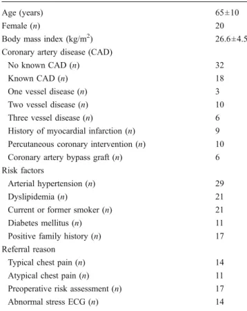
Table 1 Baseline characteristics (n=50)
Age (years) 65±10
Female (n) 20
Body mass index (kg/m2) 26.6±4.5
Coronary artery disease (CAD)
No known CAD (n) 32
Known CAD (n) 18
One vessel disease (n) 3
Two vessel disease (n) 10
Three vessel disease (n) 6
History of myocardial infarction (n) 9
Percutaneous coronary intervention (n) 10
Coronary artery bypass graft (n) 6
Risk factors
Arterial hypertension (n) 29
Dyslipidemia (n) 21
Current or former smoker (n) 21
Diabetes mellitus (n) 11
Positive family history (n) 17
Referral reason
Typical chest pain (n) 14
Atypical chest pain (n) 11
Preoperative risk assessment (n) 17
(HALF time) on a dedicated cardiac dual-head detector camera (Ventri, GE Healthcare) using a low-energy, high-resolution collimator, a 20% symmetric window at 140 keV, a 64 x 64 matrix, an elliptic orbit with step-and-shoot acquisition at 3° intervals over 180° arc (45° right anterior oblique to 45° left posterior oblique) with 30 steps (60 views). FULL time acquisitions were performed according to the
EANM guidelines [7, 8], with a scan time set to 25 s per
frame for stress and rest, resulting in a total acquisition time of 14 min 52 s for both acquisitions. For HALF, the time per frame was reduced to 15 s per frame for stress acquisition and to 10 s per frame for rest acquisition (total scan time of 9 min 42 s and 7 min 7 s for stress and rest, respectively). All other parameters remained unchanged, including the rotation time between the different angles.
For attenuation correction (AC), all patients underwent a low-dose 64-slice CT examination on a Light Speed VCT Scanner (GE Healthcare) during breathhold and after reconstruction and transfer to a Xeleris workstation (GE Healthcare) AC maps were generated as previously
reported [9].
SPECT image reconstruction
For all patients, the FULL time SPECT image set was reconstructed on a dedicated workstation (Xeleris 2.0, GE Healthcare) using the following three different reconstruc-tion algorithms: (1.) FBP, (2.) standard IR applying ordered-subset expectation maximization (OSEM) with two iterations and ten subsets without x-ray based AC (IRNC) and (3.) IR with x-ray based AC (IRAC).
The images from HALF time acquisitions were analyzed on the same platform using the recently developed Evolution for Cardiac software package (GE Healthcare) as previously
reported [5]. In brief, this algorithm, which incorporates RR
and maximum a posteriori noise regularization, allows the SPECT MPI acquisition time to be reduced by about 50% (HALF). HALF scans were reconstructed both without (IRNC) and with AC (IRAC).
All images were reconstructed in standard axis (short-axis, vertical long-(short-axis, and horizontal long-axis slices) encompassing the entire left ventricle using the Myovation software package (GE Healthcare). Polar maps of perfu-sion, wall motion, and wall thickening were produced using a commercially available software package (Cedars QGS/
QPS; Cedars-Sinai Medical Center) [10]
SPECT image analysis
Comparison of HALF time vs. FULL time
In a first step, FULL time scans were reconstructed by conventional IR (without AC) compared to the most widely
used standard reconstruction algorithm, i.e., FBP. Second, we compared IRNC and IRAC from HALF time with the respective images from FULL time.
Visual analysis
For the visual analysis, all available reconstructed images were used. We assessed the presence and the location of reversible and/or fixed perfusion defects as previously
reported [11]. Furthermore, we subjectively graded image
quality as poor, good, or excellent for all reconstructions. A clinical diagnosis based on consensus of two experienced nuclear cardiologists, blinded to each other and to the various reconstructed images, was determined from integrating both attenuation corrected and uncorrected for FULL and HALF time scans.
Quantitative analysis
Quantitative analysis was done via perfusion SPECT images using a 20-segment model for the left ventricle
[12]. Polar maps were normalized to 100% peak activity
and relative percentage counts uptake of gamma ray emissions was assessed for each segment. In addition, the uptake was automatically rated using a five-point scale (0 = normal uptake, 1 = mildly reduced uptake, 2 = moderately reduced uptake, 3 = severely reduced uptake and 4 = no uptake) to provide a summed stress (SSS) and summed rest score (SRS) for calculating a summed different score (SDS) as well as defect extent expressed in percent of the left ventricular myocardium
[10,13]. The percent uptake from each segment (n = 1,000,
20 segments x 50 patients) was included for stress and rest in the analysis. We also grouped the 20 segments into five
regions of the left ventricle as previously described [9]:
apex (segments 19 and 20), anterior (segments 1, 2, 7, 8, 13, and 14), septal (segments 3, 9, and 15), lateral (segments 5, 6, 11, 12, 17, and 18), and inferior (segments 4, 10, and 16).
Statistical analysis
Statistical analysis was performed using the SPSS software (version 12.0.1 for Windows, SPSS Inc., Chicago, Illinois). All numerical values are given as mean ± SD. Linear regression analysis and Bland-Altman limits of agreement
(BA) [14] were used to compare the different algorithms.
Segmental comparison of percent uptake was performed by paired t-test.
Analysis was performed for all segments overall (n=1,000) as well as for each of the five left ventricular regions as defined above. All p-values <0.05 were considered to be statistically significant.
Results Visual analysis
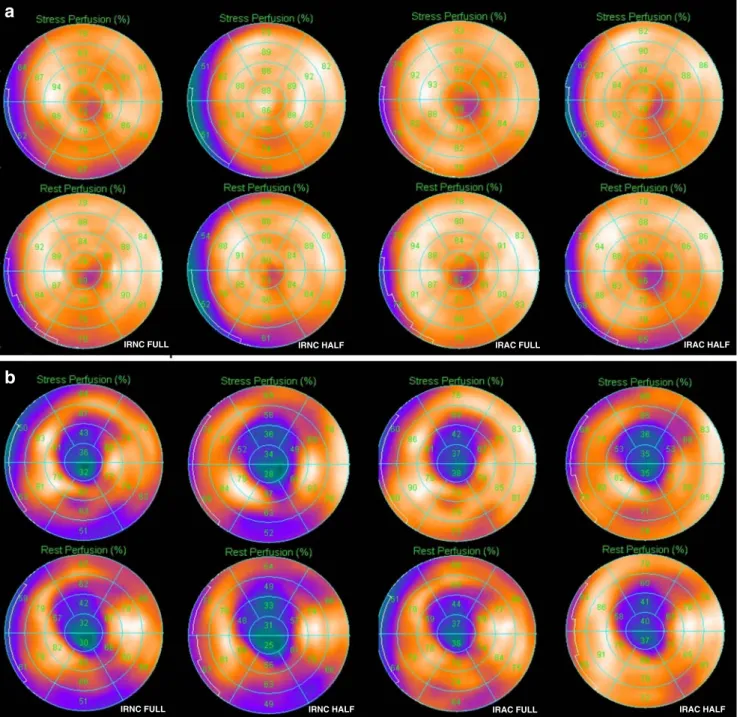
There were no differences in subjective image quality between FULL and HALF images for all reconstructions. All reconstructions revealed excellent image quality in 49 of 50 patients and classified the same patient as good image quality. Visual analysis of both acquisition proto-cols (i.e., FULL time and HALF time) revealed
compa-rable clinical information (Fig. 1). In fact, with standard
FULL as well as with HALF protocol, 38 patients were classified as normal, while there was a scar in five, an ischemia in three and mixed findings in two patients. In addition, two cases of FULL time revealed a minimal ischemia, which was not depicted by HALF time but was evaluated as equivocal clinical relevance due to the limited extent and severity. Overall, this yielded a scan readout agreement with regard to perfusion finding and its regional distribution between HALF and FULL protocol of 96%, nevertheless resulting in a clinical interpretation concordance of 100%.
IRNC HALF IRAC FULL IRAC HALF
IRNC FULL
IRAC HALF
IRNC HALF IRAC FULL
IRNC FULL
a
b
Fig. 1 a Stress/rest study with FULL versus HALF (with and without AC) showing a normal perfusion. b Stress/rest study with FULL versus HALF (with and without AC) showing an antero-apical scar
T able 2 Correlation of percent segmental uptake between FULL and HALF Segment IRNC IRAC Stress Rest Stress Rest rp BA rp BA rp BA rp BA 1 0.75 <0.001 − 1 1.90 7.78 0.68 <0.001 − 10.64 1 1.55 0.66 <0.001 − 14.60 9.64 0.64 <0.001 − 14.06 9.74 2 0.47 <0.001 − 16.70 15.18 0.55 <0.001 − 14.71 13.74 0.61 <0.001 − 17.37 16.70 0.50 <0.001 − 20.15 17.83 3 0.66 <0.001 − 14.82 15.38 0.75 <0.001 − 13.29 12.21 0.64 <0.001 − 15.52 14.00 0.58 <0.001 − 19.78 1 1.76 4 0.92 <0.001 − 8.43 8.95 0.87 <0.001 − 9.58 10.78 0.77 <0.001 − 15.95 14.35 0.78 <0.001 − 15.60 12.88 5 0.80 <0.001 − 1 1.39 10.38 0.79 <0.001 − 8.53 10.29 0.68 <0.001 − 12.01 13.85 0.73 <0.001 − 10.92 12.24 6 0.66 <0.001 − 1 1.10 9.14 0.67 <0.001 − 9.77 1 1.53 0.37 <0.01 − 14.10 14.02 0.56 <0.001 − 1 1.10 10.77 7 0.89 <0.001 − 1 1.93 7.77 0.83 <0.001 − 1 1.99 9.91 0.80 <0.001 − 12.05 7.74 0.81 <0.001 − 12.24 7.16 8 0.59 <0.001 − 12.92 12.44 0.64 <0.001 − 1 1.20 10.08 0.62 <0.001 − 12.60 1 1.60 0.67 <0.001 − 13.18 8.30 9 0.75 <0.001 − 13.57 13.49 0.86 <0.001 − 10.69 9.57 0.68 <0.001 − 13.99 1 1.1 1 0.68 <0.001 − 15.48 9.84 10 0.89 <0.001 − 1 1.14 1 1.42 0.83 <0.001 − 10.78 12.46 0.81 <0.001 − 15.10 12.10 0.80 <0.001 − 14.01 9.89 1 1 0.69 <0.001 − 12.32 13.56 0.72 <0.001 − 1 1.08 12.32 0.78 <0.001 − 9.12 10.80 0.77 <0.001 − 8.96 12.20 12 0.76 <0.001 − 1 1.98 10.30 0.90 <0.001 − 8.1 1 8.19 0.74 <0.001 − 10.80 12.60 0.85 <0.001 − 7.69 10.49 13 0.88 <0.001 − 13.70 1 1.34 0.89 <0.001 − 12.16 9.60 0.81 <0.001 − 14.13 1 1.81 0.88 <0.001 − 12.30 7.50 14 0.88 <0.001 − 8.54 10.14 0.88 <0.001 − 9.21 9.37 0.77 <0.001 − 12.95 13.35 0.88 <0.001 − 10.13 7.49 15 0.85 <0.001 − 7.94 10.82 0.84 <0.001 − 8.20 9.96 0.69 <0.001 − 12.57 9.89 0.69 <0.001 − 3.24 10.04 16 0.85 <0.001 − 13.08 10.60 0.86 <0.001 − 10.15 10.99 0.72 <0.001 − 16.50 10.30 0.76 <0.001 − 13.14 10.18 17 0.80 <0.001 − 12.36 12.20 0.87 <0.001 − 9.47 8.15 0.85 <0.001 − 8.06 10.62 0.82 <0.001 − 9.66 1 1.18 18 0.86 <0.001 − 1 1.97 10.57 0.92 <0.001 − 9.88 5.88 0.81 <0.001 − 10.2 12.80 0.91 <0.001 − 7.40 7.92 19 0.89 <0.001 − 12.40 10.56 0.88 <0.001 − 12.47 10.55 0.85 <0.001 − 12.38 1 1.66 0.90 <0.001 − 10.54 7.44 20 0.87 <0.001 − 13.49 10.21 0.90 <0.001 − 10.74 9.48 0.85 <0.001 − 13.17 9.90 0.87 <0.001 − 1 1.78 9.90
Quantitative perfusion analysis
Comparison between FBP and FULL IRNC There were no significant differences between segmental percent uptake in FBP vs. FULL IRNC (75.17 ±11.26 vs. 73.85 ±11.45, p = ns for stress and 74.76 ± 11.03 vs. 73.96 ± 11.14, p = ns for rest, respectively). For overall segment comparison, there was an excellent correlation with narrow limits of agreement for both stress (r=0.96, p<0.001;
BA−5.37 to 8.02%) and rest (r=0.96, p<0.001; BA −5.59
to 7.20%). Similarly, correlation between FBP and FULL IRNC was excellent for defect extent % at stress (r=0.96, p<0.001) and rest (r=0.98, p<0.001) as well as for SSS (r=0.93, p<0.001) and SRS (r=0.95, p<0.001).
Comparison between FULL IRNC and HALF IRNC There were no significant differences between segmental percent uptake in FULL IRNC vs. HALF IRNC (73.85±11.45 vs. 74.33±12.06, p = ns for stress and 73.96±11.14 vs. 74.14± 11.49, p = ns for rest, respectively). Furthermore, there was a good correlation of percent uptake comparing all segments
for both stress (r=0.87, p<0.001; BA−12.26 to 11.28%) and
rest (r=0.89, p<0.001; BA−10.39 to 10.75%). Results from
the comparison of each individual segment are given in
Table2.
Similarly, the correlation coefficient for each region of the left ventricular myocardium ranged from 0.80 to 0.91
for stress and from 0.74 to 0.90 for rest (Table3).
Comparison between FULL IRAC and HALF IRAC There were no significant differences between segmental per-cent uptake in FULL IRAC vs. HALF IRAC (77.09 ± 9.86 vs. 77.69 ± 10.49, p = ns for stress and 77.09 ± 10.06
vs. 78.26 ± 9.98, p = ns for rest, respectively). Overall
correlation was r = 0.80 (p < 0.001; BA−13.39 to 12.18%)
for stress and r = 0.82 (p < 0.001; BA −12.20 to 10.70%)
for rest. Results from comparison of each individual
segment are given in Table 2. The correlations for the
regions ranged from 0.77 to 0.85 at stress and from 0.79 to
0.89 at rest (Table3).
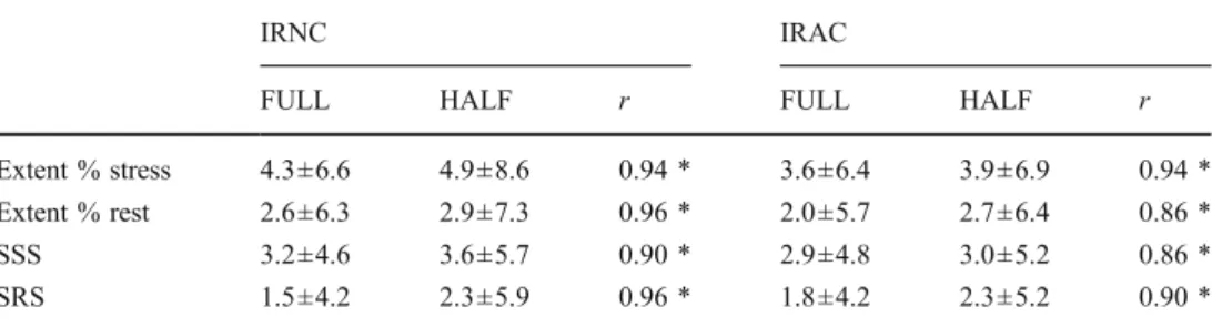
Defect extent and summed perfusion scores
The mean values for percent defect extent of rest and stress as well as the summed scores for IRNC and IRAC for
FULL and HALF are given in Table 4. There was a good
correlation for SSS and SRS as well as for defect extent between FULL time and HALF time at both stress and at rest.
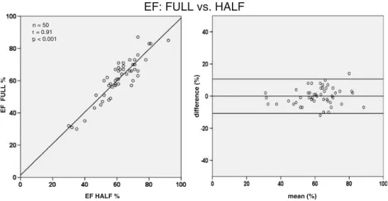
Quantitative analysis of LV function
Quantitative analysis was performed on both FULL IRNC and HALF IRNC gated rest (high dose) acquisitions and the values of left ventricular ejections fraction (LVEF) were not significantly different (61±13 vs. 61±12, p = ns). LVEF determined from FULL correlated well with that from HALF r=0.91, p<0.001. The Bland-Altman plot analysis confirmed the absence of bias (mean difference 0.04±
5.5%) and narrow limits of agreement (Fig.2)
Discussion
In the present study, two SPECT MPI acquisitions were recorded consecutively using the standard parameters first immediately followed by a shortened protocol.
IRNC IRAC
FULL HALF r FULL HALF r
Extent % stress 4.3±6.6 4.9±8.6 0.94 * 3.6±6.4 3.9±6.9 0.94 *
Extent % rest 2.6±6.3 2.9±7.3 0.96 * 2.0±5.7 2.7±6.4 0.86 *
SSS 3.2±4.6 3.6±5.7 0.90 * 2.9±4.8 3.0±5.2 0.86 *
SRS 1.5±4.2 2.3±5.9 0.96 * 1.8±4.2 2.3±5.2 0.90 *
Table 4 Comparison of mean values for percent defect extent as well as the summed scores for stress and rest
* p<0.001
Regions Segments (n) IRNC IRAC
Stress (r) Rest (r) Stress (r) Rest (r)
anterior 300 0.83* 0.74* 0.80* 0.81*
lateral 300 0.83* 0.86* 0.77* 0.82*
inferior 150 0.91* 0.86* 0.78* 0.79*
septal 150 0.80* 0.86* 0.80* 0.79*
apical 100 0.89* 0.90* 0.85* 0.89*
Table 3 Correlation of percent segment uptake grouped into regions between FULL and HALF
For reconstruction of the HALF time acquisition scans we used new software with improved IR algorithms including RR in order to compensate for the low counts due to the shortened scan time. Our results prove the validity of this HALF time reconstruction algorithm allowing shortening scan time by about 50% on a dedicated cardiac camera. Although two edge segments prone to higher variability showed only modest quantitative agreement, there was a high overall correlation with regard to segmental and also regional radionuclide uptake between the two protocols with narrow Bland and Altman limits of agreement. Consequently, this translated into an excellent agreement of clinical conclusion from FULL and HALF SPECT MPI studies. Similarly, there was excellent agreement between FULL and HALF with regard to LV function.
The introduction of 99mTc-labeled perfusion agents has
been accompanied by an impressive growth of nuclear cardiology studies in the past decade. This has led to an intensive search for options to improve the throughput of the nuclear cardiology laboratories by shortening scan time. This would also confer advantages with regard to patient comfort and potentially reduce motion artifacts. Due to technical advances, it has now become feasible in a clinical setting to use IR as a practical alternative to FBP. IR algorithms including fast OSEM are potentially of great importance in this context due to improved noise properties over FBP, such as reduced noise correlation length at low
numbers of iterations, [15]. Algorithms with RR have been
integrated from various vendors into latest-generation reconstruction algorithms, such as Evolution for Cardiac
(GE Healthcare) [5,16], Wide-beam reconstruction (WBR)
(UltraSPECT) [16], Astonish (Philips Medical System) [16,
17] and 3D Flash (Siemens Medical Solutions) [18].
In the present report we have shown that processing HALF time SPECT MPI data with IR and OSEM including RR results in clinical information in agreement to that provided by
FULL time FBP. Our results confirm findings from a recent report on the feasibility of HALF time SPECT MPI with a
general-purpose SPECT gamma camera [5] and expand the
applicability to a dedicated cardiac device. This is important not only for allowing using HALF time acquisition in cardiac imaging but also because the previous study had significant limitations: for example Ali et al. have compared FULL and HALF by analyzing differences in SSS. Thus, they have not provided any information on comparability per segment, per region, or per major coronary vessel territory. By contrast, using segmental percent tracer uptake as objective quantitative metric, our study provides evidence that agreement between FULL and HALF remains preserved after providing regional and segmental comparison, which underlines the validity of the proposed method to allow half time MPI scanning.
As an alternative to shortening the scan time, this technique may offer the opportunity to decrease patient radiation exposure while maintaining image quality. This would also help in making SPECT MPI scans most suitable for hybrid imaging in combination with CT coronary angiography.
It may be perceived as a limitation of the present study that for assessing summed scores as well as extent abnormalities the same normal limits have been used for the HALF as for the FULL time studies. However, this is in line with recent reports that also suggested the use of the same limits for such comparison as this allows for better HALF vs. FULL comparability, while changing both parameters (i.e., recon-struction and limits) might hampered this. In fact, our results substantiated the validity of this approach.
Conclusion
On the basis of these data, cardiac SPECT MPI studies can be acquired with half of the scan time and
EF: FULL vs. HALF
mean (%) difference (%) EF FULL % n = 50 r = 0.91 p < 0.001 EF HALF % Fig. 2 Linear regression
analy-sis (left panel) and Bland-Altman plots (right panel) for comparison of EF (%) between FULL and HALF at rest (high dose) with iterative reconstruc-tion (IR) without attenuareconstruc-tion correction
reconstructed using the new algorithm“Evolution for Cardiac” without compromising image quality, resulting in an excellent agreement with regard to uptake and clinical conclusion, compared to standard acquisition time and reconstruction.
References
1. Klocke FJ, Baird MG, Lorell BH, et al. ACC/AHA/ASNC guidelines for the clinical use of cardiac radionuclide imaging— executive summary: a report of the American College of Cardiology/American Heart Association Task Force on Practice Guidelines (ACC/AHA/ASNC Committee to Revise the 1995 Guidelines for the Clinical Use of Cardiac Radionuclide Imaging). J Am Coll Cardiol. 2003;42:1318–33.
2. Taillefer R, Primeau M, Costi P, Lambert R, Leveille J, Latour Y. Technetium-99 m-sestamibi myocardial perfusion imaging in detection of coronary artery disease: comparison between initial (1-hour) and delayed (3-hour) postexercise images. J Nucl Med. 1991;32:1961–5.
3. Borges-Neto S, Pagnanelli RA, Shaw LK, et al. Clinical results of a novel wide beam reconstruction method for shortening scan time of Tc-99 m cardiac SPECT perfusion studies. J Nucl Cardiol. 2007;14:555–65.
4. DePuey EG, Nichols KJ, Slowikowski JS, et al. Fast stress and rest acquisitions for technetium-99 m-sestamibi separate-day SPECT. J Nucl Med. 1995;36:569–74.
5. Ali I, Ruddy TD, Almgrahi A, Anstett FG, Wells RG. Half-Time SPECT myocardial perfusion imaging with attenuation correction. J Nucl Med. 2009;50:554–62.
6. Cerqueira MD, Verani MS, Schwaiger M, Heo J, Iskandrian AS. Safety profile of adenosine stress perfusion imaging: results from the Adenoscan Multicenter Trial Registry. J Am Coll Cardiol. 1994;23:384–9.
7. Hansen CL, Goldstein RA, Akinboboye OO, et al. Myocardial perfusion and function: single photon emission computed tomography. J Nucl Cardiol. 2007;14:e39–60.
8. Hesse B, Tagil K, Cuocolo A, et al. EANM/ESC procedural guidelines for myocardial perfusion imaging in nuclear cardiology. Eur J Nucl Med Mol Imaging. 2005;32:855–97.
9. Schepis T, Gaemperli O, Koepfli P, et al. Use of coronary calcium score scans from stand-alone multislice computed tomography for attenuation correction of myocardial perfusion SPECT. Eur J Nucl Med Mol Imaging. 2007;34:11–9.
10. Germano G, Kavanagh PB, Waechter P, et al. A new algorithm for the quantitation of myocardial perfusion SPECT. I: technical principles and reproducibility. J Nucl Med. 2000;41:712–9. 11. Berman DS, Kiat H, Friedman JD, et al. Separate acquisition rest
thallium-201/stress technetium-99 m sestamibi dual-isotope myo-cardial perfusion single-photon emission computed tomography: a clinical validation study. J Am Coll Cardiol. 1993;22:1455–64. 12. Cerqueira MD, Weissman NJ, Dilsizian V, et al. Standardized
myocardial segmentation and nomenclature for tomographic imaging of the heart: a statement for healthcare professionals from the Cardiac Imaging Committee of the Council on Clinical Cardiology of the American Heart Association. Circulation. 2002;105:539–42.
13. Sharir T, Germano G, Waechter PB, et al. A new algorithm for the quantitation of myocardial perfusion SPECT. II: validation and diagnostic yield. J Nucl Med. 2000;41:720–7.
14. Bland JM, Altman DG. Statistical methods for assessing agree-ment between two methods of clinical measureagree-ment. Lancet. 1986;1:307–10.
15. Daou D, Pointurier I, Coaguila C, et al. Performance of OSEM and depth-dependent resolution recovery algorithms for the evaluation of global left ventricular function in 201Tl gated myocardial perfusion SPECT. J Nucl Med. 2003;44:155–62. 16. DePuey EG, Gadiraju R, Clark J, Thompson L, Anstett F, Shwartz
SC. Ordered subset expectation maximization and wide beam reconstruction "half-time" gated myocardial perfusion SPECT functional imaging: a comparison to "full-time" filtered back-projection. J Nucl Cardiol. 2008;15:547–63.
17. Bateman TM, Heller GV, McGhie AI. Application of simulta-neous Gd-153 line source attenuation correction to half-time SPECT acquisitions: multi-center clinical evaluation. Paper presented at: ASNC 2007 meeting; September 6–9; San Diego, California.
18. Kritzman JN, Cahill JM, Ficaro EP, Corbett JR. Quantitative comparison of standard and reduced acquisition time attenuation corrected and non-corrected myocardial perfusion images: a phantom study utilizing a 3D iterative reconstruction [abstract]. J Nucl Cardiol. 2007;14(suppl 1):S107–8.