HAL Id: tel-02924993
https://tel.archives-ouvertes.fr/tel-02924993
Submitted on 28 Aug 2020
HAL is a multi-disciplinary open access archive for the deposit and dissemination of sci-entific research documents, whether they are pub-lished or not. The documents may come from teaching and research institutions in France or abroad, or from public or private research centers.
L’archive ouverte pluridisciplinaire HAL, est destinée au dépôt et à la diffusion de documents scientifiques de niveau recherche, publiés ou non, émanant des établissements d’enseignement et de recherche français ou étrangers, des laboratoires publics ou privés.
Nuclear organization and regulation of gene expression
in mouse Embryonic Stem Cells by long non-coding
RNAs
Alexandra Tachtsidi
To cite this version:
Alexandra Tachtsidi. Nuclear organization and regulation of gene expression in mouse Embryonic Stem Cells by long non-coding RNAs. Genomics [q-bio.GN]. Sorbonne Université, 2018. English. �NNT : 2018SORUS444�. �tel-02924993�
Nuclear organization and regulation of gene expression in
mouse Embryonic Stem Cells by long non-coding RNAs.
Alexandra TACHTSIDI
Thesis presented for the degree of Doctor of Philosophy
Directed by Pablo Navarro Gil
Epigenetics of stem cells Laboratory - Pasteur Institute- Paris
Ecole Doctorale Complexité du Vivant (ED 515)
Sorbonne Université
December 11, 2018
Thesis Defense Committee:
President : Pr MORILLON Antonin, Research director
Examiner : Pr FEIL Robert, Research director
Examiner : Pr CIAUDO Constance, Research director
Member : Dr FRANCASTEL Claire, Research director
Member : Dr CHAUMEIL Julie, Principal Investigator
Acknowledgements
I am greatly thankful to my thesis director, Pablo Navarro Gil, for giving me the chance to work in his laboratory. Thank you for always pushing me further and redefining my limits, shaping my perseverance and endurance. Thank you for the opportunity to use many different experimental techniques that were particularly enriching for me growing as a scientist while the experience of supervising a master student has been instructive for my managerial skills. I am also grateful to you for the possibility to participate in the fascinating experience of a Cold Spring Harbor Course and to attend very interesting international conferences during these years.
I would like to thank all the past and present members of our laboratory, the ECS team. It was a real pleasure to work with all of you guys these last four years, and to develop personal relationships outside the laboratory too. Thank you for all the help, advice and thoughts that we have exchanged and the moments of joy and laughter that we have shared. Agnes, thank you for keeping the lab running functional, for always being a kind and helpful colleague but also a friend. Thank you Philippe for always answering my questions and sharing so generously your wide knowledge and scientific expertise, and your office the last few months. Thank you Nicola for being an inspiring colleague and a friend from the very beginning of my thesis. I wish to express my gratitude to Thaleia for the liberating feeling of speaking daily in our mother-tongue, for bringing an extra sense of organization to the lab, for her support, suggestions and corrections. Thank you Nick for all our interesting discussions, the explanations and the analyses that you have done on my sequencing results. Inma, thank you for your positivity, support and kindness. I thank Nancy for our collaboration during her master internship. And Laurence, thank you for taking care of our administrative work.
I thank past and present members of the Department of Stem Cells and Developmental Biology of Pasteur Institute for creating a supportive and fun working environment but also Frank, Françoise and Masha for our discussions.
I would also like to express my gratitude to my Thesis Committee Members, Deborah Bourc’his, Antonin Morillon and Marco Vignuzzi, for their support and instructions during the years of the thesis.
Many friends have been surrounding me these last years to whom I am deeply grateful for their friendship and support. Especially, thank you Elma for suggesting I should come to Paris to pursue my Master and PhD studies, for being helpful and caring through the good and tough moments of our long student life.
A big, special thank you goes to you Victor, for your unlimited support and encouragement, for being always by my side loving and caring. Thank you for believing in me when I didn’t believe in myself; for making me see science and the world through a different perspective.
Finally, all my gratitude goes to my parents, Eirini and Michail. В заключение, хотела бы выразить всю мою благодарность моим родителям. Папа, Мама спасибо вам огромное за вашу бесконечную любовь, поддержку и веру в меня. За все что вы сделали для меня, и продолжаете делать до сих пор, за все чему научили меня и предоставили. Все чего я достигла, является и вашей заслугой.
1
Abstract
The nucleus is a highly structured organelle and its complex architectural organization enables and facilitates different biological processes to take place at distinct subnuclear domains. The implication of long non-coding RNAs (lncRNAs) in nuclear organization by establishing and maintaining nuclear compartmentalization is now widely accepted. Numerous examples have been shown to either participate in the structuration of subnuclear domains or in the establishment of long range interactions in the three-dimensional nuclear space. However, a robust approach for the identification of “nuclear organizers” molecules such as Xist, Neat1, and Firre that shape the nucleus is currently lacking. To that end, we established an experimental approach that would allow us to identify such “structural” lncRNAs on a genome-scale level. Based on the biochemical property of known nuclear organizing lncRNAs to resist the so called nuclear matrix preparation, where most of the DNA and soluble molecules are removed, we performed nuclear matrix fractionation on mouse Embryonic Stem Cells (mESCs), purified the RNA fraction and explored its constituents by RNA-sequencing. We identified in such a way, a subset of transcripts (non-extracted RNAs, nextRNAs) potentially involved in the functional compartmentalization of the nucleus. The group of nextRNAs identified by RNA-seq was validated by RT-qPCR and contained few transcripts that are already known and described to be “nuclear organizers” (e.g. Xist, Firre, Neat1). Notably, we detected previously non-annotated transcripts thanks to our original RNA-seq datasets and focused our work on two of them: NextC1 (Next Candidate 1) and NextC2.
We extensively described and characterized the identified NextC1 and NextC2 on a functional and phenotypical level. The expression profile of the transcripts was studied in pluripotent and differentiating culturing conditions, in mutant cell lines for pluripotency transcription factors (TFs) as well as in different embryo-derived cell types. The subcellular localization of both lncRNAs was assessed by RNA-FISH. Loss- and gain-of-function assays were performed by targeting the promoter regions of NextC1 and 2 with the canonical CRISPR/Cas9 system for genome editing and CRISPR-derived systems for transcription inhibition or activation. Many of these functional assays were subsequently RNA-sequenced and an integrative data analysis is currently under investigation.
To my parents, to whom I am deeply grateful For everything they have done for me and keep doing to date.
1
Table of Contents
Index of abbreviations... 3
Introduction ... 5
I. Nuclear organization ... 6
A. Chromosome organization and transcription ... 7
B. A and B compartments ... 7
C. Topologically associated domains (TADs) ... 8
D. Nuclear lamina and Lamina-associated domains (LADs)... 9
E. Nuclear bodies ... 11
F. Nuclear matrix ... 12
II. Long non-coding RNAs ... 14
A. Long non-coding RNAs identification and classification ... 14
B. Functional mechanisms of lncRNAs ... 16
III. Mouse Embryonic Stem Cells ... 23
A. Extrinsic pathways regulating pluripotency ... 23
B. Transcription factor-mediated pluripotency regulation... 24
C. Pluripotency states... 25
D. ES cells and nuclear organization ... 26
E. ES cells and lncRNAs ... 27
Thesis objectives ... 28
Materials and methods ... 29
Results ... 48
I. Identification of structural long non-coding RNAs ... 49
A. Establishment of the experimental approach ... 49
B. Matrix-associated transcript identification ... 55
C. Selecting candidates for functional characterization ... 64
D. Discovery of novel long non-coding RNAs ... 68
E. Discussion ... 69
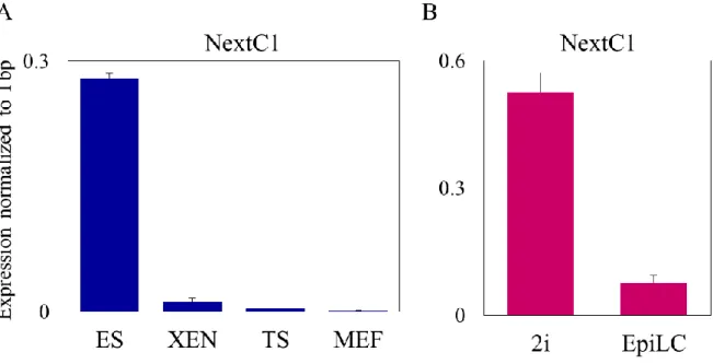
II. NextC1 (Non-extracted Candidate 1) ... 73
A. Validation of NextC1 RNA and matrix retention ... 73
B. NextC1 coding potential and conservation ... 76
2
D. NextC1 expression regulation by the pluripotency network ... 78
E. NextC1 subcellular localization ... 86
F. Functional assays ... 89
i. Loss of function ... 89
ii. Gain of function ... 99
G. Discussion ... 106
III. NextC2 (Non-extracted Candidate 2) ... 115
A. Validation of NextC2 RNA and matrix retention ... 115
B. NextC2 coding potential and conservation ... 117
C. NextC2 expression regulation by the pluripotency network ... 118
D. NextC2 subcellular localization ... 126
E. NextC2 RNA stability ... 128
F. Functional assays ... 130
i. Loss of function ... 130
ii. Gain of function ... 140
G. Discussion ... 143
3
Index of abbreviations
BFP: Blue Fluorescent Protein
ChIP: Chromatin Immunoprecipitation
CRISPR: Clustered Regularly Interspaced Short Palindromic Repeats CTCF: CCCTC-binding factor
DAPI: 4',6-Diamidino-2-Phenylindole dCas9: catalytically dead Cas9 protein DE: Differentially Expressed
EB: Embryoid Body EpiLC: Epiblast-Like Cells
FACS: Fluorescence Activated Cell Sorter FC: Fold Change
FCS: Fetal Calf Serum
FISH: Fluorescence In Situ Hybridization GFP: Green Fluorescent Protein
KO: Knock Out
LAD: Lamina-Associated Domain LIF: Leukaemia Inhibiting Factor
lincRNA: Long intergenic non-coding RNA lncRNA: Long non-coding RNAs
mESC: Mouse Embryonic Stem Cells miRNA: microRNA
4 PRC2: Polycomb Repressive Complex 2
RA: Retinoic Acid
RFP: Red Fluorescent Protein RNA-seq: RNA Sequencing
RT-qPCR: Real Time quantitatice Polymerase Chain Reaction SE: Super Enhancer
smFISH: Single Molecule Fluorescence In Situ Hybridization TAD: Topologically Associating Domain
TF: Transcription Factor Tpm: Transcripts per million TSS: Transcriptional Start Site
UCSC: University of California Santa Cruz (Database)
VP64: Four copies of VP16 (viral protein of 16 amino acids) transcriptional activator protein WT: Wild Type
5
6
I. Nuclear organization
The characteristic feature of eukaryotic cells is the presence of a nucleus; an organelle which has a complex and dynamic organization. The nuclear architecture has triggered the research from the spectrum of cellular and developmental biology for almost a century now. Yet, the nuclear compartmentalization and its functionality have not been fully characterized. The high-order organization of the mammalian nucleus allows different biological processes to take place in distinct subnuclear compartments and it serves for a precise regulation of gene expression during different developmental stages through chromatin modifications and architectural rearrangements.
The analysis of the nuclear organization was inaugurated by E. Heitz in 1928, with the observation that the transcriptionally active euchromatin is decondensed and no longer visible during interphase, whereas heterochromatin -that is transcriptionally less active- is still visible following mitosis. Later on, the establishment of electron microscopy (EM) supported his conception and also showed that nuclear organization markedly varies in different cell types or developmental stages, however, it is similar between cells of a given cell type (Pueschel et al., 2016). The first big step forward in the study of the nuclear organization was achieved with fluorescence-based microscopy that allowed the precise localization of proteins and genes in relation to nuclear landmarks. Many nuclear bodies were discovered in that way and their consisting proteins and genes were identified, such as the nucleolus, the speckles, the paraspeckles and even smaller structures like the transcription factories (Bond and Fox, 2009; Eskiw et al., 2008; Spector and Lamond, 2011). At a larger scale, chromosome territories and gene-positioning in association with transcriptional activity were described (Cremer et al., 2006; Croft et al., 1999). With the development of the chromosome conformation capture technology (Dekker et al., 2002) we have gained more insight into the higher-order organization of the chromosomes and it has been shown that chromatin has different levels of organization, ranging from the typical 10nm chromatin fiber to topologically associating domains (Dixon et al., 2012; Nora et al., 2012; Sexton et al., 2012). Today, we start to have an understanding of the three-dimensionally (3D) organized genome that is extensively compartmentalized while allowing long-range interactions to occur for gene regulation (Dekker and Mirny, 2016).
7
A. Chromosome organization and transcription
Interphase chromosomes occupy discrete territories of the nuclear space, the so-called Chromosome Territories (CT) that have a specific positioning in the nucleus depending on their gene composition; the gene-rich chromosomes have a more central position compared to the gene-poor chromosomes that are located closer to the nuclear periphery (Bolzer et al., 2005; Croft et al., 1999). In the same manner, gene positioning is tightly correlated with the transcriptional activity. Heterochromatin tends to be located at the nuclear periphery in vicinity to the nuclear lamina, whereas the distribution of euchromatin localizes in the center of the nucleus or close to nuclear pores. The transition through different developmental stages during cell differentiation causes gene reposition, depending on their transcriptional activity (Kosak et al., 2007; Meister et al., 2010; Takizawa et al., 2008; Williams et al., 2006). To that direction, genes can move to the nuclear periphery once being silenced whereas upon activation they can move to the interior of the nucleus or loop out of their CTs to the interchromosomal space (Chambeyron et al., 2005; Kosak et al., 2007; Meister et al., 2010). The first Hi-C study performed in human cells molecularly confirmed the existence of chromosome territories. The defined spatial positioning of chromosomes was shown by obtaining far more frequent interactions between distant sequences located on the same chromosome, compared to any other loci in the rest of the genome (Lieberman-Aiden et al., 2009).
B. A and B compartments
The aforementioned study additionally identified the existence of two classes of genomic compartments, the first one being gene rich, transcriptionally active, and hypersensitive to DNase I digestion, while the second was relatively gene poor, transcriptionally silent, and DNase I insensitive (Lieberman-Aiden et al., 2009). This was highly resembling the EM-observed euchromatin and heterochromatin regions in interphase cells. These two major compartments are termed compartments A and B. The A compartment, similar to euchromatin, contains more open and active chromatin whereas B is more closed, compact, harboring repressive chromatin marks, similar to heterochromatin (Pueschel et al., 2016; Rao et al., 2014). A compartment is associated with histone marks such as H3K4me3, H3K36me3, and hyperacetylation while B compartment, on the other hand, is bound by Polycomb Group (PcG) proteins and heterochromatin proteins (HP1) thus associated with repressive marks (Nagano et al., 2013; Sexton et al., 2012). Inter-chromosomal contacts
8 between domains from the same compartments (A/A, B/B) are more frequent than those between different compartments (A/B), and A compartments make more contacts than B ones (Gibcus and Dekker, 2013; Lieberman-Aiden et al., 2009).
C. Topologically associating domains (TADs)
The A/B compartments are comprised of sub-megabase-scale domains which constitute the primary units of interphase chromosome folding and are termed topologically associating domains (TADs) (Dixon et al., 2012; Nora et al., 2012; Sexton et al., 2012). The TADs are large self-associating domains of chromosomes ranging several hundreds of kilobases. TADs that show similar chromatin states, i.e. active or repressed, tend to associate with each other in cis and in trans, with TADs on the same or other chromosomes respectively, to form two genomic compartments (Dekker and Heard, 2015). They are constant throughout development and are largely conserved across different mammalian cell types. Genes within the same TAD share more similar regulation than genes in different TADs during embryonic stem cell differentiation (Nora et al., 2012) and reorganization of the genome architecture occurs at the sub-megabase scale (within TAD) during differentiation (Phillips-Cremins et al., 2013). TADs are demarcated by constitutive occupancy of CTCF (CCCTC-binding factor, an insulator protein that blocks communication between adjacent regulatory elements in an orientation-dependent manner), Cohesin complexes, transcription start sites of housekeeping genes, transfer RNAs, and short interspersed element (SINE) retrotransposons (Dixon et al., 2012; Nora et al., 2012). However, most of the CTCF-binding sites (around 85%) are actually found within the TADs, delimiting intra-TADs of an intermediate size of 100 kb–1 Mb. The spacing and orientation of CTCF-binding sites is responsible for the formation of individual loops or larger TADs was revealed by genome-wide CTCF ChIA-PET analysis (Chromatin Interaction Analysis by Paired-End Tag Sequencing) which also showed that gene regulatory interactions between promoters and their distal regulatory elements occur mostly within TADs (Tang et al., 2015).
CTCF and Cohesin co-occupied sites define and anchor the long-range interactions, i.e. the TADs and inter-TAD communications while Mediator and Cohesin co-binding establishes short-range, cell-type specific interactions within a TAD such as for enhancer-promoter interactions (Phillips-Cremins et al., 2013). Depletion of CTCF results in loop elimination between CTCF sites, and in disrupted insulation between neighboring TADs, surprisingly
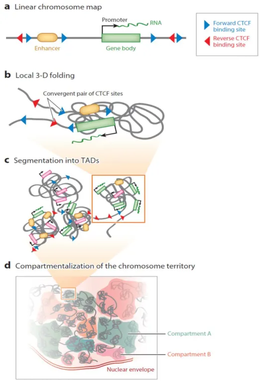
9 though the genomic compartments are unaffected and no aberrant gene activation is observed (Kubo et al., 2017; Nora et al., 2017). On the same line, Cohesin depletion disrupts looping between CTCF sites and reduces intra-TAD interactions yet leaves compartmentalization intact (Rao et al., 2017; Sofueva et al., 2013). Therefore, compartmentalization of mammalian chromosomes emerges independently of proper insulation of TADs. Chromosome folding beyond the TAD scale is disrupted only when of both CTCF and Cohesin are depleted leading to general chromatin compaction (Tark-Dame et al., 2014). The inversion of an individual CTCF motif from a convergent looping pair abrogates the loop, underscoring the importance of the orientation for interactivity between remote pairs of CTCF sites (Guo et al., 2015; de Wit et al., 2015). In addition, inversion of clustered CTCF sites (at the protocadherin and β-globin loci) has been shown to disrupt local chromatin folding and allowed the inverted CTCF cluster to contact previously insulated regions downstream of the CTCF site (Guo et al., 2015). Collectively, TADs appear to represent functional domains with boundaries that do not allow enhancers to reach genes located in adjacent TADs (Fig.1.1).
Compartments and TADs are not a constitutive feature of chromosomes, as TADs are depleted along mitotic chromosomes (Naumova et al., 2013), and compartmentalization is lost at the inactive X chromosome in mammalians (Minajigi et al., 2015; Nora et al., 2012). This implies that there are mechanisms continuously instructing the chromosome organization for the proper 3D genome folding.
D. Nuclear lamina and Lamina-associated domains (LADs)
In mammalian cells, a network of intermediate filament proteins (of lamins and lamin-binding associated proteins) exists between the nuclear membrane and the chromatin (Gruenbaum et al., 2005). This structure, known as the nuclear lamina (NL), is implicated in a broad range of biological functions such as nuclear architecture, chromatin organization, and gene expression (Goldman et al., 2002). The development of genome-wide mapping techniques has made it possible to assess the molecular interactions between the chromatin and the NL. DamID is a genome-wide assay where NL proteins are fused to a DNA adenine methyltransferase (Dam) protein from the bacteria Escherichia coli, which will methylate any piece of DNA that is in molecular contact with the NL in vivo (van Steensel and Henikoff, 2000). Through the use of DamID, chromatin-NL interaction maps have been generated for mouse, and human cells (Guelen et al., 2008; Peric-Hupkes et al., 2010) and have revealed that
10 very large (median size of 500Kb) chromosomal domains engage in interactions with the NL. These domains are termed lamina-associated domains (LADs) and over a thousand of them exist in mouse and human cells.
Figure 1.1. Illustration of the different levels of genome folding. a) Linear chromosome map. Representation of a genomic locus with an enhancer, a gene and its promoter, and CTCF binding sites in forward (blue) or reverse (red) orientation. b) Local 3-D folding. Convergent pairs of CTCF sites are brought into close spatial proximity forming a loop and enabling chromosomal contacts between the enhancer and the target promoter in the intervening domain. c) Segmentation into into topologically associating domains (TADs). This level of folding packages enhancers and promoters (resulting in transcriptional activation, in green) from the same domain together while insulating them from the
11 regulatory elements of neighboring domains. TADs also contain inactive genes (pink), which are not responsive to surrounding enhancers. d) Compartmentalization of the chromosome territory. The association of TADs from the same or different chromosomes defines two main compartments; A (blue) and B (red), which roughly correspond to the transcriptionally active and inactive fractions of the genome (with permission from Merkenschlager and Nora, 2016).
LADs are typically gene-poor and transcriptionally inert, enriched in repressive (H3K9me3, H3K27me3) and devoid of active histone marks (H3K4me3, H3K36me3) (Kind et al., 2015). Surprisingly, recent studies have demonstrated that LADs associate also with euchromatin regions and are important for the epithelial-to-mesenchymal transition in mouse (Pascual-Reguant et al., 2018). Finally, lamins have been shown to have an important role for the global three-dimensional genome organization, as their loss has been shown to cause decompaction and detachment of some LADs from the NL in mouse embryonic stem cells (mESCs) which in turn disrupts the 3D chromatin interactions of LADs and inter-TADs (Zheng et al., 2018).
E. Nuclear bodies
The mammalian nucleus is further compartmentalized into membraneless subnuclear organelles which are specialized domains (“nuclear bodies”) supporting distinct biological processes. They are defined mostly by the specific proteins and RNAs that they contain, at high local concentrations (Dundr, 2012; Dundr and Misteli, 2010; Mao et al., 2011). Some of the reported nuclear bodies with specialized functions are the nucleolus, the nuclear speckles, the paraspeckles, the Cajal and Promyelocytic (PML) bodies. The nucleolus is the largest nuclear structure where ribosomal RNAs are transcribed and ribosomes assembled (Boisvert et al., 2007). The nuclear speckles harbor the pre-mRNA splicing machinery and tend to localize near large clusters of active genes (Spector and Lamond, 2011). The paraspeckles are the domains were RNAs are sequestered for nuclear retention (Clemson et al., 2009). In the Cajal bodies the biogenesis and maturation of small nuclear RNA (snRNAs) takes place as well as the processing of histone mRNAs (Caudron-Herger and Rippe, 2012). In PML bodies divers regulatory proteins aggregate and these structures are required for heterochromatin integrity although their exact way of function is not yet deciphered (Pueschel et al., 2016). Many of these nuclear bodies are enriched in coding RNAs and some particularly in long non-coding RNAs (lncRNAs); the implication of lncRNAs in the formation of nuclear domains will be described more thoroughly in the section II.B.
Fluorescence recovery after photobleaching (FRAP) experiments have shown rapid and dynamic exchange of major protein components of nuclear bodies with the nucleoplasm,
12 suggesting an ordered assembly of these nuclear sub-organelles (Phair and Misteli, 2001). The formation and structural maintenance of nuclear bodies relies on the protein-protein and protein-RNA interactions (Dundr and Misteli, 2010; Mao et al., 2011). Very recent studies on liquid phase separation have proposed that different nuclear bodies behave like liquid-phase droplets that can condense through concentration-dependent liquid-phase separation. Nucleoli, paraspeckles and Cajal bodies form liquid-like condensates which are able to compartmentalize and concentrate proteins of similar biochemical properties and RNAs (Berry et al., 2015; Fox et al., 2018; Sawyer et al., 2018).
F. Nuclear matrix
Evidence for a non-chromatin scaffold within the nucleus first came to light half a century ago when electron microscopy (EM) and two-dimensional gel analysis revealed that the nucleus contains a large amount of non-chromatin insoluble protein and heterogeneous RNA resistant to extensive biochemical extraction (Berezney and Coffey, 1974; Capco et al., 1982; Herman et al., 1978). This fibrogranular structure revealed by DNAseI digestion and ammonium sulfate extraction, was termed nuclear matrix, and was proposed to form an architectural scaffold to support internal organization of the nucleus (He et al., 1990). Nuclear RNA was showed to be a key component of the nuclear matrix, since transcription inhibition or RNase treatment was causing the matrix fibers to collapse leaving what appeared to be largely hollow nuclei (as observed by EM) (Herman et al., 1978; Nickerson et al., 1989). After DNase digestion, further extraction with high salts of matrix proteins revealed a core filament network of RNA nature that was depleted upon RNase digestion (He et al., 1990). Different protocols have been used for the extraction of the components of the nuclear matrix, essentially differing in the salts and salt concentrations of the washes that follow the permeabilization with nonionic detergents and DNaseI digestion (Engelke et al., 2014).
The nuclear matrix was present in all the cells and tissues examined and was shown to organize the chromatin by attaching to the bases of DNA loops (Matrix/Scaffold attachment region, S/MAR) (Nickerson, 2001; Razin et al., 1981). Chromosome territories have been shown to be anchored to the nuclear matrix through the S/MARs and disruption of the matrix with RNase treatment results in the disruption of higher-order chromosome territory architecture (Ma et al., 1999). The S/MAR elements can be found inside genes and even inside exons. Some of the nuclear matrix proteins that preferentially bind to S/MAR elements are
13 lamins (Fiorini et al., 2006), SATB1 (special AT-rich binding protein 1) (de Belle et al., 1998), and SAFA/hnRNPU (Romig et al., 1992).
14
II. Long non-coding RNAs
A. Long non-coding RNAs identification and classification
Two-thirds of the mammalian genome has been shown to be pervasively transcribed, however only less than 2% is finally translated into proteins (Bertone et al., 2004; Carninci et al., 2005; Dinger et al., 2009; Djebali et al., 2012). During the last years, the advent of next generation sequencing techniques (NGS) has enabled the identification of thousands of non-coding genes that are subdivided into two categories: long and small non-non-coding RNAs. Long non-coding RNAs (lncRNAs) comprise a large family of transcripts larger than 200 nucleotides in size that often share many properties with the protein-coding mRNAs as being RNA polymerase II transcribed, spliced, capped and poly-adenylated (Quinn and Chang, 2016; Rinn and Chang, 2012). They mostly do not encode proteins, however some annotated lncRNAs have been reported to give rise to small peptides (Anderson et al., 2015; Cohen, 2014; Nelson et al., 2016). LncRNAs definition being based only on the length of the transcript and the lack of coding potential results in a broad heterogeneous family of molecules with highly diverse functional properties (Ulitsky and Bartel, 2013a).
The discovery of two long RNA molecules that had typical mRNA properties yet did not encode a protein, the first identified lncRNAs H19 and Xist, can be traced back to the early 1990’s (Brannan et al., 1990; Brockdorff et al., 1992; Brown et al., 1991). Both of these lncRNAs were shown to be functional, H19 was involved in parental imprinting (Bartolomei et al., 1991; Gabory et al., 2010) while Xist was found to be orchestrating the inactivation of X chromosome, for dosage compensation in female and male mammals, by coating the inactive X chromosome from which it is transcribed (Brockdorff et al., 1992; Brown et al., 1991, 1992; Penny et al., 1996). However, it was more than a decade later that the identification of a huge number of lncRNAs was accomplished. High-throughput RNA sequencing (RNA-seq), chromatin immunoprecipitation sequencing (ChIP-seq) analysis and ab initio transcriptome reconstruction performed in multiple cell lines in the mouse and human, resulted in the identification of thousands of lncRNAs (Guttman et al., 2009, 2010a; Mikkelsen et al., 2007; Mortazavi et al., 2008; Sultan et al., 2008; Trapnell et al., 2009; Yassour et al., 2009). To date, only a few of this plethora of molecules have been characterized, while the vast majority of them remain largely unstudied.
15 LncRNAs are commonly expressed at lower levels than mRNAs, are slightly shorter and exhibit more tissue- or cell-type specific patterns of expression (Cabili et al., 2011; Dinger et al., 2009; Guttman et al., 2009; Mercer et al., 2008; Ulitsky and Bartel, 2013b). The sequence conservation is on average much lower for lncRNAs than for their coding counterparts (Kutter et al., 2012; Necsulea et al., 2014; Ulitsky and Bartel, 2013a). Evolutionary conservation of a lncRNA suggests often functional relevance (Chen et al., 2016; Ulitsky, 2016; Ulitsky et al., 2011). Nevertheless, although exonic sequences of lncRNA are not highly conserved, many studies have shown that numerous lncRNAs are localized in syntenic regions and exhibit a conserved location in respect to adjacent orthologous coding genes (Carninci et al., 2005; Dinger et al., 2008; Hezroni et al., 2015; Ulitsky et al., 2011). This finding suggests that lncRNAs might have a function independent of their sequence and this has shown to be the case for some lncRNAs with synteny conservation which regulate their neighboring protein-coding genes (Amaral et al., 2009; Bell et al., 2016; Wang et al., 2011). Another characteristic that can be independent of the primary sequence conservation is the formation of secondary structures, since some mutations can alter the primary sequence of an RNA but still preserve base pairing (Pegueroles and Gabaldón, 2016; Washietl et al., 2005). LncRNAs often fold into complex and thermodynamically stable secondary and tertiary structures which can be of crucial importance for their function (Mercer and Mattick, 2013; Zhang et al., 2010).
A functional prediction and classification method for lncRNAs is still lacking, however a categorization based on their relative position to neighboring coding-genes is used to group this large family of transcripts. Therefore, in respect to their location to a nearby coding-gene (Fig.1.2), lncRNAs can be named antisense when they are transcribed from the opposite direction to their coding counterpart or divergent lncRNAs when they originate from bivalent promoters that also control protein-coding genes (Katayama et al., 2005). Intronic lncRNAs are transcribed from introns within protein-coding genes and are transcribed to the same direction transcripts with intronic and/or exonic overlaps (Rinn and Chang, 2012). Finally, many lncRNAs are transcribed from loci devoid of protein-coding genes, at distance at least 5kb, and are named long intergenic noncoding RNAs, lincRNAs (or large intervening) (Guttman et al., 2009).
16
Figure 1.2. Classification of lncRNAs based on their position relative to a neighboring protein-coding gene. Antisense lncRNAs are transcribed in the opposite direction of protein-coding genes at its 3’ extremity. Intronic lncRNAs initiate their transcription within introns of protein-coding genes. Divergent lncRNAs are transcribed from bivalent promoters at a reverse direction in respect to the protein-coding gene. Intergenic lncRNAs have distinct transcriptional units from protein-coding genes, they are transcribed from gene deserts, i.e. genomic locations between protein-coding genes of more than 5kb distance (with permission from Rinn and Chang, 2012).
B. Functional mechanisms of lncRNAs
The number of studies describing the involvement of individual lncRNAs in diverse biological functions is ever growing, however, the functional relevance and the mechanisms of actions for the vast majority of them is largely unknown, and a functional classification is yet to be established. Computational analyses aiming at providing tools to improve our ability to predict the functionality of a given lncRNA just start to evolve (Kirk et al., 2018). One way of summarizing lncRNAs mode of action is to divide them based on their localization and function: (i) nuclear lncRNAs regulating gene expression in cis, (ii) nuclear lncRNAs acting in trans on distant genes, and (iii) regulatory lncRNAs acting in the cytoplasm. Few examples that demonstrate different functional activities exerted by lncRNAs follow.
Cis-acting lncRNAs
The Xist RNA is the paradigm of RNA-mediated regulation of transcription. It is one of the first and definitely the most studied lncRNA to date. At the onset of X-chromosome inactivation, Xist is transcribed from the future inactive X, spreads (in cis) across the entire chromosome and forms a subnuclear compartment devoid of active transcription marks and
17 enriched in repressive (Brockdorff et al., 1992; Brown et al., 1991; Chaumeil et al., 2006; Clemson et al., 1996). Xist RNA is indispensable for silencing, compaction and repositioning of the X-chromosome to the nuclear periphery (Chaumeil et al., 2006; Plath et al., 2002). The mechanism of action of Xist for the regulation of transcription is based on its interactions with chromatin regulatory complexes and its localization to the chromatin, through its interaction with nuclear matrix protein hnRNP U (SAFA) (Hasegawa et al., 2010; McHugh et al., 2015). Few recent studies have identified the proteins that interact with Xist RNA and revealed its multilayered repression activity. More specifically, it has been shown that Xist interacts with repressor proteins SHARP and SMRT that activate the histone deacetylase HDAC3 leading to the eviction of RNA polymerase II from the X chromosome (McHugh et al., 2015) but also that Xist is interacting with cohesins in order to repulse them from the inactive and establish a chromosomal architecture that disfavors transcription (Minajigi et al., 2015). In addition, Polycomb repressive complex 2 (PRC2) is recruited by Xist RNA (through debated direct or indirect interactions) for the deposition of the H3K27me3 repressive mark and transcriptional silencing (McHugh et al., 2015; Minajigi et al., 2015).
Another process where lncRNAs can regulate transcription in cis is genomic imprinting, when a certain gene is mono-allelically expressed in a parent-of-origin specific manner (Ferguson-Smith, 2011). It has been shown that a lncRNA is often transcribed on the opposite allele of the one that is producing the mRNA suggesting a repressive role for the lncRNA. Few notable examples are Air, Kcnq1ot1 and Nespas lncRNAs at the Igfr2, Kcnq and Gnas imprinted loci respectively (Nagano et al., 2008; Pandey et al., 2008; Williamson et al., 2011). The repression can be mediated through different mechanisms by the lncRNA; the act of transcription itself of the Air lncRNA has a repressive effect on the overlapping Igfr2 gene (Latos et al., 2012) while the recruitment of chromatin modifying proteins (G9a) by Air to the locus leads to the silencing of the Slc22a3 neighboring gene within the imprinted cluster (Nagano et al., 2008).
On the other hand, lncRNAs have been shown to activate transcription in cis by recruiting histone modifying complexes, as in the case of HOTTIP lncRNA that anchors the H3K4me3 methyltransferase MLL complex through its direct binding to WDR5 protein, in order to promote the transcriptional activation of the HOXA gene cluster (Wang et al., 2011).
18 Many enhancers are transcribed bidirectionally into molecules (eRNAs) that are unspliced, unstable and correlated with the expression of their neighboring genes (Kim et al., 2010a; Santa et al., 2010). Some enhancers are found in very close proximity to the promoters of lncRNAs and result in the unidirectional transcription of the enhancer-associated lncRNAs (termed ncRNA-activating, ncRNA-a), which are spliced, poly-adenylated, and stable transcripts (Lam et al., 2014; Ørom et al., 2010). These lncRNAs can activate the expression of neighboring genes (Li et al., 2013; Marques et al., 2013; Ørom et al., 2010) by facilitating enhancer-promoter interactions through their interaction with Mediator co-activator complex (Lai et al., 2013). Such an enhancer function has been shown to be the case for several lncRNAs (Fulco et al., 2016; Paralkar et al., 2016; Yin et al., 2015) and has been suggested to be the way of action for many lncRNA genes for transcription regulation (Bonasio and Shiekhattar, 2014). Very recently, it has been proposed that the splicing process of this class of lncRNA is crucial for their enhancer activity (Gil and Ulitsky, 2018; Tan et al., 2018).
Trans-acting lncRNAs
When the lncRNA transcripts are localized and have a functional role across the genome, they can act as scaffolds for the assembly of ribonucleoprotein complexes, as decoys for proteins to prevent their binding to their targets or as guides for proteins to mediate their localization at specific genomic loci (Rinn and Chang, 2012). The TERC lncRNA belongs to the first category as it is required for structural integrity of the telomerase complex by serving as a scaffold for the protein components of the complex (Zappulla and Cech, 2006). Gas5 lncRNA is induced upon growth factor starvation and interacts directly with the DNA-binding domain of the glucocorticoid receptor, thus acting as a decoy glucocorticoid response element and inhibiting glucocorticoid-regulated transcription in growth-arrested cells (Kino et al., 2010). The lncRNA HOTAIR (HOX transcript antisense intergenic RNA) acts as a guide for the recruitment of PRC2 complex to the HOXD gene cluster. HOTAIR is expressed from the HOXC locus and it interacts with the PRC2 complex, enabling its localization and induction of H3K27me3-mediated transcriptional repression of the HOXD locus which is located on a different chromosome (Rinn et al., 2007). LncRNAs can also accumulate in specific nuclear bodies and exert their functions there; such RNAs will be detailed below in section “lncRNAs and nuclear organization”.
19
lncRNAs acting in the cytoplasm
This class of lncRNAs need to be translocated to the cytoplasm in order to exert their biological function. In the cytoplasm the principles of their action are similar to those described for nuclear lncRNAs acting in trans, i.e. as scaffold and decoy for proteins. A lncRNA that acts as a decoy is the cytoplasm is NORAD which sequestrates Pumilio proteins and therefore prevents them from binding to their target mRNAs, effectively modulating their abundance (Lee et al., 2016; Tichon et al., 2016). linc-RoR acts as a microRNA sponge, modulating the concentration of miR-145. It shares miRNA-response elements with the pluripotency TFs Nanog, Oct4 and Sox2 and prevents these TFs from miRNA-mediated suppression in self-renewing human ES cells (Wang et al., 2013b)(Wang et al., 2013).
Collectively, the cases of several lncRNAs that have been studied point out to the fact that these genes can act on any level of gene regulation, summarized in Fig.1.3.
Figure 1.3. Diverse mechanisms of lncRNAs function. LncRNAs can act (i) on a co-transcriptional level in cis where either the transcript itself or the act of transcription is important, (ii) by recruitment of proteins or molecular complexes to specific genomic loci (in cis or in trans), (iii) by forming functional nuclear domains, (iv) serving as scaffold for ribonucleoprotein complexes, or (v) as protein decoy. Depicted as green, purple and blue circles as well as red and yellow rombs are different proteins.
20
LncRNAs and nuclear organization
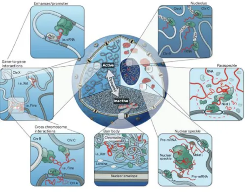
LncRNAs have been shown to be involved in the dynamic nuclear organization as key player molecules, either by their participation in the formation of nuclear subcompartments or by the establishment of three-dimensional interactions of genomic loci for the regulation of gene expression (Fig.1.4). The nuclear, highly abundant lncRNAs Neat1 and Neat2/Malat1 are the most prominent cases of lncRNA that localize to and participate in the structural integrity of nuclear bodies (Hutchinson et al., 2007). Neat1 lncRNA scaffolds the formation of paraspeckles, nuclear bodies involved in the retention of mRNAs that undergo Adenosine-to-Inosine editing, possibly by relying on Neat1’s continued transcription. Neat1 interacts with paraspeckles proteins and is required for the formation and stability of this nuclear compartment (Chen and Carmichael, 2009; Clemson et al., 2009; Mao et al., 2011; Sasaki et al., 2009; Sunwoo et al., 2009). Malat1 lncRNA localizes to nuclear speckles, a repository of transcription and splicing factors, and facilitates the proper localization of some protein components of the speckle (Bernard et al., 2010; Hutchinson et al., 2007). Malat1 is not necessary for the formation of speckles, in contrast to Neat1 RNA which is a structural component of the paraspeckles (Clemson et al., 2009). Gomafu (also known as Miat or Rncr2) is another example of lncRNA that participates in the formation of nuclear bodies (Sone et al., 2007a). It binds two splicing factors and modulates their function by sequestering these proteins into separate nuclear bodies (Ishizuka et al., 2014).
Other lncRNAs are necessary for the establishment of nuclear subcompartements locally silenced and compacted. These include Xist for the formation of the Barr body (the inactive X-chromosome), Kcnq1ot1 and Air for their imprinted loci, as previously detailed.
In addition, lncRNAs can participate in the three-dimensional structure of the genome through the establishment of long-range interactions in the nuclear space. The lncRNA Firre (Functional Intergenic Repeating RNA Element) has been demonstrated to form a domain localizing in cis over its own locus and in trans bringing five trans-chromosomal loci to proximity and orchestrating their gene expression (Hacisuleyman et al., 2014). Moreover, Firre is involved in anchoring the inactive X-chromosome to the perinucleolar region through the CTCF/cohesin complex (Yang et al., 2015). The lncRNA Charme also forms a domain in which it stabilizes long-range chromosomal interactions (in cis) on a region on its own chromosome and regulates the expression of the genes of that region (Ballarino et al., 2018). Many cases exist of chromosome looping or pairing where lncRNAs have been implicated to (Ma et al.,
21 2015; Wang et al., 2011; Yang et al., 2013). The association of lncRNA function with higher order chromatin structure by bringing distal sites into proximity have been verified by genome-wide chromosome conformation capture (Hi-C) data (Engreitz et al., 2013; Lieberman-Aiden et al., 2009).
Figure 1.4. Cross-section of nucleus showing different organizational levels: active (A) and inactive (B) compartments, nuclear bodies and their associated ncRNAs, short- and long-range genomic interactions. Clockwise: nucleolus with the rRNAs, paraspeckle with Neat1 RNAs, nuclear speckles with Malat1 RNAs, the inactive X chromosome (Barr body) repositioned to B compartment and localized to the nuclear periphery, Firre RNA established long-range inter-chromosomal interactions, short-range interactions of enhancer-promoter elements (with permission from Rinn and Guttman,
2014).
LncRNAs and nuclear matrix
Many of the aforementioned lncRNAs, which actively participate in the nuclear organization, seem to share a common characteristic: their association with the nuclear matrix. Xist RNA resists the biochemical fractionation procedure of the removal of cellular DNA, protein and mRNAs, resulting in the insoluble nuclear matrix (Clemson et al., 1996). Additionally, Xist directly interacts with nuclear matrix hnRNPU/SAF-A protein (Hasegawa et al., 2010; McHugh et al., 2015) and requires the SATB1 nuclear matrix protein for the X-inactivation initiation (Agrelo et al., 2009). Therefore, Xist RNA seems to be strongly associated to the nuclear matrix. Furthermore, Firre RNA has been shown to interact with the hnRNPU/SAF-A protein and this interaction is necessary for the formation of the Firre domain
22 (Hacisuleyman et al., 2014), and Gomafu RNA has been shown to be fractionating with the insoluble nuclear matrix upon nuclear matrix preparation (Sone et al., 2007a). It is thus possible, that the association of a lncRNA with the nuclear matrix could be an indication that they might participate in higher-level chromatin organization.
23
III.
Mouse Embryonic Stem Cells
The Embryonic Stem (ES) cells are isolated from the inner cell mass (ICM) of pre-implantation embryos at the blastocyst stage, and can be kept in culture under defined media. ES cells are characterized by two defining properties: self-renewal and pluripotency. These abilities to undergo unlimited cell divisions while maintaining their identity and to give rise to cells of all three germ layers (endoderm, mesoderm and ectoderm) as well as the germ lineage in vitro (Evans and Kaufman, 1981; Thomson et al., 1998) make them an interesting model system to study pluripotency and cellular differentiation. Mouse ES cells are able to give rise to teratocarcinomas when injected in adult compartments, and to produced chimeric animals when injected back in pre-implantation embryos contributing also to the germ line of the newborn (Bradley et al., 1984).
A. Extrinsic pathways regulating pluripotency
Mouse ES cells were originally cultured on feeder layers derived from mouse embryonic fibroblasts (MEF). Later it was found that Leukaemia Inhibitory Factor (LIF), a member of the Interleukin-6 cytokines produced by MEFs, was the key factor to maintain pluripotency of mouse ES cells by inhibiting their differentiation (Smith et al., 1988; Williams et al., 1988). ES cells require extrinsic growth factors to maintain their pluripotency in culture. These extrinsic modulators act on different signaling pathways to regulate intrinsic transcription factor networks to sustain ES cells in the undifferentiated state.
Binding of LIF to its receptor induces the activation of the JAK (Janus tyrosine kinase) pathway which subsequently can activate different signaling pathways: STAT3 (signal transducer and activator of transcription), Ras/ERK1/2 (extracellular-signal-related kinases 1/2) and PI3K (phosphoinositide 3-kinase). STAT3 is the effector of the self-renewal response (Han et al., 2013). In mouse ES cells, LIF can substitute MEF feeder layers in maintaining pluripotency in the presence of animal serum, by activating STAT3. However, in serum-free cultures, LIF is insufficient to block neural differentiation and maintain pluripotency. To that direction, it was found that BMP is able to replace serum short-term to maintain pluripotency of mouse ES cells in the presence of LIF, by activating inhibitors of differentiation (Id) genes, which block neural differentiation by promoting endo- and mesoderm differentiation (Ying et al., 2003).
24 FGF/MAPK (fibroblast growth factor/mitogen-activated protein kinases) signaling pathway triggers differentiation in mouse ES cells and its activation is antagonistic to self-renewal. The use of inhibitors of FGF receptor and ERK results in promotion of mouse ES cell pluripotency (Burdon et al., 1999; Kunath et al., 2007; Stavridis et al., 2007). Independence from the Erk pathway is a defining feature of mouse ES cells because it is a basic signaling module that is essential in many cell types.
In the canonical Wnt pathway, β-Catenin binds to Tcf3 in the nucleus and leads to the abolition of its repressing effects on stemness genes, by dissociating Tcf3 from its DNA-binding sites (Shy et al., 2013; Wu et al., 2012). Tcf3 has been shown to colocalize with many pluripotency factors on key regulatory regions to directly repress pluripotency factors as Nanog and is defined as an important factor to instruct early differentiation in mouse ES cells (Cole et al., 2008; Guo et al., 2011; Leeb et al., 2014; Martello et al., 2012; Pereira et al., 2006). Intracellular β-catenin is targeted for proteasome destruction due to phosphorylation by GSK3 (glycogen synthase kinase-3). Inhibition/deletion of GSK3 in presence of LIF allows efficient ES cell self-renewal, indicating GSK3 as its antagonist (Sato et al., 2004).
The chemical inhibition of Erk (by PD0325901) sustains robust ES cell self-renewal in the presence of LIF and chemical inhibition of GSK3β (by CHIR99021) mainly prevents β-catenin degradation. These two inhibitors constitute the 2i medium, in which spontaneous differentiation of mouse ES cells is abolished, LIF and serum stimulation become facultative, and the pluripotent state is considered as naïve, or ground state of pluripotnecy (Ying et al., 2008).
B. Transcription factor-mediated pluripotency regulation
The intrinsic regulators of pluripotency are forming a complex network of pluripotency factors. Pluripotency factors orchestrate the maintenance of the ES cell state and have been identified by relatively specific expression in ES cells and early embryos, and through genetic screens. The central pluripotency factor is Oct4 (a POU-domain transcription factor, POU5F1). Oct4 is expressed in oocytes, during the first cleavages of the embryo, then restricted to the inner cell mass of the blastocyst and later, in the post-implantation embryo, exclusively detected in the germ cell lineage. Its repression results in loss of pluripotency and dedifferentiation to trophectoderm lineage (Niwa et al., 2000). Interestingly, Oct4 overexpression does not reinforce pluripotency. Even a mild overexpression of Oct4 causes
25 differentiation into primitive endoderm and mesoderm (Niwa et al., 2000). Consequently, its level needs to be tightly regulated to sustain ES cell self-renewal since its up- or downregulation induce divergent developmental programs. Another essential TF for ES cell self-renewal is Sox2, and as for Oct4, its levels should be constrained for efficient self-renewal. Sox2 depletion results in trophoblast differentiation while overexpression might lead in neuroectoderm, mesoderm, and trophectoderm but not endoderm differentiation (Kopp et al., 2008; Masui et al., 2007; Zhao et al., 2004) Sox2 physically interacts with Oct4 protein and binds DNA together with Oct4 at Oct/Sox elements (Chen et al., 2008; Pardo et al., 2010). It acts synergistically with Oct4 to activate Oct-Sox enhancers, which regulate the expression of pluripotent stem cell-specific genes, including Oct4 and Sox2 themselves, and Nanog, the third core pluripotency TF. Although Nanog is not required for maintenance of pluripotency of ES cells (Chambers et al., 2007) it is necessary for the in vivo pluripotency to develop the ICM (Silva et al., 2009). These core TFs are highly interconnected and interdependent with one another and with others such as Esrrb, Klf4, Tfcp2l1, and Tbx3 forming the complex pluripotency transcription network (Chen et al., 2008; Marson et al., 2008).
C. Pluripotency states
Mouse ES cells are derived from the early blastocyst between E3.5 and E4.5 originating from the ICM and the pre-implantation epiblast, respectively. Mouse pluripotent cell lines could as well be derived from post-implantation epiblasts, called Epiblast stem cells (EpiSCs). EpiSCs show dependency to different signaling pathways, exhibit a distinct expression profile of pluripotent markers and a different epigenetic profile compared to ES cells. They can form teratocarcinomas showing multiple lineage origins, a hallmark of pluripotent cells, but are unable to contribute to chimeras upon blastocyst injection (Brons et al., 2007; Tesar et al., 2007). EpiSCs are still able to contribute to the development of various lineages, including the germ line, when grafted at specific locations in the post-implantation embryos (Huang et al., 2012; Kojima et al., 2014).Therefore, ES cells and EpiSC constitute two pluripotency states, the former are naïve and the latter are primed pluripotent cells (Smith, 2017).
Reprograming of differentiated cells has been achieved through the artificial expression of pluripotency-associated TFs (reprogramming factors) (Takahashi and Yamanaka, 2006). The induced pluripotent stem cells (iPSCs) show identical properties to ES cells. The reprogramming process involves the combination of the silencing of the somatic program and the induction of the pluripotency-associated gene network.
26
D. ES cells and nuclear organization
Self-renewal requires that the ES cell genome maintains a cellular memory that defines its pluripotent capacity. At the same time, the ES cell genome must exhibit high plasticity to be able to enter any distinct differentiation pathway. However, the architectural integrity of the nucleus is important for faithful genome function (Francastel et al., 2000; Lamond and Earnshaw, 1998). The gene repositioning through different developmental stages and during differentiation occurs during mitosis, when the nuclear-envelope breaks down and the condensation of chromosomes disrupts the organization of the nuclear architecture, and thereby allows repositioning of chromosomal regions (Walter et al., 2003). Many nuclear compartments with specific biological functions are subjected to massive rearrangement upon differentiation. The nuclear lamina, the nucleolus, heterochromatin structure and nuclear speckles are a few of them to undergo significant morphological changes comparing ESC to neuronal progenitor cells (NPC) (Meshorer and Misteli, 2006). Such changes include heterochromatin that is confined to fewer and larger foci in ESC, the nuclear speckles form smaller and more dispersed foci in ESC than in NPCs, while the ill-defined lamina of the ESCs becomes distinct and round in the NPCs.
Chromatin structure can influence gene function by affecting the accessibility of regulatory proteins to their target sites and by modulating the affinity of transcriptional regulators with their targets. In ES cells chromatin is globally decondensed, and as cells differentiate, regions of condensed heterochromatin are formed (Francastel et al., 2000; Melcer and Meshorer, 2010). ES cell chromatin is overall more active, and differentiation is accompanied by a transition to transcriptionally less-permissive chromatin by increase of H3K9me3 and decrease in H3, H4 acetylation (Meshorer et al., 2006).
Pluripotency transcription factors have been shown to have a role in the establishment of interactions that take place within a TAD (intra-TAD level). High levels of Oct4, Nanog, Sox2, Klf4, Esrrb are co-binding with Mediator coactivator (Med1) and RNA pol II at certain genomic hotspots, called super enhancers (SEs), which seem to control cell identity genes (Hnisz et al., 2013; Whyte et al., 2013). On the other hand, Sox2 enhancers form 3D-clusters that are segregated from heterochromatin but overlap with a subset of Pol II enriched regions. Such an enhancer clustering may increase the speed at which Sox2 finds its target sequences within individual clusters (Liu et al., 2014).
27 Pluripotent cells have been shown to contain almost entirely euchromatin, with highly mobile open chromatin and relative lack of nuclear structure (Dang-Nguyen and Torres-Padilla, 2015; Meshorer et al., 2006). In contrast, differentiated cells contain a heterochromatin footprint unique to their specific cell type that is visible by various imaging techniques; heterochromatin is compacted and relocated to the nuclear periphery (Hathaway et al., 2012). During differentiation, the organization and localization of chromocenters (heterochromatic clusters of centromeres from different chromosomes) changes and commonly they are observed on the nuclear periphery and the perinucleolar zone in differentiated cells (Mayer and Grummt, 2005; Wijchers et al., 2015). Pluripotent cells have smaller blocks of heterochromatin as imaged by microscopy and based on DAPI distribution, chromocentres are poorly compacted in ES and full iPS cells compared to MEFs (Ahmed et al., 2010; Fussner et al., 2011).
E. ES cells and lncRNAs
Many large-scale screens have been performed in ES cells in order to identify lncRNAs that are expressed in pluripotent cells and could be important for ES cell biology. Mostly loss-of-function techniques were applied and then the effect on pluripotency, differentiation or reprogramming properties of the mouse ES cells was monitored (Bergmann et al., 2015a; Bogu et al., 2016; Guttman, 2009; Guttman et al., 2010b, 2011a; Lv et al., 2015). Despite the high number of lncRNAs found to be specifically expressed in mouse ES cells, very few of them have been individually studied and functionally characterized so far. The few examples that have been studied demonstrated that lncRNAs can act through different mechanisms to exhibit a functional role relevant to ES cell biology. TUNA lncRNA (Tcl1 upstream neuron-associated lncRNA) has been shown important for the ES cells self-renewal and neural differentiation but also for reprogramming efficiency when overexpressed (Lin et al., 2014). Panct1 lncRNA was shown to associate with Tobf1 protein and affecting the recruitment of Oct4 at common gene targets (Chakraborty et al., 2017). The Linc-RoR (regulator of reprogramming) lncRNA, as mentioned before, acts as a sponge for miRNAs thus eliminating their negative effect on the core pluripotency TFs (Wang et al., 2013b). Last, the lincU lncRNA has been demonstrated to repress the ERK1/2 signaling pathway by stabilizing Dusp9 ERK-specific phosphatase (Jiapaer et al., 2018).
28
Thesis objectives
A number of studies have revealed the implication of lncRNAs in nuclear organization by forming functional domains in the nucleus. Xist, Neat1, and Firre lncRNAs are a few of the known functional lncRNAs that actively participate in the compartmentalization of the nucleus. To date, there is no robust way of identifying such molecules. The objective of my thesis is to be able to identify and characterize lncRNAs with a functional relevance for 3D genome organization.
Nuclear organization is tightly related to gene expression regulation and severely affected during differentiation of ES cells. Upon differentiation, mouse ES cells show massive genome architecture reconstruction. Combining the fields of lncRNA and ES cell biology under the prism of nuclear organization, the aim of my studies is to identify lncRNA genes that would play a role in the establishment of nuclear domains orchestrating in that way the necessary changes that need to occur for differentiation or maintenance of pluripotency.
29
30
Cell culture
Culture media
FCS/LIF medium
Dulbecco’s Modified Eagle Medium DMEM + GlutaMAX-I (Gibco, cat. 31966-021) 10% fetal calf serum FCS (Gibco, cat. 10270-098)
1X MEM non-essential amino acids (Gibco, cat. 1140- 035) 0.1 mM 2-mercaptoethanol (Gibco, cat. 31350-010)
10 ng/ml recombinant LIF (MILTENYI BIOTEC, 130-099-895)
2i/LIF medium
50% DMEM/F-12(1:1v/v, Gibco, cat. 31331-028) 50% Neurobasal (Gibco, cat. 21103-049)
0.5X N2 supplement (Gibco, cat. 17502-048) 0.5X B27 supplement (Gibco, cat. 17504-044). 1X L-Glutamine (Gibco, cat. 25030-024)
10µg/mL Insulin (Sigma I1882-100MG) 37.5µg/mL BSA (Sigma A3311-10G)
0.1 mM 2-mercaptoethanol (Gibco, cat. 31350-010) 1μM PD0325901 (Axon, cat. 1408)
3μM CHIR99021 (Axon, cat. 1386)
10 ng/ml recombinant LIF (MILTENYI BIOTEC, 130-099-895) ES cell passaging
ES cells were cultured on plastic coated with 0.1% gelatin (SIGMA, cat. G1890-100G) in FCS/LIF media (or 2i/LIF when mentioned) and incubated at 37°C in 7% CO2. For the “matrix prep” (see below) cells were grown on gelatinized slides (Superfrost Plus, Thermo Fisher, cat. 4951PLUS4) placed in 15cm plates. Cells were passaged every 2-3 days, when they reached
31 70-80% confluence. Medium was changed every one or two days. Culture plates/flasks were treated with 0.1% gelatin in PBS 1X for 10 min before use. ES cells were washed with pre-warmed PBS and incubated with 1X trypsin-EDTA 0.05% (Thermo 25300062) at 37°C for 3 min. ES cells were quickly resuspended by pipetting up and down and a volume of DMEM/FCS medium equivalent to 5 times the volume of trypsin was added to block the reaction. Cells were transferred to a falcon tube and centrifuged for 5 min at 1000rpm. The pellet was resuspended in FCS/LIF (or 2i/LIF) and cells were split 1:5 or 1:10 at each passage.
Colony formation assay
After collecting the ES cells by trypsinization (as described for cell passaging), the cell pellets were resuspended in DMEM/FCS and counted. 600 cells were plated in a gelatinized well of a six well plate. Cells were cultured for 7 days in DMEM/FCS media with or without LIF and/or Doxycycline. Cells were washed with DMEM/FCS once and medium was replaced every day. Following, cells were washed in PBS and incubated for 45 sec in fixative solution (25ml of citrate solution, 8ml of formaldehyde solution and 65 ml of acetone). Fixed plates were washed in distilled water and stained for alkaline phosphatase activity using a leukocyte alkaline phosphatase kit (AP staining) (Sigma, cat. 86R-1KT). After a last water wash and an air drying step, the number of undifferentiated, mixed and differentiated colonies was assessed on a stereo-microscope (NIKON-SMZ1500).
Proliferation rate assay
0.3 million cells were counted and plated in appropriate medium in a well of a six-well plate. After 3 days, cells were trypsinized and counted to evaluate the total number of cells obtained. This procedure was repeated 4 times for each assessed cell line. Cell lines that were planned to be compared were always cultured in parallel to ensure comparable culture condition.
ES cell retinoic acid (RA) differentiation
Cells from FCS/LIF culture were counted and 105 cells were replated in 25cm2 flasks in
DMEM/FCS media without LIF. 24h later, media was changed and retinoic acid (RA, final concentration 10-6M) was added to DMEM/FCS. DMEM/FCS+RA medium was changed
every day for 3 days. RNA samples were collected over the course of the assay with Day 0 being the plating point when cells come from +LIF medium and days 1, 2, 3 corresponding to RA treatment.
32
ES cell differentiation upon LIF withdrawal
Cells from FCS/LIF culture were counted and 0.3 x 10⁶ cells/well were replated in six-well plates in two media conditions: DMEM/FCS with or without LIF. Culture lasted 48h with every day media change.
Embryoid bodies differentiation
Cells were washed once with PBS 1X and treated with pre-warmed trypsin. Partial dissociation of ES cells colonies was evaluated under the microscope and inactivation with a large volume of DMEM/FCS was done 1 or 2 min after trypsinization to allow small clumps of cells to be maintained. Cells were carefully recovered with a 10 mL pipette and transferred in a 50 mL falcon tube to avoid further dissociation. After few minutes, when the clumps progressively reached the bottom of the tube, as much as possible supernatant was gently removed without perturbing the accumulated clumps of cells. 10 mL of DMEM/FCS medium was gently added to the tube and the cells clumps were precociously resuspended and transferred into bacterial Petri dishes thus precluding cell adhesion (Day 0). Dishes were incubated at 37°C in 7% CO2. Medium was changed every day by carefully collecting the clumps of cells with a 10 mL pipette and replacing them into bacterial Petri Dishes in DMEM/FCS medium for 6 additional days and by splitting them, if necessary, into several dishes. At Day 6, the biggest embryoid bodies were collected by allowing the clumps to decant for a short time followed by the quick aspiration of the supernatant. They were subsequently replated on gelatinized surfaces at low density to allow for bodies adhesion and cell differentiation. One day later, adhesion of the embryoid bodies was checked under the microscope. Medium was changed every day and differentiating samples were collected at Day 6 and Day 10 for RNA extraction and gene expression analysis.
EpiLC differentiation
Cells cultured in FCS/LIF medium were adapted to 2i/LIF medium for 3 passages (9 days in total) before starting the differentiation protocol. The EpiLCs differentiation was induced by plating 0.23 million ES cells on a well of a 6-well plate coated with human plasma fibronectin (16.7 mg/ml) in N2B27 medium containing activin A (20 ng/ml), bFGF (12 ng/ml), and KSR (1%). Medium was changed every day until day 3 of differentiation. Cells were harvested along the assay (FCS/LIF, 2i/LIF 1st passage, 2i/LIF 3 passages, Day 1, 2 and 3 EpiLCs) for RNA extraction and gene expression analysis or in situ hybridization experiments.
33
Oct4 depletion assay
Zhbtc4 cells (Hitoshi Niwa, Miyazaki, and Smith, 2000) were used for Oct4 loss of function assay. This transgenic cell line was generated from the WT E14Tg2a line. Both endogenous loci of Oct4 gene have been invalidated and replaced by antibiotics resistance. In addition, two exogenous transgenes have been randomly integrated in the genome: one constitutively expressing the Doxycycline-repressed transcriptional activator (tTA) and another one harboring a tTA responsive promoter driving Oct4 cDNA expression. Therefore, upon Dox addition in the medium, the constitutive expression of Oct4 is quickly abolished to be already undetectable by 12 hours after treatment. 3 million cells were plated in FCS/LIF medium in a T75 flask and treated or not with Dox for 12, 24 of 48 hours and collected at the end of the treatment for RNA extraction and gene expression analysis.
Nanog depletion assay and Nanog KO cell line
44iN cells (Festuccia et al., 2012) were used for Nanog loss of function assay. This transgenic cell line was generated from the WT E14Tg2a line. Both endogenous loci of Nanog gene have been invalidated and replaced by antibiotics resistance. In addition, two exogenous transgenes have been randomly integrated in the genome: one constitutively expressing the Doxycycline-activated transcriptional activator (rtTA) and another one harboring an rtTA responsive promoter driving Nanog cDNA expression. Therefore, upon Dox withdrawal from the medium, the constitutive expression of Nanog is quickly abolished to be already undetectable by 12 hours after Dox removal. 3 million cells were plated in FCS/LIF medium in a T75 flask and treated or not with Dox for 12, 24 of 48 hours and collected at the end of the treatment for RNA extraction and gene expression analysis. To culture 44iN cells in the absence of Nanog long-term (Nanog KO cells), the cells were maintained under G418 selection, as previously described (Festuccia et al. 2012).
Nanog overexpression cell line
EF4 cells (Chambers et al., 2003) were used as Nanog overexpressing cells. This transgenic cell line was generated from the WT E14Tg2a line. An exogenous transgene have been randomly integrated in the genome harboring a Nanog cDNA cassette downstream of a CAG promoter leading to the stable and strong overexpression of Nanog. 1 million EF4 cells were plated in a T25 in FCS/LIF medium and lysed after 3 days of culture for RNA extraction and gene expression analysis.