HAL Id: hal-02678518
https://hal.inrae.fr/hal-02678518
Submitted on 31 May 2020
HAL is a multi-disciplinary open access
archive for the deposit and dissemination of
sci-entific research documents, whether they are
pub-lished or not. The documents may come from
teaching and research institutions in France or
abroad, or from public or private research centers.
L’archive ouverte pluridisciplinaire HAL, est
destinée au dépôt et à la diffusion de documents
scientifiques de niveau recherche, publiés ou non,
émanant des établissements d’enseignement et de
recherche français ou étrangers, des laboratoires
publics ou privés.
Distributed under a Creative Commons Attribution - NonCommercial| 4.0 International
License
chicken embryonic stem cells
Hervé Acloque, Anne Mey, A.M. Birot, H. Gruffat, Bertrand Pain, Jacques
Samarut
To cite this version:
Hervé Acloque, Anne Mey, A.M. Birot, H. Gruffat, Bertrand Pain, et al.. Transcription factor cCP2
controls gene expression in chicken embryonic stem cells. Nucleic Acids Research, Oxford University
Press, 2004, 32 (7), pp.2259-2271. �hal-02678518�
Transcription factor cCP2 controls gene expression
in chicken embryonic stem cells
Herve Acloque, Anne Mey, Anne Marie Birot, Henri Gruffat
1, Bertrand Pain and
Jacques Samarut*
Laboratoire de Biologie MoleÂculaire de la Cellule, CNRS-INRA UMR5161, Ecole Normale SupeÂrieure de Lyon, IFR128 BioSciences Lyon-Gerland, 46 AlleÂe d'Italie, 69364 Lyon Cedex 07, France and1Unite de Virologie
Humaine, U412 INSERM, Ecole Normale SupeÂrieure de Lyon, IFR128 BioSciences Lyon-Gerland, 46 AlleÂe d'Italie, 69364 Lyon Cedex 07, France
Received January 9, 2004; Revised and Accepted March 24, 2004 DDBJ/EMBL/GenBank accession no. AY298725
ABSTRACT
cENS-1/cERNI genes have been shown to be expressed very early during chicken embryonic development and as well as in pluripotent chicken embryonic stem (CES) cells. We have previously identi®ed a promoter region, which is speci®cally active in CES cells compared to differentiated cells. In order to understand the molecular mechanisms which regulate the cENS-1/cERNI promoter, we analyzed the cis-acting elements of this promoter in CES and differentiated cells. We identi®ed a short sequence, named the B region, 5¢-CAAG TCCAGG CAAG-3¢, that exhibits a strong enhancer activity in CES and differentiated cells. Mutation of the B region in the whole cENS-1 promoter strongly decreases the promoter activity in CES cells, suggesting that this region is essential for activat-ing the promoter. The B region is similar to the previously described response element for the tran-scription factor CP2 and we show by supershift experiments that a protein complex containing CP2 is bound to this B response element. All these results identify a nuclear factor belonging to the CP2 transcription factor family that is crucial for the activation of the cENS-1/cERNI promoter. The pat-tern of expression of cCP2 in early chicken embryo before gastrulation is very similar to that of cENS-1/ cERNI which strongly suggests that cCP2 also plays an essential role in gene expression early in embry-onic development.
INTRODUCTION
Chicken embryonic stem (CES) cells are derived from the epiblast of chicken early embryos [stage X according to Eyal-Giladi and Kochav (1)]. These cells are pluripotent, can grow in vitro for long term and can contribute to many
embryonic tissues including germ line when they are grafted into an host embryo (2). They then provide a very good model to investigate molecular mechanisms underlying the control of pluripotency and differentiation commitment. In addition to the mouse embryonic stem (ES) cell model, it could help to de®ne more precisely the pathways conserved and involved in the maintenance of pluripotency during embryonic develop-ment.
In the mouse embryo, few regulators of ES cell pluripotency have been characterized so far. It has been shown that Oct-3/4 is a key regulator of ES cells pluripotency and is involved in the control of other genes like FoxD3 or FGF4 (reviewed in 3). Sox2, FoxD3, FGF4, FGFR2 and recently Nanog have also been shown to be involved in the regulation of embryonic cell pluripotency in vivo and in vitro (4±9). The in vivo knock-out of these genes leads to a slight decrease in the number of cells originating from the epiblast and to a rapid embryonic lethality. Moreover, it has been impossible to derive in vitro ES cells from these mutant embryos.
In order to identify new sets of critical genes for ES cell biology, we used a gene trap based strategy in CES cells. We have identi®ed a gene family which is speci®cally expressed in CES cells and early embryo (10). The Embryonic Normal Stem (ENS) genes are strongly expressed in CES cells, but their expression rapidly decreases after the induction of CES cell differentiation by retinoic acid, or during embryoid body formation. In the embryo, ENS genes are highly expressed in the epiblast prior to gastrulation, and their expression is then restricted to the prospective neural plate, and later to the border of the neural plate. Up to now, three genes of this family have been characterized (10): 1) cENS-1 which contains an open reading frame identical to cERNI (Early Response to Neural Induction) (11), inserted between two repeated sequences; 2) cENS-2 which is a truncated form of cENS-1; 3) cENS-3 which possesses two open reading frames corresponding to pol and env-retrovirus related gene. The coding region for ENS-1 does not show any known homology with retroviral gene and does not seem to be conserved among birds other than galliforms. However, the occurrence of LTR-like repeated structures at both ends of ENS genes
*To whom correspondence should be addressed. Tel: +33 4 7272 8536; Fax: +33 4 7272 8080; Email: [email protected] Present address:
Herve Acloque, Instituto Cajal, Avenida Doctor Arce 37, 28002 Madrid, Spain
Nucleic Acids Research, 2004, Vol. 32, No. 7 2259±2271 DOI: 10.1093/nar/gkh545
suggested that these genes might be derived from an ancestral retrovirus or transposon structure (10). In the putative U3 region of the LTR-like structures, we have previously isolated a promoter region, and shown that its activity is much stronger in CES cells than in differentiated cells (10). This result was surprising because it has been shown that retroviral LTRs are usually silenced in mouse ES cells and early mouse embryo, and correlated with a high methylation of these sequences (12,13). This silencing has not yet been observed in chicken blastula. Early chicken blastodermal cells (from which CES cells are derived) are permissive for infection by Rous Sarcoma Virus (RSV) and readily expressed RSV genes (14), suggesting that CES cells could be more permissive to retroviral gene expression than mouse ES cells.
It is also possible that ENS LTRs are protected from gene silencing by speci®c molecular regulators or by a positive position effect. The restricted expression pattern of ENS genes in embryonic stem cells and in the early embryo is of particular interest, and led us to study the mechanisms underlying ENS gene expression. Based on a systematic analysis of the regulatory elements of cENS genes, we report here that one cis-acting element, belonging to the ®rst 310 bp of the cENS-1/cERNI promoter, is responsible for its activity in CES cells. These results as a whole reveal the existence of a nuclear factor, which belongs to the CP2 transcription factor family. Moreover, we observed that the pattern of expression of CP2 in early embryo before gastrulation is very similar to that of cENS/cERNI which strongly suggests that CP2 also plays an essential role in gene expression early in embryonic development.
MATERIALS AND METHODS Cell culture and DNA transfection
The 9N2-5 CES cell line was maintained in vitro and transfected as previously described (10) with several modi®-cations. Brie¯y, CES cells were cultivated in Dulbecco's modi®ed Eagle's medium (DMEM)/F12 (Invitrogen) contain-ing 5% fetal bovine serum (FBS; PAN, Austria) supplemented with 1 ng/ml IGF1 (Sigma), 0.5 ng/ml mSCF (R&D), 1 ng/ml hIL-6 (Peprotek), 1 ng/ml hIL6-sR (Peprotek 10±4 M),
b-mercaptoethanol, sodium pyruvate and non-essential amino acids on an irradiated feeder of STO cells at 104
cells/cm2. QT6 cells (15) were cultivated in HamF12 medium
(Invitrogen) containing 2% FBS complemented with 10% TPB (Tryptose Phosphate Broth, Difco). The day before transfection, the cells were plated in 12-well dishes at a density of 5 3 104cells per well for the 9N2-5 cells or 1 3 105
per well for QT6 cells. Cells were cotransfected using Lipofectamine reagent (Invitrogen) with 300 ng of DNA construct and 30 ng of the Renilla Luciferase Reporter construct, pRL-TK, to normalize for transfection ef®ciency. After 5 h of incubation in diluted culture medium containing only 1% FBS, the cells were washed twice with phosphate-buffered saline (PBS) and regular medium was added to each well. On the following day, cells were washed twice with PBS and lysed with passive lysis buffer (Promega), then Fire¯y and Renilla luciferase luminescence assays were successively performed using Dual Luciferase Assay (Promega) as described by the manufacturer. Fire¯y reporter gene values
were normalized to the activity of the Renilla luciferase, used as internal control.
DNA constructs
All the pGL2 luciferase reporter vectors were purchased from Promega. ENS reporter constructs were produced using PCR ampli®cation with multiple primers that introduced a 5¢-MluI restriction site at positions ±738, ±456, ±308, ±179 and ±32, respectively paired with a single 3¢-antisense primer that generated a 3¢-MluI restriction site at position +83. Primer sequences are available in Table 1. PCR products and pGL2-basic vector were digested with MluI, puri®ed and then ligated to produce, respectively, p738-luc, p456-luc, p308-luc, p179-luc and p32-p179-luc. Reporter constructs and the pGL2 basic negative control vectors were used for transient transfection studies. Monomers or trimers of the ±313/±180 region were constructed as follows: the ±313/±180 region was ampli®ed by PCR with BamHI ±313 S and BglII ±180 AS primers (Table 1). After digestion with BamHI/BglII, PCR products were ligated in the BamH1 site of pGEM-7Z. BglII restriction enzyme was then added to the ligation mix to eliminate multimers with various orientations. Multimers were then excised from pGEM-7Z by NsiI/KpnI and ligated into p179-luc or pGL2-promoter (Promega). Multimer copy numbers and identities were con®rmed by DNA sequencing. Multimers (one or three copies) of the wild-type or mutated A and B response element (Table 1) were cloned in the SacI restriction site of the p179-luc construct or of pGL2-promoter. Insert identities were con®rmed by DNA sequencing. Targeted mutagenesis on p456-luc for the B response elements has been performed with the Quick Change Mutagenesis kit (Stratagene) using the primers: B mutagenesis S and B mutagenesis AS.
Nuclear extracts
Nuclear extracts were prepared with the Active Motif nuclear extract kit (Active Motif) and stored at ±80°C until further use. Protein concentrations were estimated using the Bio-Rad Protein Assay Kit I.
DNA binding assays
Probes used for DNA-binding assay were labeled using T4 polynucleotide kinase and [g±32P]ATP (Amersham). The
labeled fragments were puri®ed by acrylamide gel electro-phoresis. Approximately 2 3 104 c.p.m. of the probe were
added to a 30-ml reaction mixture in the presence or absence of 10 mg of nuclear extract in a buffer containing 20 mM HEPES (pH 7.9), 20% glycerol, 100 mM KCl, 2 mM MgCl2, 0.2 mM EDTA, 0.5 mM dithiothreitol and 1 mg of poly(dI±dC)-poly(dI±dC). For competition experiments, a 100-fold molar excess of unlabeled double-stranded oligonucleotide was incubated for 10 min with nuclear extract prior to the addition of labeled double-stranded probe. After incubation on ice for 30 min, the DNA±protein complexes and unbound probes were separated by electrophoresis on a 4% glycerol and 5% polyacrylamide gel in 0.53 TBE at 160 V for 120 min. The gels were dried and autoradiographed. EMSA supershift assays were performed with a mouse monoclonal antibody directed against CP2 (anti-LSF from BD Biosciences). Then 250, 25 or 2.5 ng of this antibody was added to the reaction mix after the addition of the labeled probe.
Methylation interference assays
Probes were labeled as previously described (16). For sense or antisense oligoprobes, only the sense or antisense oligo-nucleotide was labeled. For the guanine methylation proced-ure, 1 3 106c.p.m. of the probe was methylated using 1 ml of
dimethyl sulfate (DMS) (Merck) for 3 min at room tempera-ture in 200 ml of 50 mM sodium cacodylate (pH 8.0)±1 mM EDTA. The reaction was stopped by the addition of 50 ml of stop buffer [5 M sodium acetate (pH 7.0), 1 M b--mercaptoethanol]. The DNA was then precipitated with ethanol, and resuspended in 30 ml of water. The electro-phoretic mobility shift assays were carried out as described above, but with 2 3 105c.p.m. of modi®ed32P-labeled probe
and 30 mg of crude nuclear extracts. After autoradiography, the DNA was electro-eluted from the bands corresponding to the retarded probe and the free unbound probe. After precipitation with ethanol, the amounts of radioactivity of the bound and free probe were adjusted and the DNA was cleaved in 100 ml of 1 M piperidine for 30 min at 90°C. The
cleaved products were separated by electrophoresis through a denaturing 18% polyacrylamide gel and were autoradio-graphed.
Real time PCR
Total mRNA were extracted from CES cells using RNeasy kits (Qiagen). Real-time RT±PCR was performed on a Light-Cycler (Roche) using the QuantiTect SYBR Green PCR Kit (Qiagen). The obtention of the single denaturation peak upon heating and of a single band after agarose gel electrophoresis (data not shown) con®rmed that the real time RT±PCR reaction was speci®c. The PCR conditions used were: 15 min denaturation at 95°C followed by 50 cycles of 95°C for 15 s denaturation step, 58°C for 15 s and 72°C for 15 s; followed by a melting curve from 60 to 95°C.
The primers used for real time RT±PCR are: (i) QPCR GAPDH S: 5¢-TGGGTGTCAACCATGAGAAA-3¢; (ii) QPCR GAPDH AS: 5¢-CATCCACCGTCTTCTGTGTG-3¢; (iii) QPCR CP2 S: 5¢-CTATCTGGAGGAGCTGACGG-3¢; (iv) QPCR CP2 AS 5¢-TCTGAATCATCTCGTCGCTGA-3¢.
Table 1. Oligonucleotides used in this study
Oligonucleotide Sequence (5¢ ® 3¢) ±738 S acgcgtgtggatgtttattaggaagc ±456 S acgcgttgaaacatttgattccac ±308 S acgcgtagtttgctgaggaacaag ±179 S acgcgtaggggcatcgagagagag ±32 S acgcgttggaggataaaagaggt +83 AS acgcgttggcagagaacccct BamHI ±313 S ggatccctcaaagtttgctgagga BglII ±180 AS agatctggccttgtccatctgtgc KpnI ±456 S ggtacctgaaacatttgattccac SacI ±238 AS gagctccagtgtcctcaagacaaa A RE wild-type S cactgatggacaggagct A RE wild-type AS cctgtccatcagtgagct A RE mutant S cactaatggataggagct A RE wild-type x3 S cactgatggacaggactgatggacaggactgatggacaggagct A RE wild-type x3 AS cctgtccatcagtcctgtccatcagtcctgtccatcagtgagct A RE mutant x3 S cactaatggataggactaatggataggactaatggataggagct A RE mutant x3 AS cctatccattagtcctatccattagtcctatccattagtgagct B RE wild-type S caagtccaggcaagtccgagct B RE wild-type AS cggacttgcctggacttgagct B RE mutant S caattccaggcaattccgagct B RE mutant AS cggaattgcctggaattgagct B RE wild-type x3 S caagtccaggcaagtccgcaagtccaggcaagtccgcaagtccaggcaagtccgagct B RE wild-type x3 AS cggacttgcctggacttgcggacttgcctggacttgcggacttgcctggacttgagct B RE mutant x3 S caattccaggcaattccgcaattccaggcaattccgcaattccaggcaattccgagct B RE mutant x3 AS cggaattgcctggaattgcggaattgcctggaattgcggaattgcctggaattgagct Probe 1 S gatccttcttttatatcattgactcaaagtttgcta Probe 1 AS gatctagcaaactttgagtcaatgatataaaagaag Probe 2 S gatcaggtttgctgaggaacaagtccaggcaagtcctgggcaaa Probe 2 AS gatctttgcccaggacttgcctggacttgttcctcagcaaacg Probe 3 S gatccaggcaagtcctgggcaaaggcagagaaa Probe 3 AS gatcatttctctgcctttgcccaggacttgcct Probe 4 S gatccgggcaaaggcagagaaatcttttgtcta Probe 4 AS gatctagacaaaagatttctctgcctttgcccg Probe 5 S gatctcttttgtcttgaggacactgatggacagg Probe 5 AS gatccctgtccatcagtgtcctcaagacaaaaga Probe 6 S gatccttgaggacactgatggacaggtcctggca Probe 6 AS gatctgccaggacctctccatcagtgtcctcaag Probe 7 S gatctcctggctaaggattgtgaaatccttta Probe 7 AS gatctaaaggatttcacaatccttagccagga B mutagenesis S ctcaaagtttgctgaggacacagtccaggcaagtcctg B mutagenesis AS caggacttgcctggactgtgtcctcagcaaactttgag
In situ hybridization of whole-mount embryos
Whole-mount in situ hybridization was carried out as described by Wilkinson and Nieto (17) and revised by Streit et al. (18). The probe used for the experiment was ampli®ed by PCR with the following primers: CP2 S 5¢-gaatt-catggcctgggcgctgaa-3¢, CP2 QPCR AS 5¢-cccttggttgaggtaggt-ga-3¢ and covers the ®rst 280 bp of the CP2 coding sequence. RESULTS
A ±308/+83 cENS/cERNI promoter fragment is speci®cally active in CES cells
In a previous study, we identi®ed a promoter region of ENS genes, which was speci®cally active in CES cells (10). The promoter sequence is shown in Figure 1. Analysis of the promoter sequence using MatInspector Professional release 6.1 (19) revealed several putative cis-acting elements for factors implicated in regulating constitutive gene expression (like AP1). It also revealed three well conserved binding elements for GATA-1 in the proximal region of the promoter. However, the analysis did not identify any candidates known
to be involved in regulating ES cell speci®c genes. In order to localize functional cis-acting element(s) in the cENS-1 promoter, luciferase reporter constructs were designed and generated for use in transient transfection assays. By using 5¢ deletion mutants of the cENS1 promoter fused to the luciferase reporter gene (cENS-luc), we carried out DNA transfection experiments in CES cells, CES cells induced to differentiate and QT6 cells, a quail ®broblast cell line. Our largest construct contained the ®rst 738 bp of the promoter. We chose this fragment because it includes the putative U3 region of ENS genes (10). Successive deletions of cENS-luc from ±738 to ±456 did not reduce the expression level of the reporter gene in CES cells (Fig. 2). An additional deletion to position ±308 did not decrease the promoter activity in these cells (not shown). In contrast, a further deletion to position ±179 gave a 20-fold decrease, indicating that critical cis-acting elements are located between ±308 and ±179. None of these constructs showed a strong transcriptional activity either in CES cells induced to differentiate with retinoic acid for 48 h or in QT6 cells, a quail immortalized ®broblast line (15) (Fig. 2). QT6 cells were used in the following experiments as a model for differentiated cells.
Figure 1. Structure of cENS-1 promoter. The sequence from ±738 to +83 relative to the transcription start site (TSS) is presented. The TSS is marked with right-angled arrows and the putative TATA box in bold. This sequence was analyzed by MatInspector Professional release 6.1. Transcription factors with 1.00 core similarity and more than 0.95 matrix similarities are highlighted in gray and labeled above the sequence. (+) and (±) indicate, respectively, the sense and antisense strands. The A and B response elements identi®ed in this study are underlined.
A ±308/±180 region is a key region for promoter activation in CES cells
To address the possible enhancer function of the DNA sequences that are located between ±308 and ±179, we cloned this region as one or three copies upstream of a SV40 basal promoter, which drives the expression of the luciferase reporter gene (SV40 basal promoter in pGL2-promoter). These constructs were transfected in CES cells or in QT6 cells. Three copies of the ±308/±180 region increased luciferase activity 17-fold in QT6 cells and 13-fold in CES cells (Fig. 3A). One copy of the ±308/±180 region also increased luciferase activity in both cell lines. This stimulatory effect was independent of the orientation of the ±308/±180 region too. To test this enhancer activity on the cENS-1 promoter, we placed one or three copies of the ±308/±180 region upstream of the ±179/+83 cENS-1 promoter region (p179-luc construct). This minimal promoter contains a TATA-like box as a known cis-acting element, and has a weak transcriptional activity in CES cells. As with the SV40 basal promoter, one or three copies of the ±308/±180 region increased luciferase activity in CES and QT6 cells (Fig. 3B). However when the ±308/±180 region was placed upstream of the ±179/+83 cENS-1 pro-moter, in QT6 cells, the activation level decreased strongly compared to the basal SV40 promoter (from 17-fold to 4-fold with three copies of the ±308/±180 region and from 10-fold to 3-fold with one copy). In contrast, the level of activation was not altered in CES cells (compare Fig. 3A and B).
As a whole, these results indicate that the ±308/±180 region acts as a transcriptional enhancer for the expression of a reporter gene in undifferentiated CES cells as well as in QT6 cells. Moreover, when located upstream of the ±179/+83 region, this ±308/±180 region is speci®cally active in undif-ferentiated CES cells.
Two distinct protein±DNA interacting regions are located in the ±308/±180 region
To investigate potential DNA±protein binding sites located in the ±308/±180 region and involved in cENS-1/ERNI promoter activity, we performed gel retardation experiments using CES cells nuclear extracts. Seven overlapping oligonucleotides were designed to cover the ±308/±180 region (Fig. 4A). The putative AP-1 site identi®ed in silico and located in probe I was apparently not bound in vitro by nuclear proteins from CES cells (Fig. 4B, lane 2). In contrast, the oligonucleotides II, V and VI were bound by CES nuclear factors (Fig. 4B, lanes 5, 14 and 17). These three complexes were competed for by 100-fold molar excess of the respective unlabeled oligonu-cleotides (Fig. 4B, lanes 6, 15 and 18). The DNA±protein complexes observed with probes V and VI migrated to the same positions suggesting that they are identical and located on a DNA sequence situated between positions ±251 and ±230 of the ENS-1 promoter, in the overlapping region of the V and VI oligonucleotides. Complexes migrating to the same positions were also observed when probes II, V and VI were incubated with QT6 nuclear extracts (data not shown). These results are consistent with our previous data showing that the ±313/±180 region was active in both cell lines when placed upstream of a SV40 basal promoter. The overlapping sequence of oligonucleotides V and VI was called the A region and the sequence located on oligonucleotide II was designated the B region.
Fine mapping of the A and B response elements
To de®ne more accurately the nucleotides involved in DNA± protein interaction on A and B regions, we used double-stranded oligonucleotides chemically modi®ed with dimethyl sulfate (methylation interference analysis). On both the coding
Figure 2. Functional analysis of deletion mutants of the cENS-1 promoter. Deletions were generated as described in Materials and Methods; a schematic representation of each reporter construct is shown on the left. These constructs were transfected into CES cells, CES cells induced to differentiate with retinoic acid for 48 h and into QT6 cells. Fire¯y luciferase activity was normalized for transfection ef®ciency using the Renilla luciferase activity. Values are relative to the activity obtained with the largest promoter fragment (p738-luc) in CES cells for three independent experiments, each performed in triplicate.
and non-coding strands of the A region, strong interferences were detected on guanine residues which allowed us to delimit a binding region between positions ±240 and ±232 in the sequence 5¢-Tgatggaca-3¢ (Fig. 5A).
The same experiment was performed on both the coding and the non-coding strands of the B region, where strong interferences were detected on some guanine residues between positions ±296 and ±280 in the sequence 5¢-AACAAGTCCAGGCAAGT-3¢ (Fig. 5A). This response element contains a direct repeat (underlined) 5¢-CAAGT CCAGG CAAGT-3¢, which is important for DNA±protein interaction, as suggested by the strong interferences on the same guanine residues on each of the two CAAGT boxes (Fig. 5A). The respective places of these two response elements in the cENS-1 promoter are shown in Figures 1 and 5B.
The A region (±240/±232) binds nuclear factors present in CES cells but is devoid of enhancer activity.
To con®rm that the residues identi®ed by methylation interference analysis were essential for DNA±protein
interactions, we performed band shift experiments with a labeled probe containing the A region, in competition with wild-type or mutated oligonucleotides for the A region (not shown). These results con®rmed that the residues identi®ed by methylation interference analysis in the A region were essential for DNA±protein interaction.
To investigate the role of the A binding site in cENS-1 promoter activity, we placed multimers of the A sequence upstream of the p179-luc construct or the SV40 basal promoter. The different constructs generated were transfected in CES or in QT6 cells and the enhancer activity was estimated with respect to the activity of empty vectors. None of these constructs revealed any enhanced activity as compared to empty vectors (not shown). These data then suggest that the A element does not have any enhancer function in the cENS-1/ cERNI promoter region at least in the two types of cells tested. Characterization of the B region (±294/±280)
After having identi®ed the residues by the methylation interference assay, we con®rmed that they were essential for
Figure 3. Functional analysis of the ±308/±180 region. (A) One or three copies of the ±308/±180 region fused to the pGL2 promoter construct were assayed by transfection experiments in CES cells and in QT6 cells. Values are relative to the activity obtained in each cell line with the empty pGL2-promoter (SV40) construct. (B) One or three copies of the ±308/±180 region fused to the p179-luc construct were assayed by transfection into CES and QT6 cells. Values are relative to the activity obtained with p179-luc construct. Data represent ratios of ®re¯y luciferase versus Renilla luciferase activities and values are means of at least three independent transfection experiments. The orientation of the ±308/±180 is shown by the sense of the arrows.
DNA±protein interaction through gel shift experiments with an oligonucleotide-labeled probe covering the B region (B RE) (Fig. 6A); the results are shown in Figure 6B. A unique DNA± protein complex was detected with the labeled probe incu-bated with CES nuclear extracts (Fig. 6B, lane 2). A competition with 10- or 100-fold molar excess of wild-type B oligonucleotide strongly decreased the signal corresponding to the DNA±protein complex, con®rming the speci®city of this interaction (Fig. 6B, lanes 3 and 4). A competition with a 100-fold molar excess of oligonucleotides mutated, respect-ively, on the C1, G4, C11and G14residues (Fig. 6A), which are important for DNA±protein interaction as proven by methyla-tion interference analysis, did not decrease the signal (Fig. 6B, lanes 5 to 8). These results con®rmed that each of these four nucleotides are essential for the interaction with nuclear binding proteins. It also demonstrates that each part of the direct repeat is necessary for DNA±protein complex forma-tion. To determine whether each of the nucleotides of the B sequence participates in this interaction, mutated labeled probes covering the ®rst part of the repeat were used in gel shift retardation assays (Fig. 6A and C). Results showed that the interaction with nuclear proteins was abrogated only when the C1or the G4residues were mutated (Fig. 6C, lanes 1 and 4). A probe, in which the T5residue is mutated, exhibited a weaker signal compared to the wild-type probe (Fig. 6C, lane 5), suggesting that this nucleotide improves the stabilization of the DNA±protein complex. The same results were obtained with probes mutated in the second part of the direct repeat (not shown). The two parts of this direct repeat are separated by a
linker of ®ve nucleotides (CCAGG). In order to check the function of this spacing sequence, we tested probes containing spacers of different lengths. Band shift assays performed with these oligonucleotides are shown in Figure 6D. Deletion or addition of three nucleotides to the linker decreased the interaction between DNA and nuclear factors (Fig. 6D, lanes 6 and 10), while the addition of six bases totally abolished the interaction (Fig. 6D, lane 14). The optimal B response element is then summarized by the sequence CAAGTNNNNNCAAGT where bold characters indicate bases essential for the DNA±protein complex formation. The B region is essential for promoter activation in CES cells
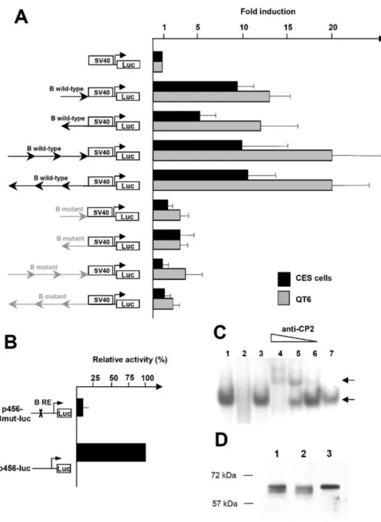
To test the enhancer activity of the B response element, multimers of this region were placed upstream of the SV40 basal promoter. The different constructs were transfected in parallel into CES cells and QT6 cells. One copy of the B region in sense or antisense orientation increased the activity of the basal SV40 promoter from 5±7 fold in CES cells to 10-fold in QT6 cells. Three copies of the B region in sense or antisense orientation also strongly increased the activity of the basal SV40 promoter from 10-fold in CES cells to 20-fold in QT6 cells (Fig. 7A). In comparison, the mutated B response element was inactive in the two cell lines. These data prove that the B region exhibits an enhancer activity, like the ±308/± 180 region, and strongly suggest that it could be a key activator of the cENS-1 promoter. This hypothesis is also con®rmed by targeted mutagenesis of the B response element
Figure 4. The ±308/±180 region contains two binding sites for CES cells nuclear factors. (A) Map of the seven probes used to cover the ±308/±180 region. Each probe is designated with a roman numeral. (B) EMSAs were performed with each double-stranded radiolabeled oligonucleotide probe and nuclear extracts prepared from CES cells as described in Materials and Methods. The positions of DNA±protein complexes throughout are indicated by arrows. Lanes 1, 4, 7, 10, 13, 16, 19: labeled probes alone; lanes 2, 5, 8, 11, 14, 17, 20: labeled probes with nuclear extracts from CES cells; lanes 3, 6, 9, 12, 15, 18, 21: competition experiments with 100-fold molar excess of wild-type unlabeled probes.
in the p456-luc construct. In this construct, the cytosine residue of the ®rst CNRG box in the B response element was substituted by an adenine residue (see Materials and Methods), leading in vitro to the loss of the protein±DNA interaction (Fig. 6C). Compared to the wild-type ±456/+83 cENS-1 promoter, the promoter with a mutated B element exhibited a strong decrease in its activity in CES cells (Fig. 7B) showing that the B response element plays an essential role within its
natural sequence environment. All these data support the fact that the B element is essential for the cENS-1 promoter activation in CES cells.
The B response element is bound by the transcription factor CP2
Analysis of the B response element revealed that it is similar to the CP2 consensus binding sequence CNRG-N6-CNRG (20).
Figure 5. Fine mapping of A and B binding sites by methylation interference. (A) Methylation interference assays were performed with methylated ±251/±223 (A region) or ±297/±277 (B region) probes radiolabeled at the 5¢-end of the sense or antisense strands. The probes were incubated with CES cell nuclear extracts. The free (F) and retarded (R) probes were then sequenced and compared with the G+A sequence obtained with the non-methylated probes. The G residues implicated were identi®ed by a disappearance or a decrease in the signal. (B) Summary of the interactions. The guanine nucleotides identi®ed by methylation interference assays are presented in bold on a double-stranded DNA fragment.
The CP2 response element, like the B response element, is composed of a direct repeat essential for protein binding. In order to test whether the B response element is bound in vitro by the transcription factor CP2, we performed supershift experiments using a mouse monoclonal antibody, which recognizes human CP2. Addition of anti-CP2 to the reaction after complex formation generated antibody-dependent super-shifts (Fig. 7C, lanes 4 to 6, top arrow) which were not seen with use of an IgG control antibody (Fig. 7C, lane 7). Increasing dilutions of the anti-CP2 progressively restored the unshifted DNA±protein complex (Fig. 7C, lanes 5 to 6, bottom arrow). The anti-CP2 incubated with nuclear extracts of CES cells did not interfere with the DNA±protein complex formed with the A response element (not shown). To con®rm the speci®city of the anti-CP2, we performed immunoblots using protein extracts from CES and HeLa cells (Fig. 7D). A band of 64 kDa was detected in undifferentiated and differentiated CES cells (lane 1 and 2) and HeLa cells (lane 3) at the
described size of CP2 (21). These results strongly suggest that the B response element binds the chicken CP2 transcription factor.
Expression pattern of chicken CP2 in CES cells and early embryo
Chicken CP2 has been previously shown to regulate the aA-crystallin promoter (21). Based on the cCP2 cDNA sequence published in that reported work, we ampli®ed the chicken CP2 by PCR from RNAs extracted from CES cells indicating that cCP2 is expressed in CES cells. In silico transduction of this cCP2 coding sequence (GenBank accession no. AY298725) revealed an amino acid sequence, which is very similar to those of human (22) and mouse (23) and is also identical to the published sequence of chicken CP2 (21), but with a small deletion of 10 amino acids (from position 323 to position 332). This small deletion is localized in the oligomerization domain (from position 266 to 403) (24) but preserved the DNA
Figure 6. Binding of chicken nuclear proteins to the B region. (A) Sequences of the oligonucleotides used. DNA binding sequence is underlined and mutations are marked with an asterisk and bold characters. Mutated nucleotides on the B region are numbered. (B) EMSAs were performed with the B wild-type labeled probe alone (lane 1) or with nuclear extracts prepared from CES cells (lane 2). The position of the DNA±protein complex is indicated by an arrow. Competition experiments were performed with a 10-fold molar excess (lane 3) or 100-fold molar excess (lane 4) of the unlabeled wild-type B oligonucleotide or with a 100-fold molar excess of the B mutated C1, G4, C11 and G14unlabeled oligonucleotide (lanes 5, 6, 7 and 8, respectively).
(C) B mutated C1, A2, A3, G4, T5, C6, C7oligonucleotides were labeled and used for EMSAs with CES cell nuclear extracts (lanes 1, 2, 3, 4, 5, 6 and 7,
respectively). (D) EMSAs were performed with each double-stranded radiolabeled probe alone (lanes 1, 5, 9, 13) or with CES cell nuclear extracts (lanes 2, 6, 10, 14). The position of the DNA±protein complex is indicated by an arrow. Competition experiments were performed with a 100-fold molar excess of either the mutated B unlabeled oligonucleotide (lanes 3, 7, 11, 15) or the B wild-type unlabeled oligonucleotide (lanes 4, 8, 12, 16).
binding domain (from position 189 to 239) (21) and the serine 291, whose phosphorylation is important for CP2 activation (25). CP2 expression was detected in undifferentiated and differentiated CES cells by immunoblot using a mouse monoclonal anti-CP2 antibody (Fig. 7D). These results are
consistent with the fact that the B region is active in undifferentiated and differentiated cells when placed upstream of the SV40 basal promoter. The spatial pattern of the cCP2 gene expression was examined during embryogenesis by whole-mount in situ hybridization. By using a probe located in
Figure 7. Functional analysis of the B region. (A) Multimerized wild-type or mutated B region fused to the pGL2-promoter construct were assayed by transfection into QT6 and CES cells. For each cell line, values are relative to the activity obtained with the pGL2-promoter construct. Data represent ratios of ®re¯y luciferase versus Renilla luciferase activities, and values are the means of three independent experiments. (B) The B region was disrupted by targeted mutagenesis on the p456-luc construct to give p456 Bmut-luc. The cytosine residue of the ®rst CNRG box in the B response element was substituted by an adenine residue (see Materials and Methods). These two constructs were transfected into CES cells and promoter activity assayed. Values are relative to the activity obtained with the p456-luc. (C) Labeled wild-type B oligonucleotide was used as a probe with CES cell nuclear extracts (lane 1). Complex is indicated by a black arrow. Competition with 100-fold molar excess of unlabeled wild-type or mutated B oligonucleotides is shown, respectively, on lanes 2 and 3; 250, 25 and 2.5 ng of monoclonal mouse anti-CP2 or 250 ng of IgG control antibody were incubated with CES nuclear extracts after the addition of B labeled probe (lanes 4 to 7, respectively). Antibody-dependent supershift is indicated by the top black arrow. (D) Immunoblots realized with nuclear extracts from undifferentiated and differentiated CES cells (lanes 1 and 2, respectively) and HeLa cells (lane 3) incubated with a monoclonal mouse anti-CP2.
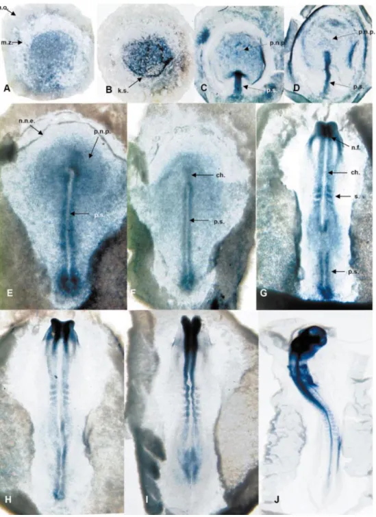
the coding sequence of cCP2, we observed a particular expression pattern during early chick embryogenesis. cCP2 is expressed in the epiblast of embryos at the pre-primitive streak stage (Fig. 8A and B). In embryos at the primitive streak stage, it is also expressed in the extending primitive streak and in the prospective neural plate (11) (Fig. 8C±F). At stages 7 and 8, the cCP2 expression was mostly localized in the neural folds and the somites of the embryo (Fig. 8G±I) but also in the regressing primitive streak. Later at stage12, cCP2 was
ubiquitously expressed in the whole embryo (Fig. 8J). The pattern of expression before stage 12 is quite similar to that previously described for cENS-1/cERNI with few differences (10,11).
DISCUSSION
The cENS-1/cERNI gene is expressed speci®cally in CES cells, in the epiblast of chicken blastula as well as in the
Figure 8. Expression of CP2 in chicken embryo. Whole-mount in situ hybridization of CP2 mRNA. Embryos were hybridized to an RNA antisense probe lo-cated in the coding sequence of CP2. (A) Stage X embryo (EG) dorsal side; a.o., area opaca; m.z., marginal zone. (B) Stage XIII embryo (EG) dorsal side; k.s., koller's sickle. (C) Stage 3 embryo dorsal side; p.s., primitive streak; p.n.p., prospective neural plate. (D) Stage 3+ embryo dorsal side. (E) Stage 4+ em-bryo; n.n.e., non-neural ectoderm; n.p., neural plate. (F) Stage 5 emem-bryo; ch., chord. (G) Stage 7+ emem-bryo; n.f., neural fold; s., somite. (H) Stage 8 embryo. (I) Stage 9 embryo. (J) Stage 12 embryo.
prospective neural plate of chick gastrula (10,11). Even though the functions of cENS-1/cERNI remain elusive either for the biology of CES cells or for the neural induction of embryonic ectoderm, its speci®c expression pattern is of particular interest to understand the mechanisms which lead to both the formation of the neural plate and the maintenance of cell pluripotency. These genes are so far the only genes identi®ed with a speci®c expression in undifferentiated CES cells (10). They can then be considered as endogenous reporter genes to identify transcription mechanisms and signaling pathways that might account for determining the pluripotency state of embryonic stem cells. Few mechanisms which regulate ES-speci®c gene expression have been identi®ed so far. Oct-3/4 is one of the genes whose speci®c expression in pluripotent cells (26±28) and implication for transcriptional regulation of ES-speci®c genes (3) are the best understood in the mouse. Analyzing the transcriptional regulation of cENS-1/cERNI will then provide new insights on ES-speci®c transcription regulatory mechanisms but also on neural induction. Therefore, the primary aim of our studies was to identify ®rst speci®c regulatory elements located on the ENS promoter and then trans-activating factors that might be involved in the cell-speci®c activity of this promoter. Functional dissection of the cENS-1 promoter through testing in pluripotent ES cells versus differentiated cells, either differentiated cells derived from ES cells or ®broblast cells, lead us to identify two main ways of regulating the cENS-1 promoter. One way, described in this study, is mediated by the CP2 transcription factor, which works as a transcriptional activator. Another still not fully identi®ed pathway, induces a selective repression of the cENS-1 promoter in differentiated cells.
Identi®cation of CP2 transcription factor as essential for cENS-1 promoter activation
Molecular and functional assays clearly led to identifying a consensus CP2 responsive element (5¢-CAAGTCC-AGGCAAGT-3¢) in the cENS-1 promoter. Speci®c mutation of this response element strongly abrogates the function of the promoter in transient transfection assay in CES cells. Moreover, bandshift assay clearly demonstrated the presence of the nuclear factor CP2 in CES cells. Cloning of the chicken CP2 product in CES cells con®rmed the identity and expression of cCP2 in these cells. CP2 (also known as LSF or LBP-1c) belongs to a transcription factor family whose founding member is the Drosophila grainyhead (also known as NTF-1 or Elf-1) gene, an important factor in developmental patterning in the ¯y (29±31). The phylogenetic tree of this transcription factor family is composed of two distinct branches (32). The ®rst contains grainyhead and its homo-logue mammalian grainyhead (MGR), brother of MGR (BOM) and sister of MGR (SOM). The second contains dCP2 and the mammalian CP2, LBP1-a (NF2d9 for the mouse homologue) and LBP-9 (CRTR-1 in the mouse) genes. CP2 controls the expression of a wide range of genes in human, mouse or chicken cells (21±23,33±38). RT±PCR ampli®cation of cCP2 and immunoblots using two distinct anti-CP2 antibodies revealed that it is expressed in QT6 and CES cells (not shown), in addition to published data indicating that cCP2 is expressed ubiquitously in chick embryonic tissues from stage 13 (21). Our present data show that cCP2 is differentially
expressed in cell layers and tissues of the early chicken embryo. First, cCP2 is expressed in the epiblast of chick gastrula in the tissue from which CES cells are derived. However, during gastrulation, cCP2 expression is stronger in the primitive streak and the Hensen node and is absent in the non-neural ectoderm of stage 3 and 4 embryos. This is consistent with a previous study which demonstrated that a graft of the Hensen node induced cENS-1/cERNI expression in the area opaca of chicken embryo (11). This also suggests that cCP2 could be essential for cENS-1/cERNI expression in vivo because its absence in the non-neural ectoderm is correlated with the absence of cENS-1/cERNI in this tissue. As expres-sion of the cENS-1 promoter in transient transfection assays strongly depends on the integrity of the CP2-response element, we might consider that CP2 is the major transcription activator of cENS-1 gene. Nevertheless, we cannot exclude a contribu-tion from other transcripcontribu-tion factors, whose binding sites were identi®ed in silico in the promoter sequence. However, the observation that major expression of cCP2 in the early embryo (before and around gastrulation) strongly correlates with that of cENS-1 (10), is highly supportive of a major role attributable to cCP2 in the regulation of the expression of cENS-1 during early chicken embryogenesis. In the mouse, the expression pattern of CP2 is not described at gastrulation stages. Preliminary experiments indicate that CP2 is expressed in mouse ES cells and that the B region is also active in mouse ES cells (unpublished results). Together, these data support the idea that CP2 could be an important regulator of gene expression in embryonic stem cells.
ACKNOWLEDGEMENTS
We are grateful to Anne Mabillon, BeÂrengeÁre Dalmais, AmeÂlie Chavreau for technical assistance and to GeÂrard Triquenaux, Bruno Rinaldi and members of the oncogenesis and development group for helpful advice. We thank Robert G. Roeder for the anti-CP2 serum and Angela Nieto for her helpful comments on the manuscript. This work was supported by a grant from the Ligue National contre le Cancer. REFERENCES
1. Eyal-Giladi,H. and Kochav,S. (1976) From cleavage to primitive streak formation: a complementary normal table and a new look at the ®rst stages of the development of the chick. I. General morphology. Dev. Biol., 49, 321±337.
2. Pain,B., Clark,M.E., Shen,M., Nakazawa,H., Sakurai,M., Samarut,J. and Etches,R.J. (1996) Long-term in vitro culture and characterisation of avian embryonic stem cells with multiple morphogenetic potentialities. Development, 122, 2339±2348.
3. Niwa,H. (2001) Molecular mechanism to maintain stem cell renewal of ES cells. Cell Struct. Funct., 26, 137±148.
4. Avilion,A.A., Nicolis,S.K., Pevny,L.H., Perez,L., Vivian,N. and Lovell-Badge,R. (2003) Multipotent cell lineages in early mouse development depend on SOX2 function. Genes Dev., 17, 126±140. 5. Hanna,L.A., Foreman,R.K., Tarasenko,I.A., Kessler,D.S. and
Labosky,P.A. (2002) Requirement for Foxd3 in maintaining pluripotent cells of the early mouse embryo. Genes Dev., 16, 2650±2661. 6. Feldman,B., Poueymirou,W., Papaioannou,V.E., DeChiara,T.M. and
Goldfarb,M. (1995) Requirement of FGF-4 for postimplantation mouse development. Science, 267, 246±249.
7. Arman,E., Haffner-Krausz,R., Chen,Y., Heath,J.K. and Lonai,P. (1998) Targeted disruption of ®broblast growth factor (FGF) receptor 2 suggests a role for FGF signaling in pregastrulation mammalian development. Proc. Natl Acad. Sci. USA, 95, 5082±5087.
8. Chambers,I., Colby,D., Robertson,M., Nichols,J., Lee,S., Tweedie,S. and Smith,A. (2003) Functional expression cloning of Nanog, a pluripotency sustaining factor in embryonic stem cells. Cell, 113, 643±655. 9. Mitsui,K., Tokuzawa,Y., Itoh,H., Segawa,K., Murakami,M.,
Takahashi,K., Maruyama,M., Maeda,M. and Yamanaka,S. (2003) The homeoprotein Nanog is required for maintenance of pluripotency in mouse epiblast and ES cells. Cell, 113, 631±642.
10. Acloque,H., Risson,V., Birot,A.M., Kunita,R., Pain,B. and Samarut,J. (2001) Identi®cation of a new gene family speci®cally expressed in chicken embryonic stem cells and early embryo. Mech. Dev., 103, 79±91. 11. Streit,A., Berliner,A.J., Papanayotou,C., Sirulnik,A. and Stern,C.D.
(2000) Initiation of neural induction by FGF signalling before gastrulation. Nature, 406, 74±78.
12. Jahner,D., Stuhlmann,H., Stewart,C.L., Harbers,K., Lohler,J., Simon,I. and Jaenisch,R. (1982) De novo methylation and expression of retroviral genomes during mouse embryogenesis. Nature, 298, 623±628. 13. Gautsch,J.W. and Wilson,M.C. (1983) Delayed de novo methylation in
teratocarcinoma suggests additional tissue-speci®c mechanisms for controlling gene expression. Nature, 301, 32±37.
14. Reddy,S.T., Stoker,A.W. and Bissell,M.J. (1991) Expression of Rous sarcoma virus-derived retroviral vectors in the avian blastoderm: potential as stable genetic markers. Proc. Natl Acad. Sci. USA, 88, 10505±10509.
15. Moscovici,C., Moscovici,M.G., Jimenez,H., Lai,M.M., Hayman,M.J. and Vogt,P.K. (1977) Continuous tissue culture cell lines derived from chemically induced tumors of Japanese quail. Cell, 11, 95±103. 16. Gruffat,H., Manet,E., Rigolet,A. and Sergeant,A. (1990) The enhancer
factor R of Epstein±Barr virus (EBV) is a sequence-speci®c DNA binding protein. Nucleic Acids Res., 18, 6835±6843.
17. Wilkinson,D.G. and Nieto,M.A. (1993) Detection of messenger RNA by in situ hybridization to tissue sections and whole mounts. Methods Enzymol., 225, 361±373.
18. Streit,A., Sockanathan,S., Perez,L., Rex,M., Scotting,P.J., Sharpe,P.T., Lovell-Badge,R. and Stern,C.D. (1997) Preventing the loss of competence for neural induction: HGF/SF, L5 and Sox-2. Development, 124, 1191±1202.
19. Quandt,K., Frech,K., Karas,H., Wingender,E. and Werner,T. (1995) MatInd and MatInspector: new fast and versatile tools for detection of consensus matches in nucleotide sequence data. Nucleic Acids Res., 23, 4878±4884.
20. Lim,L.C., Fang,L., Swendeman,S.L. and Sheffery,M. (1993) Characterization of the molecularly cloned murine alpha-globin transcription factor CP2. J. Biol. Chem., 268, 18008±18017. 21. Murata,T., Nitta,M. and Yasuda,K. (1998) Transcription factor CP2 is
essential for lens-speci®c expression of the chicken alphaA-crystallin gene. Genes Cells, 3, 443±457.
22. Yoon,J.B., Li,G. and Roeder,R.G. (1994) Characterization of a family of related cellular transcription factors which can modulate human immunode®ciency virus type 1 transcription in vitro. Mol. Cell. Biol., 14, 1776±1785.
23. Lim,L.C., Swendeman,S.L. and Sheffery,M. (1992) Molecular cloning of the alpha-globin transcription factor CP2. Mol. Cell. Biol., 12, 828±835.
24. Shirra,M.K. and Hansen,U. (1998) LSF and NTF-1 share a conserved DNA recognition motif yet require different oligomerization states to form a stable protein±DNA complex. J. Biol. Chem., 273, 19260±19268. 25. Volker,J.L., Rameh,L.E., Zhu,Q., DeCaprio,J. and Hansen,U. (1997)
Mitogenic stimulation of resting T cells causes rapid phosphorylation of the transcription factor LSF and increased DNA-binding activity. Genes Dev., 11, 1435±1446.
26. Fuhrmann,G., Chung,A.C., Jackson,K.J., Hummelke,G., Baniahmad,A., Sutter,J., Sylvester,I., Scholer,H.R. and Cooney,A.J. (2001) Mouse germline restriction of Oct4 expression by germ cell nuclear factor. Dev. Cell, 1, 377±387.
27. Yeom,Y.I., Fuhrmann,G., Ovitt,C.E., Brehm,A., Ohbo,K., Gross,M., Hubner,K. and Scholer,H.R. (1996) Germline regulatory element of Oct-4 speci®c for the totipotent cycle of embryonal cells. Development, 122, 881±894.
28. Barnea,E. and Bergman,Y. (2000) Synergy of SF1 and RAR in activation of Oct-3/4 promoter. J. Biol. Chem., 275, 6608±6619.
29. Bray,S.J. and Kafatos,F.C. (1991) Developmental function of Elf-1: an essential transcription factor during embryogenesis in Drosophila. Genes Dev., 5, 1672±1683.
30. Liaw,G.J., Rudolph,K.M., Huang,J.D., Dubnicoff,T., Courey,A.J. and Lengyel,J.A. (1995) The torso response element binds GAGA and NTF-1/Elf-1 and regulates tailless by relief of repression. Genes Dev., 9, 3163±3176.
31. Huang,J.D., Dubnicoff,T., Liaw,G.J., Bai,Y., Valentine,S.A.,
Shirokawa,J.M., Lengyel,J.A. and Courey,A.J. (1995) Binding sites for transcription factor NTF-1/Elf-1 contribute to the ventral repression of decapentaplegic. Genes Dev., 9, 3177±3189.
32. Wilanowski,T., Tuck®eld,A., Cerruti,L., O'Connell,S., Saint,R., Parekh,V., Tao,J., Cunningham,J.M. and Jane,S.M. (2002) A highly conserved novel family of mammalian developmental transcription factors related to Drosophila grainyhead. Mech. Dev., 114, 37±50. 33. Huang,H.C., Sundseth,R. and Hansen,U. (1990) Transcription factor LSF
binds two variant bipartite sites within the SV40 late promoter. Genes Dev., 4, 287±298.
34. Powell,C.M., Rudge,T.L., Zhu,Q., Johnson,L.F. and Hansen,U. (2000) Inhibition of the mammalian transcription factor LSF induces S-phase-dependent apoptosis by downregulating thymidylate synthase expression. EMBO J., 19, 4665±4675.
35. Bing,Z., Reddy,S.A., Ren,Y., Qin,J. and Liao,W.S. (1999) Puri®cation and characterization of the serum amyloid A3 enhancer factor. J. Biol. Chem., 274, 24649±24656.
36. Casolaro,V., Keane-Myers,A.M., Swendeman,S.L., Steindler,C., Zhong,F., Sheffery,M., Georas,S.N. and Ono,S.J. (2000) Identi®cation and characterization of a critical CP2-binding element in the human interleukin-4 promoter. J. Biol. Chem., 275, 36605±36611. 37. Zhang,Y., Niu,Z., Cohen,A.J. and Adams,S.L. (1999) The internal
chondrocyte-speci®c promoter of the chick type III collagen gene is activated by AP1 and is repressed in ®broblasts by a complex containing an LBP1-related protein. Nucleic Acids Res., 27, 4090±4099.
38. Kang,J.S., Lee,H.B., Rhee,S.G., Park,K. and Yoo,O.J. (1997) The 5¢-upstream region of the rat phospholipase C-beta 3 gene contains two critical Sp1 sites and an HIV Inr-like element. Gene, 197, 19±28.