Protein Engineering vol.9 no.4 pp.365-37O, 1996
Production and characterization of anti-human interferon y
receptor antibody fragments that inhibit cytokine binding to the
receptor
Angela Bridges, Fiona Stuart, Julia Spath, Stefan Lang, Christoph Henke, Ashley Birch and John A.Robinson1 Institute of Organic Chemistry, University of Zurich, Winterthurerstrasse 190, 8057 Zurich, Switzerland
'To whom correspondence should be addressed
Three single-chain antibody fragments that recognize the extracellular human interferon y receptor a-chain (IFNyR), and inhibit the binding of human IFNy, have been produced in Escherichia coli. These fragments are derived from murine anti-receptor monoclonal antibodies, and comprise the variable heavy (VH) domain linked to the variable light
(VJ chain through a 15 amino acid linker [(GGGGS)3].
Using surface plasmon resonance technology (BLAcore), the soluble proteins were shown to retain a high affinity for recombinant IFNyR, and by radiohnmunoassay to possess high inhibitory activity towards IFNy-binding to human Raji cells. The antibody fragments most likely recognize epitopes that overlap the cytokine binding site on the receptor surface. Attempts to dissect further the antibodies to isolated VH- and VL-chains and to synthetic
linear and cyclic peptides derived from the individual complementarity determining regions failed to afford frag-ments with significant IFNyR binding affinity. Nevertheless, these native-like variable region fragments and petidomi-metics derived from them are of interest in the design of novel IFNyR antagonists.
Keywords: antagonist/epitope/Fv fragment/ligand design/
mimetic/monoclonal antibody
Introduction
The physico-chemical basis of the binding of protein ligands at specific sites on the surface of receptor proteins is not well understood. The extracellular portion of the human interferon y receptor a-chain (IFNyR) is an interesting target for studies of protein—protein recognition, since both the natural ligand and several surrogate ligands in the form of neutralizing anti-receptor monoclonal antibodies (mAbs) are available for investigation (Aguet and Merlin, 1987; Garotta et al, 1990). Moreover, the crystal structure of the IFNy-IFNyR was solved recently, which revealed in atomic detail the residues on the receptor that are buried upon complex formation with the cytokine (Walter et al, 1995). It is then of interest to enquire how the structural epitopes bound by neutralizing anti-IFNyR mAbs are related to the ligand binding site on the receptor surface. The study of such neutralizing mAbs is also fuelled by the prospect that fragments derived from them may provide a useful starting point in the design of novel small molecule EFNyR antagonists.
In some cases much smaller fragments of an antibody may retain the ability to interact selectively with a protein antigen. This includes the Fv fragment (Raag and Whitlow, 1995), which contains an intact copy of the antigen binding site, as
well as the individual H-chain or L-chain variable regions (Ward et al, 1989; Hamers-Casterman et al., 1993; Davies and Riechmann, 1995; Monfardini et al., 1995), each with just three complementarity determining regions (CDRs). Even relatively small linear and cyclic peptides derived from the sequences of individual CDRs of some mAbs have been identified that retain a weak antigen-binding activity (Saragovi
et al, 1992; Doring et al, 1994).
The diverse functions of IFNy in host defence, inflammation and autoimmunity arise through its association with the IFNyR (Farrar and Schreiber, 1993). This -90 kDa transmembrane glycoprotein binds IFNy with high affinity (KD = 10~10 M),
and consists of an extracellular portion of 229 amino acid residues folded into two Ig-like domains, a single transmem-brane region of 22 residues and an intracellular region of 221 residues. The recombinant extracellular portion alone, as well as the intact membrane-bound receptor, binds IFNy tightly in a 2:1 receptonligand stochiometry (Fountoulakis et al., 1992; Greenlund et al, 1993), as revealed recently in the X-ray crystal structure of the IFNy-IFNyR complex (Walter et al, 1995). IFNy-induced signal transduction, however, requires a species-specific interaction of the a-chain-hormone complex with a separate transmembrane receptor p" subunit (Hemmi
et al, 1994; Soh et al, 1994; Marsters et al, 1995), and
additional receptor subunits may be required to mediate the anti-viral actions of IFNy (Soh et al, 1994). IFNyR antagonists are of potential value in the treatment of autoimmune diseases, chronic inflammations, allograft rejection and delayed-type hypersensitivity.
An understanding of IFNyR recognition by mAbs will require high-resolution structural data on mAb fragment-receptor complexes, as well as the ability to produce mutants for structure-function studies. For both goals, an important step is the production in Escherichia coli of soluble scFv fragments. The variable region cDNAs encoding the H- and L-chains of the neutralizing anti-IFNyR mAbs A6, yR38 and yR99, studied here, have been described in previous work (Bridges et al, 1995). The first two mAbs bind overlapping epitopes contained entirely widiin the N-terminal Ig-like domain of the IFNyR, between residues 1 and 108, whereas the last mAb binds an epitope within the membrane proximal Ig-like domain, between residues 105 and 229 (Ruegg et al, 1995; Williams et al, 1995). In all three cases, the mAbs recognize conformational (i.e. discontinuous) epitopes.
Materials and methods
Polymerase chain reaction
Amplifications were carried out in a reaction volume of 100 (il under mineral oil containing the following: dNTPs (0.2 mM of each), MgCl2 (1-5 mM), sense and antisense primers (1 \xM),
plasmid or cDNA, and the recommended buffer (AmpliTaq or Vent). Filter tips were used throughout. The reactions were performed using a Perkin-Elmer 480 thermal cycler as follows: 5 min at 95°C, 1 min at 94°C, 2 min at 55°C and 2 min at 72°C. 365
DNA sequencing and analysis
The sequences of all the scFv and individual VH and VL genes
cloned in pHENl were verified by dideoxy sequencing (Sanger
et al, 1977) of plasmid DNA isolated from E.coli TGI using
Sequenase (USB) and the following primers:
HENLEFT 5'-GCTATGACCATGATTACGCCAGCTT-3' HENRIGHT 5'-CGATCTAAAGTTTTGTCGTCTTTCC-3' LINKFORNEW 5'-TGGAGACTGAGTGAGCTCGATGTC-3' LINK- 5'-GGCACCACGGTCACCGTGTGGTGA-3' BACKNEW
Variable region gene assembly
To assemble scFv genes, VH and VL cDNAs (Bridges et al.,
1995) were amplified by PCR using the Heavy and Light Primer Mixes in the Mouse scFv Module of the Pharmacia (Pharmacia Biotech, Uppsala, Sweden) Recombinant Phage Antibody System. The scFv gene was then assembled by PCR using the Linker-Primer Mix and RS-Primer Mix, according to the manufacturer's instructions (Pharmacia). The resulting product was rendered blunt-ended with T4 DNA polymerase and concatenated by ligation to improve subsequent digestion with Notl and Sfil, before ligation into the vector pHENl (Hoogenboom et al, 1991).
The individual VH and VL cDNAs were amplified by PCR,
using the appropriate M13 clone (Bridges et al, 1995) as template, and the following primers (Hoogenboom etal, 1991)
(Notl and Sfil restriction sites underlined):
VH1BACKSFI15 5 '-CATGCCATGACTCGCGGCCCAGCCGGCCATGG-CCS AGGTSMARCTGC AGSAGTCWGG-3' VH1FOR2NOT1 5'-TTCTGCGGCCGCTGAGGAGACGGTGACCGTG-GTCCCTTGGCCCC-3' VLPHENSFI 5'-ACGCGGCCCAGCCGGCCATGGCTGACGTCGT-GATGACMCARWCKCCA-3' VLPHENNOTI 5 '-TTCTGCGGCCGCCCGTTTCAGCTCCAGCTTGG-TCCC-3'
The PCR products were then digested with Notl and Sfil and cloned in pHENl, as above for the scFv genes.
Site-directed mutagenesis
Camelization of H-chains was carried out by site directed mutagenesis using the Altered Sites II in vitro Mutagenesis System (Promega, Madison, WI). VH genes were amplified by
PCR using the appropriate cDNA as template, the products were digested with Xbal and AspllS and the products ligated into the vector pALTER-1. The following oligonucleotides were used for mutagenesis of A6, T R 3 8 and yR99 H-chains, respectively: A6VHCAMEL1 5'-CGTCAGTCTTCAGGGAAGGAACGCGAGGGGC-TGGCACACATTTGGTGGG - 3 ' R38VHCAMEL1 5'-CCGCCAGTCTCCAGAGAAGGAGCGCGAGGGG-GTTGCTGAAATTAGATTGA-3' R99VHCAMEL1 5'-AAAACAGAGGCCTGGACAGGAACGCGAAGG-GATTGGATACATTAATCCTA-3'
Clones carrying the desired mutation were identified by DNA sequencing, and used as template for PCR using the primers VH1BACKSFI15 and VH1FOR2NOT1. The products were subsequently cloned into pHENl (see above).
Production and purification of scFvs
Escherichia coli HB2151 was transformed with phagemids
pOCI455 (pHENl + A6 scFv), pOCI456 (pHENl + yR38 scFv) and pOCI461 (pHENl + yR99 scFv), and transformants were selected on 2XTY agar containing 1% glucose+100 Hg/ml ampicillin at 30°C. Colonies were then grown in 2XTY 366
medium, also with 1% glucose+100 Jig/ml ampicillin at 30°C. Cells were pelleted after growth to OD550 = 0.5-0.8, washed
once with 2TY medium and resuspended in TY medium with 100 |ig/ml ampicillin and 0.01 mM IPTG. After growth for a further 16 h at 30°C, scFv was purified either from the culture supernatant (for A6 and yR99) or from cells (for "yR38). For the former, PMSF (1 mM) benzamidine (1 mM) and EDTA (1 mM) were added to the culture, cells and debris were removed by centrifugation and the supernatant was applied to an imrr.unoaffinity column prepared by cross-linking anti-c-myc tag mAb 9E10 (Evan et al, 1985) to protein-G Sepharose (Pharmacia). The column was washed with Tris-HCl (50 mM, pH 7.5), EDTA (1 mM) and NaN3 (0.04%), and bound protein
was eluted with glycine-HCl (50 mM, pH 2.5), EDTA (1 mM) and NaN3 (0.04%). This was neutralized immediately with sodium phosphate (4 M, pH 7). The protein was then applied to a second immunoaffinity column, prepared by immobilizing a thioredoxin-IFNYR1"108 (for A6 scFv), or a
thioredoxin-IFN7R100"229 (for T « 9 9 scFv) fusion protein (Ruegg et al,
1995; Williams et al, 1995) on CNBr-activated Sepharose. The bound scFv was eluted with glycine-HCl (50 mM, pH 2.5), EDTA (1 mM) and NaN3 (0.04%) and neutralized
immediately with sodium phosphate (4 M, pH 7). For the •yR38 SCFV, a cell lysate was prepared by resuspending E.coli cells in sucrose (20% v/v), Tris-HCl (pH 8.0), EDTA (1 mM) and Triton X-100 (0.1%) for 5 min, followed by centrifugation (27 000 g) to remove cellular debris. The 7R38 SCFV was then purified as for the A6 scFv. scFv concentrations were determined by quantitative amino acid analysis.
Gel filtration
Analytical gel filtration chromatography was performed on an Ultropac TSK G3000SW column (7.5X600 mm) equilibrated with MOPS (40 mM, pH 7.5) and Ca(OAc)2 (100 mM) at a
flow rate of 0.7 ml/min. The column was calibrated using BSA (Mr 67 000), ovalbumin (Mr 43 000), carbonic anhydrase
(Mr 29 000) and ribonuclease A (Mr 13 700).
Biosensor measurements
The intact extracellular IFN7R produced in insect cells (Gentz
et al, 1992) was immobilized on CM5 sensor chips by the
random amine coupling method described in the BIAcore methods manual (Pharmacia). Binding curves, measured in duplicate, were recorded under the following conditions: A6 scFv—injection volume 40 (il, flow rate 10 jil/min, HBS buffer pH 8, HEPES (10 mM), NaCl (150 mM), EDTA (3.4 mM), surfactant P20 (0.005%), concentrations of scFv tested 38, 47, 66, 76, 85, 95, 104, 114 nM; ?R38 scFv—injection volume 30 |xl» flow rate 10 (il/min, HBS buffer pH 7.4, concentrations of scFv tested 20, 40, 60, 80, 100, 140, 180, 220 nM; and 7R99 scFv—injection volume 30 ul, flow rate 20 |il/min, phosphate buffer (50 mM, pH 7.2), NaCl (150 mM), with P20 (0.005%) concentrations of scFv tested 6, 8, 10, 12, 14, 16, 18,20 nM. After each measurement the surface was regenerated with glycine buffer (10 \il, 0.1 M, pH 2.0). The specificity of each interaction was tested using a control surface, and by preincubation of each scFv with the intact IFN7R or with thioredoxin (Trx)-IFNYR fragment fusion proteins (Ruegg
et al, 1995; Williams et al, 1995). Preincubation of A6 and
-yR38 scFvs with the intact IFNyR or the Trx-IFNTR1"108 fusion
protein inhibited binding of scFv to the biosensor, whereas Trx-IFN7«100-229 had no effect. For the yR99 scFv, the
intact IFNjR and Trx-IFNiR100-229 inhibited binding and
Anti-lnterferon y receptor antibody fragments
Radioimmunoassays
Raji cells (107 cells/ml) in Hanks BSS with 1% BSA and
HEPES buffer (15 mM, pH 7.2) were aliquoted (100 nl = 106
cells) into wells in a COSTAR 96U-well microtitre plate each containing [1 2 5I]IFNY (-2.5 ng, 1.5X10⁴ c.p.m./ng) and scFv
protein in the concentration range 0.1-10"6 mg/ml). After
incubation at 4°C for 90 min, the plates were centrifuged (1500 r.p.m., 5 min) and the supernatant was discarded. The cells were washed twice with Hanks BSS with 0.1% BSA in HEPES buffer (15 mM, pH 7.2) and 0.01% Triton X100. The amount of [l25I]IFNy bound to the cells was determined by
liquid scintillation counting (Garotta et al, 1990). The assays were performed in triplicate and the results given are the mean values (Table IT).
Peptide synthesis
Linear peptides were assembled on an ABI430A synthesizer (Applied Biosystems, Foster City, CA), using Fmoc chemistry. Each HPLC-purified peptide gave a satisfactory amino acid analysis, a molecular ion (M + H or M + Na) on electrospray mass spectrometry and 600 MHz 'H NMR spectra that were consistent with the expected connectivity. Cyclic peptides were assembled in the same way except that Fmoc-Cys(Trt)-COO-p-alkoxybenzyl alcohol resin (Bachem, Switzerland) was used to initiate the synthesis, the fully assembled peptides were bromoacetylated, the. resin was then treated with TFA con-taining water (5% v/v), thioanisole (5% v/v), phenol (5% v/v) and triisopropylsilane (5% v/v), and cyclization was performed in acetonitrile-water (1:1, ~1 mg/ml) at pH 8 (NH3), with stirring under N2 for 14 h. The cyclic peptides were also
purified by reversed-phase-HPLC and characterized by elec-trospray mass spectrometry and 600 MHz ID and 2D NMR spectrometry.
Inhibition assays
Competitive ELISA was performed by minor variation of an established method (Ozmen et ai, 1992). Competitive binding assays were performed with the BIAcore apparatus. Intact antibody and free (linear or cyclic) peptide were co-injected over immobilized receptor. No inhibition of antibody binding to the biosensor in the presence of up to ~500 (JM peptide concentration could be detected.
Results
Cloning and expression of scFv genes
Genes encoding scFv fragments derived from the mAbs A6, ?R38 and T R 9 9 were assembled by PCR (Marks et al, 1991) from cDNAs encoding individual H- and L-chains (Bridges
et al., 1995). Each VH and VL was connected via a linker
encoding the peptide (GGGGS)3, in the order Vn-linker-VL. The three scFv genes were each cloned as Sfil-Notl fragments into the phagemid vector pHENl, in-frame behind a pelB leader sequence, and before a linker encoding a peptide c-myc tag, such that the tag is attached to the C-terminus of VL in
the scFv (Hoogenboom et al, 1991). The anti-myc tag mAb 9E10 (Evan et al, 1985) was used to detect secretion of soluble scFv after growth in E.coli HB2151 and induction with IPTG at 30°C. No significant improvement in yield was observed by induction at lower temperatures. Production of the A6 and the yR99 scFv gave rise to extensive cell lysis, whereas cell division continued after induction of the -yR38 scFv, which could subsequently be isolated from a per-iplasmic extract. A28O
0.001
AuJ
D M A6scFv 7R38 scFv 0.01 AU0.001
AUJ
7R99 scFv 0 5 10 15 20 25 Elution Vol. (ml)Fig. 1. Elution profile of each scFv from a gel filtration chromatographic column (see Results and Materials and methods sections). S = salt peak; M=elution volume expected for monomeric scFv; D=elution volume expected for dimeric scFv.
Purification and characterization of scFvs
An immunoaffinity column of 9E10 mAb coupled to Sepharose was used to isolate scFv fused to the peptide tag from the culture supernatant or a periplasmic extract. Functionally active scFv protein was further purified by affinity chromatography using recombinant IFN7R fragments immobilized on Sepharose. The native-like N-terminal IFN7R domain (Willi-ams et al, 1995) fused to thioredoxin (Trx-IFN-yR1-108) was
used for the A6 and yR38 scFvs, and the membrane proximal IFN7R domain as a thioredoxin fusion protein (Ruegg et al, 1995) (Trx-rFN-yft100-229) for -yR99 scFv. This affinity
purifica-tion step afforded in each case > 9 5 % homogeneous scFv, with the correct relative mass by SDS-PAGE, and the expected N-terminal amino acid sequence. The yields were yR99 SCFV (<0.2 mg/1), A6 scFv (-0.3 mg/1) and yR38 scFv (-1-2 mg/1). Upon gel filtration chromatography, >95% of each protein eluted close to the volume expected for monomeric scFv (Figure 1). The A6 scFv eluted slightly slower than expected, but this property was also shared by A6 Fab fragment. The yR99 scFv and to a lesser extent the A6 scFv precipitated irreversibly from even dilute solutions (<1 mg/ml) upon storage at 4°C. The yR38 scFv was stable over several weeks at 4°C and remained soluble in more concentrated solutions (up to several mg/ml).
Isolated Vfj- and VL-chains
Each VH and VL gene from A6, Y R 3 8 and yR99 was amplified separately by PCR and cloned as a Sfil-Notl fragment into the phagemid vector pHENl (Hoogenboom et al, 1991), as described above for the scFvs. Production of tag-positive material after growth in E.coli HB2151 and induction with IPTG was detected with the 9E10 mAb. Although in several cases soluble tag-positive proteins were found, none was able 367
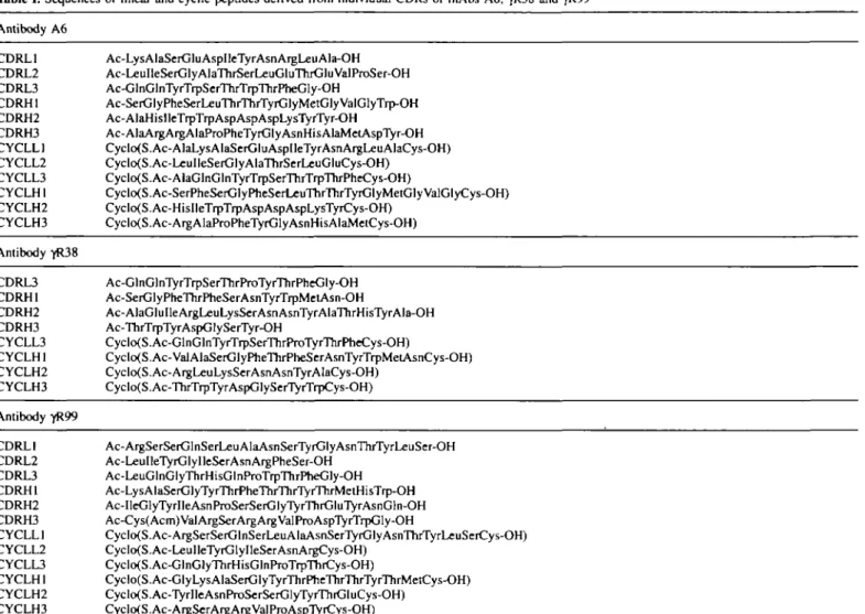
Table I. Sequences of linear and cyclic peptides derived from individual CDRs of mAbs A6, "jfR38 and 7R99 Antibody A6 CDRL1 Ac-LysAlaSerGluAspIleTyrAsnArgLeuAla-OH CDRL2 Ac-LeuIleSerGlyAlaThrSerLeuGluThrGluValProSer-OH CDRL3 Ac-GlnGlnTyrTrpSerThrTrpThrPheGly-OH CDRH1 Ac-SerGlyPheSerLeuThrThrTyrGlyMetGly ValGlyTrp-OH CDRH2 Ac-AlaHisIleTrpTrpAspAspAspLysTyrTyr-OH CDRH3 Ac-AlaArgArgAlaProPheTyrGlyAsnHisAlaMetAspTyr-OH CYCLL1 Cyclo(S.Ac-AlaLysAlaSerGluAspIleTyrAsnArgLeuAlaCys-OH) CYCLL2 Cyclo(S.Ac-LcuIleSerGlyAlaThrSerLeuGluCys-OH) CYCLL3 Cyclo(S.Ac-AlaGlnGlnTyrTrpSerThrTrpThrPheCys-OH) CYCLH1 Cyclo(S.Ac-SerPheSerGlyPheSerLeuThrThrTyrGlyMetGlyValGlyCys-OH) CYCLH2 Cyclo(S.Ac-HisIleTrpTrpAspAspAspLysTyrCys-OH) CYCLH3 Cyclo(S.Ac-ArgAlaProPheTyrGlyAsnHisAlaMetCys-OH) Antibody yR38 CDRL3 Ac-GlnGlnTyrTrpSerThrProTyrThrPheGly-OH CDRH1 Ac-SerGlyPheThrPheSerAsnTyrTrpMetAsn-OH CDRH2 Ac-AlaGluIleArgLeuLysSerAsnAsnTyrAlaThrHisTyrAla-OH CDRH3 Ac-ThrTcpTyrAspGlySerTyr-OH CYCLL3 Cyclo(S.Ac-GlnGlnTyrTrpSerThrProTVrThrPheCys-OH)
CYCLH 1 Cyclo(S. Ac-Val AlaSerGlyPheThrPheSerAsnTyrTrpMetAsnCys-OH) CYCLH2 Cyclo(S.Ac-ArgLeuLysSerAsnAsnTyrAIaCys-OH) CYCLH3 Cyclo(S.Ac-ThrTrpTyrAspGlySerTyrTrpCys-OH) Antibody yR99 CDRL1 Ac-ArgSerSerGlnSerLeuAlaAsnSerTyrGlyAsnThrTyrLeuSer-OH CDRL2 Ac-LeuIleTyrGlylleSerAsnArgPheSer-OH CDRL3 Ac-LeuGlnGlyThrHisGlnProTrpThrPheGly-OH CDRH1 Ac-LysAlaSerGlyTyrThrPheThrThrTyrThrMetHisTrp-OH CDRH2 Ac-IlcGlyTyrlleAsnProSerSerGlyTyrThrGluTyrAsnGln-OH CDRH3 Ac-Cys(Acm)VaJArgSerArgArgValProAspTyrTrpGly-OH CYCLL 1 Cyclo(S.Ac-ArgSerSerGlnSerLeuAlaAsnSerTyrGlyAsnThrTyrLeuSerCys-OH) CYCLL2 Cyclo(S.Ac-LeuIleTyrGlyIleSerAsnArgCys-OH) CYCLL3 Cyclo(S.Ac-GlnGlyThrHisGlnProTrpThiCys-OH) CYCLH 1 Cyclo(S.Ac-GlyLysAlaSerGlyTyrThrPheThrThrTyrThrMetCys-OH) CYCLH2 Cyclo(S.Ac-TyrIleAsnProSerSerGlyTyrThrGluCys-OH) CYCLH3 Cyclo(S.Ac-ArgSerArgArgValProAspTyrCys-OH)
The linear peptides are labelled according to H-chain (HI-3) or chain (LI-3) CDR, and CYCL indicates the corresponding cyclic peptides. Note that the L-chains of A6 and -yR38 have identical LI and L2 CDR sequences since both mAbs use the same Vk34C germline gene (Bndges et al., 1995).
to bind the IFN7R fragments immobilized on Sepharose. Triple G44E, L45R and W47G mutations were introduced into each VH-chain by site-directed mutagenesis. All three mutated VH
-chains could be produced in a soluble form in E.coli HB2151 following induction with IPTG. However, none of these proteins retained significant IFNYR binding activity.
CDR peptides
Sequence comparisons of each Fv with the structural database (Chothia et al, 1989; Bridges et al, 1995) were used to determine which amino acid residues are likely to be located in CDR loops. These, together with one or more residues from the flanking framework regions, were incorporated into synthetic linear peptides, as shown in Table I. Cyclic derivatives were made separately by starting the synthesis with cysteine at the C-terminus, addition of a bromoacetyl group at the N-terminus and subsequent cyclization by displacement of bromine with the S atom of cysteine, to afford a thioether.
The ability of each linear and cyclic peptide to compete with intact mAb for binding recombinant IFN-yR was analysed on a BIAcore instrument and by ELISA. In both assays, no inhibitory activity (up to ~0.5 mM peptide) was found amongst the peptides investigated.
Biosensor measurements and radioimmunoassay
The receptor affinity and inhibitory activity of each scFv protein was analysed on a BIAcore instrument and by radio-immunoassay using native receptor on human Raji cells. For BIAcore measurements, the amount of recombinant insect cell-derived (Gentz et al, 1992) WNyR immobilized on the biosensor chip was selected to give responses that could be fitted accurately to a monoexponential function (Karlsson et al., 1991). The dissociation rate constants were determined by non-linear fitting of the dissociation phase to an integrated form of the first-order rate equation. Two methods of fitting the association phases were then used. The first makes use of the fcoff value to determine k^ by direct non-linear fitting of the instrument response to an integrated form of the rate equation, using protocols supplied by the manufacturer (Pharm-acia). In the second method (Karlsson et al, 1991), the response is used to calculate k^, where k^ = konC + koff, and a linear regression of a secondary plot of ks against C yields
km and fc^. Within experimental error, both methods gave the same values for the interaction constants. Binding curves from duplicate assays at eight analyte concentrations were analysed for each scFv (see Figure 2). Owing to the high k^ observed for the 7R99 scFv [seen also for the intact mAb in earlier
Anti-interferon y receptor antibody fragments A6 scFv ) 120 240 360 480 600 42 120- yR38 scFv 200 400 600 100 150 Time (s)
Fig. 2. Sensorgrams from a Pharmacia BIAcore instrument for scFv binding
to immobilized recombinant IFNyR. The amount of IFNyR immobilized on the biosensor was varied by adjusting the exposure time of biosensor surface to activation reagent and of protein to the activated surface. Activation of the sensor surface was with NHS (50 mM) and EDC (0.2 M), and coupling to IFN7R was performed in sodium citrate buffer (10 mM, pH 3.95).
Itoble II. Kinetic and thermodynamic parameters for binding of scFvs
derived from mAbs A6, yR3& and 7R99 to the recombinant human IFN7R by BIAcore measurements and IC50S for inhibition of IFNy binding to the native receptor on human Raji cells
scFv (X10"3 * „ , (X105 M - ' s-1) KD (nM) IC50 (nM) A6 10.4 ± 0.1 7R38 9.2 ± 0.3 7R99 6.9 ± 0.5 5.3 ± 0.2 3.3 ± 0.4 63.8 ± 0.5 20.0 27.8 1.1 10.3 25.8 8.5
work (Williams et al., 1995)], the level of immobilized receptor had to be reduced to a low level to avoid mass transport limitation. Binding reactions run at different flow rates, and use of the BIAsimulation software indicated that none of the data used for estimation of K&S was influenced significantly by mass transport limitation. The interaction constants deter-mined in this work are given in Table n.
The ability of each scFv to inhibit the binding of I25
I-labelled IFNy to human Raji cells (Aguet and Merlin, 1987), which overexpress the IFNyR, was also tested by radioimmuno-assay (Figure 3). The concentration of scFv needed to reduce the amount of bound [I 2 5I]IFNY by 50% (IC50) is also given
in Table n. A6 scFv yR38 scFv SCFV c
E
* 3 O oconcentration nM
Fig. 3. Concentration-dependent inhibition of ['^IJIFNy binding to human
Raji cells by the A6, "|fR38 and yR99 scFvs using radioimmunoassay (see Materials and methods).
Discussion
The Fv region, comprising the entire variable region of the H-and L-chains, is the smallest fragment of an antibody that contains an intact copy of the antigen binding site. As such, it can be expected to maintain consistently the antigen-binding specificity and affinity of the whole antibody (Raag and Whitlow, 1995). Further dissection of the Fv fragment may lead to isolated VH- or VL-chains, which in some cases have
been observed to retain a high antigen-binding affinity (Ward
et al, 1989; Monfardini et al, 1995). However, the production
of individual VH-chains can be problematic owing to low
expression and poor solubility. Attempts have been made to engineer the exposed interface on VH for the light chain
(Davies and Riechmann, 1994) to increase hydrophilicity, as seen in camelid antibodies which occur naturally as heavy chains without a light chain partner (Hamers-Casterman
et al, 1993).
In this work, three scFv fragments, derived from mAbs A6, yR38 and yR99, have been produced in E.coli, and isolated as soluble monomeric proteins that retain a high affinity for the recombinant human IFNyR (Table II). The relative affinities of the intact mAbs for the IFNyR, measured in earlier work using a laboratory-built SPR biosensor (Williams et al, 1995), are similar within a factor of 2-3 to those measured in this work for the scFvs on a Pharmacia BIAcore instrument. The A6 and yR38 scFvs, which bind the N-terminal Ig-like domain of the IFNyR, have dissociation constants (KQS) of 20 and 28 nM, respectively. The yR99 scFv binds an epitope in the membrane proximal domain of the receptor, which includes residues in the interdomain linker region (Ruegg et al, 1995), with a KD of ~1 nM. Moreover, as found in earlier work with intact mAbs, the higher relative affinity for the immobilized receptor shown by the yR99 SCFV, under these conditions, arises largely from an increased A^,, rather than a lower off-rate. The soluble scFvs also retain the ability of the whole mAbs to inhibit the binding of IFNy to the native receptor on human Raji cells (Table II). The inhibitory activities expressed as IC50 values lie between 9 and 26 nM, which is approximately an order of magnitude weaker than the IC50S seen for the intact mAbs (Bridges et al, 1995). This is most likely due to an 369
avidity effect with the whole antibody, since the Fab fragments derived from each mAb have IC50S that, within a factor of 2, are similar to those observed for the scFvs (data not shown). These results also show that the inhibitory effect of each mAb is caused by the association of the variable domain with the receptor, rather than steric exclusion of the hormone binding site by the antibody constant regions. The X-ray crystal structure of the IFNy-IFN'yR complex has revealed that the hormone binding site is centrally located in the rod-shaped IFN7R molecule and comprises residues from both Ig-like domains (Walter et ai, 1995). Most likely the structural epitopes bound by each mAb overlap, at least partially, the hormone binding site on the receptor, although other mechanisms of inhibition, such as allosteric changes in receptor conformation upon antibody binding, cannot be ruled out at present.
Attempts to produce individual, native-like VH- and VL
-chains from each mAb failed to afford soluble protein that bound to the IFN7R. Of the VH proteins, only those from
antibodies A6 and yR38 could be isolated directly in a soluble form, following immunoaffinity chromatography using an anti-C-terminal peptide tag mAb. The *yR99 VH gave no soluble
tag-positive protein in the expression system used here. To overcome potential problems arising from low solubility and non-specific interactions through the VL interface, triple G44E,
L45R and W47G mutations were introduced into each VH
-chain by site-directed mutagenesis, in analogy with camelid antibodies (Davies and Riechmann, 1994). All three 'camelized' VH-chains could be produced in a soluble form in
E.coli HB2151 following induction with IPTG. However, none
of these proteins retained significant IFN7R binding activity. No IFN7R binding activity was observed with VL-chains
produced in this way, or with linear and cyclic peptides derived from the individual CDR sequences of each mAb (Table I).
The inability of the linear and cyclic peptides derived from individual CDR sequences (see Table I) to bind the IFN7R is most Likely due to the absence in each of a sufficient number of the key residues that contribute significantly to the energetics of receptor binding, and/or to the fact that these are not displayed in the correct conformation. This point may be addressed quantitatively in future work by detailed structural and thermodynamic studies on native and mutant Fv-receptor complexes. This should now be feasible with the ability to produce both the mAb Fv fragments and native-like IFN7R fragments in E.coli, combined with the application of NMR and crystallographic techniques for structure determination.
The de novo design of new small-molecule ligands for the IFN7R, that block the cytokine binding site and act as antagon-ists, remains a considerable challenge. Conceivably, such molecules may be derived by design of small-molecule peptide mimetics of the key CDR loops derived from the mAbs described here. However, detailed mutagenesis studies on two different Fv fragment-protein antigen complexes (Hawkins
et al., 1993; Kelley and O'Connell, 1993) have shown already
that the antibody residues which appear to be most important for antigen binding are grouped near the centre of the interface, and are located in more than one CDR loop. If these findings prove to be common, it seems likely that a general approach to mimetic design must take into account the necessity of retaining groups located in several CDRs to achieve high antigen-binding affinity. Possible solutions to this design prob-lem may be found by grafting multiple loops on to alternate protein frameworks, for example a designer minibody (Martin
et al, 1994), or possibly by their incorporation into
template-assembled synthetic proteins (Mutter and Vuilleumier, 1989).
Acknowledgements
The authors thank the Swiss National Science Foundation for financial support, Drs G. Garotta and R. Gentz (Hoffmann-La Roche) for recombinant IFNyR from insect cells and Annehes Meier for technical assistance.
References
Aguet,M. and Merlin.G. (1987) J. Exp. Med., 165, 988-999.
BridgesA, BirchA, Williams.G., Aguet.M., Schlatter.D., Huber.W., Garotta.G. and RobinsonJ.A. (1995) Mol. Immunol., 32, 1329-1338. Chothia.C. et al. (1989) Nature, 342, 877-883.
DaviesJ. and Riechmann,L. (1994) FEBS Utt., 339, 285-290. DaviesJ. and Riechmann.L. (1995) Biotechnology, 13, 475-479.
Doring.E., Sugler,R., Grutz.G., Vonbaehr.R. and SchneidermergenerJ. (1994)
Mol. Immunol, 31, 1059-1067.
Evan.G.I., Lewis.G.K., Ramsay.G. and BishopJ.M. (1985) Mol. Cell. Bioi, 5, 3610-3616.
Farrar.M.A. and Schreiber.R.D. (1993) Annu. Rev. Immunol., 11, 571-611. Fountoulakis.M., Zulauf.M., LustigA and Garotta,G. (1992) Eur. J. Biochem.,
208,781-787.
Garotta.G., Ozmen,L., Fountoulakis.M., DembicZ., Van Loon.A.P.G.M. and Stuber.D. (1990) J. Biol. Chem., 265, 6908-6915.
Gentz,R., HayesA, Grau.N., Fountoulakis.M., Lahm.H -W., Ozmen.L. and Garotta,G. (1992) Eur. J. Biochem., 210, 545-554.
GreenlundAC, Schreiber.R.D., Goeddel.D.V. and Pennica,D. (1993) J. Biol.
Chem., 268, 18103-18110.
Hamers-Casterman.C, Atarhouch.T., Muyldermans.S., Robinson,G., Hamers.C, Bajyana Songa,E., Bendahman.N. and Hamers.R. (1993) Nature, 363,446-448.
Hawkins.R.E., Russell.SJ., Baier.M. and Winter.G. (1993) /. Mol Biol., 234, 958-964.
Hemmi.S., Bohni.R., Stark.G., Di Marco.F. and Aguet,M. (1994) Cell, 76, 803-810.
Hoogenboom.H.R., Griffiths A D . , Johnson.K.S., Chiswell.DJ., Hudson.P. and Winter,G. (1991) Nucleic Acids Res., 19, 4133-4137.
Karlsson.R., Michaelsson.A and Mattsson.L. (1991) J. Immunol. Methods,
145, 229-240.
Kelley.R.F. and O'Connell.M.P. (1993) Biochemistry, 32, 6828-6835. MarksJ.D., Hoogenboom.H.R., Bonnert.T.P., McCaffertyJ., Griffiths.A.D. and
Winter.G. (1991) J. Mol. Biol., 222, 581-597.
Marsters.SA, Pennica.D., Bach.E., Schreiber.R D. and AshkenaziA (1995)
Proc. Natl Acad. Sci. USA, 92, 5401-5405.
Martin.F., Toniatti.C, Salvati.A.L., Venturini.S., Ciliberto.G., Cortese.R. and Sollazzo.M. (1994) EMBO J., 13, 5303-5309.
Monfardini.C. et al. (1995) J. Biol. Chem., 270, 6628-6638.
Mutterjvi. and Vuilleumier.S. (1989).4nseH>. Chem.. Int Ed. Engl.,2S, 535-554. Ozmen.L., Fountoulakis.M. and Garotta,G. (1992) J. Immunol. Methods, 147,
261-270.
Raag.R. and Whitlow.M. (1995) FASEB J., 9, 73-80.
Ruegg.N., Williams.G., Birch.A., RobinsonJ.A., Schlatter.D. and Huber.W. (1995) J. Immunol. Methods, 183, 95-101.
Sanger,F., Nicklen.S. and CoulsonA (1977) Proc. Natl Acad. Sci. USA, 74, 5463-5467.
Saragovi.H.U., Greene.M.I., Chmsciel.R.A. and Kahn.M. (1992)
BioTechnology, 10, 773-778.
SohJ. et al. (1994) Cell, 76, 793-802.
Walter.M.R., Windsor,W.T., Nagabhushan.T.L., Lundell.D.J., Lunn,CA, Zauodny.PJ. and Narula.S.K. (1995) Nature, 376, 230-235.
Ward.E.S., Gussow.D., Griffiths A D . , Jones,P.T. and Winter.G. (1989) Nature,
341, 544-546.
Williams.G. et al. (1995) Biochemistry, 34, 1787-1797.