HAL Id: hal-02983697
https://hal.inrae.fr/hal-02983697
Submitted on 30 Oct 2020
HAL is a multi-disciplinary open access
archive for the deposit and dissemination of
sci-entific research documents, whether they are
pub-lished or not. The documents may come from
teaching and research institutions in France or
abroad, or from public or private research centers.
L’archive ouverte pluridisciplinaire HAL, est
destinée au dépôt et à la diffusion de documents
scientifiques de niveau recherche, publiés ou non,
émanant des établissements d’enseignement et de
recherche français ou étrangers, des laboratoires
publics ou privés.
Distributed under a Creative Commons Attribution - NonCommercial - NoDerivatives| 4.0
intestinal stem cell niche in vitro with pharmacological
inhibitors or L-WRN conditioned medium
Eloïse Mussard, Cécile Pouzet, Virginie Helies, Géraldine Pascal, Sandra
Fourre, Claire Cherbuy, Aude Rubio, Nathalie Vergnolle, Sylvie Combes,
Martin Beaumont
To cite this version:
Eloïse Mussard, Cécile Pouzet, Virginie Helies, Géraldine Pascal, Sandra Fourre, et al.. Culture
of rabbit caecum organoids by reconstituting the intestinal stem cell niche in vitro with
pharmaco-logical inhibitors or L-WRN conditioned medium. Stem cell research, Elsevier, 2020, 48, pp.1-10.
�10.1016/j.scr.2020.101980�. �hal-02983697�
Contents lists available atScienceDirect
Stem Cell Research
journal homepage:www.elsevier.com/locate/scr
Culture of rabbit caecum organoids by reconstituting the intestinal stem cell
niche in vitro with pharmacological inhibitors or L-WRN conditioned
medium
Eloïse Mussard
a, Cécile Pouzet
b, Virginie Helies
a, Géraldine Pascal
a, Sandra Fourre
c,
Claire Cherbuy
d, Aude Rubio
e, Nathalie Vergnolle
e, Sylvie Combes
a, Martin Beaumont
a,⁎aGenPhySE, Université de Toulouse, INRAE, ENVT, F-31326 Castanet Tolosan, France
bFRAIB-TRI Imaging Platform Facilities, Université de Toulouse, CNRS, 31320 Castanet-Tolosan, France cGeT‐PlaGe, Genotoul, INRAE, Castanet Tolosan, France
dMicalis Institute, INRAE, AgroParisTech, Université Paris-Saclay, 78350 Jouy-en-Josas, France
eIRSD, Université de Toulouse, INSERM, INRAE, ENVT, UPS, U1220, CHU Purpan, CS60039, 31024 Toulouse, France
A R T I C L E I N F O Keywords: Enteroid Spheroid Epithelium Culture medium Monolayer A B S T R A C T
Intestinal organoids are self-organized 3-dimensional (3D) structures formed by a single layer of polarized epithelial cells. This innovative in vitro model is highly relevant to study physiology of the intestinal epithelium and its role in nutrition and barrier function. However, this model has never been developed in rabbits, while it would have potential applications for biomedical and veterinary research. Here, we cultured rabbit caecum organoids with either pharmacological inhibitors (2Ki medium) or L-WRN cells conditioned medium (L-WRN CM) to reconstitute the intestinal stem cell niche in vitro. Large spherical organoids were obtained with the 2Ki medium and this morphology was associated with a high level of proliferation and stem cells markers gene expression. In contrast, organoids cultured with L-WRN CM had a smaller diameter; a greater cell height and part of them were not spherical. When the L-WRN CM was used at low concentration (5%) for two days, the gene expression of stem cells and proliferation markers were very low, while absorptive and secretory cells markers and antimicrobial peptides were elevated. Epithelial cells within organoids were polarized in 3D cultures with 2Ki medium or L-WRN CM (apical side towards the lumen). We cultured dissociated organoid cells in 2D monolayers, which allowed accessibility to the apical compartment. Under these conditions, actin stress fibers were observed with the 2Ki medium, while perijonctionnal localization of actin was observed with the L-WRN CM suggesting, in 2D cultures as well, a higher differentiation level in the presence of L-WRN CM. In conclusion, rabbit caecum organoids cultured with the 2Ki medium were more proliferative and less differentiated than organoids cultured with L-WRN CM. We propose that organoids cultured with the 2Ki medium could be used to rapidly generate in vitro a large number of rabbit intestinal epithelial stem cells while organoids cultured with the L-WRN CM used at low concentration represent a suitable model to study differentiated rabbit epithelium.
1. Introduction
The intestinal epithelium is formed by a monolayer of cells located at the surface of the gut mucosa, facing the luminal environment (Peterson and Artis, 2014). Intestinal epithelial cells (IEC) play a pivotal role for both nutrition (digestion, nutrient absorption, hormone secre-tion) and barrier function (formation of tight junctions, secretion of
mucus and antimicrobial peptides and detection of microbes). This pleiotropic function of the intestinal epithelium is linked to the pre-sence of specialized differentiated IEC (absorptive, paneth, goblet and enteroendocrine cells) that arise from intestinal stem cells (ISC) located at the bottom of epithelial crypts (Barker, 2014). Recent progress in the characterization of the ISC niche (i.e. microenvironment) allowed the development of intestinal organoid culture in vitro (Sato et al., 2009).
https://doi.org/10.1016/j.scr.2020.101980
Received 18 March 2020; Received in revised form 19 August 2020; Accepted 28 August 2020
Abbreviations: 2D, 2 dimensions; 2Ki, name of the organoid culture medium containing pharmacological inhibitors only; 3D, 3 dimensions; BMP, bone morphogenic
protein; CM, conditioned medium; DMEM, Dulbecco's Modified Eagle Medium; FBS, fetal bovine serum; IEC, intestinal epithelial cell; ISC, intestinal stem cell; L-WRN, L cell line engineered to secrete mouse Wnt3a, R-spondin 3 and Noggin; P/S, penicillin/streptomycin
⁎Corresponding author.
E-mail address:martin.beaumont@inrae.fr(M. Beaumont).
Stem Cell Research 48 (2020) 101980
Available online 04 September 2020
1873-5061/ © 2020 The Author(s). Published by Elsevier B.V. This is an open access article under the CC BY-NC-ND license (http://creativecommons.org/licenses/BY-NC-ND/4.0/).
These self-organized 3D structures are formed by a closed lumen sur-rounded by a single layer of polarized IEC (Sato et al., 2009). Organoids closely replicate epithelial physiology and have been used as an effi-cient model to study nutrient transport, host-microbe interactions or pathological processes (Clevers, 2016; Lukovac et al., 2014; Zietek et al., 2015). They can also be used for pharmacological studies (Sébert et al., 2018).
Organoids are obtained by seeding tissue-derived ISC in an extra-cellular matrix (e.g. Matrigel) with growth factors reproducing in vitro the main ISC niche signaling pathways, i.e. high Wnt and low bone morphogenic protein (BMP) activation (Date and Sato, 2015; Gehart and Clevers, 2019; Holmberg et al., 2017). Thus, organoid culture medium usually contains three proteins: Noggin (inhibiting the BMP pathway), Wnt3a and R-spondin (activating the Wnt pathway) (Sato et al., 2009, 2011). Conditioned medium (CM) of engineered cell lines is a cost-effective and efficient source for these factors, when compared to purified recombinant proteins (Holmberg et al., 2017). For instance, the L-WRN cells are mouse L cells secreting Wnt3a, R-spondin 3 and Noggin which CM can be used to grow intestinal organoids (Miyoshi and Stappenbeck, 2013; VanDussen et al., 2015; 2019). Alternatively, pharmacological inhibitors can be used to reconstitute ISC niche sig-naling pathways in vitro. Indeed, organoids can be cultured with the GSK3 inhibitor CHIR-99021 (activating the Wnt pathway) and the BMP type I receptor inhibitor LDN-193189 (inhibiting the BMP pathway) (Li et al., 2018a).
Recently, the methods used to grow human or mouse intestinal organoids were successfully adapted to several other animal species (pig, cow, sheep, chicken, horse and dog) (Derricott et al., 2019; Hamilton et al., 2018; van der Hee et al., 2018; Powell and Behnke, 2017; Yin et al., 2019). However, a method to grow rabbit intestinal organoids is lacking despite the usefulness of such model in the context of biomedical or veterinary research (e.g. study of human bacillary dysentery or of epizootic rabbit enteropathy) (Licois et al., 2005; Yum et al., 2019). Herein, we present for the first time a method to grow rabbit caecum organoids in 3D. This digestive segment was chosen since it is an important site for interaction with the microbiota in rabbits (Beaumont et al., 2020). Our results show that replicating the ISC niche with pharmacological inhibitors or with L-WRN CM differ-entially regulates epithelial proliferation, differentiation and barrier function. We also show that rabbit caecum organoid cells can be cul-tured in 2D monolayers to facilitate the access to the apical side of IEC, which is needed to study their interactions with luminal components such as microbes, nutrients or xenobiotics.
2. Materials and methods
2.1. L-WRN cell culture
L-WRN cells (ATCC, Cat# CRL-3276 Manassas,VA) (passages 1–11) were seeded in T75 flasks at 37 °C under 5% CO2 atmosphere in
“complete DMEM” containing: Dulbecco’s Modified Eagle Medium (DMEM) high glucose GlutaMAXTM Supplement pyruvate (Thermo
Fisher Scientific, Waltham, MA), 10% fetal bovine serum (FBS) (Thermo Fisher Scientific) and 1% penicillin/streptomycin (P/S) (Sigma-Aldrich, St-Louis, MO). The original method for L-WRN cells culture used 20% FBS (Miyoshi and Stappenbeck, 2013), but we re-duced it to 10% in order to have the same FBS concentration than in the 2Ki organoid culture medium described below (table 1). Cells con-taining plasmids for expression of mouse Wnt3a, R-spondin 3 and Noggin were selected during the 3 first days post seeding by adding 0.5 mg/mL G418 (Sigma-Aldrich) and 0.5 mg/mL Hygromycin B (In-vivoGen, San Diego, USA) to the complete DMEM (Miyoshi and Stappenbeck, 2013) (Fig. 1A). Then, cells were washed with PBS to remove residual antibiotics and cultured in complete DMEM without G418 nor Hygromycin B. The conditioned medium (L-WRN CM) was collected every day for 4 days. After centrifugation (300 g, room
temperature, 5 min), L-WRN CM were kept at −20 °C before thawing and pooling when 500 mL were produced. The pool of L-WRN CM was then stored at −20 °C until use. L-WRN cells were weekly sub-cultured (dilution 1:10 v/v) by partial digestion with EDTA-trypsin 0.25% w/v (Thermo Fisher Scientific).
2.2. Isolation of intestinal crypts
Rabbits (line INRAE 1777) were bred at the PECTOUL Experimental Unit (INRAE, Castanet-Tolosan, France). Animals were handled ac-cording to the European Union recommendations on the protection of animals used for scientific purpose (2010/63/EU) and in agreement with French legislation (NOR:AGRG1238753A 2013). Animal experi-ments received the approval of the local ethical committee (SSA_2018_010). Caecal tissues used in this study were obtained from 30-day-old male rabbits slaughtered by electronarcosis and ex-sanguination. Epithelial crypts were isolated from rabbit caecal tissue fragments by incubation in a dissociation solution (PBS without Ca2+
-Mg2+ (Thermo Fisher Scientific), 9 mM EDTA (Thermo Fisher
Scientific), 3 mM 1,4-Dithiothréitol (DTT) (Roche, Basel, Switzerland), 10 µM ROCK inhibitor Y27632 (ATCC® ACS-3030™) and 1% P/S (Sigma Aldrich)) under agitation during 30 min at room temperature. Then, crypts were detached by manual shaking (1 min) in cold PBS. After centrifugation (24g, 4 °C, 5 min), the crypt pellet was re-suspended in cold DMEM and used immediately for organoid culture. A fraction of the crypts was lyzed in TRI Reagent (Zymo Research, Irvine, CA) and kept at −80 °C until total RNA extraction.
2.3. Organoid culture
Crypts were counted and re-suspended in ice-cold Matrigel (Corning, Cat #354234, Corning, NY) and seeded in pre-warmed 48-well plates at a density of 150 crypts/25 µL of Matrigel. After poly-merization (37 °C, 5% CO2, 20 min), 250 µL warm organoid culture
media (2Ki medium or 50% L-WRN CM, detailed intable 1) was added as indicated inFig. 1B. The media was replaced every 2–3 days. After 7 days of culture, organoids were counted according to their spherical or non-spherical morphology by bright field optical microscopy (Eclipse Ts2, Nikon Instruments, Melville, NY). The diameter of organoids and epithelial cell height were measured with Image J software (Schneider et al., 2012).
Every week, after washing with warm PBS, organoids were broken by pipetting in EDTA-trypsin 0.25% w/v (Thermo Fisher Scientifc) before incubation (37 °C, 5% CO2, 5 min). Six wells were pooled before
centrifugation (24g, 4 °C, 5 min) and the pellet was re-seeded in Matrigel with a dilution ratio 1:8 and cultured as previously described. For organoids cryoconservation, the media was removed and freezing solution was added (80% complete DMEM, 10% FBS, 10% DMSO, 10 µM Y27632). Matrigel domes were scratched with a pipet tip and the pool of 6 wells were transferred in a cryotube and then placed in an isopropanol freezer box, allowing the cells to be gradually frozen at −80 °C, before being kept in liquid nitrogen for long term storage. After fast thawing at 37 °C, organoids were re-cultured with a dilution ratio 1:8 as described above.
2.4. Two-dimensional monolayers of organoid cells
Organoids cultured in 3D with 2Ki medium or 50% L-WRN CM for 1 week were collected from a pool of 6 wells (about 1000 organoids) in complete DMEM before centrifugation (95 g, room temperature, 5 min). Organoids were dissociated into single cells by incubation in pre-warmed TrypLE Express Enzyme (Thermo Fisher Scientific) for 10 min at 37 °C. After centrifugation (300 g, room temperature, 5 min), dis-sociated cells were resuspended in organoid culture medium and seeded in one Transwell (0.33 cm2culture area) (Thermo Fisher Scientific).
with diluted Matrigel (1:50 v/v in DMEM) before drying (room tem-perature, 10 min). 2Ki medium or 50% L-WRN CM was added at the apical (400 µL) and basal (600 µL) side. After 24 h of incubation, the medium was replaced by complete DMEM for 24 h to allow IEC dif-ferentiation, as previously described (Moon et al., 2014; VanDussen et al., 2015). Transepithelial electrical resistance (TEER) was measured with Millicell ERS-2 Volt-Ohm Meter (Merck Millipore, Burlington, MA).
2.5. Gene expression
Organoids were washed with PBS and 6 wells/condition were pooled in 300 µL of TRI Reagent (Zymo Research, Irvine, CA) and kept at −80 °C until total RNA extraction. After thawing, crypts and orga-noid samples were centrifuged (12 000 g, 4 °C, 10 min) to remove particles. The supernatant was collected and total RNA were extracted with Direct-zol RNA MiniPrep Plus kit (Zymo Research) according to the manufacturer’s instructions. A DNAse I digestion step was included to remove genomic DNA. After elution in RNase-free water, RNA con-centration was determined using a Nanodrop before storage at −20 °C until use. cDNA were prepared from 1 µg RNA with GoScript Reverse
Transcription Mix, Random primer (Promega, Madison, Wisconsin) following the manufacturer’s instructions. cDNA were diluted 1:2 (v/v) and stored at −20 °C. Gene expression was analyzed by real-time qPCR using QuantStudio 6 Flex Real-Time PCR System (Thermo Fisher Scientific) or Biomark microfluidic system using a 48.48 Dynamic Array IFC for gene expression (Fluidigm, San Francisco, CA) according to the manufacturer recommendations. The sequences of the primers used are presented in table S1. Data were analyzed with the 2−ΔΔCtmethod with
Atp5b gene expression used as a reference.
2.6. Staining and confocal microscopy of organoids
Organoids were cultured as described above in 3D in Nunc Lab-Tek Chamber Slide system (Thermo Fisher Scientific). For proliferative cells staining, organoids (after 1 week of culture) were incubated (37 °C, 5% CO2, 1 h) with 10 µM EdU (5-ethynyl-2′-deoxyuridine, Thermo Fisher
Scientific), a nucleoside analog of thymidine incorporated into DNA during active DNA synthesis. After fixation with 4% paraformaldehyde (Electron Microscopy Sciences, Hatfield, PA) during 20 min under agitation, organoid cells were permeabilized with 0.5% triton X-100 (Sigma Aldrich) during 20 min under agitation. Between each step,
Table 1
Composition of 2Ki and 50% or 5% L-WRN CM medium. L-WRN CM: conditioned medium produced by L-WRN cells.
Component Supplier Function Concentration
2ki medium (pharmacological inhibitors)
DMEM Thermo Fisher
Scientific Provides growth factors and nutrients
P/S (Penicillin/Streptomycin) Sigma Aldrich Antibiotics with large spectra preventing bacterial contamination 1% FBS (Fetal bovine serum) Thermo Fisher
Scientific Growth factors/vitamins 10% HEPES Sigma Aldrich Buffer (pH 7) 1 mM N-acetylcysteine Sigma Aldrich Antioxidant preventing cellular damages and apoptosis 500 µM CHIR99021 Sigma Aldrich Inhibitor of glycogen synthase kinase 3 (GSK3): Wnt pathway activation 10 µM Y27632 ATCC Associated Roh kinase (ROCK) inhibitor: prevents anoikis 10 µM SB431542 Sigma Aldrich Inhibitor of the TGF-β pathway 10 µM LDN193189 Sigma Aldrich BMP pathway receptor inhibitor 0.2 µM
50% L-WRN conditioned medium
DMEM, 10% FBS, 1% P/S Complete DMEM 50% L-WRN conditioned medium Conditioned medium from L-WRN cells secreting mouse Wnt3A, R-spondin 3 (Wnt pathway
activation) and Noggin (BMP pathway inhibition) 50% Y27632 ATCC Associated Roh kinase (ROCK) inhibitor: prevents anoikis 10 µM SB431542 Sigma Aldrich Inhibitor of the TGF-β pathway 10 µM
5% L-WRN conditioned medium
DMEM, 10% FBS, 1% P/S Complete DMEM 95% L-WRN conditioned medium Conditioned medium from L-WRN cells secreting mouse Wnt3A, R-spondin 3 (Wnt pathway
activation) and Noggin (BMP pathway inhibition) 5%
Fig. 1. Schematic representation of methods used for organoid culture. A. L-WRN cells were seeded in complete DMEM (DMEM, 10% SVF, 1% P/S) with
Hygromycin B and G418 to select cells containing expression plasmids for mouse Wnt3a, R-spondin 3 and noggin. After 3 days of culture, cells were washed with PBS and cultured with complete DMEM only. Conditioned medium (L-WRN CM) was collected every 24 h. B. Organoids were cultured for 7 days in 3 different conditions: (i) with pharmacological inhibitors (2Ki) for 7 days or (ii) with 50% L-WRN CM for 7 days or (iii) with 50% L-WRN CM for 5 days and then 5% L-WRN CM for 2 days. E. Mussard, et al. Stem Cell Research 48 (2020) 101980
wells were washed twice with 3% bovine serum albumin (Euromedex, Souffelweyersheim, France) in PBS. EdU was detected with Alexa 488 fluorochrome according to the protocol of Click-iT EdU Cell Proliferation kit (Thermo Fisher Scientific). For actin staining, orga-noids were fixed and permeabilized as described above before incuba-tion with 10 µM phalloidin coupled with TRITC fluorochrome (Sigma Aldrich), under agitation (room temperature, 30 min). After staining, culture chambers were removed and mounting medium (Vector Lab, Burlingame, CA) supplemented with DAPI was added. Slides were stored in the dark at 4 °C until observation. For organoid cell mono-layers cultured in 2D, EdU incubation, fixation and staining were per-formed as described above directly in Transwells before mounting the membrane on a microscope slide. Fluorescence staining was analyzed with a confocal laser scanning microscope (TCS SP8; Leica, Germany) using a x10 dry immersion objective lens or a x25 water immersion objective lens. DAPI, Alexa 488 and TRITC fluorescence was excited with the 405, 488 and 552 nm laser line and recorded in one of the confocal channels in the 415–475 / 500–550/ 560–620 nm emission range, respectively. Images were acquired in the sequential mode using LAS X software (version 3.0, Leica).
2.7. Protein sequence alignment
Mouse and rabbit orthologs of Wnt3a (NP_033548.1, XP_002723899.1), R-spondin 3 (NP_082627.3, XP_002714851.1) and Noggin (NP_032737.1, XP_002719325.1) were aligned with EMBOSS Needle (EMBL-EBI). The overall percentage of identity between mouse (GRCm38.p6) and rabbit (OryCun2.0) orthologous genes (ortholog_o-ne2one, ortholog_one2many, ortholog_many2many) were obtained using the Biomart tool of the ENSEMBL database (Cunningham et al., 2019).
2.8. Statistics
All statistical analysis were performed using the R software (version 3.5.1) with the packages car, lme4 and emmeans (version 3.5.1). A linear model was used to test the effect of each component of the 2Ki medium on organoid number or diameter. Mean values of each group (i.e. each composition of culture medium) were then compared to the mean value of the complete 2ki condition and P-values were adjusted with the Tukey method. In experiments comparing characteristics of organoids cultured with 2ki medium or L-WRN CM (50% or 50–5%), all conditions were tested on organoids from 4 rabbits and several condi-tions were tested on each culture plate. Thus a linear mixed model was used with medium composition as a fixed effect and rabbit and culture plate as random effects. Mean values of each group were compared pairwise and P-values were adjusted with the Tukey method. P < 0.05 was considered significant.
3. Results
3.1. Reconstitution of the ISC niche with pharmacological inhibitors produces spherical rabbit caecum organoids
Since purified recombinant proteins (Wnt3a, R-spondin or Noggin) usually used to reconstitute the epithelial stem cell niche are not commercially available in rabbits, we first tested an organoid culture method adapted from a previously developed medium called “2Ki” replacing these proteins by pharmacological inhibitors (Li et al., 2018a). After 7 days of culture in the 2Ki medium, spherical rabbit caecum organoids were obtained with a mean diameter of about 300 µm (Fig. 2A-C). These organoids could be sub-cultured, cryopre-served and resuscitated (data not shown). Then, we removed one by one each component of the 2Ki medium to analyze their effects on the formation and growth of rabbit caecum organoids. FBS presence in the media was an absolute requirement to observe the formation of
organoids. When Y-27632 (ROCK inhibitor preventing IEC anoikis (Sato et al., 2009)) was removed, the number and the diameter of organoids tended to be lower than in complete 2Ki medium, although these changes were not statistically significant. In absence of CHIR-99021 (inhibitor of GSK3, activating of the Wnt pathway), only few organoids were observed and their diameter was very small (about 40 µM, 7-fold smaller than organoids cultured with the complete 2Ki medium). Re-moval of the TGF-β/BMP pathway inhibitors (SB-431542 and LDN-193189) did not modify significantly the number of organoids but re-duced their diameter (1.5-fold smaller than organoids cultured with the complete 2Ki medium). Altogether, our results show that FBS and pharmacological inhibitors reproducing the stem cell niche (high Wnt/ low BMP signaling pathway) are necessary for optimal growth of rabbit caecum organoids.
3.2. Reconstitution of the ISC niche with L-WRN CM produces rabbit caecum organoids with spherical and non-spherical morphologies
The spherical morphology of rabbit caecum organoids cultured with pharmacological inhibitors (2Ki medium) (Fig. 2C), suggested highly proliferative IEC with a low differentiation level (Merker et al., 2016). Such poorly differentiated culture might not be relevant to test inter-actions with nutrients or microbes. Thus, in an attempt to grow more differentiated organoids, we developed a second culture method to reconstitute the ISC niche by using the CM of L-WRN cells (L-WRN CM) secreting mouse proteins Wnt3a, R-spondin 3 (Wnt signaling po-tentiator) and Noggin (inhibitor of the TGF-β superfamily, inhibiting the BMP pathway) (Miyoshi and Stappenbeck, 2013). The amino acid sequence is highly similar between mouse and rabbit orthologs of Wnt3a (94%), R-spondin 3 (93.9%) and Noggin (98.3%) (figure S1, knowing that the overall percentage of identity between mouse and rabbit orthologous genes is ~ 86%). This high level of conservation suggested that L-WRN CM could support the growth of rabbit caecum organoids, as previously observed for several other animal species (Powell and Behnke, 2017). We used the L-WRN CM at two con-centrations: 50% for 7 days (50% L-WRN CM condition) or 50% for 5 days followed by 5% for 2 days (50–5% L-WRN CM condition) (Fig. 1B) based on a previous publication (VanDussen et al., 2015). When compared to the 2Ki medium, slightly more organoids were ob-tained in the 50% L-WRN CM condition but not with the 50–5% L-WRN CM condition (Fig. 3A). In contrast, the diameter of organoids cultured in L-WRN CM (50% or 50–5%) was significantly smaller than in orga-noids cultured with the 2Ki medium (Fig. 3B). L-WRN CM produced organoids with more diverse morphologies than the 2Ki medium since both spherical and non-spherical organoids (i.e. mulitlobulated, ~12%) were observed with L-WRN CM (50% or 50–5%) (Fig. 3C and E). Of note, non-spherical organoids were observed only at later time points (5–7 days after passaging). Moreover, the height of organoid cells was significantly greater with L-WRN CM (50% or 50–5%) compared to organoids cultured with 2Ki medium (Fig. 3D and E). Of note, there was no significant differences in the morphological features of organoids cultured with L-WRN CM used at 50% or 50%-5%. Overall, our results indicate that mimicking the ISC niche with L-WRN CM (containing mouse Wnt3a, R-spondin 3 and Noggin) allowed the growth of rabbit caecum organoids with spherical and non-spherical morphologies.
3.3. L-WRN CM induces less proliferation and more differentiation in rabbit caecum organoids than pharmacological inhibitors
The presence of non-spherical structures, the increased thickness of epithelial layer and the smaller diameter of organoids cultured with L-WRN CM suggested a lower proliferation rate and a higher differ-entiation level than in organoids cultured with pharmacological in-hibitors (2Ki) (Merker et al., 2016). Therefore, to verify this hypothesis, we analyzed proliferation and differentiation markers in organoids grown in 2Ki medium or L-WRN CM. EdU staining after a short
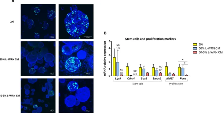
incubation period (1 h) suggested that less organoids were positive for proliferation in the 50% and 50–5% L-WRN CM conditions, when compared to 2Ki condition (Fig. 4A). However, in proliferating orga-noids (EdU+), the staining intensity was not different between the three
conditions of growth media. Of note, we did not observe EdU positive cells in non-spherical organoids (L-WRN CM condition). The mRNA level of the stem cell marker Lgr5 was the highest in organoids grown with 2Ki medium, while the expression of this gene was not detected in 1/4 samples with 50% L-WRN CM and 3/4 samples with 50–5% L-WRN CM (Fig. 4B). Similar trends were obtained for the other stem cell markers Olfm4, Sox9 and Smoc2. The expression of Lgr5 and Olfm4 tended to be higher in epithelial crypts than in organoids, especially when cultured with L-WRN CM (figure S2A). In addition, the mRNA level of the proliferation marker Pcna was significantly reduced in or-ganoids grown in 50–5% L-WRN CM, when compared to the 2Ki medium and 50% L-WRN CM (Fig. 4B). In contrast, markers of differ-entiated epithelial cells were highly expressed in organoids grown in 50–5% L-WRN CM, when compared to the 2Ki medium and, to a lower extent in 50% L-WRN CM (Fig. 5A and B). The gene expression of the absorptive cells markers Krt20, Alpi, Vil1 and Aqp8 were significantly higher in organoids grown in 50–5% L-WRN CM, when compared to the 2ki medium (Fig. 5A). Vil1 was also significantly more expressed when L-WRN CM was used at 50–5% compared to 50%. The expression of
Vil1 and Ca2 was similar in epithelial crypts and in organoids (figure S2B). The transcription factor involved in secretory lineage commit-ment Atoh1 was significantly more expressed in organoids grown in L-WRN CM than in 2Ki medium (Fig. 5B). Goblet cells markers (Muc2 and
Spdef) and the enteroendocrine cells associated gene Pyy were more
expressed in 50–5% L-WRN CM organoids compared to the 2Ki condi-tion (Fig. 5B). The expression of Muc2 was similar in epithelial crypts and in organoids when grown in 50–5% L-WRN CM (figure S2C). Al-together, our results show that rabbit caecum organoids grown with
pharmacological inhibitors (2Ki medium) were highly proliferative and enriched in stem cells, while organoids grown in L-WRN CM were more differentiated, notably when the L-WRN CM was used at low con-centration for two days.
3.4. Rabbit caecum organoids differentiation regulates the expression of key components of the epithelial barrier
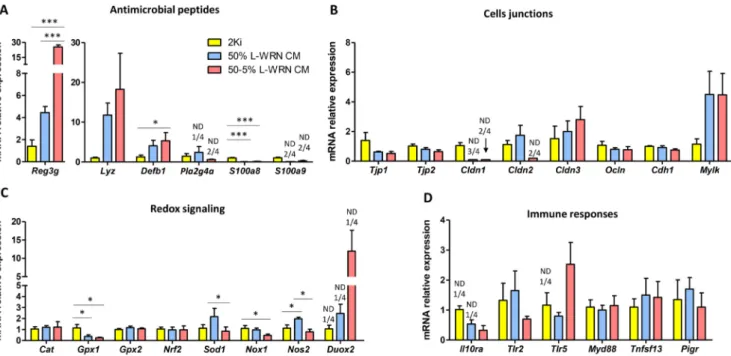
As a next step, we explored whether the greater differentiation level observed in organoids cultured with L-WRN CM was associated with a modulation of the epithelial barrier function. The antimicrobial pep-tides Reg3g and Defb1 were significantly more expressed in organoids grown in 50–5% L-WRN CM than in 2ki condition (Fig. 6A). Moreover, the expression level of Reg3g was also higher when the L-WRN CM was used at 50–5%, when compared to 50%. The mean expression of the antimicrobial peptide Lyz was more than 10-fold higher in organoids grown with L-WRN CM than in 2Ki medium, but this effect was not significant due to high variability. The expression of Lyz was also higher in organoids grown in L-WRN CM than in epithelial crypts, but this difference was not significant (figure S2C). In contrast, the gene ex-pression of the two monomers of the antimicrobial protein calprotectin (S100a8/S100a9) was strongly reduced or undetectable in organoids grown in L-WRN CM compared to 2Ki. The mRNA levels of most of the genes involved in epithelial junctions were not modified according to the culture condition excepted for Cldn1 and Cldn2 which mRNA levels was low or undetectable in organoids grow with L-WRN CM (Fig. 6B). Regarding genes involved in redox signaling in epithelial cells, there was a downregulation of the expression of Gpx1 in organoids grown in L-WRN CM compared to the 2Ki medium (Fig. 6C). A similar effect was observed for the superoxide generating enzyme Nox1 when L-WRN CM was used at 50%. In contrast, the nitric oxide producing enzyme Nos2 was upregulated in organoids grown in 50% L-WRN CM compared to
Fig. 2. Characteristics of organoids cultured with pharmacological inhibitors (2Ki medium). Organoids were cultured during 7 days with complete 2Ki
medium or without one of its components. A. Number of organoids per well (n = 3 wells/condition). Data are presented as means + SEM. B. Diameter of organoids. Individual values are presented with dots and means with bars (complete medium: n = 34, without FBS: no organoid, without Y-27632: n = 33, without CHIR-99021: n = 7, without SB-431542: n = 34, without LDN-193189: n = 33). A linear model was used to test the effect of the medium composition. Mean value of each group was compared to the mean value of the complete 2Ki condition. ***: P < 0.001, *: P < 0.05 (adjusted with the Tukey method). C. Representative images of organoids in each condition. The scale bar represents 100 µm.
E. Mussard, et al. Stem Cell Research 48 (2020) 101980
2Ki and 50–5% L-WRN CM. The gene expression of the antioxidant enzyme Sod1 was higher when the L-WRN CM was used at 50% com-pared to 50–5%. Finally, we observed no effect of organoid growth conditions on the mRNA levels of genes involved in cytokine signaling (Il10ra), toll-like receptor signaling (Tlr2, Tlr5, Myd88) or IgA secretion pathway (Tnfsf13, Pigr) (Fig. 6D). Altogether, our results show that the modulation of epithelial differentiation level in organoids induced by growth condition is associated with an alteration of the expression of several key components of the epithelial barrier (antimicrobial pep-tides, redox defenses and tight junctions).
3.5. Culture of rabbit caecum organoid cells in 2D facilitates the access to the apical side of epithelial cells
Accessibility of the apical side of epithelial cells is an essential feature of intestinal organoids, especially to study interaction between luminal compounds (nutrients, microorganisms) and IEC. Thus, we analyzed the polarity of rabbit caecum organoids cultured with phar-macological inhibitors (2Ki) or with 50% or 50–5% L-WRN CM. Actin (a marker of the apical side of epithelial cells (Co et al., 2019)) was de-tected towards the lumen of the organoids when cultured in 3D, in all culture condition tested (Fig. 7A). We then aimed at developing a model in which the apical compartment of rabbit cultured epithelial cells would be accessible. Rabbit organoids grown in 3D for 7 days with 2Ki medium or 50% L-WRN CM were dissociated into single cells before seeding in Transwells previously coated with a thin layer of Matrigel. Two days later, we observed a contiguous monolayer of epithelial cells (Fig. 7B). TEER increased between 24 and 48 h post-seeding which
indicated the gradual formation of an intact epithelium (Fig. 7C). TEER was similar in both growth conditions. EdU incorporation assay in-dicated that organoid epithelial cells were not proliferating in Trans-well monolayers 48 h post-seeding (figure S3). Actin was located to-wards the upper compartment and thus accessible in IEC from organoids grown in 2Ki medium or 50% L-WRN CM (Fig. 7B, Z-section). Interestingly, actin was localized exclusively at cell junctions in IEC of organoids cultured with 50% L-WRN CM while actin stress fibers were observed with the 2Ki medium. In conclusion, the culture of rabbit caecum organoid cells in 2D allows the accessibility to the apical side of epithelial cells using both 2Ki and L-WRN CM culture media. 4. Discussion
Our results show that rabbit caecum organoids can be obtained by seeding epithelial crypts in Matrigel with a growth culture medium reproducing in vitro the ISC niche either with pharmacological in-hibitors or with L-WRN CM. Independently of the culture conditions, rabbit caecum organoids can be passaged, cryopreserved and re-suscitated. These organoids contain the main epithelial cell types ac-cording to gene expression of markers for ISC and differentiated IEC (absorptive, goblet and enteroendocrine cells). Moreover, the gene ex-pression of key components of the epithelial barrier (e.g. antimicrobial peptides, tight junction proteins, toll-like receptors) suggests that epi-thelial functionality is reproduced in vitro. Overall, the newly developed rabbit caecum organoid model closely replicates epithelial physiology and could thus be used to study rabbit epithelium in the context of biomedical or veterinary research.
Fig. 3. Comparison of characteristics of organoids cultured with 2Ki medium or L-WRN CM. Organoids were cultured during 7 days in 2Ki, 50% L-WRN CM
(conditioned medium produced by L-WRN cells) or 50–5% L-WRN CM conditions. A. Number of organoids per well (expressed relatively to the number of organoids obtained for the same rabbit with 2Ki medium). B. Diameter of organoids. C. Number of organoids according to their spherical or non-spherical morphology. D. Cell height of organoids. A-D. Data are presented as means + SEM, n = 7. A linear mixed model was used with medium composition as a fixed effect and rabbit and culture plate as random effects. Mean value of each group were compared pairwise. ***: P < 0.001, **: P < 0.01, *: P < 0.05 (adjusted with the Tukey method).
To culture rabbit caecum organoids, we reconstituted the ISC niche
in vitro either with pharmacological inhibitors or with L-WRN CM,
based on previous publications (Li et al., 2018a; Miyoshi and Stappenbeck, 2013; Powell and Behnke, 2017; VanDussen et al., 2015, 2019). High Wnt/low BMP signaling pathways activation is needed for ISC maintenance and proliferation while low Wnt/high BMP signaling pathways activation leads to IEC differentiation (Gehart and Clevers, 2019). In our experiments, Wnt signaling pathway was activated by the GSK3 inhibitor CHI99021 in the 2Ki medium and by Wnt3a and R-Spondin 3 in L-WRN CM. BMP signaling was inhibited by LDN-193189 in the 2Ki medium and by Noggin in the L-WRN CM. Pharmacological inhibitors or mouse proteins secreted in L-WRN CM probably resulted in differential Wnt and BMP signaling pathway activation in rabbit caecum organoids which could explain the phenotypic differences ob-served according to the culture method. Moreover, the use of L-WNR CM at low concentration (5%) probably resulted in a low activation of
Wnt signaling pathway and a low inhibition of the BMP pathway, as previously shown in human and mouse organoids (VanDussen et al., 2015; 2019).
The greater diameter of organoids cultured with 2ki medium, when compared to L-WRN CM indicated a faster epithelial growth with pharmacological inhibitors, suggesting a high Wnt/low BMP signaling pathways activation. Accordingly, removal of the Wnt activator CHIR-99021 or of the BMP inhibitor LDN-193189 from the 2Ki medium re-duced the diameter of organoids and thus limited their growth. Organoids cultured with 2Ki medium were exclusively spherical/cystic, this spheroid morphology being associated with an immature pheno-type and a high proliferation rate (Merker et al., 2016; Stelzner et al., 2012; VanDussen et al., 2019). In contrast, organoids cultured with L-WRN CM were not all spheroids, some of them being multilobulated, suggesting that these organoids were more mature, less proliferative than organoids cultured with 2Ki medium. Indeed, we did not observe
Fig. 4. Comparison of proliferation markers in organoids cultured with 2Ki medium or L-WRN CM. Organoids were cultured during 7 days in 2Ki, 50% L-WRN
CM (conditioned medium produced by L-WRN cells) or 50–5% L-WRN CM conditions. A. Proliferating cells were stained for 1 h by EdU (green) in organoids after 7 days of culture. DAPI staining (blue) shows nuclei. The scale bar represents 100 µm. B. Relative gene expression of stem cells and proliferation markers. Gene expression was analyzed by high throughput microfluidic qPCR. Data are presented as means + SEM, n = 4. A linear mixed model was used with medium composition as a fixed effect and rabbit and culture plate as random effects. Mean value of each group were compared pairwise. *: P < 0.05 (adjusted with the Tukey method). ND: non-detected.
Fig. 5. Comparison of differentiation markers in organoids cultured with 2Ki medium or L-WRN CM. Gene expression was analyzed by high throughput
microfluidic qPCR in organoids cultured during 7 days in 2Ki, 50% L-WRN CM (conditioned medium produced by L-WRN cells) or 50–5% L-WRN CM conditions.
A-B. Relative gene expression of absorptive cells markers (A) and secretory cells markers (B). Data are presented as means + SEM, n = 4. A linear mixed model was
used with medium composition as a fixed effect and rabbit and culture plate as random effects. Mean value of each group were compared pairwise. **: P < 0.01, *: P < 0.05 (adjusted with the Tukey method). ND: non-detected.
E. Mussard, et al. Stem Cell Research 48 (2020) 101980
Fig. 6. Comparison of epithelial barrier markers in organoids cultured with 2Ki medium or L-WRN CM. Gene expression was analyzed by high throughput
microfluidic qPCR in organoids cultured during 7 days in 2Ki, 50% L-WRN CM (conditioned medium produced by L-WRN cells) or 50–5% L-WRN CM conditions.
A-D. Relative expression of genes involved in antimicrobial defenses (A), cell junctions (B), redox signaling (C) and immune response (D). Data are presented as
means + SEM, n = 4. A linear mixed model was used with medium composition as a fixed effect and rabbit and culture plate as random effects. Mean value of each group were compared pairwise. ***: P < 0.001, *: P < 0.05 (adjusted with the Tukey method). ND: non-detected.
Fig. 7. Polarization of organoids cultured with 2Ki medium or L-WRN CM. A. Organoids were cultured in 3 dimensions during 7 days in 2Ki, 50% L-WRN CM
(conditioned medium produced by L-WRN cells) or 50–5% L-WRN CM condition. B. Organoids dissociated cells were cultured in 2 dimensions in 2Ki or 50% L-WRN CM conditions in insert coated with Matrigel. Phalloidin staining (red) shows actin and DAPI staining (blue) shows nuclei. The scale bar represent 100 µm (X-Y plan) or 10 µm (Z-section). C. Transepithelial electrical resistance (TEER) of organoid cell monolayers in 2Ki or 50% L-WRN CM conditions 24 and 48 h after seeding. Data are presented as means + SEM, n = 6–7. A linear mixed model was used with medium composition and time as a fixed effect and transwell as a random effect. **: P < 0.01, (adjusted with the Tukey method).
EdU+cells (i.e. proliferating) in non-spherical organoids cultured with
L-WRN CM. It would be interesting to test whether these non-spherical organoids could still propagate after subculture. In contrast, spherical organoids positive for EdU staining were observed both with 2Ki medium and L-WRN CM, but their frequency seemed to be higher with the 2Ki medium. Accordingly, the gene expression of the ISC markers
Lgr5 and Olfm4 (Flier et al., 2009; Sato et al., 2009) and of the pro-liferation marker Pcna was very low or not detectable when the L-WRN CM was used at low concentration. Our results are in agreement with a previous study in human organoids showing that reducing the L-WRN CM from 50% to 5% downregulated the expression of Lgr5 and Olfm4 (VanDussen et al., 2015). In another study, mouse colon organoids cultured with 2Ki medium had higher expression of Lgr5 than organoid cultured with a medium containing recombinant Noggin and R-spondin (Li et al., 2018a), suggesting high Wnt/low BMP signaling pathways activation in organoids cultured with 2Ki medium. Besides Wnt and BMP signaling pathway, epidermal growth factor (EGF) is a key reg-ulator of IEC proliferation and is an essential component of organoid culture media (Gehart and Clevers, 2019). Our two culture methods (2Ki and L-WRN CM) contained 10% FBS which is a source of EGF (Miyoshi and Stappenbeck, 2013). This could explain why we did not obtain organoids when the 2Ki medium did not contain FBS. However, a study showed that EGF was not needed to grow mouse colon orga-noids with the 2Ki medium containing the supplements N-2 and B-27 (Li et al., 2018a). Thus, in our culture conditions, FBS might bring es-sential but undefined compounds such as insulin or vitamins contained in N-2 and B-27 supplements. Intestinal organoids from other species (e.g. mouse and human) grown in 50% L-WRN CM are typically highly proliferative (Miyoshi and Stappenbeck, 2013; VanDussen et al., 2015), which is not the case of our rabbit caecum organoids cultured with L-WRN CM, at least when compared to organoids grown in 2Ki medium. Previous studies used 20% FBS in L-WRN CM (Miyoshi and Stappenbeck, 2013) while we used 10% FBS in order to have the same concentration than in the 2Ki medium and thus allowing the compar-ison between both culture conditions. It remains to be elucidated whether this reduction of FBS concentration in L-WRN CM explains why rabbit caecum organoids appear less proliferative than previously described for mouse and human intestinal organoids grown in L-WRN CM. Altogether, our results show that the 2Ki medium is a more potent inducer of ISC maintenance and proliferation in rabbit caecum orga-noids than the L-WRN CM, especially when the latter is used at low concentrations.
Cell height was greater in organoids cultured with L-WRN CM than with 2Ki medium, this characteristic being typical of differentiated and highly polarized IEC (Altay et al., 2019). Accordingly, the gene ex-pression of absorptive cells markers (Krt20, Alpi, Aqp8 and Vil1) was higher in organoids cultured with L-WRN CM (notably when used at 50–5%) compared to the 2Ki medium. These results obtained in rabbit caecum organoids are consistent with previous data obtained in mouse organoids showing that the 2Ki medium reduced the gene expression of
Alpi when compared to a growth medium containing recombinant EGF,
Noggin and R-spondin (Li et al., 2018a). Upregulation of absorptive cells markers in human organoids were also found when L-WRN CM was used at low concentration (VanDussen et al., 2015). The gene ex-pression of Atoh1 coding for the transcription factor Math1, required for the commitment of epithelial progenitors into the secretory lineage (Yang et al., 2001), was upregulated in organoids cultured with L-WRN CM compared to the 2Ki medium, suggesting a higher number of se-cretory cells. This result was associated with an upregulation of goblet cells markers (Muc2 and Spdef) and of the enteroendocrine cell gene Pyy in organoids cultured with L-WRN CM, especially when used at 50–5%. The low expression level of secretory cell markers in organoids cultured with the 2ki medium was not observed in the study byLi et al. (2018a). It might be due to the species difference (rabbit vs mouse) or slight modifications in growth media composition. Altogether, our results indicated that epithelial cells were more differentiated when cultured
with L-WRN CM used at low concentration compared to the 2Ki medium. IEC differentiation is usually associated with a modulation of epithelial barrier function (Sun et al., 2017). Indeed, in our experi-ments, differentiation of rabbit caecum organoid was associated with the regulation of antimicrobial peptides and proteins involved in cell junction and redox signaling. Overall, organoids cultured with 2Ki medium were highly proliferative and contained few differentiated cells while the opposite was observed for organoids cultured with L-WRN CM. These phenotypes are consistent with a higher activation of Wnt pathway and more potent inhibition of BMP signaling pathway in rabbit caecum organoids cultured with the 2Ki medium than with L-WRN CM. The development of validated antibodies targeting rabbit proteins used as markers for each epithelial cell types will be required for the validation of our results by using immunostaining. When rabbit caecum organoids were cultured in 3D with 2Ki medium or L-WRN CM, the apical side of the epithelium was oriented toward the lumen of organoids, according to actin localization. Similar results were pre-viously obtained in 3D intestinal organoids from human, mouse or farm animals (Co et al., 2019; Derricott et al., 2019). Thus, the physiological route of exposure to luminal compounds (microbes or nutrients) is not easily accessible in 3D rabbit caecum organoids. To circumvent this issue, we seeded dissociated cells from 3D organoids onto Matrigel-coated Transwell permeable supports, based on previous publications (van der Hee et al., 2018; Moon et al., 2014; VanDussen et al., 2015). We obtained 2D contiguous monolayers, in which IEC apical side was facing upwards, making it accessible to experimental treatments. In-terestingly, the presence of actin stress fibers in 2D monolayers cultured with 2Ki medium but not with L-WRN CM is consistent with our results indicating a lower differentiation level when the 2Ki medium is used. Indeed, actin stress fibers are typically observed in IEC with a low differentiation level, while the perijunctionnal localization of actin (as observed with the L-WRN CM) is associated with a more differentiated state (Li et al., 2018b; Soubeyran et al., 1999). The high TEER of or-ganoid epithelial cell monolayers indicated the formation of an intact epithelium. Further work is needed to develop functional assays to use this model to study epithelial barrier function (e.g. permeability mea-surement, immunoglobulin A transcytosis). Another perspective of our work would be to develop rabbit organoids from other digestive re-gions.
In summary, reconstitution of the ISC niche with pharmacological inhibitors or with L-WRN CM allowed the growth of rabbit caecum organoids. Due to the large phenotypic differences observed according to the method used, we anticipate different applications of organoids cultured with 2Ki medium or L-WRN CM. Rabbit caecum organoids cultured with 2Ki medium could be used to generate rapidly a large number of rabbit ISC in vitro. In contrast, the L-WRN CM used at low concentration can produce organoids composed mainly of differ-entiated IEC and thus appropriate to study rabbit epithelium phy-siology. Our results also show that intestinal organoid culture methods developed in mice or human can be easily adapted to other animal species, probably due to the high evolutionary conservation of the key regulators of ISC niche signaling pathways. Thus, we believe that the culture of intestinal organoids should be feasible for most animal spe-cies for which in vitro models of IEC are not available yet.
Funding
The present work received the financial support from INRAE PHASE division (“MiniGut” project). The Organoid Core Facility of the IRSD is supported by a “Fond Unique Interministeriel” (FUI) program, from the Région Midi-Pyrénées (now Occitanie), Toulouse Métropole, the Banque Publique d’Investissement (BPI) de France. Equipments of the IRSD Organoid Core facility were obtained thanks to the Fonds Européens de Développement Régional (FEDER), and the region Occitanie funds.
E. Mussard, et al. Stem Cell Research 48 (2020) 101980
Author contribution
EM, SC and MB: conceived the experiments. EM, CP, VH, SF and MB performed experiments. CC, AR and NV shared expertise and biological resources. EM, SC, GP and MB analyzed the data. EM, SC and MB wrote the manuscript. All authors contributed to manuscript editing. Declaration of Competing Interest
The authors declare that they have no known competing financial interests or personal relationships that could have appeared to influ-ence the work reported in this paper.
Acknowledgements
The authors thank the staff of the rabbit experimental unit PECTOUL for animal care. We also acknowledge Yvette Lahbib-Mansais and Christelle Marrauld (GenPhySE, INRAE) for help with cell culture and confocal imaging experiments. We are grateful to the Genotoul bioinformatics platform Toulouse Occitanie (Bioinfo Genotoul, doi: 10.15454/1.5572369328961167E12) for providing computing and storage resources.
Appendix A. Supplementary data
Supplementary data to this article can be found online athttps:// doi.org/10.1016/j.scr.2020.101980.
References
Altay, G., Larrañaga, E., Tosi, S., Barriga, F.M., Batlle, E., Fernández-Majada, V., Martínez, E., 2019. Self-organized intestinal epithelial monolayers in crypt and villus-like do-mains show effective barrier function. Sci. Rep. 9, 1–14.
Barker, N., 2014. Adult intestinal stem cells: critical drivers of epithelial homeostasis and regeneration. Nat. Rev. Mol. Cell Biol. 15, 19–33.
Beaumont, M., Paës, C., Mussard, E., Knudsen, C., Cauquil, L., Aymard, P., Barilly, C., Zemb, O., Fourre, S., Gautier, R., et al. (2020). Gut microbiota derived metabolites contribute to the gut barrier maturation at the suckling-to-weaning transition. Gut Microbes 10.1080/19490976.2020.1747335.
Clevers, H., 2016. Modeling Development and Disease with Organoids. Cell 165, 1586–1597.
Co, J.Y., Margalef-Català, M., Li, X., Mah, A.T., Kuo, C.J., Monack, D.M., Amieva, M.R., 2019. Controlling Epithelial Polarity: A Human Enteroid Model for Host-Pathogen Interactions. Cell Reports 26, 2509–2520.e4.
Cunningham, F., Achuthan, P., Akanni, W., Allen, J., Amode, M.R., Armean, I.M., Bennett, R., Bhai, J., Billis, K., Boddu, S., et al., 2019. Ensembl 2019. Nucleic Acids Res. 47, D745–D751.
Date, S., Sato, T., 2015. Mini-Gut Organoids: Reconstitution of the Stem Cell Niche. Annu. Rev. Cell Dev. Biol. 31, 269–289.
Derricott, H., Luu, L., Fong, W.Y., Hartley, C.S., Johnston, L.J., Armstrong, S.D., Randle, N., Duckworth, C.A., Campbell, B.J., Wastling, J.M., et al., 2019. Developing a 3D intestinal epithelium model for livestock species. Cell Tissue Res. 375, 409–424.
Flier, L.G.van der, Haegebarth, A., Stange, D.E., van de Wetering, M., Clevers, H., 2009. OLFM4 Is a Robust Marker for Stem Cells in Human Intestine and Marks a Subset of Colorectal Cancer Cells. Gastroenterology 137, 15–17.
Gehart, H., Clevers, H., 2019. Tales from the crypt: new insights into intestinal stem cells. Nat. Rev. Gastroenterol. Hepatol. 16, 19–34.
Hamilton, C.A., Young, R., Jayaraman, S., Sehgal, A., Paxton, E., Thomson, S., Katzer, F., Hope, J., Innes, E., Morrison, L.J., et al., 2018. Development of in vitro enteroids derived from bovine small intestinal crypts. Vet. Res. 49, 54.
van der Hee, B., Loonen, L.M.P., Taverne, N., Taverne-Thiele, J.J., Smidt, H., Wells, J.M.,
2018. Optimized procedures for generating an enhanced, near physiological 2D culture system from porcine intestinal organoids. Stem Cell Res. 28, 165–171.
Holmberg, F.E., Seidelin, J.B., Yin, X., Mead, B.E., Tong, Z., Li, Y., Karp, J.M., Nielsen, O.H., 2017. Culturing human intestinal stem cells for regenerative applications in the treatment of inflammatory bowel disease. EMBO Mol. Med. 9, 558–570.
Li, Y., Liu, Y., Liu, B., Wang, J., Wei, S., Qi, Z., Wang, S., Fu, W., Chen, Y.-G., 2018a. A growth factor-free culture system underscores the coordination between Wnt and BMP signaling in Lgr5 + intestinal stem cell maintenance. Cell Discovery 4, 49.
Li, B.-R., Wu, J., Li, H.-S., Jiang, Z.-H., Zhou, X.-M., Xu, C.-H., Ding, N., Zha, J.-M., He, W.-Q., 2018b. In Vitro and In Vivo Approaches to Determine Intestinal Epithelial Cell Permeability. J Vis Exp.
Licois, D., Wyers, M., Coudert, P., 2005. Epizootic Rabbit Enteropathy: experimental transmission and clinical characterization. Vet. Res. 36, 601–613.
Lukovac, S., Belzer, C., Pellis, L., Keijser, B.J., de Vos, W.M., Montijn, R.C., Roeselers, G., 2014. Differential Modulation by Akkermansia muciniphila and Faecalibacterium prausnitzii of Host Peripheral Lipid Metabolism and Histone Acetylation in Mouse Gut Organoids. MBio 5, e01438–e1514.
Merker, S.R., Weitz, J., Stange, D.E., 2016. Gastrointestinal organoids: How they gut it out. Dev. Biol. 420, 239–250.
Miyoshi, H., Stappenbeck, T.S., 2013. In vitro expansion and genetic modification of gastrointestinal stem cells in spheroid culture. Nat. Protoc. 8, 2471–2482.
Moon, C., VanDussen, K.L., Miyoshi, H., Stappenbeck, T.S., 2014. Development of a primary mouse intestinal epithelial cell monolayer culture system to evaluate factors that modulate IgA transcytosis. Mucosal Immunol. 7, 818–828.
Peterson, L.W., Artis, D., 2014. Intestinal epithelial cells: regulators of barrier function and immune homeostasis. Nat. Rev. Immunol. 14, 141–153.
Powell, R.H., Behnke, M.S., 2017. WRN conditioned media is sufficient for in vitro pro-pagation of intestinal organoids from large farm and small companion animals. Biology Open 6, 698–705.
Sato, T., Vries, R.G., Snippert, H.J., van de Wetering, M., Barker, N., Stange, D.E., van Es, J.H., Abo, A., Kujala, P., Peters, P.J., et al., 2009. Single Lgr5 stem cells build crypt-villus structures in vitro without a mesenchymal niche. Nature 459, 262–265.
Sato, T., Stange, D.E., Ferrante, M., Vries, R.G.J., van Es, J.H., van den Brink, S., van Houdt, W.J., Pronk, A., van Gorp, J., Siersema, P.D., et al., 2011. Long-term Expansion of Epithelial Organoids From Human Colon, Adenoma, Adenocarcinoma, and Barrett’s Epithelium. Gastroenterology 141, 1762–1772.
Schneider, C.A., Rasband, W.S., Eliceiri, K.W., 2012. NIH Image to ImageJ: 25 years of image analysis. Nat. Methods 9, 671–675.
Sébert, M., Denadai-Souza, A., Quaranta, M., Racaud-Sultan, C., Chabot, S., Lluel, P., Monjotin, N., Alric, L., Portier, G., Kirzin, S., et al., 2018. Thrombin modifies growth, proliferation and apoptosis of human colon organoids: a protease-activated receptor 1- and protease-activated receptor 4-dependent mechanism. Br. J. Pharmacol. 175, 3656–3668.
Soubeyran, P., André, F., Lissitzky, J.-C., Mallo, G.V., Moucadel, V., Roccabianca, M., Rechreche, H., Marvaldi, J., Dikic, I., Dagorn, J.-C., et al., 1999. Cdx1 promotes differentiation in a rat intestinal epithelial cell line. Gastroenterology 117, 1326–1338.
Stelzner, M., Helmrath, M., Dunn, J.C.Y., Henning, S.J., Houchen, C.W., Kuo, C., Lynch, J., Li, L., Magness, S.T., Martin, M.G., et al., 2012. A nomenclature for intestinal in vitro cultures. Am. J. Physiol. Gastrointest. Liver Physiol. 302, G1359–G1363.
Sun, X., Yang, Q., Rogers, C.J., Du, M., Zhu, M.-J., 2017. AMPK improves gut epithelial differentiation and barrier function via regulating Cdx2 expression. Cell Death Differ. 24, 819–831.
VanDussen, K.L., Marinshaw, J.M., Shaikh, N., Miyoshi, H., Moon, C., Tarr, P.I., Ciorba, M.A., Stappenbeck, T.S., 2015. Development of an enhanced human gastrointestinal epithelial culture system to facilitate patient-based assays. Gut 64, 911–920.
VanDussen, K.L., Sonnek, N.M., Stappenbeck, T.S., 2019. L-WRN conditioned medium for gastrointestinal epithelial stem cell culture shows replicable batch-to-batch activity levels across multiple research teams. Stem Cell Res. 37, 101430.
Yang, Q., Bermingham, N.A., Finegold, M.J., Zoghbi, H.Y., 2001. Requirement of Math1 for secretory cell lineage commitment in the mouse intestine. Science 294, 2155–2158.
Yin, Y.-B., Guo, S.-G., Wan, D., Wu, X., Yin, Y.-L., 2019. Enteroids: Promising in Vitro Models for Studies of Intestinal Physiology and Nutrition in Farm Animals. J. Agric. Food Chem. 67, 2421–2428.
Yum, L.K., Byndloss, M.X., Feldman, S.H., Agaisse, H., 2019. Critical role of bacterial dissemination in an infant rabbit model of bacillary dysentery. Nat. Commun. 10, 1–10.
Zietek, T., Rath, E., Haller, D., Daniel, H., 2015. Intestinal organoids for assessing nutrient transport, sensing and incretin secretion. Sci. Rep. 5, 1–10.