by
Sharon R. Grossman
B.A. Chemical and Physical Biology and Mathematics Harvard University
Submitted to the Department of Biology
in Partial Fulfillment of the Requirements for the Degree of Doctor of Philosophy
at the
MASSACHUSETTS INSTITUTE OF TECHNOLOGY June 2019
© 2019 Massachusetts Institute of Technology. All rights reserved.
Signature redacted
Signature of Author ... .../
.Sharon'R.-GrossmanSignature redacted
Department of BiologyCertified by ...
-Signature redacted
Accepted by ... MASSACHUSETTS INSTITUTE OF TECHNOLOGY .MAY 2 3 2019
... Eric S. Lander Professor of Biology Thesis Supervisor .... .... .... ... .. ... Amy Keating Professor of Biology Co-Chair, Graduate CommitteeMITLibraries
DISCLAIMER NOTICE
The pagination in this thesis reflects how it was delivered to the Institute Archives and Special Collections.
The Table of Contents does not accurately represent the
by Sharon R. Grossman
Submitted to the Department of Biology on April 2 nd, 2019 in Partial Fulfillment of the
Requirements for the Degree of Doctor of Philosophy
ABSTRACT
Combinatorial gene regulation is encoded in enhancers and promoters in the form of binding sites for transcription factors (TFs), which collaboratively recruit the
transcriptional machinery and drive gene expression. Using high-throughput and quantitative technologies developed by our lab and others, we studied TF binding sites in enhancers from numerous different cell types and regulatory systems, shedding light general principles of motif composition and organization in typical cellular regulatory elements.
We find extensive synergy between TF binding sites, some with organizational
constraints and some with flexible positioning. We demonstrate that different TFs bind at distinct positions within regulatory elements, suggesting a new type of architectural constraint in enhancers. Importantly, our analysis of both TF organization and
cooperativity revealed distinctive patterns that separates TFs into potential functional classes.
Together, our results suggest a structure of the regulatory code at the level of TF function and generate new hypotheses about regiospecific binding patterns and functions of TF classes within enhancers.
Thesis Supervisor: Eric S. Lander Title: Professor of Biology
Graduate school has been a period of enormous scientific and personal growth for me. I could not have done it without the support of my colleagues, mentors, friends, and family, who were there for me through the highs and lows. In particular, I would like to thank the following people:
• I cannot express enough gratitude for my advisor, Eric Lander, who inspired me
with his love of biology and genuine curiosity about life, taught me both scientific rigor and vision, and supported and believed in me through some of the most difficult personal challenges of my life. His confidence in my ability to grow and succeed made me into the scientist and person I am today.
• My thesis committee, Chris Burge, Aviv Regev, and Brad Bernstein, for their
scientific guidance and insight.
• Jesse Engreitz, who has been my colleague and friend since the very beginning
of our rotations, and spent countless hours discussing science and life. Having a comrade through graduate school meant more than I can say.
• Tim Wang, Charlie Fulco, and Brian Cleary for scientific discussion and
commiseration throughout our graduate careers.
• Nicole Brellethin and Kate Mulherin, for making everything run smoothly and
always being there for a smile or cup of coffee.
• Pardis Sabeti, whose advice shaped my scientific path, and who had been the
best mentor and friend anyone could hope for.
• My parents, sister, and grandparents, whose unwavering love and support
inspired me to follow my passions and gave me the confidence and strength to get to where I am today.
• Kristin Knouse, Eric Bent, Jeremiah Wala, Will Gibson, Xenos Mason, Jennifer
Lo, Belinda Wang, Dave and Yakir Reshef, and Hilary Finucane, who friendship and laughter made graduate school fun and inspiring, and taught me so much about life.
Title page 1
Abstract 3
Acknowledgements 5
CHAPTER 1: Introduction 7
CHAPTER 2: Systematic dissection of PPARy enhancers 92
CHAPTER 3: Positional specificity of TF binding sites 168 CHAPTER 4: TF regulatory activity across six cell types 217
Chapter 1
Introduction
SECTION I. Overview
Control of gene expression plays a key role in a multitude of biological processes, from organismal development and cell differentiation to response to environmental stresses. Since the discovery of regulatory DNA in the 1960s,
deciphering the cis-regulatory "code" that specifies how gene expression is encoded in regulatory sequences has been a major research focus. In addition to shedding light on core biological process, the ability to "read" and "write" the regulatory code has broad implications for human health: deviations from the appropriate expression patterns due to mutations in regulatory sequences or transcriptional regulators underlie the formation of many cancers and other diseases (1-3), and the ability to design novel regulatory sequences with desired properties opens the way to targeted, specific treatments. Furthermore, the majority of evolutionary changes in animal morphology and other traits stem from mutations in developmental regulatory sequences (4-7).
Information about when, where, and to what level each gene should be
expressed is encoded in cis-regulatory sequences such as promoters and enhancers, in the form of organized arrays of binding sites for sequence-specific transcription factors (TFs). Cellular differentiation and cues from the environment trigger lead to the
activation of specific sets of TFs, which bind to enhancers and interact with cofactors to stabilize the transcription-initiation machinery to enable gene expression (8, 9).
Compared to bacteria, metazoan systems employ vastly more elaborate protein machineries to carry out transcription (10). TFs almost never function alone, instead relying on combinatorial input from multiple partner TFs to drive robust expression. In addition, the general protein machineries required for core promoter recognition and
basal transcription in metazoans include numerous diverse and functionally specialized components. Complex systems of cofactors are involved in mediating signals between TFs and the transcriptional machinery and remodeling local chromatin structure, adding a final layer of regulation (11).
Over the past four decades, the molecular players in transcription have largely been identified, but the detailed mechanistic steps involved in enhancer-mediated transcriptional activation remain unclear. The vast majority interactions between TFs, cofactors, and basal transcriptional machinery are not known. Without knowing the
biochemical activities of each bound TF, it is hard to interpret dependencies observed between TF binding sites in functional characterizations of regulatory sequences. Basic mechanistic questions about enhancer structure and function remain, such as: how many distinct biochemical activities are required for transcription? Do individual TF binding sites contribute to different functional steps? If so, is there a universal formula at the level of TF function?
From a systems perspective, cells "compute" information from input TFs via regulatory sequences to determine the output expression pattern. In keeping with the intricate molecular underpinnings of transcription, the computations performed by enhancers are remarkably complex, generating many different relationships between the combination and concentration of input TFs and the output gene expression (12,
13). Many aspects of the enhancer sequence can affect transcriptional output, including the identity, number and affinity of TF binding sites, their arrangement and spacing within the enhancer, and potential synergies between the bound TFs (14). With only a
handful of well-characterized enhancer sequences and little mechanistic understanding of combinatorial TF function, a global cis-regulatory code has remained elusive (15-17). A major breakthrough in our ability to investigate regulatory sequences and how they encode gene expression came with the development of high-throughput methods to identify enhancers by mapping genome-wide chromatin states (18-20) and TF occupancy (21, 22). However, until recently these predicted enhancers could only be functionally characterized using low-throughput methods, precluding the systematic dissection of functional elements within regulatory sequences and the generation of large datasets for computational modeling. A second breakthrough occurred shortly before I began my Ph.D., when our lab and others developed high-throughput parallel enhancer assays enabling measurements of thousands of regulatory elements in one experiment (23-25).
In this thesis, I present our contribution towards deciphering regulatory DNA and uncovering a general cis-regulatory code using the unprecedented ability to
comprehensively identify native genomic enhancers, rapidly characterize their
regulatory activity, and dissect their functional properties. In Chapter 1, we develop a generalizable framework for deciphering cis-regulatory grammar through (i) systematic identification of functional TF binding sites, (ii) experimental quantification of the activity of sites corresponding to each TF, and (iii) analysis of patterns of cooperativity between TFs and architectural constraints. We used this approach to comprehensively
characterize PPARy-response elements (PPREs) in adipocytes. Comparing of the patterns of cooperativity between different factors revealed clusters of TFs marked by similar interaction patterns with other TFs. This "modular epistasis" suggests the intriguing possibility of functional "equivalency" classes of TFs, which might contribute different regulatory functions. Consistent with this idea, the classes showed similar effects on expression when perturbed and were associated with known biological processes. In Chapter 2, we dig further into functional distinctions between TFs using a different feature of enhancer architecture: binding site position within nucleosome-depleted regions (NDRs). We examine the positional distribution of binding sites in NDRs for 103 TFs across 47 cell types, and show that TFs fall into six distinct classes with strikingly different spatial localization within enhancers. These positional classes bring together factors that have a number of similar properties, and suggests specific functional roles for each class consistent with their NDR localization. This suggests that architectural constraints in enhancers can arise not only from well-known cooperative binding interactions, but also to optimally position a TF to carry out its function. Finally, in Chapter 3, we describe work in progress to experimentally validate the positional classes by showing motif sites in "optimal" positions are more likely to be occupied by their cognate TF and contribute more strongly to expression output in enhancer assays. Moreover, the relative effect sizes of the different classes are consistent with their hypothesized roles.
To provide context for thinking about how gene regulation is encoded in DNA, I first present a brief overview of the eukaryotic transcriptional machinery controlling the activity of RNA polymerase at core promoters and the transcriptional cycle. I review the
discovery and initial characterization of enhancers, and how known properties of enhancers are used today by various methods to predict regulatory elements in the genome. Next, I discuss TF binding and function, examining intrinsic TF DNA-binding specificities, how TFs bind in the genome, and various effector functions of TFs after DNA binding. Finally, I discuss models of enhancer architecture and regulatory grammar, drawing upon deeply characterized test cases as well as recent high-throughput experiments manipulating various features of enhancer architecture.
SECTION 1l. Molecular mechanisms of transcription at promoters
Expression of protein-coding genes begins with the transcription of the gene's DNA sequence into RNA by RNA polymerase II (RNAPIl). Transcription usually begins at a defined location at the 5' end of the gene, the transcription start site (TSS). The TSS is embedded in a core promoter, extending -50 bp upstream and downstream of the TSS, which constitutes the minimal DNA sequence needed for basal transcription. The core promoter contains recognition sites for the transcriptional machinery,
comprising RNAPII and general transcription factors (GTFs). Basal transcription from core promoters is generally low, and can be further suppressed by closed chromatin. Robust transcription requires activation by additional regulatory DNA elements located at varying distances from the TSS called enhancers (Fig. 1A). Enhancers contain binding sites for TFs, which recruit cofactors and can promote or repress transcription.
Transcription from a core promoter is a tightly regulated multistep process resulting in a specified expression output. Since all the processes controlling RNAPII transcription must ultimately lead to the basal transcriptional machinery at the core
essential to the study of gene regulation. I will therefore begin by reviewing the structure of core promoters and the molecular mechanisms underlying the assembly and
activation of the transcriptional machinery. These basic promoter properties of core promoters provide the foundation for understanding how this machinery is regulated by distal enhancers through various combinations of TFs and cofactors.
Properties of gene core promoters Sequence elements
The core promoter serves as a binding platform to position and support the assembly of the pre-initiation complex (PIC), which includes RNAPII and GTFs (26). Metazoan core promoters contain several interchangeable sequence elements known as core-promoter motifs, which are positioned at fixed distances from the TSS and recruit various GTFs and mediate the assembly of the PlC (Fig. 1B).
The most commonly occurring (though not universal) core-promoter motif that has been identified to date is the initiator (Inr) motif (27). The Inr directly overlaps the TSS and is recognized by several components of the TEIlD complex, a GTF involved in recruiting RNAPII and mediating PIC assembly (28, 29). Another well-characterized core-promoter motif is the TATA-box motif, located -30 bp upstream of the TSS, which is recognized and bound by the TATA-box binding protein (TBP) (30). TBP is a
component of the TFIID complex, Although the TATA box is conserved from yeast to humans (30), however, it is found in only a minority of core promoters (e.g. -5% of core
Enhww Trs TSS Be
I
T T 2 SiCAAACC~AS>~
a
OK~I
ooeo TsuW-ap.wmI genes ...ug. 3.«w1...f 2I1T~
a ?use..r. Wood...l Om Vwseorut.a
nvvb
Key dgWI---nWgws Itolt PrtWg IOM A~bs H-R C AellL ."41
Figure 1. Regulation of transcription. (A) A summary of promoter elements and regulatory signals. Chromatin is comprised of DNA wrapped around histones to form nucleosomes. The structure of chromatin can be tightly wrapped or accessible to proteins (such as TF). The region around the transcription start site (TSS) is often divided into a larger proximal promoter upstream of the TSS and a smaller core promoter just around the TSS. The exact boundaries vary between studies. To recruit RNAPII and to activate transcription of the gene, TFs bind to specific sequence patterns (TFBSs) that are near to the TSS (proximal elements) or that are far away from it
(enhancers). (B) Sequence patterns in core promoters. The region around the TSS has several over-represented sequence patterns; the TATA box and initiator (Inr) are the most studied and most prevalent. The location of patterns relative to the TSS and their sequence properties are shown as boxes and as associated sequence logos based on the JASPAR database. The Inr pattern is not shown as it varies considerably between studies. Importantly, most promoters only have one or a few of these patterns, and some patterns are mostly found in certain species. BRE, B recognition element; DCE, downstream core element; DRE, DNA recognition element; MTE, motif ten element. (C) Mapping endogenous transcription initiation at single nucleotide resolution revealed striking differences between core promoters, leading to the classification of 'focused' or 'sharp' core promoters, which have a single, well-defined TSS (left), and 'dispersed' or 'broad' promoters, which have multiple closely spaced TSSs that are used with similar frequency (middle and right). Figure modified from (31) and (32).
promoters in flies (33, 34) and 10-15% in humans (35-37)). In promoters that lack a TATA-box, the Inr motif is often paired with the downstream promoter element (DPE) instead (38). DPE occurs a fixed distance downstream of the Inr motif, and this spacing facilitates cooperative binding of TFIID to the two elements (29, 39).
In addition to these three relatively abundant core-promoter motifs, several other motifs with fixed positions relative to the TSS have been identified, such as the TFIIB recognition elements (BREs) (40, 41), the motif ten element (MTE) (42), and
downstream core elements (DCEs) (43). Like TATA, Inr and DPE, these motifs have been shown to bind specific GTFs in vitro (41, 44), and thus might contribute to PIC positioning and assembly in vivo.
Different combinations of core promoter motifs are associated with different transcription initiation patterns (35, 45, 46). Promoters with TATA boxes and Inr
elements are associated with a single, well-defined TSS (35, 47). These promoters are generally associated with highly cell-type specific genes with restricted expression patterns (48). In contrast, core promoters of broadly-expressed genes tend to lack a TATA box and instead contain CpG islands (CGIs), regions with high densities of CpG dinucleotides. These promoters are associated with broad, dispersed transcription initiation from multiple closely spaced TSS (35, 45). Promoters of key developmental genes also show dispersed initiation patterns in mammals, and tend to lack TATA boxes and contain long CGIs (35).
Chromatin features
Active core promoters are marked by distinctive chromatin features that make them accessible to the transcriptional machinery and facilitate the assembly of the PIC. Active core promoters reside in nucleosome-depleted regions (NDRs) flanked by
precisely positioned and phased nucleosomes downstream of the TSS (49-51). The first few nucleosomes downstream of the TSS (particularly the +1 nucleosome) are enriched for the histone variant H2A.Z, which has been shown to present a lower barrier to RNAPII transcription than canonical histones (52, 53). Housekeeping gene promoters with broad initiation patterns in particular are associated with clearly defined NDRs and stably positioned flanking nucleosomes (54). Promoters of highly regulated genes with focused initiation patterns are often occluded by nucleosomes before activation, and require nucleosome disassembly by ATP-dependent chromatin remodeling complexes
for activation (55, 56). After activation, focused promoters tend to have more imprecisely positioned nucleosomes (54).
Chromatin surrounding promoters is marked by distinctive patterns of histone modifications. The nucleosomes downstream of active promoters display a high level of tri-methylation on H3 Lys4 (H3K4me3) and acetylation of H3 Lys27 (H3K27ac). The role of these histone modifications in promoter function remains unclear. Histone acetylation can decrease the affinity of DNA for nucleosomes (57), and thus may promote open chromatin (58). Histone modifications may also assist in recruiting elements of the transcriptional machinery (59). For example, a subunit (BPTF) of the nucleosome remodeling factor (NURF) contains two domains that can recognize H3K4me3 and H3K27ac respectively, spanned by a rigid linker that positions them to correctly contact nucleosomes with these modifications (60, 61), and TAF1, a subunit of TFIID, shows increased affinity for acetylated histone H4 (62). However, several studies suggest H3K4me3 and H3K27ac may not be required for transcription. Drosophila cells
expressing non-methylatable forms of canonical and variant H3 histones can properly regulate transcription (63), and a Lys-to-Arg mutation at position 27 does not reduce transcription (64). Some of these effects may reflect functional redundancy among marks, but further studies of the mechanistic role of chromatin modifications is needed to distinguish causation from correlation and determine their functionality in
RNAPII transcription cycle
PIC assembly and promoter opening
RNAPII transcription begins with the ordered assembly of GTFs at the core promoter to form the PIC (Fig. 2A,B). Over the past two decades, the crystal structures of many of the components of PIC have been resolved, and the PIC structure and assembly has been extensively probed biochemically through mutagenesis and crosslinking mapping (65). Recently, crystallographic and cryo-electron microscopy (EM) studies have elucidated the structures of RNAPII complexes with GTFs (66, 67), verifying the earlier biochemically-derived models (68, 69). Together, these studies
provide remarkable structural and mechanistic insights into how RNAPII cooperates with the GTFs to bind to and open promoter DNA, initiate RNA synthesis, and escape the promoter.
The first step in PIC assembly is core promoter recognition and binding by TFIID, which contacts multiple core promoter sequence elements as well as histones with promoter-associated chromatin marks (62, 70, 71). The TFIID complex canonically consists of TBP and 13 TAFs, although cell-type specific variants of TBP and TAFs can form alternate complexes in metazoans. The saddle-shaped TBP binds to the DNA minor groove at the TATA box (if present) and induces a -90-degree bend in the DNA. The remaining TFIID subunits (TAFs) contact additional core promoter elements (as discussed above), arching from the TBP binding site 30 bp upstream of the TSS to the DPE located 30 bp downstream of the TSS and looping back to contact the Inr at the TSS (29). TFIID functions as a molecular ruler to position RNAPII at a precise position relative to the core promoter elements, and can also regulate TBP activity (72, 73) and
facilitate TEIlF and TFIE recruitment (29). TEIlD subunits are the target of various activating TFs, which can expedite TFIlD recruitment to promoters (74, 75).
TEIlD binding to the core promoter is stabilized by TFIIA (76), which bind the core promoter upstream of TBP (77-79) and inhibits a repressive domain in TFIID that occludes the TBP DNA-binding surface when TEIlD is not bound to promoter DNA (80). Notably, this anti-repression effect can be augmented by certain activating TFs,
resulting in a considerable increase in TFIlD DNA-binding activity (81).
TFIIB also facilitates TBP binding and DNA bending (82), and is required for RNAPII recruitment to promoters (83, 84). The C-terminal domain of TFIIB interacts with sequences upstream and downstream of the TBP binding site (BREs)(40, 41, 85, 86), orienting the assembly of the initiation complex (87, 88). The N-terminal domain binds to the dock domain of RNAPII to recruit the polymerase (89). TFIIB also functions in
establishing the TSS using two distinct functional domains, the linker helix and B-reader helix (67). The B-linker helix is positioned above the cleft where DNA opening commences and contributes to the establishment and maintenance of open DNA in the transcriptional bubble (67), and the B-reader helix binds the template strand to position DNA for the initiation of mRNA synthesis (90).
RNAPII binds to the TFIIB-TBP-DNA complex with the assistance of TIF to complete the core initiation complex (91). TEIlF interacts with unbound RNAPII to prevent non-specific DNA binding (92), and after binding stabilizes the PIC, particularly
TFIIB (93, 94). TFIIF stabilizes DNA downstream of the polymerase active central cleft and inducing a minor opening of the RNAPII clamp domain (66), contributing to the establishment of the TSS (95) and maintenance of the transcriptional bubble (96).
After the assembly of core initiation complex at the promoter, the final two components of the PIC, TFIlE and TFIIH, bind sequentially to form the complete PIC. TFIE and TFIIH are required for promoter DNA opening. Structural studies showed that TIlE binds to RNAPII and spans over the polymerase cleft containing loaded DNA, anchoring melted DNA in the polymerase cleft and stabilizing the open promoter complex (66, 97, 98). TIIE also contains an acidic domain that interacts strongly with TFIIH, facilitating the recruitment of TFIIH to the initiation complex (99) and stimulating
its activity (100).
TFIIH possesses DNA-dependent ATPase activity required for transcriptional initiation (101, 102), and functions in promoter opening and escape (103-105) as well as the DNA nucleotide excision repair pathway (106). TFIIH comprises 10 subunits, three of which have catalytic activity, including two ATPases (XPB and XPD) and a kinase (CDK7). The structure of TFIIH-containing PIC has recently become available (66), showing the TFIIH core contacts TEIlE while the XPB domain stretches to contact DNA downstream of the PIC. Transcription initiation requires the complete TFIIH (107), but only XPB catalytic activity is required for promoter opening (105, 108-110), whereas XPD catalytic activity is required for DNA opening in the repair pathway (111). CDK7 phosphorylates the C-terminal domain (CTD) of RNAPII (112, 113), but CTD
phosphorylation is not required for transcription initiation in vitro (108).
Figure 2. Schematic of Pol II transcription initiation. (A) Depicted is the canonical model for stepwise pre-initiation complex (PIC) assembly from general transcription factors (various colors) and RNAPII (grey) on promoter DNA. The names for the
intermediate complexes that form during the initiation-elongation transition are provided to the left of the images. TEIlD or its TATA box-binding protein (TBP) subunit binds to promoter DNA, inducing a bend. The TBP-DNA complex is then stabilized by TFIIB and TFIlA, which flank TBP on both sides. The resulting upstream promoter complex is
joined by the RNAPII-TFIIF complex, leading to the formation of the core PIC.
Subsequent binding of TIE and TFIIH complete the PIC. In the presence of ATP, the DNA is opened (forming the 'transcription bubble') and RNA synthesis commences. Finally, dissociation of initiation factors enables the formation of the Pol 11 elongation complex, which is associated with transcription elongation factors (blue). TAF,
TBP-A Upsveamn promow Open PMc comptne compta £~ainazts.m.
B Yeat PIC Human PIC
C Fb" .1 o D Recrumet PrenAwon comple (PIC) inatmon Elongahon RNA
*61*...
• PVometr UNA
Pwiao
i-h
Scaffod complex MTPS
associated factor. (B) Ribbon models of the yeast PIC (114) and the human PIC (66) (cryo-electron microscopy density is shown as a transparent surface). The TFIIF winged helix (WH) domain is apparently mobile because it is located near the protrusion in the yeast PIC model (114), whereas it is positioned above DNA in the yeast core initiation complex (91). The ATPase subunit of TFIIH (known as XPB in humans and Ss12 in yeast) is located at downstream DNA. Pol 11 is depicted in grey. (C) Models of closed (left) and open (right) RNAPII-TFIIB-TBP-DNA complexes based on the RNAPII-TFIIB crystal structure (RCSB Protein Data Bank code 3K1 F) are shown (67). The location of Ssl2 at downstream DNA and the apparent movement of DNA during promoter opening are indicated by arrows. According to the current model for DNA opening, the ATPase Ss12 functions as a translocase that threads downstream DNA into the active center cleft of RNAPII while upstream DNA remains fixed. (D) The pathway of transcription initiation and reinitiation for RNAPIl. Initiation of transcription begins with synthesis of the first phosphodiester bond of RNA. After synthesis of -30 bases of RNA, RNAPII releases its contacts with the core promoter and the rest of the transcription machinery and enters the stage of transcription elongation. After initiation of transcription by RNAPII, many of the general transcription factors remain behind at the promoter in the scaffold complex. This scaffold complex bypasses the typically slow step of PIC
recruitment for subsequent rounds of transcription. Figure modified from (115) and (116).
Initiation and promoter escape
A dramatic conformation change occurs once the complete PIC is bound to the promoter, melting 11-15 bp of DNA surrounding the TSS and positioning the template strand in the active cleft of RNAPII (117). The current model suggests that the TFIIH XPB domain functions as an ATP-dependent translocase, threading the downstream DNA into the active cleft of polymerase while upstream DNA remains fixed, and the resulting strain instigates DNA melting and promoter opening (65).
The opening of the PIC allows polymerase to begin transcribing the first few nucleotides of the nascent transcript. As the nascent RNA grows past 4 nt, it helps to displace TFIIB from the RNAPII exit channel (67, 118, 119). The elongating RNA helps drive conformational changes of the initiating complex, allowing RNAPII to dissociate
from the GTFs and escape the promoter (120). Phosphorylation of the RNAPII CTD by TFIIH is thought to aid this process by destabilizing the interactions between RNAPII and the promoter-bound factors (121). At 8-9 nt, the upstream transcriptional bubble collapses and the nascent transcript reaches the stable full-length RNA-DNA hybrid
present in the elongating complex (118, 122). By -20 nt, the nascent RNA is capped at the 5' end (123), increasing RNA stability and facilitating RNA export and translation
(124).
Elongation and pausing
At many genes, the early elongating RNAPII complex pauses after transcribing -30-50 nt (125, 126). Promoter-proximal pausing was first observed at inactive heat shock response genes (127). The paused RNAPII is rapidly released upon heat shock,
resulting in immediate robust gene expression. Pause release thus presents an opportunity for regulation that bypasses the slow steps of PIC recruitment and transcription initiation (128, 129). Subsequent studies found that pause release
comprised the rate-limiting step of transcription in other contexts requiring synchronous or rapid changes in gene expression, such as tissue morphogenesis and cell cycle control (130-132).
Promoter-proximal pausing is effected by the negative elongation factor (NELF), which is recruited to RNAPII and the nascent RNA by DRB sensitivity-inducing factor (DSIF) and inhibits transcriptional elongation (133). The promoter-proximal pause site and duration are influenced by the DNA sequence of the gene. RNAPII is prone to transiently pause, especially in A/T rich regions that form less stable DNA-RNA hybrids (134). Instead of inducing novel pauses, NELF and DSIF work by prolonging these
intrinsic, sequence-dependent pauses (133, 135). Several TFs bind NELF, including BRCA1, ERR1 and AP-1, and may help recruit NELF to promoters (136-138).
Paused RNAPII is released by the phosphorylation of NELF and DSIF by positive transcription elongation factor b (P-TEFb), ejecting NELF from the transcriptional
complex and relieving elongation inhibition (135, 139-141). P-TEFb also phosphorylates the RNAPII CTD on Ser2 (142), creating a platform for the assembly of factors that travel with the elongating polymerase, including chromatin remodelers and regulators of elongation, RNA processing and termination (142, 143). Like NELF, P-TEFb can be recruited to pause sites by transcriptional activators like NFKB and c-myc (144-146), as well as Bromodomain protein Brd4, which binds acetylated histones (147, 148).
Moreover, tethering P-TEFb to the Drosophila hsp70 gene enhanced expression (149), suggesting that recruitment of P-TEFb may constitute an important function of TFs. Moreover, inhibiting the P-TEFb kinase causes global downregulation of transcription, even at genes with no detectable RNAPII pausing, indicating that a pause-like transition between initiation and elongation is a universal feature of RNAPII-mediated transcription (150-152).
Once RNAPII transitions into productive elongation, additional factors such as FACT and SPT6 regulate its catalytic rate and help it to move through nucleosomes. The histone remodeling complex FACT (facilitates chromatin transcription) travels with RNAPII and functions to disassemble an H2A-H2B dimer from nucleosomes, allowing RNAPII to transcribe through, and redeposit the dimer in its wake (153, 154). SPT6 interacts with histones H3 and H4 and acts as a histone chaperone (155). In addition, SPT6 can increase the elongation rate of RNAPII on naked DNA (156). Recently, global
run-on sequencing (GRO-seq) assays tracking the progress of RNAPII through gene bodies determined that elongation rates between cell types and genes can vary by almost 10-fold, suggesting elongation is another important point of regulation in the transcriptional cycle (157).
Reinitiation using scaffold complex
When RNAPII clears the promoter, Mediator and most of the GTFs remain behind at the core promoter in a scaffold complex (158). This scaffold complex
bypasses slow steps of PIC recruitment and allows reinitiation by RNAPII in successive rounds of transcription (Fig. 2D). Eventually, the scaffold complex dissociates from the promoter, preventing further rounds of transcription until the PIC is reassembled.
Certain TF activation domains and core promoter elements such as the TATA box have been shown to stabilize the scaffold complex in vitro (159-162), resulting in a higher transcriptional output (159, 163).
Single-gene transcriptional dynamics and bursting
The reinitiation cycle results in probabilistic switching of promoters between active and inactive states (164, 165). This process is reflected in transcriptional kinetics: transcription occurs in short, intense bursts comprising clusters of initiation events separated by periods of inactivity (166, 167). Transcriptional output is thus determined
by the combination of the amplitude of the burst (i.e. the number of initiation events per burst) and the frequency of the bursts. Studies have shown that burst amplitude is a fixed intrinsic property of the core promoter sequence, which mediates GTF binding
(164, 168, 169). Consistent with its role in stabilizing the scaffold complex, the presence of the TATA box motif is associated with larger burst sizes in yeast (168), with a tradeoff
of greater transcriptional noise and cell-to-cell variability (170). In contrast, enhancers appear to modulate transcription by influencing the frequency of transcriptional bursts, without affecting the burst sizes (165, 171).
SECTION Il1. Transcriptional regulation by enhancers
The first paradigms of gene expression regulation were worked out in bacteria and phage. In these organisms, the core promoter and sequences in the immediate vicinity (50-60 bp) contain all the regulatory elements necessary to determine when a gene is on or off. When scientists began to investigate eukaryotic gene regulation in the 1970s, many believed it would mirror the situation in bacteria, though likely with more complexity in activators and repressors (172). In order to scrutinize an example of a eukaryotic gene promoter, one of the scientists studying gene regulation, William Schaffner, cloned a DNA fragment containing the rabbit
p-globin
gene and its promoterinto two plasmids, one of which contained a partial copy of the SV40 virus genome. Surprisingly, the plasmid containing the SV40 genomic DNA robustly expressed
p-globin when introduced into human HeLa cells, while the other plasmid showed no expression. Schaffner and his team narrowed down this effect to a 200 bp segment of the SV40 genome located immediately upstream of the SV40 early promoter, which controls expression of the viral genes required for replication. This segment was shown to enhance the expression of
P-globin
nearly 100-fold over a distance of 10 kb, longer than the entire SV40 genome (173). Moreover, this "enhancer" worked in either orientation relative to the promoter, and from upstream or downstream of the gene.A few years after the discovery of the SV40 enhancers, the first cellular enhancer was identified in an intron of the IgH gene (174, 175). Unlike the SV40 enhancer, the
IgH enhancer was cell-type specific, enhancing IgH transcription only in B lymphocytes. This discovery provided one of the first clues that distal cis-regulatory elements might play a central role in controlling differential expression of genes in multicellular
organisms across different cell types and environmental conditions. The existence of regulatory DNA located at long genomic distances from their target promoter is a distinctive feature of metazoan genomes, as most regulatory elements in yeast and other simple eukaryotes are located immediately adjacent (100-200 bp) of the promoter (176). In contrast, most metazoan regulatory elements are distal enhancers that
regulate their target gene over a distance (177).
The uncoupling of regulatory DNA from promoters allows a gene to be regulated by multiple distal enhancers active in different cell types and conditions, facilitating the complex combinatorial expression patterns necessary to generate a wide array of cellular states. For example, the Drosophila segmentation gene even-skipped is
expressed in seven stripes along the embryo, controlled by five enhancers with distinct spaciotemporal activities (178). Similarly, the Hoxd gene in mice is controlled by dozens of enhancers in two separate topological-associated domains (TADs), with enhancers in the 5' TAD controlling expression in the distal region of the developing limb forming the digits and those in the 3' controlling expression in the proximal region (179). Strikingly, morphological and behavioral complexity in organisms correlates with increased regulatory complexity rather than increased number of genes (180), and deletions or modifications of enhancers underlie many morphological changes between species (5). Thus, the modular organization and distal locations of metazoan enhancers played a key role the development of multiple cell types and the rise of animal diversity.
General properties of enhancers
The ability of the SV40 enhancer to function independently from its distance and orientation to the target gene has turned out to be a general hallmarks of enhancers. They can function at distances up to 2 Mb (181, 182), looping to contact the target promoter (183-185). In addition, enhancers can activate heterologous promoters and function independent of their sequence context, enabling their characterization in
reporter constructs. In the genome, they are marked by distinctive chromatin properties, including TF binding, increased DNA accessibility, modifications such as H3K27ac and H3K4me1 on histones surrounding the enhancer, and production of short unstable RNA transcripts, discussed below.
Enhancers comprise clusters of TF binding sites
Functional enhancers contain organized arrays of TF recognition motif sites (Fig. 3A). TFs bound to enhancers in turn recruit non-DNA binding cofactors that regulate transcription through a variety of mechanisms, including modifying and remodeling the local chromatin environment, and recruiting Mediator and the basal transcription
machinery. Enhancer activity often requires the binding of multiple TF, including ubiquitously active TFs and lineage- and signal-specific TFs, thus enabling them to serve as information integration hubs. This cooperativity contributes to both DNA binding and post-binding functions (discussed further in section IV).
At the level of binding, cooperativity between TFs can stem from both direct and indirect interactions. TFs can directly interact with each other through protein-protein contacts to increase their mutual DNA affinity and specificity, called "direct
which binds DNA either as a homodimer or a heterodimers with Jun (186). Another type of cooperativity stems from the chromatin context of enhancers. Enhancer sequences tend to have a high affinity for nucleosomes, which prevent TFs from accessing the underlying DNA. Binding of multiple TFs in close proximity (e.g. within the same nucleosomal unit) helps to overcome the energy barrier for nucleosome eviction, a process referred to as "indirect cooperativity" or "collaborative competition" (187, 188).
Finally, some TFs, called "pioneer factors" possess distinct biochemical properties that allow them to bind to DNA wrapped around nucleosomes. These TFs can bind first to enhancer DNA and facilitate the subsequent binding of additional TFs. Examples of well-characterized pioneer factors include FoxA, which has a domain whose structure mimics the linker histone H1 (189-191), and the Drosophila TF Zelda (192, 193).
TFs also function synergistically after DNA binding. An early experiment demonstrating this type of cooperativity showed that TFs from widely divergent
organisms (yeast TF GAL4 and mammalian glucocorticoid receptor (GR)) could activate transcription synergistically with the mammalian, despite being unlikely to bind
cooperatively, and this synergy persists even when TF binding is saturated (194). The existence of pioneer factors as a special class of TFs suggests there may be other specialized classes of TFs that contribute different molecular functions. Consistent with this idea, studies have shown that combinations of different TFs are more effective at activating transcription than multiple copies of a single TF. Moreover, a recent functional study of -500 Drosophila TFs with standardized DNA binding showed identified several classes of TFs that were active in different contexts, suggesting they possess
This extensive cooperativity between TFs at the levels of DNA binding and function explain why enhancers tend to contain clusters of multiple TF recognition sites, all required for enhancer activity. The often-complex interplay between different
regulators allows for the encoding of intricate patterns of gene expression patterns in enhancers using various combinations of enhancers, as discussed in more detail in section V. A P1(2 Cor B C ANAs ANA% HOC"
Adapted from Habone et a 20f8
D
1wt TAD 2 TAO3
Adapted from Zabid f a; 2016
Figure 3. Properties and function of enhancers. (A) Transcription from core
promoters is activated by enhancers, which can be located distally and bind sequence-specific transcription factors (TF), which recruit cofactors (COF) that convey the
activating cues to the PIC at the core promoter. (B) Nucleosomes surrounding active enhancers are marked by characteristic modifications, such as H3K4mel and H3K27ac. (C) Active enhancers exhibit divergent transcription of short, unstable enhancer RNAs (eRNAs) from two separate transcription start sites located at the edges of the NDR where the enhancer resides. (D) Enhancer function is typically restricted to activate core promoters within the same TAD, the boundaries of which are enriched in insulator protein binding. Figure modified from (32) and (196).
,70-
,
11 i _ z" Cd
Chromatin context and enhancer function
As a result of the generally high affinity of enhancer sequences for nucleosomes, inactive enhancers are typically occupied by nucleosomes (197, 198). Nucleosomes block TF binding, maintaining a default "off' state (199-201). Enhancer activation is thought to involve pioneer factors that bind to and displace nucleosomes with the help of ATP-dependent chromatin remodelers (202), and collaborative competition by additional TFs to maintain the open state. Since DNA accessibility is required for TF binding and enhancer activity, it has often been used to predict enhancers. DNA accessibility can be mapped genome-wide using methods that preferentially target accessible chromatin, such as DNasel treatment or transposon insertion, followed by sequencing (DNase-seq (18) and ATAC-seq (203), respectively). However, enhancer activity is not perfectly correlated with DNA accessibility, as inactive enhancers and promoters as well as other genomic regions such as insulators can also be accessible.
The histones flanking the accessible enhancer region are frequently marked with characteristic post-translational modifications, similar to histones near promoter NDRs (Fig. 3B). Two marks commonly used to identify enhancers are H3K4me1 and
H3K27ac, deposited by the enzymes M113/4 and p300/CBP, respectively. While these and other chromatin modifications are predictive of enhancer activity, their role in enhancer function remains unclear.
Histone modifications could contribute to transcriptional activation in several ways. Modifications of histone residues that contact DNA including H3K27ac have been shown to decrease nucleosome stability (204, 205), potentially assisting the
"reader" proteins that might enhance the binding of the transcriptional machinery or recruit other chromatin remodelers (61, 206, 207).
Recent data, however, suggests that enhancer-associated histone marks may not be required for enhancer activity. Catalytically inactivating M113/4 resulted in a
depletion of H3K4me1 but minimally affected gene expression (208, 209), and cells with hyperactive Trr, the fly ortholog of M113/4, showed similarly mild expression changes (209). Similarly, replacing the canonical H3 in Drosophila with a mutant that could not be acetylated on H3K27 did not cause widespread loss of transcription (64). However, the acetyltransferase activity of p300/CBP is required for transcription activation (210, 211), suggesting p300/CBP might target other histone residues (212) or non-histone proteins such as TFs (213-215). One possible explanation for the seeming dispensibility of enhancer-associated histone marks is that several marks may contribute to activity through parallel mechanisms, such that loss of any one mark has little effect on the overall function. For example, each modification may confer a weak binding affinity for a certain activating complex, but multivalent binding to multiple marks simultaneously results in dramatically increased affinity and specificity (59). It is also possible that some modifications could be functionally irrelevant off-target effects of enzymes. Future
studies should help to clarify the role of histone modifications and distinguish causation from correlation (216).
Transcription initiation at enhancers
Widespread transcription initiation at mammalian enhancers has been detected in many cell types (217-219), resulting in the production of short, unstable enhancer RNAs (eRNAs; Fig. 3C). eRNA production correlates with target gene transcription
across different cell types (218) and in inducible systems (220, 221). In some cases, it appears eRNA transcription precedes mRNA production (219, 222), while in others, both transcripts appear at the same time (223, 224).
The functional role of eRNAs has proven difficult to establish, and several hypotheses have been proposed. As discussed in section 1, RNAPII can help to displace nucleosomes and establish a NDR, and eRNA might contribute to enhancer activity through this mechanism (225, 226). Alternatively, the nascent eRNA transcripts could play a function role, for example by stabilizing TF binding (227), recruiting
cofactors (223, 228, 229), promoting pause release through NELF (221), or fostering enhancer-promoter contacts mediated by cohesion (220, 230, 231). However, none of these RNA-dependent functions appear as yet to occur generally across many eRNA. Most recently, eRNAs have been proposed to mediate the formation of specialized transcriptionally active compartments via phase transition (232, 233), based on RNA-mediated phase transitions seen for nucleolar rRNA and RNAs with tandem repeats (234, 235). Finally, it is possible that enhancer transcription is merely a byproduct of accessible DNA and the presence of activating factors that recruit and stabilize polymerase and the basal machinery (236, 237).
Enhancer targeting to core promoters
A hallmark of enhancers is their ability to regulate promoters over long genomic distances. The dynamic communication of distal enhancers with their target promoters is an area of active research. I first review the current view of the spatial organization of mammalian genomes, and then discuss how enhancers find the correct target promoter within this framework.
Spatial organization of genomes into topological-associated domains
In recent years, the development of high-throughput chromosome conformation capture methods has shed light on the three-dimensional structure and distal
interactions of metazoan genomes (238, 239). These methods involve the digestion and re-ligation of chromatin, allowing the read out of DNA sequences in three-dimensional proximity to each other in the nucleus (reviewed in (240)). Applying these methods to Drosophila (241, 242) and mammalian (243, 244) genomes revealed that metazoan chromosomes are organized into self-associating topological-associated domains (TADs). Mammalian TADs comprise a median of 185 kb, containing 1-5 genes and dozens of enhancers (245, 246). Strikingly, TAD boundaries are largely unchanged across cell types and are conserved across a wide swath of evolution, suggesting TADs and their boundaries represent fundamental structural features of chromatin
organization (244, 247, 248). TAD boundaries are enriched for insulator-binding proteins such as CTCF and cohesion, and convergent CTCF motif sites are present at >90% of chromatin loop anchors (249, 250). These convergent CTCF-cohesin sites are thought to drive genome segmentation into TADs.
TADs form "regulatory neighborhoods," largely restricting the activity of enhancers and other regulatory elements to genes that fall within the same domain (243, 244). The partitioning of the genome into TADs limits the enhancer search space of possible targets, ensuring high regulatory specificity. Accordingly, disruption of TAD
boundary elements can result in the dysregulation of gene expression due to the
structural variation and aberrant DNA methylation at CTCF binding have been linked to congenital malformations and cancer (252, 253).
Enhancer-promoter communication within TADs
The classic model of enhancer-promoter communication is through the looping of the distal enhancer to the proximal promoter. Several enhancers have been shown to
contain promoter-targeting sequences required for enhancer activity at a distance, which likely mediate enhancer-core promoter spatial proximity or contact (254, 255).
One of the best characterized examples is the loop formed between the p-globin promoter and the locus control region (LCR), a large distal enhancer that controls temporal switching between embryonic, fetal and adult p-type globin genes. Formation
of the LCR-promoter loop is mediated by the TFs GATA1 and LBD1 (256). GATA1 binds to both the LCR and
P-globin
promoter and forms a complex with the non-DNA binding LBD1, allowing the self-interacting domain of LBD1 to establish and stabilize the loop between the LCR and promoter (257). Additional examples of loops mediated by homotypic TF interactions have been documented, including loops established and stabilized by SP1 in mammals (185, 258) and GAGA in Drosophila (259).Another mode of loop formation involves cell type-specific "genomic organizers" such as SATB1/2 and ARIDA3 that anchor chromatin at specialized A/T rich DNA sequences, organizing it into distinct loops required for proper gene expression (260, 261). SATB1 is the best-characterized tissue specific chromatin organizer. Unstimulated
mature T cells do not express SATB1, but its expression is rapidly induced upon TH2 activation (262). SATB1 forms a transcriptionally active chromatin configuration at the TH2 cytokine locus, with numerous small loops between the 115, 114 and 1113 promoters
and distal enhancers, and this active chromatin structure is required for cytokine gene expression (262). SATB1-deficient cells show dysregulation of a large number of genes located near anchored loci (261, 263), suggesting it contributes globally to chromatin looping and gene regulation. The cell type-restricted expression of SATB1 and its homologue SATB2 and ARIDA3 may facilitate cell type-specific chromatin organization despite the fixed TAD structure discussed above (263-267).
Despite these well-documented examples of enhancer-promoter loops, emerging evidence suggests that most enhancer-promoter contacts are less stable and more transient. Most active enhancers and promoters are not detected as peaks (i.e. loop anchors), even in high-resolution Hi-C maps (246). Genes within the same TAD are often co-regulated (243, 268), suggesting enhancers move between multiple active promoters in a TAD. Several recent findings support this highly dynamic model. A single enhancer was shown to simultaneously activate transcription from two promoters that were 15 kb apart in a transgenic system in Drosophila. Even the supposedly stable
LCR-p-globin promoter loops are formed and released with rapid kinetics (171). Furthermore, intronic enhancers can function while RNAPII crosses them during
transcription (269, 270), suggesting rigid protein-protein interactions between enhancers and promoters are not required.
Despite the promiscuous contact between enhancers and promoters within a TAD, many genes in the same TAD exhibit distinct expression patterns, raising the question of how cells achieve enhancer-promoter specificity. Promoters may be turned on or off by promoter-specific mechanisms, rendering them impervious to enhancer activity. There is also evidence for different biochemical compatibilities between
enhancers and different classes of promoters. For example, reporter genes with TATA-box containing promoters or with DPE-containing promoters responded differently to the same enhancer landscape (271), suggesting enhancers have specificities or
preferences towards certain kinds of promoters. Consistent with this idea, Drosophila core promoters from housekeeping and developmental genes (which generally contain DPE and TATA boxes, respectively) placed into a reporter construct responded to different sets of enhancers (272). These mechanisms may help to effect greater regulatory specificity between enhancers and promoters within the same TAD.
Integrating the activity of multiple enhancers
Most developmental genes that have been studied are regulated by multiple enhancers with specific and overlapping spatiotemporal activities. The number of active enhancers in mammalian cells vastly outnumbers the number of expressed genes, indicating that regulatory interactions between enhancers are common. How the inputs provided by distinct regulatory elements are integrated at promoters remains an area of active investigation.
Modes of regulatory interactions between enhancers
The classic model of enhancers as autonomous and modular predicts that combinations of enhancers will behave additively. Modular and additive behavior has been demonstrated in a number of cases for enhancers controlling the same gene across different tissues and for enhancers in dense clusters, often called
superenhancers (273-275).
In other cases, however, the overall regulatory output differs quantitatively or qualitatively from the sum of the individual enhancer activities. For example, detailed
studies of the endo16 regulatory landscape in sea urchins demonstrated that some enhancers within the region are required for the activity of other enhancers, and observed strong synergic activity among enhancers (276, 277). Synergy between enhancers acting together to amplify expression output has been subsequently observed in many other contexts (for example (278, 279)), and a comparison of enhancer activity in reporter assays and endogenous gene-expression patterns estimated that the activity of more than a third of Drosophila enhancers may be regulated by their native surrounding regulatory context. Notably, antagonistic
interactions between enhancers are also commonly observed, often serving to restrict the overall gene expression to the proper spatiotemporal pattern. Examples of
cross-repressive interactions include enhancers regulating proper gap-gene-expression patterns in Drosophila (280) and correct developmental and cell-specific expression of Fgf8 in mice (281).
In some cases, interactions between enhancers appear to follow a hierarchical regulatory logic, as was observed for the endo16 regulatory region. For instance, an enhancer near the PU.1 gene in myeloid cells is required to establish a permissive chromatin environment, allowing for the activation of an adjacent enhancer (282).
Repressive hierarchical interactions also occur, often functioning to enforce correct spatiotemporal activity patterns. For example, the Drosophila bithorax complex (BX-C) regulatory region contains nine chromosome domains responsible for expression
patterns in different parasegments, each containing several enhancers active only in the appropriate segments (274). Interestingly, only one or two enhancers from each
the native locus, while the rest were active across all segments. In the native context, the initiator elements are sufficient to silence or allow the activity of all the enhancers in the regulatory domain (283, 284).
Enhancer redundancy and competition
Another common mode of non-additive enhancer behavior is enhancer redundancy. Functionally redundant enhancers, often referred to as "shadow enhancers," display overlapping (though not necessarily identical) spatial activity
patterns (reviewed in (285)). Shadow enhancers are thought to confer robustness against fluctuating environmental conditions or genetic variability (286, 287). Such
robustness is essential for highly deterministic processes such as embryonic development (285).
One mechanism that could underlie this robustness is competition between multiple enhancers for the same promoter, resulting in buffering of the activity of individual enhancers. For example, strongly activated shadow enhancers at the Drosophila hunchback and snai loci were observed to behave sub-additively,
suggesting the regulatory input to the promoter might be saturated. In contrast, weaker activation resulted in additive behavior (288), concordant with a competitive
mechanism,. In this model, loss-of-function in one enhancer through mutation or dysregulation would be compensated for by increased promoter contacts of the remaining enhancers, maintaining constant expression levels.
SECTION IV. Transcription factor binding and function
Transcription factors (TFs) lie at the heart of gene regulation, reading the
control gene expression. Some serve as "master regulators" that play a core role in driving cell differentiation and embryonic development, and others effect cellular responses to environmental signals and immune stimuli. In accordance with their essential functions, their protein sequences and regulation are often deeply conserved among metazoans (289, 290), and mutations in TFs and their binding sites cause many human diseases (291). And yet, in other ways TFs appear to be highly dynamic: there is rapid evolutionary turnover of TF binding sites in regulatory sequences (292, 293), and the same TF can regulate different genes in different cell types. Understanding how TFs achieve their tasks of recognizing specific binding sites and controlling transcription is thus a complicated but crucial research goal.
The defining features of TFs are the abilities to (1) bind DNA in a sequence-specific manner and (2) regulate transcription (294). In this section, I first summarize the discovery of sequence-specific TFs and their intrinsic DNA-binding activity. I next review in vivo TF binding and additional features that influence genomic binding specificity in cells. Finally, I discuss the current knowledge of TF effector functions and mechanisms of post-binding cooperativity between multiple TFs.
Sequence-specific DNA binding by TFs
Identification of sequence-specific DNA binding factors
Soon after the discovery enhancers in the late 1970s, functional dissection of enhancer and promoter sequences uncovered the first eukaryotic sequence-specific TFs. Early studies used DNasel footprinting assays (leveraging DNasel's preferential digestion of naked DNA) to reveal protein-binding sites in enhancer DNA (295), and gel-shift assays (utilizing the difference in gel mobility between unbound and protein-bound
DNA) to assay sequence-specific binding activity of nuclear fractions and isolate the DNA-binding proteins (296). Some of the earliest eukaryotic RNAPII-associated TFs identified using these approaches include the glucocorticoid receptor (GR) and Sp1, observed to bind to sequences in a murine virus (MMTV) and GC box motifs in SV40 respectively (258, 295, 297-299).
Shortly after the first TF were identified, the development of sequence-specific DNA-affinity chromatography enabled the purification of GR and Sp1, as well as numerous other eukaryotic TFs (300-306). The cloning of these TFs soon followed (307-312), opening the door for the functional characterization through perturbations of TF structures and amino acids sequences. New TFs continue to be identified by DNA affinity purification-mass spectrometry (313) and protein microarrays that screen for DNA binding (314), although currently most TFs are identified by sequence homology to known TF domains (discussed below).
TF DNA binding domains
One striking feature of TF function is their modular nature. Most TFs comprise separable DNA binding domains (DBDs) linked to one or more activation or repression domain. This modularity was first demonstrated by fusing the yeast TF GAL4 to the DBD of the bacterial TF LexA. The fused protein could activate transcription for genes with LexA binding sites, showing that the GAL4 activation domain can function
autonomously when recruited to DNA by a heterologous DBD (315). Comparisons of the DBDs of different TFs revealed frequent similarities in the amino acid composition and structure. Most eukaryotic DBDs fall into a small set of common structural families, such as basic helix-loop-helix (bHLH), basic leucine zipper (bZIP), C2H2-zinc finger
(ZF), and nuclear hormone receptor (NR). TFs in each DBD family recognize DNA using common structural motifs. Currently there are -100 known DBD types (catalogued in (316-318)), which are used to scan protein sequences to identify novel DNA-binding proteins and classify TFs (319). All but a handful of functionally-characterized metazoan TFs contain a known DBD (320).
Structures of DBD bound to DNA have been solved for many mammalian TFs (321), shedding light on how TFs recognize specific DNA sequences (322). Two
different modes of protein-DNA recognition contribute to TF binding specificity. The first mode, called 'base readout,' is governed by physical interactions such as hydrogen bonds and hydrophobic contacts between the amino acid side chains of the DBD and the base pairs that are recognized (323). Many of these interactions happen in the major groove of DNA, although minor groove and sugar/phosphate backbone
interactions also occur (319). TFs can also preferentially bind specific DNA sequences through 'shape readout' (Fig. 4B). This mode of protein-DNA recognition involves the
readout of sequence-specific structural features such as DNA bending and unwinding (324-326). Most TF DBD use an interplay of these two modes to recognize their cognate binding sites (327, 328).
Determining TF DNA-binding specificity in vitro
TFs can have 1,000-fold higher affinity for their cognate binding sequences relative to other sequences. TFs generally have low-affinity non-sequence specific
interactions with the DNA backbone (~10-3-10-1 M), allowing them to slide along the
genome scanning for target sites. At target sites, high affinity sequence specific


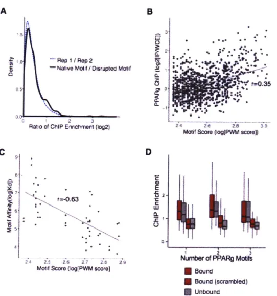
![Figure S1. Validation of PPARy ChIP. (A) Reproducibility of PPARy ChIP enrichment ([reads in ChIP]/[reads in WCE]) of candidate enhancers in two biological replicates](https://thumb-eu.123doks.com/thumbv2/123doknet/14375500.505038/109.917.121.748.142.829/figure-validation-reproducibility-enrichment-candidate-enhancers-biological-replicates.webp)


![Figure 2. Elements in flanking sequence govern enhancer activity. (A) Left, ratio of expression (log 2 [RNA/DNA]) for each genomic sequence with an intact vs](https://thumb-eu.123doks.com/thumbv2/123doknet/14375500.505038/117.917.116.769.133.662/figure-elements-flanking-sequence-enhancer-activity-expression-sequence.webp)

