HAL Id: hal-02641172
https://hal.inrae.fr/hal-02641172
Submitted on 28 May 2020HAL is a multi-disciplinary open access
archive for the deposit and dissemination of sci-entific research documents, whether they are pub-lished or not. The documents may come from teaching and research institutions in France or abroad, or from public or private research centers.
L’archive ouverte pluridisciplinaire HAL, est destinée au dépôt et à la diffusion de documents scientifiques de niveau recherche, publiés ou non, émanant des établissements d’enseignement et de recherche français ou étrangers, des laboratoires publics ou privés.
Baddreddine Boussadia, Giuseppe Gangarossa, Laila Mselli-Lakhal,
Marie-Claude Rousset, Frédéric de Bock, Frédéric Lasserre, Chaitali Ghosh,
Jean-Marc Pascussi, Damir Janigro, Nicola Marchi
To cite this version:
Baddreddine Boussadia, Giuseppe Gangarossa, Laila Mselli-Lakhal, Marie-Claude Rousset, Frédéric de Bock, et al.. Lack of CAR impacts neuronal function and cerebrovascular integrity in vivo. Exper-imental Neurology, Elsevier, 2016, 283, pp.39-48. �10.1016/j.expneurol.2016.05.018�. �hal-02641172�
Version postprint
Baddreddine Boussadia, Giuseppe Gangarossa, Laila Mselli-Lakhal, Marie-Claude Rousset, Frederick de Bock, Frederic Lassere, Chaitali Ghosh, Jean-Marc Pascussi, Damir Janigro, Nicola Marchi
PII: S0014-4886(16)30136-4
DOI: doi:10.1016/j.expneurol.2016.05.018
Reference: YEXNR 12295
To appear in: Experimental Neurology
Received date: 16 February 2016 Revised date: 13 April 2016 Accepted date: 12 May 2016
Please cite this article as: Boussadia, Baddreddine, Gangarossa, Giuseppe, Mselli-Lakhal, Laila, Rousset, Marie-Claude, de Bock, Frederick, Lassere, Frederic, Ghosh, Chaitali, Pascussi, Jean-Marc, Janigro, Damir, Marchi, Nicola, Lack of CAR impacts neuronal function and cerebrovascular integrity in vivo, Experimental Neurology (2016), doi: 10.1016/j.expneurol.2016.05.018
This is a PDF file of an unedited manuscript that has been accepted for publication. As a service to our customers we are providing this early version of the manuscript. The manuscript will undergo copyediting, typesetting, and review of the resulting proof before it is published in its final form. Please note that during the production process errors may be discovered which could affect the content, and all legal disclaimers that apply to the journal pertain.
Version postprint
ACCEPTED MANUSCRIPT
Lack of CAR impacts neuronal function and
cerebrovascular integrity in vivo
Baddreddine Boussadia1, Giuseppe Gangarossa4,Laila Mselli-Lakhal2, , Marie-Claude Rousset1, Frederick de Bock1, Frederic Lassere2, Chaitali Ghosh5, Jean-Marc Pascussi3, Damir
Janigro6 and Nicola Marchi1
1Laboratory of Cerebrovascular Mechanisms of Brain Disorders, Department of Neuroscience, Institute of functional Genomics, France. 2INRA Toxalim, Toulouse, France; 3Laboratory Signalisation, plasticite et cancer, Department of Cancer Biology, Institute of functional Genomics, France; 4Center for Interdisciplinary Research in Biology (CIRB), College de France, Paris, France; 5Cerebrovascular Research, Lerner Research Institute, Cleveland Clinic, USA; 6Flocel Inc., Cleveland, Ohio, USA
Running title: Nuclear Receptors and neurovascular dysfunction
Keywords: development, cerebrovascular, nuclear receptors, memory, stress Number of pages: 28 Number of words: 5098 Number of Fig: 6 Supplemental Tables: 2 Supplemental Fig: 2 Supplemental Movies: 2
Corresponding Authors: Dr. Nicola Marchi, nicola.marchi@igf.cnrs.fr. Cerebrovascular
Mechanisms of Brain Disorders, Institut de Génomique Fonctionnelle, CNRS UMR5203, INSERM U1191, Université Montpellier, 141 rue de la Cardonille, 34094 Montpellier, Cedex 5, France.
Acknowledgements: Supported by NIH R01 NS078307, FFRE-TF1 and FFRE Prix Valerie Chamaillard. IPAM imaging Platform (IGF) and electron Microscopy Platform (UM).
Version postprint
ACCEPTED MANUSCRIPT
Abstract
Nuclear receptors (NR) are a group of transcription factors emerging as key players in
normal and pathological CNS development. Clinically, an association between the
constitutive androstane NR (CAR) and cognitive impairment was proposed, however never
experimentally investigated. We wished to test the hypothesis that the impact of CAR on
neurophysiology and behavior is underlined by cerebrovascular-neuronal modifications. We
have used CAR-/- C57BL/6 and wild type mice and performed a battery of behavioral tests
(recognition, memory, motor coordination, learning and anxiety) as well as longitudinal
video-electroencephalographic recordings (EEG). Brain cell morphology was assessed using
2-photon or electron microscopy and fluorescent immunohistochemistry.
We observed recognition memory impairment and increased anxiety in CAR-/- mice,
while locomotor activity was not affected. Consistent with the memory deficits, EEG
monitoring revealed a decrease in 3.5-7 Hz theta waves during the awake/exploration and
sleep periods. Behavioral and EEG abnormalities in CAR-/- mice mirrored structural changes,
including tortuous fronto-parietal penetrating vessels and a few localized vascular
micro-leakages. At the cellular level we found reduced ZO-1, but not CLDN5, tight junction protein
expression in cortical and hippocampal isolated microvessel preparations. Interestingly, the
neurotoxin kainic acid, when injected peripherally, provoked a rapid onset of generalized
convulsions in CAR-/- as compared to WT mice, supporting the hypothesis of vascular
permeability. The morphological phenotype of CAR-/- mice also included some modifications
of GFAP/IBA1 glial cells in the parenchymal or adjacent to collagen-IV+ or FITC+
Version postprint
ACCEPTED MANUSCRIPT
density, hippocampal granule cell dispersion and increased NPY immunoreactivity in the
CA1 region in CAR-/- mice. The latter may contribute to the in vivo phenotype.
Our results indicate that behavioral and electroencephalographic changes in adult CAR-/- mice are concomitant to discrete developmental structural brain defects. The latter could also increase the vulnerability to neurotoxins. The possibility that interfering with nuclear receptors during development could contribute to adulthood brain changes is proposed.
Version postprint
ACCEPTED MANUSCRIPT
Introduction
Nuclear receptors (NR) are a superfamily of transcription factors. Recent evidence is
expanding the breadth of NR functions to include a role in brain cortical development (Fan et
al., 2008), memory formation (Hawk et al., 2012; Kaur and Sodhi 2015; Yang and Wang
2014), anxiety (Tan et al., 2012), vascular or barrier integrity and tissue homeostasis
(Banerjee et al., 2015; Ogura et al., 2012; Venkatesh et al., 2014; Wang et al., 2014; Zhao
and Bruemmer 2010; Zhao et al., 2010). Among the existing NR, xenobiotic receptors, such
as the Pregnane X Receptor (NR1I2) and the Constitutive Androstane Receptor (CAR,
NR1I3), are sensors of toxic byproducts (Tolson and Wang 2010). Experimental evidence
supports a functional impact of a NR1I2 receptor in memory function in mice upon receptor
activation (Kaur and Sodhi 2015). Interestingly, regulation of vascular inflammation and
mechanisms of homeostasis were shown to encompass pathways controlled by NR (Banerjee
et al., 2015; Wang et al., 2014; Zhao and Bruemmer 2010; Zhao et al., 2010). For instance,
loss of NR1I2 receptor was associated with a disrupted gastro-intestinal barrier and decreased
tight junction levels resulting in increased susceptibility to toxic insults (Venkatesh et al.,
2014).
No studies have investigated whether loss of CAR may impact neuronal functions in
vivo. The latter is clinically relevant as a missense CAR mutation was reported in patients
presenting with intellectual disability and comorbidities including spontaneous seizures
(Kleefstra et al., 2012). It is unknown whether, and consistent with CAR ablation, cellular
brain changes exist in the adulthood, including developmental vascular barrier modifications
(Venkatesh et al., 2014; Wang et al., 2014). Remarkably, experimental and clinical evidence
Version postprint
ACCEPTED MANUSCRIPT
including psychiatric illness (Khandaker et al., 2015), seizure onset and progression
(Friedman 2011; Marchi et al., 2007; Marchi et al., 2012; van Vliet et al., 2014), and
cognitive decline (Abbott et al., 2006; Obermeier et al., 2013; Snyder et al., 2015; Zhao et al.,
2015). Both developmental or acquired pathological factors can affect cerebrovascular
permeability or the inflammatory basal state, thus perturbing brain homeostasis and
neurophysiology in the adult brain (Obermeier et al., 2013).
Xenobiotic receptors are functionally expressed at the cerebrovasculature, as demonstrated by the modulation of specific downstream gene targets (Bauer et al., 2006; Wang et al., 2010). We therefore tested the hypothesis that genetic ablation of CAR is associated with modifications of basal in vivo neuronal functions in the adult brain (Dai et al., 2012; Fan et al., 2008; Hawk et al., 2012; Kaur and Sodhi 2015; Tan et al., 2012). We also evaluated whether neuro-vascular morphological changes exist in CAR-/- brains concomitant to in vivo functional modifications. The possible contribution of maldevelopmental or homeostatic mechanisms is discussed.
Version postprint
ACCEPTED MANUSCRIPT
Materials and methods
Animals
All experiments followed European Union (Council directive 86/609EEC) and
institutional guidelines for the care and use of laboratory animals. Mice were hosted at the
IGF or INRA animal facilities (institutional license approved by the French Ministry of
Agriculture N° D34-172-13 and B31-555013). Mice were housed on a 12h lightdark cycle
with food and water ad libitum. The animal experiment protocols were approved by the local
ethical committee for animal testing (05185.01 and 00846.01). CAR−/− mice and wild type on
a C57BL/6J genetic background (Wei et al., 2000) were originally established and previously
used by the authors of this manuscript (INRA, see (Roques et al., 2013)). Colony founders
were provided to LK (INRA) by Pr. Urs A Meyer (Biozentrum, University of Basel,
Switzerland). We used adult male mice (8 - 12 weeks old).
Behavioral studies
A total of n = 12 CAR-/- and n = 12 WT mice were used for behavioral testing (see Fig.
1 and 2). Spatial Object Recognition. The spatial object recognition test was performed as
previously described (Gangarossa et al., 2014). Briefly, mice were habituated to the
experimental arena for 10 min. The following day, mice were allowed to freely explore 2
different objects (A and B) located in the same site of the arena. Object interaction was
defined as approaching the object with the nose (> 1 cm). Following a retention interval of 24
h, mice underwent a 5 min recall session when the arena contained one of the two objects
Version postprint
ACCEPTED MANUSCRIPT
were cleaned with 70% ethanol. The experiments were videotaped and the time spent
exploring the objects was scored. The percentage of exploration time was calculated as %
exploration = ((Time B)/(Time A+ Time B))*100. Rotarod test. Balance and motor
coordination as well as motor learning were assessed using a mouse accelerating rotarod (Ugo
Basile, Comerio, Italy) as previously described (Gangarossa et al., 2014). Mice were placed
on the rotating drum accelerating from 4 to 40 rpm over 5 min for 3 trials a day and for 3
consecutive days. The trial interval was 45 min for all the mice. Rotarod sessions were scored
for latency to fall or ride around the rod. Spontaneous locomotor activity. Locomotor activity
was measured as previously described (Gangarossa et al., 2014; Gangarossa et al., 2014).
Horizontal and vertical activities were measured in a circular corridor (Imetronic, Pessac,
France). Counts for horizontal activity were incremented by consecutive interruption of two
adjacent beams placed at a height of 1 cm per 90° sector of the corridor (mice moving through
one-quarter of the circular corridor) and counts for vertical activity (rearing) as interruption of
beams placed at a height of 7.5 cm along the corridor (mice stretching upwards).
Elevated-Plus Maze (EPM). The elevated plus maze was elevated 1 m above the floor (black plastic
with 2 open arms 5 cm width x 35 cm length 0.5 cm height; 2 closed arms 5 width 35 cm length 15 cm height). Mice were placed in the center of maze facing one of the open arms and were allowed to explore the maze for 10 min. Experiments were videotaped and scored
for entries and time spent in the closed arms (4 paws within closed arm) or open arms (4 paws
within open arms). Open field. Spontaneous exploratory behavior was monitored in an open
field (white plastic arena with 35 cm width 50 cm length 20 cm height) for 10 min. The center zone was defined as a virtual perimeter within 5 cm from the sides of the arena.
Experiments were videotaped and an observer scored the time spent in the center (4 paws
Version postprint
ACCEPTED MANUSCRIPT
Video-encephalography and signal analysis
Baseline EEG and frequency analyses. n=4 CAR-/- C57/BL6j and n=4 WT mice were
implanted in the fronto-parietal cortex and were used to characterize baseline EEG activity.
Each mouse was monitored using Video-EEG for a total of 50 hours (50% night and 50%
day) over a period of 1 week. EEG signals were acquired at 200-600 Hz using band pass filter
(50-100Hz), stored and analyzed using Neuroscore and MatLab. Ten minutes EEG samples
were extracted from all recordings and: i) classified as day or night; ii) separated by
awake/exploratory vs. sleep (Video); iii) video analysis ruled out motion artifacts associated
with scratching, eating, drinking or chewing. As a result we obtained the following number of
EEG extracts: n = 39 WT awake/exploratory, n = 43 WT sleep, n = 34 CAR
-/-awake/exploratory and n= 31 CAR-/- sleep. The latter corresponded to the EEG time: 390
minutes WT awake/exploratory, 430 minutes WT sleep, 340 minutes CAR
-/-awake/exploratory and 310 minutes CAR-/- sleep. All EEG samples were processed using
Neuroscore (Periodogram Power Bands; epoch duration 10 sec) to calculate the relative value
(0-100%) within each 0.5Hz increments (0.5 – 30 Hertz).
Kainic acid injection. A total of n = 16 CAR-/- C57/BL6j and n = 16 C57/BL6j WT
mice were used for Video-EEG or Racine behavioral monitoring of status epilepticus (SE)
induced using i.p. kanic acid (KA, 25mg/Kg; stock solution 10mg/ml KA in PBS, pH=7). In
particular, n=7 CAR-/- C57/BL6j and n=7 C57/BL6j WT mice were implanted with
fronto-parietal cortical electrodes and EEG recordings. Mice were monitored up to 24 hours after
KA. The following endpoints were determined: i) number of mice developing SE; ii) time of
SE onset; iii) time spent in generalized SE normalized by the total duration of each EEG
recording (from KA injection to death or up to 5 hours to follow SE onset and evolution).
Version postprint
ACCEPTED MANUSCRIPT
skull exposed. A preamplifier (2 differential channels, Pinnacle Inc., USA) was connected to
EEG leads. Mice were left unrestrained for one week.
CINPA1 injections. A cohort of male adult C57/BL6j WT mice (n = 3) was injected
with CINPA1 (5605, Tocris Bioscience), a novel and potent CAR antagonist exhibiting high
selectivity for CAR over other xenobiotic NR1I (Cherian et al., 2015; Cherian et al., 2015).
CINPA1has low molecular weight (395 Da) and it is lipophilic (LogP>3), therefore
penetrating cell membranes (Cherian et al., 2015; Cherian et al., 2015). A dose response was
performed (1mg/Kg, 10mg/Kg and 50mg/kg) based on available IC50 (Cherian et al., 2015).
Video-EEG was recorded for 24 hours following each injection. Control group refers to
video-EEG (12 to 24 hours) recorded before CINPA1injections in each animal. Video-EEG
data were sorted and extracted as described above (see Supplemental Table 1). The following
number of EEG extracts were analyzed: n = 31 awake/exploration and n = 33 sleep (control);
n = 52 awake/exploration and n = 87 sleep (1 mg/Kg CINPA1); n = 63 awake/exploration and
n = 97 sleep (10 mg/Kg CINPA1).
Microvessels isolation, western blot and quantifications
Capillary Isolation. We performed 2 experiments pulling together the cortical and
hippocampal extracts obtained from: i) n=5 CAR-/- and n=5 WT; ii) n=6 CAR-/- and n=6 WT
mice (see Fig. 4). The latter was implemented to obtain a sufficient protein yield, amenable
for subsequent western blot analysis. A detailed procedure is described in (Shawahna et al.,
2011). Briefly, white matter, meninges, midbrain, choroid plexus, blood vessels, and
olfactory lobes were removed from the brains under and the remaining tissue was
homogenized. Tissue was kept in cold PBS (2.7 mM KCl, 1.5 mM KH2PO4, 136.9 mM
Version postprint
ACCEPTED MANUSCRIPT
sodium pyruvate) throughout the isolation procedure. An aliquot of 30% Ficoll was
added to an equal volume of brain homogenate and capillaries were separated from the
parenchyma by centrifuging at 5800g for 20 min. Capillaries were isolated using selective
filtrations (>20 um and < 100um). Whole brain tissues. Briefly, cortical, and hippocampal
tissues were homogenized using a buffer containing 0.1% sodium dodecyl sulfate (SDS),
protease inhibitor cocktail (Promega, Madison, WI, USA), 50 mM Tris–HCl (pH 7.4), 10
mM EDTA, 1 mM Na3VO4, 40 mM sodium pyrophosphate, 50 mM NaF, and 1 mM
dithiothreitol (DTT). Samples where then centrifuged (12,000 rpm for 10 min) and protein
content quantified as previously describe (REF). Western Blots. Samples were separated by
electrophoresis and then transferred onto a nitrocellulose membrane. After 1 h of
blocking in skimmed milk, the membranes were probed overnight at 4 °C with a
mouse anti-Zona Occludens 1 (1:400; 339100, Invitrogen), mouse anti-Claudin 5 (1:800;
352500 , Invitrogen) or mouse anti-Actin (1:10,000; ab6276, Abcam). Secondary goat
anti-mouse horseradish peroxidase (HRP)-conjugated (1:4000) antibody was used.
Relative band densities were measured and normalized using ImageJ. WB bands obtained
using isolated microvessels (Fig. 4D) are representative of i) n=5 and ii) n=6 mice/group.
Transparent SeeDB brain, Electron Microscopy and Immunohistochemistry
Transparent brain preparation was performed on CAR-/- and WT mice following the
procedures described by (Ke et al., 2013). Briefly, mice were perfused intracardially using a
25mg/ml solution of FITC-Albumin (300 ul / mouse). The fronto-parietal cortices were
isolated, treated using the SeeDB protocol (Ke et al., 2013) and analyzed using 2-photon
microscopy (see Fig. 4A and Supplemental Movies 1-2). Electron microscopy was performed
Version postprint
ACCEPTED MANUSCRIPT
Tight junctions. A total of n = 6 WT and n = 6 CAR-/- mice were used specifically for this
immunohistochemistry. After perfusion with PBS mice brains were dissected and immerged
in sucrose 30%. Brains were then snap frozen and stored at -80C. Slices (20um) were
obtained using a cryostat. Slices were post-fixed directly on the slide using Methanol/Acetone
(v/v 50/50) at -20C and immerged in the solution for 1 minute. Immunohistochemistry is
performed after PBS washes. Slices were added with blocking solution (PBS, triton 0.3%,
goat or horse serum depending on the secondary antibody used) for 1hour at 4C. Primary
antibody is diluted in blocking solution (Z01 or CLDN5, see Table 2) overnight at 4C. After
PBS washes, secondary antibody was added in PBS (1:2000, donkey anti-rabbit Cy3, Jackson
Immunoresearch 711-165-152) for 2 hour at room temperature. Slices were mounted using
moviol. Neuro-peptide Y. Immunostaining was performed of PFA 4% fixed brains. Slices (30
um) were cut using a vibratome and stored in cryoprotectant solution at -20C. Free floating
slices were rinsed using PBS and blocking solution (PBS, BSA 2%, triton 0.25% and horse or
goat serum 3%) added for 2 hour at room temperature. Primary antibody (anti-NPY see
Supplemental Table 2) is added for 36 hours at 4C. After PBS washed, secondary antibody
(1:2000, donkey anti-rabbit Cy3, Jackson Immunoresearch 711-165-152) was added (PBS +
triton 0.25%). Collagen IV. Basal lamina was stained using Collagen IV (polyclonal rabbit
anti-collagen IV antibody, ab6586, 1:100, Abcam,). Staining was performed as previously
described, using an antigen retrieval technique based on citrate buffer (pH = 6). GFAP/IBA1
inflammation. PFA fixed brain slices were incubated with blocking solution (PBS, triton
0.25% and horse serum 20%) for 1 hour at room temperature. Primary antibodies were then
added in PBS (see Supplemental Table 2) overnight at 4C. After PBS washes, secondary
antibodies were added (1:2000, donkey anti-rabbit Cy3, Jackson Immunoresearch
Version postprint
ACCEPTED MANUSCRIPT
immunohistochemistry (Fig. 5A-A1) a goat anti-mouse Alexafluor 350 was used for GFAP
(see Supplemental Table 2).
Statistics
Data were analyzed using one-way or two-way ANOVA followed by Bonferroni post
hoc test for specific comparisons. F(x,y) indicates cumulative distribution and degree of
freedom (Prism). Student’s t-test with equal variances was used for groups of 2, when relevant. In all cases, significance threshold was set at p ˂ 0.05. Statistical analyses were
performed using GraphPad Prism 5.0 (GraphPad Prism Software Inc., San Diego, USA) or
Version postprint
ACCEPTED MANUSCRIPT
Results
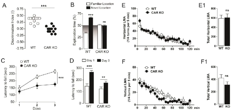
Impaired memory function and increased anxiety in CAR-/-mice
We performed a battery of behavioral tests investigating whether CAR ablation may
impact memory, learning and mood-related modifications such as anxiety. We measured the
integrity of recognition and motor/procedural memory processes. In the object place
recognition test CAR-/- mice did not show preference for the familiar or relocated object
during the recall session (24 h after exploratory phase) as opposed to WT mice (Fig. 1A and
B). This was not due to an impairment of the exploratory drive since exploration of the two
objects was not affected in CAR mice during the familiarization phase (data not shown).
Motor skill learning was also assessed using the accelerating rotarod. No differences were
observed during the first day of training, suggesting unaltered motor function and
coordination in CAR-/- mice (Fig. 1C). Interestingly, CAR-/- mice showed a decreased
improvement of rotarod motor performance compared to WT (Fig. 1C-D); this is consistent
with decreased learning ability. Thus, all mice gradually acquired motor skills following
prolonged training, even though the learning index was higher in WT as compared to CAR
-/-mice (3 days; Fig. 1C and D). No differences in the spontaneous horizontal (Fig. 1E-E1) and
vertical activity (Fig. 1F-F1) were observed in CAR-/- mice, thus indicating intact exploratory
drive. Our results suggest a link between lack of CAR and memory or learning performances,
although sparing locomotor coordination and activity.
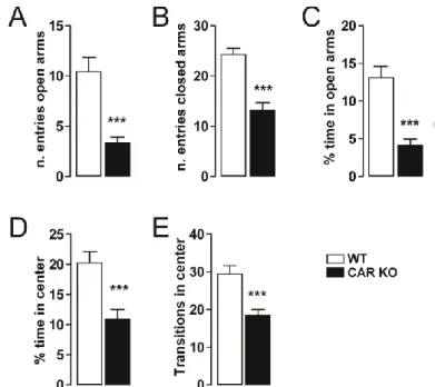
Anxiety-like behavior was also observed in CAR-/- mice; this was evaluated by the
elevated plus maze (EPM) and the open field tests (Fig. 2). In the EPM test, CAR-/- mice spent
Version postprint
ACCEPTED MANUSCRIPT
of entries in both closed and open arms. We further measured the spontaneous behavior using
the open field test (novel and stressful environment; Fig. 2D-E). CAR-/- mice spent less time
and had a reduced number of transitions in the center of the field as compared to WT,
indicating increased anxiety (Fig. 2). These results indicate a link between deletion of the
nuclear receptor CAR and anxiety-like behaviors in the adulthood.
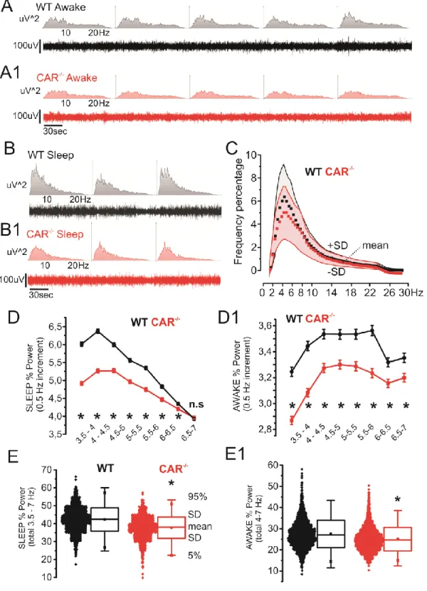
Constitutive decrease of 3.5-7Hz theta activity in CAR-/- mice in vivo
We performed a longitudinal video-EEG study to test whether the observed behavioral
changes were associated with abnormal electrographic activity (Chauviere et al., 2009).
Compared to WT, CAR-/- mice displayed a constitutive reduction in relative 3.5-7 Hz theta
power during sleep and awake/exploratory states. Fig. 3A-B shows examples (10 minutes) of
EEG recordings during awake/exploratory or sleep periods and relative frequency abundances
(uV2;0-30 Hz). Data relative to CAR-/- and WT mice (50 hours Video-EEG recordings each mouse; see Methods for details) are shown in Fig. 3C (means +/-SD). Note that the
contribution of 3.5-7 Hz theta waves was decreased in CAR-/- mice. Fig. 3D-D1 details the
changes in theta activity in CAR-/- mice observed during awake/exploratory and sleep stages.
Data were obtained using the following cumulative EEG durations: 390 minutes WT
awake/exploratory, 430 minutes WT sleep, 340 minutes CAR-/- awake/exploratory and 310
minutes CAR-/- sleep. All data are pulled together in Fig. 3E-E1. Changes in theta activity in
Version postprint
ACCEPTED MANUSCRIPT
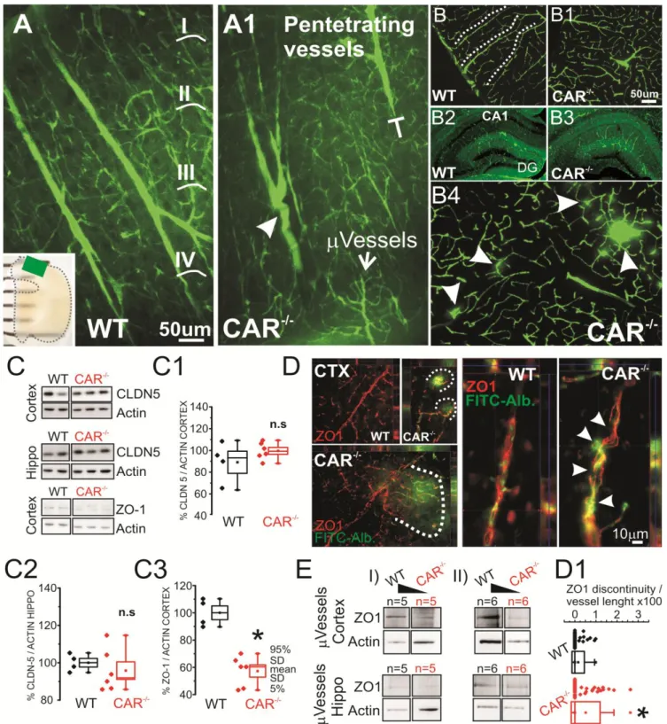
Cerebrovascular and parenchymal cells changes in CAR-/- mice
Loss of the xenobiotic receptor NR1I2 has been associated with altered barrier
function in peripheral organs (Venkatesh et al., 2014). We tested the hypothesis that, when
CAR is deleted, congenital cerebrovascular changes are concomitant to the behavioral and
EEG modifications. Cerebrovascular dysfunction underlies changes in neuronal activity in
brain diseases as demonstrated in human and experimental models (Khandaker et al., 2015;
Marchi et al., 2014). Using a transparent brain preparation and 2-photon reconstruction we
found the presence of truncated penetrating cortical vessels in CAR-/- mice (Fig. 4A-A1 and
Supplemental Movies 1 and 2). Sporadic and point form FITC-Dextran leakages (10KDa)
were observed in CAR-/- mice (Fig. 4B4 and D). At the molecular level, we quantified the
expression of the tight junctions ZO1 and CLDN5 proteins in brain homogenates (Fig. 4C) or
in microvessels isolated from hippocampi and fronto-parietal cortices (Fig. 4E). While no
significant changes in CLND5 expression were observed, ZO1 levels were reduced in CAR
-/-mice. Immunohistochemistry indicate distinct perivascular regions of discontinuous ZO1
lining the FITC+ microvessels (Fig. 4D-D1) in CAR-/- mice.
Vascular modifications were accompanied with discreet changes in neuronal
architecture. The latter included an increased fronto-parietal cortical NEUN density
(Supplemental Fig. 2A-A1 and D-D3) and dispersion of hippocampal granule cells in CAR
-/-mice (Supplemental Fig. 2B-C). In addition, staining with the neurotransmitter Neuropeptide
Y (NPY) was increased in CA1 pyramidal interneurons (Supplemental Fig. 2E-F).
Interestingly, changes in brain neuronal architecture were also reported in other NR deficient
mice (Fan et al., 2008). The latter is consistent with impaired neuronal functional (Thorsell et
Version postprint
ACCEPTED MANUSCRIPT
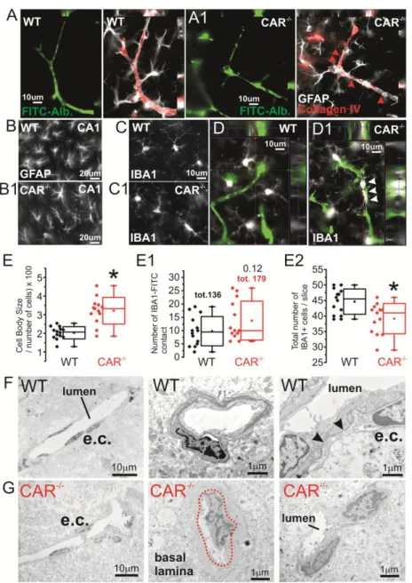
We also found signs of GFAP+ astrocytes and IBA+ microglial cells morphological abnormalities in CAR-/- mice. In CAR-/- mice hippocampal astrocytes presented with dishomogenous morphology and irregular perivascular distribution as compared to WT where astrocytes were uniformly arranged (Fig. 5A-B). GFAP+ cell rarefication was observed in CAR-/- mice. Quantification of total GFAP fluorescence indicated no significant changes between WT and CAR-/- mice (data not shown) (Wilhelmsson et al., 2006). IBA+ microglia displayed increased cell body size and a trend increase of microglial-microvascular contacts in CAR-/- mice (Fig. 5C, D and E). Interestingly, the total number of IBA+ cells was decreased in CAR-/- as compared to WT. Analysis of microvessels using electron microscopy indicated localized micro-morphological changes consistent with signs of microvascular inflammation (Fig. 5F-G) in CAR-/- mice. Our results are in accordance with a role of NR in inflammatory vascular reactivity (Ogura et al., 2012; Zhao and Bruemmer 2010; Zhao et al., 2010).
CAR-/- mice are susceptible to neurotoxins
We then tested the hypothesis that morphological changes observed in CAR-/- mice
are associated with increased susceptibly to systemically injected neurotoxins (kainic acid;
KA). CAR-/- mice developed generalized status epilepticus (SE) more rapidly as compared to
WT (Fig. 6A-B). CAR-/- mice also experienced longer SE episodes (Fig. 6C). Although only
an indirect association can be hypothesized, a more severe seizure outcome could be sustained
by the cellular changes described in Fig. 4-5 and Supplemental Fig. 1-2. Changes in vascular
permeability may favor passage of systemically circulating neuro-toxins. The latter is in
Version postprint
ACCEPTED MANUSCRIPT
2012; Tan et al., 2012; Venkatesh et al., 2014) and a role of NR in barrier homeostasis
Version postprint
ACCEPTED MANUSCRIPT
Discussion
We report an unexplored role of the nuclear receptor CAR in brain pathophysiology. The phenotype associated with loss of CAR in mice endorses future studies on the cellular mechanisms underlying memory deficits and anxiety-like behavior in the absence of NR as well as the potential role of CAR during development. Interestingly, a recent clinical study identified an uncharacterized missense mutant of CAR in subjects affected by intellectual disabilities (Kleefstra et al., 2012). However, significant differences exist between the human data and our experimental approach as in our study we used CAR knockout mice. CAR might have both genomic and non-genomic function, resulting in dissimilar functional outcomes.
Our results bear clinical significance as a number of xenobiotic and environmental toxins, including pesticides, modulate CAR activity (Banerjee et al., 2015; Wei et al., 2000). Activation or inhibition of CAR occurring during the gestation period could promote developmental changes impacting basal functions in the adult brain. We
found signs of constitutive cerebrovascular barrier dysfunctions and parenchymal changes
possibly reflecting homeostatic modifications (Wilhelmsson et al., 2006). The latter is
clinically relevant as the association between cerebrovascular permeability, the immune
system and neuronal dysfunction is gaining momentum (Friedman 2011; Khandaker et al.,
2015; Marchi et al., 2014). Thus, loss of cerebrovascular integrity impacts cognition, affection
(Falcone et al., 2015; Khandaker et al., 2015), behavior and susceptibility to seizures
(Friedman 2011; Khandaker et al., 2015; Marchi et al., 2014). Recent studies also indicated
Version postprint
ACCEPTED MANUSCRIPT
neurotransmitters in other NR deficient mice (Huang et al., 2015; Tan et al., 2012) including
PXR, the CAR cognate xenobiotic receptor (Frye et al., 2013; Zhou et al., 2009).
CAR loss and behavioral-electroencephalographic changes in the adult brain
We show agreement between behavioral results and EEG recordings, pointing to a link
between loss of CAR and memory deficits. The decreased contribution of the theta
component during sleep and awake/exploration is a neurophysiological substrate of memory
impairment (Chauviere et al., 2009). Lack of CAR was also associated with increased anxiety
scores while no changes in locomotor activity were detected. Our results indicate a localized
increased of hippocampal NPY in CAR-/- mice. Interestingly, increased NPY expression has
been associated to impaired spatial learning in mice (Thorsell et al., 2006; Thorsell et al.,
2000). The impact of CAR in brain development is further supported by initial data obtained
using a cohort of adult WT mice treated with the specific CAR antagonist CINPA1 (Cherian
et al., 2015). Our results (Supplemental Table 1) indicate that CINPA1did not recapitulate the
EEG phenotype observed in CAR-/- mice. Thus, at dosage of 1 and 10mg/Kg CINPA1did not
provoke consistent changes in the EEG frequency spectra, particularly in the 3-7Hz range (see
Supplemental Table 1). Video review showed that, at these dosages, CINPA1did not alter the
awake/sleep cycles and pattern, while 50 mg/Kg did. The fact that CINPA1 in adult WT
mice did not recapitulate the EEG phenotype observed in CAR-/- could reflect the difference between pharmacologic inhibition of CAR transcriptional activity and the genetic absence of the entire CAR protein or even developmental changes associated with the lack of this NR.
Version postprint
ACCEPTED MANUSCRIPT
The developmental role of CAR expression follows evidences obtained for other NR
(Fan et al., 2008; Hawk et al., 2012; Tan et al., 2012; Tan et al., 2010). Our behavioral
read-out shares similarities with other NR deficiencies, including ablation of X liver receptor (Tan
et al., 2012) and the NR4A (Hawk et al., 2012). It was also suggested that PXR, the closest
relative of CAR, plays a role in memory performance (Huang et al., 2015; Kaur and Sodhi
2015). Changes in neuro-steroid levels were proposed as a mechanism contributing to
behavioral changes via xenobiotic NR modulation (Frye et al., 2013). The homeostasis of
multiple neurotransmitters is altered in Farnesoid X Receptor deficient mice (Huang et al.,
2015). It remains to be investigated whether these mechanisms apply to loss of CAR.
Do NRs control developmental barrier integrity?
CAR is involved in a plethora of cellular functions extending beyond its classic role in
controlling downstream metabolic enzymes (Yang and Wang 2014). Lack of the cognate
CAR receptor PXR is associated with gastro-intestinal barrier permeability and reduced tight
junction protein levels (Venkatesh et al., 2014). This indirectly supports our results since
peripheral and CNS barrier have overlapping molecular machinery controlling paracellular
permeability. We report decreased microvascular ZO1 expression in CAR-/- mice. As reported
for peripheral barriers (Venkatesh et al., 2014), increase permeability could constitute a risk
factors for xenotoxicity. Overwhelming experimental and clinical evidences indicate that loss
of BBB-mediated CNS homeostasis is a recognized etiological factor in behavioral and
seizure disorders (Falcone et al., 2015; Khandaker et al., 2015; Marchi et al., 2012; Snyder et
al., 2015). Our results are in agreement with this evidence showing a rapid onset of status
epilepticus following intra-peritoneal injection of a prototype pro-convulsant toxin in CAR
Version postprint
ACCEPTED MANUSCRIPT
Nuclear receptors are also implicated in peripheral organ hypertrophy (Ross et al.,
2010), cell growth or differentiation, including tumors, (Chakraborty et al., 2011) and barrier
structure (Venkatesh et al., 2014). Interestingly, altered radial glia architecture was reported
in the absence of NR, which may explain an abnormal neuro-vascular development (Fan et
al., 2008; Tan et al., 2010). Negative NR modulation could promotes activation of
pro-inflammatory pathways and a defective neurovascular homeostasis (Banerjee et al., 2015;
Venkatesh et al., 2014; Wang et al., 2014; Zhao and Bruemmer 2010; Zhou et al., 2009). It
remains to be elucidated whether these effects are linked to cerebrovascular permeability,
neuronal dysfunction or both.
Final remarks
Accumulating evidence support a link between NR and inflammatory processes,
including vascular inflammation (Ogura et al., 2012; Venkatesh et al., 2014; Zhao and
Bruemmer 2010; Zhou et al., 2009). Specific evidence connects nuclear receptors to
endothelial cell proliferation (Zhao and Bruemmer 2010). Vascular inflammatory processes
also include mechanisms of monocyte recruitment to the vascular wall under the control of
NR (Zhao et al., 2010). PXR was demonstrated to be sensitive to changes in flow and
hemodynamics, impacting the expression of detoxification genes in vascular endothelial cells
(Wang et al., 2013). Mice deficient for PXR displayed over-expression of NF-kB target genes
in multiple tissues accompanied by intestinal inflammation. NF-kB modulation impact NR
activity and the expression of its target genes (Zhou et al., 2009). A link between NR and
pro-inflammatory cytokines was also reported. Finally, gene array data indicate increased genes
linked to cell proliferation, including extracellular matrix, in the absence of CAR (Li et al.,
Version postprint
ACCEPTED MANUSCRIPT
Our results introduce CAR among the xenobiotic nuclear receptors possibly involved
in neuro-vascular pathology (Fan et al., 2008; Frye et al., 2013; Hawk et al., 2012; Huang et
al., 2015; Kaur and Sodhi 2015; Litwa et al., 2015; Tan et al., 2012; Tan et al., 2010).
Behavioral and electrographic changes in CAR-/- mice may be sustained by constitutive
neuro-vascular defects. The exact cellular pathological pathways involved in the behavioral
and morphological changes observed CAR-/- mice remains to be fully investigated.
Acknowledgement
We would like to thank Emmanuel Valjent for the significant discussion and sharing
equipment used for the behavioral tests. We would like to thank Xavier DeCleves for sharing
Version postprint
ACCEPTED MANUSCRIPT
REFERENCES
Abbott NJ, Ronnback L, Hansson E Astrocyte-endothelial interactions at the blood-brain barrier. Nat Rev Neurosci 2006; 7: 41-53.
Banerjee M, Robbins D, Chen T Targeting xenobiotic receptors PXR and CAR in human diseases. Drug Discov Today 2015; 20: 618-628.
Bauer B, Yang XD, Hartz AMS, Olson ER, Zhao R, Kalvass JC, et al. In vivo activation of human pregnane X receptor tightens the blood-brain barrier to methadone through P-glycoprotein up-regulation. Molecular Pharmacology 2006; 70: 1212-1219.
Chakraborty S, Kanakasabai S, Bright JJ Constitutive androstane receptor agonist CITCO inhibits growth and expansion of brain tumour stem cells. British Journal of Cancer 2011; 104: 448-459.
Chauviere L, Rafrafi N, Thinus-Blanc C, Bartolomei F, Esclapez M, Bernard C Early deficits in spatial memory and theta rhythm in experimental temporal lobe epilepsy. J Neurosci 2009; 29: 5402-5410.
Cherian MT, Chai SC, Chen T Small-molecule modulators of the constitutive androstane receptor. Expert Opin Drug Metab Toxicol 2015; 11: 1099-1114.
Cherian MT, Lin W, Wu J, Chen T CINPA1 is an inhibitor of constitutive androstane receptor that does not activate pregnane X receptor. Mol Pharmacol 2015; 87: 878-889.
Dai YB, Tan XJ, Wu WF, Warner M, Gustafsson JA Liver X receptor beta protects dopaminergic neurons in a mouse model of Parkinson disease. Proc Natl Acad Sci U S A 2012; 109: 13112-13117.
Falcone T, Janigro D, Lovell R, Simon B, Brown CA, Herrera M, et al. S100B blood levels and childhood trauma in adolescent inpatients. J Psychiatr Res 2015; 62: 14-22.
Fan X, Kim HJ, Bouton D, Warner M, Gustafsson JA Expression of liver X receptor beta is essential for formation of superficial cortical layers and migration of later-born neurons. Proc Natl Acad Sci U S A 2008; 105: 13445-13450.
Friedman A Blood-brain barrier dysfunction, status epilepticus, seizures, and epilepsy: a puzzle of a chicken and egg? Epilepsia 2011; 52 Suppl 8: 19-20.
Frye CA, Koonce CJ, Walf AA Pregnane xenobiotic receptors and membrane progestin receptors: role in neurosteroid-mediated motivated behaviours. J Neuroendocrinol 2013; 25: 1002-1011. Gangarossa G, Ceolin L, Paucard A, Lerner-Natoli M, Perroy J, Fagni L, et al. Repeated stimulation of
dopamine D1-like receptor and hyperactivation of mTOR signaling lead to generalized seizures, altered dentate gyrus plasticity, and memory deficits. Hippocampus 2014; 24: 1466-1481.
Gangarossa G, Laffray S, Bourinet E, Valjent E T-type calcium channel Cav3.2 deficient mice show elevated anxiety, impaired memory and reduced sensitivity to psychostimulants. Front Behav Neurosci 2014; 8: 92.
Hawk JD, Bookout AL, Poplawski SG, Bridi M, Rao AJ, Sulewski ME, et al. NR4A nuclear receptors support memory enhancement by histone deacetylase inhibitors. J Clin Invest 2012; 122: 3593-3602.
Huang F, Wang T, Lan Y, Yang L, Pan W, Zhu Y, et al. Deletion of mouse FXR gene disturbs multiple neurotransmitter systems and alters neurobehavior. Front Behav Neurosci 2015; 9: 70. Kaur P, Sodhi RK Memory recuperative potential of rifampicin in aluminum chloride-induced
dementia: role of pregnane X receptors. Neuroscience 2015; 288: 24-36.
Ke MT, Fujimoto S, Imai T SeeDB: a simple and morphology-preserving optical clearing agent for neuronal circuit reconstruction. Nat Neurosci 2013; 16: 1154-1161.
Khandaker GM, Cousins L, Deakin J, Lennox BR, Yolken R, Jones PB Inflammation and immunity in schizophrenia: implications for pathophysiology and treatment. Lancet Psychiatry 2015; 2: 258-270.
Version postprint
ACCEPTED MANUSCRIPT
Kleefstra T, Kramer JM, Neveling K, Willemsen MH, Koemans TS, Vissers LE, et al. Disruption of an EHMT1-associated chromatin-modification module causes intellectual disability. Am J Hum Genet 2012; 91: 73-82.
Li D, Mackowiak B, Brayman TG, Mitchell M, Zhang L, Huang SM, et al. Genome-wide analysis of human constitutive androstane receptor (CAR) transcriptome in wild-type and CAR-knockout HepaRG cells. Biochem Pharmacol 2015; 98: 190-202.
Litwa E, Rzemieniec J, Wnuk A, Krzeptowski W, Lason W, Kajta M RXRalpha, PXR and CAR xenobiotic receptors mediate the apoptotic and neurotoxic actions of nonylphenol in mouse hippocampal cells. J Steroid Biochem Mol Biol 2015.
Marchi N, Angelov L, Masaryk T, Fazio V, Granata T, Hernandez N, et al. Seizure-promoting effect of blood-brain barrier disruption. Epilepsia 2007; 48: 732-742.
Marchi N, Granata T, Ghosh C, Janigro D Blood-brain barrier dysfunction and epilepsy: pathophysiologic role and therapeutic approaches. Epilepsia 2012; 53: 1877-1886.
Marchi N, Granata T, Janigro D Inflammatory pathways of seizure disorders. Trends Neurosci 2014; 37: 55-65.
Obermeier B, Daneman R, Ransohoff RM Development, maintenance and disruption of the blood-brain barrier. Nat Med 2013; 19: 1584-1596.
Ogura J, Terada Y, Tsujimoto T, Koizumi T, Kuwayama K, Maruyama H, et al. The decrease in farnesoid X receptor, pregnane X receptor and constitutive androstane receptor in the liver after intestinal ischemia-reperfusion. J Pharm Pharm Sci 2012; 15: 616-631.
Roques BB, Leghait J, Lacroix MZ, Lasserre F, Pineau T, Viguie C, et al. The nuclear receptors pregnane X receptor and constitutive androstane receptor contribute to the impact of fipronil on hepatic gene expression linked to thyroid hormone metabolism. Biochem Pharmacol 2013; 86: 997-1039.
Ross J, Plummer SM, Rode A, Scheer N, Bower CC, Vogel O, et al. Human constitutive androstane receptor (CAR) and pregnane X receptor (PXR) support the hypertrophic but not the hyperplastic response to the murine nongenotoxic hepatocarcinogens phenobarbital and chlordane in vivo. Toxicol Sci 2010; 116: 452-466.
Shawahna R, Uchida Y, Decleves X, Ohtsuki S, Yousif S, Dauchy S, et al. Transcriptomic and quantitative proteomic analysis of transporters and drug metabolizing enzymes in freshly isolated human brain microvessels. Mol Pharm 2011; 8: 1332-1341.
Snyder HM, Corriveau RA, Craft S, Faber JE, Greenberg SM, Knopman D, et al. Vascular contributions to cognitive impairment and dementia including Alzheimer's disease. Alzheimers Dement 2015; 11: 710-717.
Tan XJ, Dai YB, Wu WF, Warner M, Gustafsson JA Anxiety in liver X receptor beta knockout female mice with loss of glutamic acid decarboxylase in ventromedial prefrontal cortex. Proc Natl Acad Sci U S A 2012; 109: 7493-7498.
Tan XJ, Fan XT, Kim HJ, Butler R, Webb P, Warner M, et al. Liver X receptor beta and thyroid hormone receptor alpha in brain cortical layering. Proc Natl Acad Sci U S A 2010; 107: 12305-12310. Thorsell A, Karlsson RM, Heilig M NPY in alcoholism and psychiatric disorders. EXS 2006: 183-192. Thorsell A, Michalkiewicz M, Dumont Y, Quirion R, Caberlotto L, Rimondini R, et al. Behavioral
insensitivity to restraint stress, absent fear suppression of behavior and impaired spatial learning in transgenic rats with hippocampal neuropeptide Y overexpression. Proc Natl Acad Sci U S A 2000; 97: 12852-12857.
Tolson AH, Wang H Regulation of drug-metabolizing enzymes by xenobiotic receptors: PXR and CAR. Adv Drug Deliv Rev 2010; 62: 1238-1249.
van Vliet EA, Otte WM, Gorter JA, Dijkhuizen RM, Wadman WJ Longitudinal assessment of blood-brain barrier leakage during epileptogenesis in rats. A quantitative MRI study. Neurobiol Dis 2014; 63: 74-84.
Version postprint
ACCEPTED MANUSCRIPT
Venkatesh M, Mukherjee S, Wang H, Li H, Sun K, Benechet AP, et al. Symbiotic bacterial metabolites regulate gastrointestinal barrier function via the xenobiotic sensor PXR and Toll-like receptor 4. Immunity 2014; 41: 296-310.
Wang SL, Lei T, Zhang K, Zhao WX, Fang L, Lai BC, et al. Xenobiotic Pregnane X Receptor (PXR) Regulates Innate Immunity via Activation of NLRP3 Inflammasome in Vascular Endothelial Cells. Journal of Biological Chemistry 2014; 289: 30075-30081.
Wang X, Fang X, Zhou J, Chen Z, Zhao B, Xiao L, et al. Shear stress activation of nuclear receptor PXR in endothelial detoxification. Proc Natl Acad Sci U S A 2013; 110: 13174-13179.
Wang XQ, Sykes DB, Miller DS Constitutive Androstane Receptor-Mediated Up-Regulation of ATP-Driven Xenobiotic Efflux Transporters at the Blood-Brain Barrier. Molecular Pharmacology 2010; 78: 376-383.
Wei P, Zhang J, Egan-Hafley M, Liang S, Moore DD The nuclear receptor CAR mediates specific xenobiotic induction of drug metabolism. Nature 2000; 407: 920-923.
Wilhelmsson U, Bushong EA, Price DL, Smarr BL, Phung V, Terada M, et al. Redefining the concept of reactive astrocytes as cells that remain within their unique domains upon reaction to injury. Proc Natl Acad Sci U S A 2006; 103: 17513-17518.
Yang H, Wang H Signaling control of the constitutive androstane receptor (CAR). Protein Cell 2014; 5: 113-123.
Zhao Y, Bruemmer D NR4A orphan nuclear receptors: transcriptional regulators of gene expression in metabolism and vascular biology. Arterioscler Thromb Vasc Biol 2010; 30: 1535-1541. Zhao Y, Howatt DA, Gizard F, Nomiyama T, Findeisen HM, Heywood EB, et al. Deficiency of the NR4A
orphan nuclear receptor NOR1 decreases monocyte adhesion and atherosclerosis. Circ Res 2010; 107: 501-511.
Zhao Z, Nelson AR, Betsholtz C, Zlokovic BV Establishment and Dysfunction of the Blood-Brain Barrier. Cell 2015; 163: 1064-1078.
Zhou C, Verma S, Blumberg B The steroid and xenobiotic receptor (SXR), beyond xenobiotic metabolism. Nucl Recept Signal 2009; 7: e001.
Version postprint
ACCEPTED MANUSCRIPT
Figure 1. Memory and motor functions in CAR-/- mice. A) Discrimination index in CAR-/- mice (spatial object recognition test). Data (means ± SEM) were analyzed using one-way ANOVA: F(2, 35) = 20.85, *** p < 0.001. B)
Time of exploration of the novel location in CAR-/- mice. Data were analyzed using two-way ANOVA: (Object exploration x Genotype: F(2, 66) = 36.77, P < 0.0001). Specific comparisons: *** p < 0.001 (WT-new vs WT-familiar).
C) Coordination and motor learning over a training period of 3 days in CAR-/- mice (rotarod). Data were analyzed using two-way ANOVA (Time x Genotype: F(4, 66) = 1.93, P = 0.11; Time: F(2, 66) = 25.67, P < 0.0001; Genotype: F(2, 66) = 15.63, P < 0.0001). Specific comparisons: *** p < 0.001 (CAR KO-Day3 vs WT-Day3). D) Specific
comparison of motor learning in WT (n = 12) and CAR KO (n = 12) at Day3. Data were analyzed using within t-test. Specific comparisons: *** p < 0.001 (WT-Day3 vs WT-Day1) and ** p < 0.01 (CAR KO-Day3 vs CAR KO-Day1).
E-F) Spontaneous horizontal and vertical (rearing) locomotor activity in CAR-/- mice (novel non-stressful environment). Data were analyzed using two-way ANOVA: (Time x Genotype: F(46, 528) = 0.79, P = 0.83; Time: F(23, 528) = 20.01, P < 0.0001; Genotype: F(2, 528) = 33.89, P < 0.0001) (E). Data were analyzed using two-way ANOVA:
(Time x Genotype: F(46, 528) = 0.76, P = 0.88; Time: F(23, 528) = 34.64, P < 0.0001; Genotype: F(2, 528) = 18.94, P <
Version postprint
ACCEPTED MANUSCRIPT
Figure 2 – Anxiety-like behavior in CAR deficient mice. (A-B) Histograms indicate the number of entries
of CAR KO (n = 12) and WT mice (n = 12) in the open and closed arms of the EPM. (C) Histograms show the percentage of time CAR KO (n = 12) and WT mice (n = 12) spent in the open arms. (D) Histograms show the percentage of time CAR KO (n = 12) and WT mice (n = 12) spent in the center zone of the open field. (E) Histograms indicate the number of transitions CAR KO (n = 12) and WT mice (n = 12) made in the center zone of the open field. All data (means ± SEM) were analyzed using Student’s t-test: *** p < 0.001.
Version postprint
ACCEPTED MANUSCRIPT
Figure 3. Decreased theta EEG activity in CAR-/- mice. A-B) Examples of EEG and
correspondent spectrogram (WT and CAR-/-,awake/exploration and sleep; 0-30Hz). C) Relative power (percentage; n=4 mice / group) show a decrease in the 3.5-7 hertz range (red = CAR-/-, grey = WT; data points and shadows indicate mean and +/-SD respectively). D-D1) detailed frequency analysis (0.5 Hz increment) relative to sleep and awake/exploration stages. E-E1) Representation of D-D1 data accumulation (see Methods for details).
Version postprint
ACCEPTED MANUSCRIPT
Figure 4. Cerebrovascular morphology and tight junction expression in CAR-/- mice. A-A1) 2-photon images
obtained from SeeDB transparent brain preparations (ROI in the insert). Truncated penetrating cortical vessels (corresponding to layers I-IV) were found in CAR-/- as compared to WT mice (t-shape line and arrowhead). See Supplemental Movies 1 and 2. B-B1) Differential patterns of FITC+ penetrating (dotted lines) in WT vs CAR
-
mice. B2-B3) At the macroscopic level hippocampal microvessel anatomy was marginally affected. See
Supplemental Figure 2 for quantifications. B4) Example of localized microvascular permeability visualized using FITC leakages (arrows). C-C3) Decreased ZO1 expression and not CLDN-5 in total brain tissues. D) Immunohistochemistry displaying FITC-albumin leakages (dotted lines) consistent with decreased tight junction levels. An example of discontinuous ZO1 microvascular signal in provided (arrowheads). D1) Quantification of ZO1 discontinuity on FITC-Albumin microvessels. Each data point refers to one vessel. See methods for details.
Version postprint
ACCEPTED MANUSCRIPT
Figure 5. Signs of microvascular and parenchymal inflammation in CAR-/- mice. A-A1) Collagen IV immunoreactivity and astrocyte
end-feet consistently define the FITC-Albumin microvessels in WT and not in CAR-/- mice. B-B1) Signs of inflammation included hippocampal GFAP morphological changes and C-D) presence of perivascular (white arrowheads) IBA+ cells. E-E1) Quantification indicates increase in cell body size, a trend increase in the number of IBA1+ ramifications lining the microvessels but a decrease in the number of IBA1+ cells. The total number of microglial cells was diminished. F-G) Microvascular changes in CAR-/- mice also included: i) dishomogenous cellular (e.c.) distribution around the lumen, ii) basal lamina remodeling and iii) irregular cell-to-cell contact.
Version postprint
ACCEPTED MANUSCRIPT
Figure 6. CAR-/- are more susceptible to systemic KA as compared to WT. A-C) CAR-/- mice rapidly developed status epilepticus (SE) after i.p. KA. Video-EEG confirmed CAR-/- mice susceptibility to KA as indicated by increased EEG % time spent in SE (see methods). D-E) Examples (SE) of EEG and time-joint frequency analysis observed WT and CAR-/- mice.
Version postprint
ACCEPTED MANUSCRIPT
Highlights
1) Lack of the nuclear receptor CAR impacts adulthood neuronal functions in vivo
2) Lingering cerebrovascular pathology could reflect behavioral and electroencephalographic changes observed in the absence of CAR
3) The involvement of nuclear receptors in neuro-vascular development and a link to toxin exposure and inflammatory processes is proposed.
4) Nuclear receptors may represent a mechanistic entry point for chronic or neurodegenerative diseases as well as anxiety, schizophrenia and addiction.