Publisher’s version / Version de l'éditeur:
ACS Biomaterials Science & Engineering, 3, 10, pp. 2215-2222, 2017-08-28
READ THESE TERMS AND CONDITIONS CAREFULLY BEFORE USING THIS WEBSITE.
https://nrc-publications.canada.ca/eng/copyright
Vous avez des questions? Nous pouvons vous aider. Pour communiquer directement avec un auteur, consultez la
première page de la revue dans laquelle son article a été publié afin de trouver ses coordonnées. Si vous n’arrivez pas à les repérer, communiquez avec nous à [email protected].
Questions? Contact the NRC Publications Archive team at
[email protected]. If you wish to email the authors directly, please see the first page of the publication for their contact information.
NRC Publications Archive
Archives des publications du CNRC
This publication could be one of several versions: author’s original, accepted manuscript or the publisher’s version. / La version de cette publication peut être l’une des suivantes : la version prépublication de l’auteur, la version acceptée du manuscrit ou la version de l’éditeur.
For the publisher’s version, please access the DOI link below./ Pour consulter la version de l’éditeur, utilisez le lien DOI ci-dessous.
https://doi.org/10.1021/acsbiomaterials.7b00383
Access and use of this website and the material on it are subject to the Terms and Conditions set forth at
Developing hybrid polymer scaffolds using peptide modified
biopolymers for cell implantation
Abraham, Sinoj; Kuppan, Purushothaman; Raj, Shammy; Salama, Bassem;
Korbutt, Gregory S.; Montemagno, Carlo D.
https://publications-cnrc.canada.ca/fra/droits
L’accès à ce site Web et l’utilisation de son contenu sont assujettis aux conditions présentées dans le site LISEZ CES CONDITIONS ATTENTIVEMENT AVANT D’UTILISER CE SITE WEB.
NRC Publications Record / Notice d'Archives des publications de CNRC:
https://nrc-publications.canada.ca/eng/view/object/?id=97d4519c-52c4-406c-8f01-7bd5c45f4269 https://publications-cnrc.canada.ca/fra/voir/objet/?id=97d4519c-52c4-406c-8f01-7bd5c45f4269Developing Hybrid Polymer Scaffolds Using Peptide Modified
Biopolymers for Cell Implantation
Sinoj Abraham,
*
,†,‡Purushothaman Kuppan,
§Shammy Raj,
†,‡Bassem Salama,
§Gregory S. Korbutt,
*
,§and Carlo D. Montemagno
*
,†,‡†
IngenuityLab, National Institute for Nanotechnology, 11421 Saskatchewan Drive NW, Edmonton, Alberta T6G 2M9, Canada
‡
Department of Chemical and Materials Engineering, University of Alberta, Edmonton, Alberta T6G 2R3, Canada
§
Alberta Diabetes Institute, Department of Surgery, University of Alberta, Edmonton, Alberta T6G 2E1, Canada
*
S Supporting InformationABSTRACT: Polymeric scaffolds containing biomimics offer exciting therapies with broad potential impact for cellular therapies and thereby potentially improve success rates. Here we report the designing and fabrication of a hybrid scaffold that can prevent a foreign body reaction and maintain cell viability. A biodegradable acrylic based cross-linkable polycaprolactone based polymer was developed and using a multihead electrospinning station to fabricate hybrid scaffolds. This consists of cell growth factor mimics and factors to prevent a foreign body reaction. Transplantation studies were performed
subcutaneously and in epididymal fat pad of immuno-competent Balb/c mice and immuno-suppressed B6 Rag1 mice and we demonstrated extensive neo-vascularization and maintenance of islet cell viability in subcutaneously implanted neonatal porcine islet cells for up to 20 weeks of post-transplant. This novel approach for cell transplantation can improve the revascularization and allow the integration of bioactive molecules such as cell adhesion molecules, growth factors, etc.
KEYWORDS: biopolymer, transplantation, biomimics, subcutaneous, vascularization
R
ecently, cellular transplantation has gained wide recog-nition as a method of treatment for various diseases, particularly diabetes. A potentially effective alternative to daily insulin injections in type 1 diabetes is to transplant insulin-producing tissue to re-establish the natural physiological system for glucose homeostasis. Successful transplantation of human islet cells into the liver by the “Edmonton Protocol” has opened a new treatment strategy for curing diabetes mellitus1,2 and other similar diseases. However, the requirement of substantial amount of cells, repeated transplants due to attrition, lack of angiogenesis and the occurrence of immune rejection, are some of the obstacles of this approach, indicating that the strategies need to be further optimized.3,4 For example, in clinical intraportal islet transplantation, it is not impossible to image the graft post-transplant or surgically retrieve the graft if required.5 Transplant site is also important in this mode of treatment, for example, the transplantation of islets into the portal vein has been associated with life-threatening intra-peritoneal bleeding,6 portal vein thrombosis and hepaticsteatosis.7,8 Research is currently being conducted to identify suitable biomaterials for islet cell delivery, more optimal transplant sites, and methods to improve post-transplant revascularization and prevent islet inflammation and immune rejection.9−11
Currently, studies are focusing on using the subcutaneous space as an islet transplant site, but because of poor
vascularization and insufficient oxygen supply, this site has proven to be undesirable.12−14Studies are also being conducted
to develop a vascularized subcutaneous site and promote angiogenesis by incorporating a encapsulation device and/or growth factors with moderate short-term success.15 Subcuta-neous transplants of encapsulated biomaterials also trigger inflammatory and foreign body responses that cause protein adsorption and therefore, cannot be considered a viable approach for this treatment.16
Strategies relying on nonencapsulating, biodegradable micro-porous scaffolds are often more successful in subcutaneous transplantation by promoting vascularization, but are still limited.17 This treatment model was successfully employed with murine islets in syngeneic models18,19 and in tolerance-inducing allotransplant models.20Biocompatible polymers, like poly(vinyl alcohol) (PVA), polyglycolic acid (PGA),15 poly-(lactides-co-glycolide) (PLG),18and polycaprolactone (PCL),21 were utilized to fabricate scaffolds. Architectural modifications in their structure helped to enhance cell infiltration for a better vascularization of the islets and to reduce possible inflammatory responses.
Received: June 27, 2017 Accepted: August 28, 2017 Published: August 28, 2017
Even though cell replacement therapy has been identified as the future treatment strategy for various diseases, to date, a robust and translatable platform that does not rely on life-long systemic immunosuppression to protect transplanted cells has yet to be identified. Multidisciplinary research in biology, chemistry, chemical engineering, and materials science are essential to develop next generation biomaterials that are suitable for case-to-case tissue engineering and regenerative medicine. In this report, a hybrid polymeric material is synthesized for developing a cell delivery scaffold for the long-term function and survival of insulin producing β-cell grafts. These porous, retrievable scaffolds are prepared by polymerizing functionalized caprolactone monomers into functional polycaprolactone (PCL) conjugated with biomimics for promoting quick vascularization and to prevent a foreign body rejection. PCL and other lactones have been used in FDA-approved medical devices and have demonstrated long-term compatibility in numerous animal models.22−25 In
addition, the biodegradability of PCL can be selected and tuned for the specific cell type to be transplanted.26,27Binding growth factor mimics (GF) and grafting zwitterionic polymers improve cell proliferation, promote vascularization and reject blood protein adsorption, all of which prevents immunor-ejection and enhances the graft survivability. Cells are sensitive to topography of the scaffolds. Electrospinning can produce nonwoven meshes containing fibers of polymeric materials with micro level porous topography, similar to the natural extracellular matrix (ECM).
In this study, we adopted the electrospinning method equipped with multiple spin heads to deliver biomimetic geometries with multiple scale webs of spun polymer fibers and growth factor mimics to encourage cells to adhere, proliferate, and secrete matrices in short time (seeScheme 1).
Caprolactone monomers bearing pendant-protected carboxyl group were synthesized and further modified chemically to couple with hydroxyethyl methacrylate (HEMA), rendering
vinyl functionality for further conjugative reactions.28,29 The
scheme of the synthesis of the caprolactone monomer from para-dioxaspirodecanone is shown inFigure 1a. The detailed experimental procedures and characterizations are included in theSupporting Information. The polycaprolactone used in this study was synthesized by copolymerizing this functionalized monomer with normal caprolactone at a ratio of 2:8 by ring opening polymerization (ROP). Stannous octoate (Sn(Oct)2)
was utilized as the catalyst for ROP and the copolymers were obtained with a molecular weight of 18 000. Molecular weight obtained is comparatively lower than the commonly using commercially available PCL for similar scaffold applications, the consecutive steps of inter- and intra- photo-cross-linking establish additional functionality and reinforcement to these fabricated hybrid scaffolds. The schematic structure of the functional polycaprolactone is depicted in Figure 1a. The polymerization procedures and characterizations are included as
Supporting Information.
Zwitterionic sulfobetaine methacrylate (SBMA) polymers exhibit excellent antifouling properties because of their ionic nature with surface neutral charge. The surface behavior imparted by polySBMA helps to repel blood proteins and platelets.30Proteins like fibrinogen and other clotting enzymes commonly get adsorbed on the implants as soon as they come in contact with blood and induce thrombus formation.31In a similar fashion, platelets also adhere to these surface adsorbed proteins via platelet membrane integrins, followed by platelet activation causing thrombin formation and platelet aggrega-tion.32,33 Oligomers with 6−8 SBMA monomers were synthesized by atom transfer radical polymerization (ATRP) using a hydroxyl initiator. After polymerization, the hydroxyl end group was converted to vinyl group for enabling photo-cross-link reaction with PCL. The synthesis scheme is shown in
Figure 1b\ and ATRP procedures and characterizations are included in theSupporting Information.
Scheme 1. Schematic Representation of the Scaffold Made Using Vinyl Functionalized Lactone Monomers, with Covalently Bounded Bioactive Molecules Such As Growth Factor Mimics to Accelerate Cell Proliferation and Zwitterionic Polymers to Reduce Biofouling
ACS Biomaterials Science & Engineering Letter
DOI:10.1021/acsbiomaterials.7b00383
ACS Biomater. Sci. Eng.2017, 3, 2215−2222
SBMA with acrylic functionality cross-link with lactone surfaces by photoirradiation thereby provide an optimal zwitterionic surface. This will enhance the antifouling capability of these scaffolds by inhibiting the adsorption of blood proteins on it. By this functionalization, the foreign body response is minimized essentially by rejecting the blood proteins from adhering on the scaffold surface by the repulsive effect facilitating by these zwittwerionic molecules.
To enhance the biochemical signaling in tissue engineering and regenerative transplants, we needed surface modification of biomaterials with biomimicsand demonstrated recently.34,35By incorporating functional peptide sequences corresponding to biological functions, cells attach efficiently to the surface of
biomaterials and proliferate within a short span of time. In this study, we have prepared vascular endothelial growth factor (VEGF)-mimicking KLTWQELYQLKYKG (QK) peptides to achieve surface modification. A QK peptide sequence derived from VEGF was previously shown to have similar effects as the VEGF protein and support the cell and its proliferation.36
For facilitating photo cross-linking, we prepared the QK sequence with octenyl alanine [(octenyl)Ala] at the N-terminal of the peptide for facilitating the vinyl moiety as shown in
Figure 1c. These vinyl functions of the peptides can enable the covalent bond formation with the PCL scaffold along with SBMA by photoirradiation. These peptides were synthesized using a microwave assisted peptide synthesizer and purified up
Figure 1.(a) Synthesis scheme for producing caprolactone monomer with vinyl groups suitable to copolymer, with normal caprolactone monomer to produce functional polycaprolactone. (b) Atom transfer radical polymerization of SBMA and its end group modification. (c) VEGF mimic sequence and its modification for surface photo-cross-linking.
to 96% purity. Preparation and characterization data are in the
Supporting Information.
Hybrid 3D scaffolds using these synthesized materials ([2], [3], and [4]) have been developed using an electrospinning technique. Recently, this method has been identified as an efficient technique for fabricating polymeric nanofibers for tissue engineering scaffolds.37 Parameters such as nanofiber diameter, physical properties of the matrices, pore size, and compatibility can be efficiently tuned by controlling the spinning parameters of the electrospinning process. Mostly, these electrospun polymeric matrices closely resemble the collagen phase of naturally forming ECM. The high surface area, high porosity, interconnected porous structures, tunable mechanical properties, and customizable rate of degradation make these matrices ideal candidates for developing scaffolds for tissue engineering.
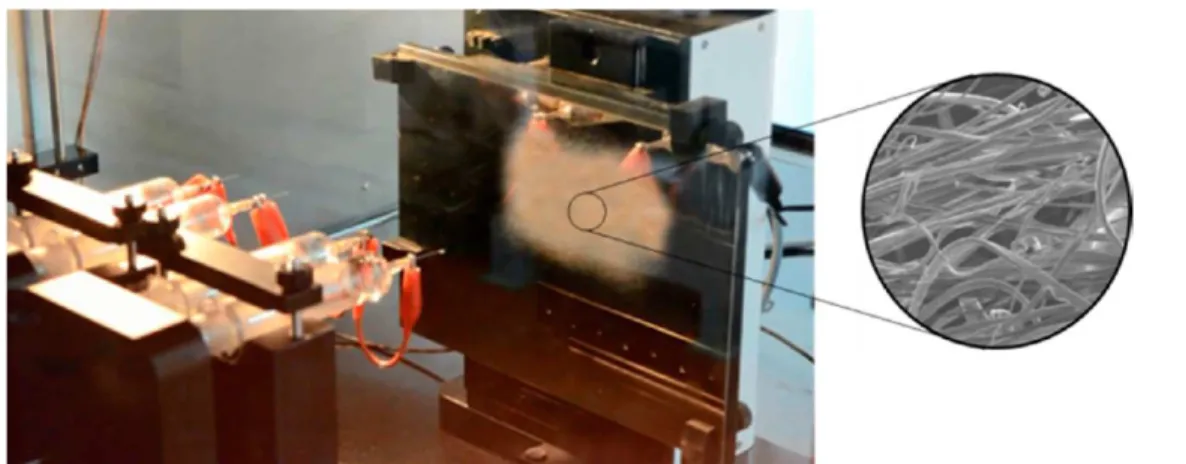
In this work, we developed a scaffold using a hybrid electrospinning machine equipped with multiple syringe pumps on an x-y stage to spin fibers of various materials simultaneously. Functional PCL, poly-SBMA and vinyl-QK were dissolved individually in chloroform, deionized water and ethanol, respectively at 20% concentration and then spun onto a single matrix. The spinning was carried out at a flow rate of 0.05 mL/min at 10 kV at a working distance of 15 cm between syringe and collector. The final compositions of these components are 90:8:2 respectively with a thickness of ∼130 μm. Experiments were conducted with and without a photo initiator (Irgacure-2959) by dissolving it with the poly SBMA
solution before electrospinning. Presence of a photo initiator enhances the curing rate and reduced the irradiation time.
Figure 2illustrates an image of the electrospinning machine that produces this hybrid matrix on the collector board. A real time video of the fabrication process is included asVideo S1.
This hybrid nano fiber matrix was photo-cross-linked by exposure to 365 nm UV light at a distance of 100 mm with approximately 1 mW/cm2power for about 15 min. The scaffold
was then rinsed with water and frozen at −80 °C for lyophilizing. The photo-cross-linking of the matrix was also evaluated without photo initiator. Exposure to UV light for up to 30 min produced scaffolds with the required degree of cross-linking. At this stage, the peptides or SBMA will not washed out while rinsing with water under sonication. Therefore, any adverse foreign body responses due to the presence of irgacure can also be avoided during this fabrication step.Figure 3shows the SEM micrograph of the electrospun hybrid nanofibrous structures.
By combining these multiple synthesis strategies and the UV light-driven cross-linking method, a novel poly lactone based scaffold was developed. The light induced cross-linking approach permits the incorporation of growth factors for the cell proliferation without any catalyst contamination or thermal degradation, as opposed to catalyst or thermal cross-linking. Moreover, due to covalent bond formation between the components, they will be permanently bonded to each other, rather than a normal physical blending, which cause the blends to get separated. The presence of zwitterionic charges imparted from the pSBMA improves the overall biocompatibility. The
Figure 2.Electrospinning process to obtain nanofiber matrices as scaffolds.
Figure 3.SEM micrograph of an electrospun PCL-PSBMA-QK nanofibers.
ACS Biomaterials Science & Engineering Letter
DOI:10.1021/acsbiomaterials.7b00383
ACS Biomater. Sci. Eng.2017, 3, 2215−2222
major advantage of this combined electrospinning and UV cross-linking fabrication process is that it enables customization of the scaffolds with respect to pore size, thickness, and
mechanical strengths. Compressive mechanical analysis was performed to determine the viscoelastic properties of this hybrid material. The instantantanous compressive modulus
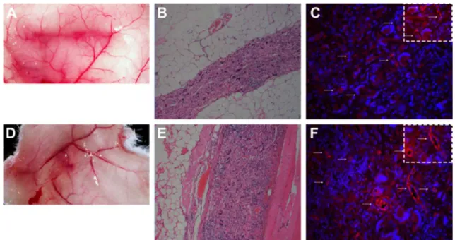
Figure 4.(A, D) Vascularization, (B, E) HE staining, and (C, F) CD-31 staining of neo-vascularization of nonfunctionalized (no VEGF) electrospun scaffolds recovered from the (A−C) fat pad and (D−F) subcutaneous sites following 5 weeks implantation in Balb/c mice. Arrows indicate CD31 positive cell forming the lumen of newly formed blood vessels.
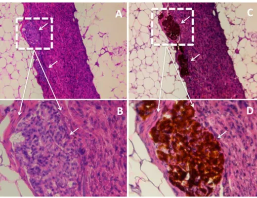
Figure 5. Comparative representation of vascularization post-transplant at subcutaneous space of B6 Rag1 mice with nonfunctionalized and functionalized scaffolds. Arrows indicate CD-31+ cells forming the lumen of newly formed blood vessels of (A−C) PCL and (D−F) PCL-VEGF nanofibers after 1, 2, and 4 weeks of implantation subcutaneously in B6 Rag1 mice. (G) Only those CD 31 positive cells located along the lumen of newly formed blood vessels were quantified by ImageJ after 1, 2, and 4 weeks. (Nucleus, blue; CD31, red; white arrow, vessels; objective, 20×; scale bar, 50 μm; *P < 0.001 Students t test.)
(slope of the stress strain curve) increased with increasing strain. The range of modulus is from 45 MPa at 1% strain to 116 MPa at 12% strain. The equilibrium modulus also increased linearly with increasing strain ranging from 28 to 23.5 MPa. Dynamic mechanical analysis revealed a phase lag of 3.6 constituting 6% energy loss from viscoelasticity.
These scaffolds were subjected to transplantation studies to evaluate the degree of neo-vascularization.
In order to determine the cell-compatibility of the polymer alone, scaffolds without being cross-linked with any growth factor mimics were prepared. These control samples were subjected to transplantation studies as explained in Figure 4. These scaffolds were implanted subcutaneously and in the epididymal fat pad of immunocompetent Balb/c mice. The scaffolds without any VEGF-mimics were initially transplanted in order to determine the post-transplant vascularization. After 5 weeks of postimplant, the gross morphology and histological assessment of the grafts was determined (Figure 4).
Retrieval of these transplanted scaffolds from both the subcutaneous zone and fat pad at 5 weeks post-transplant revealed extensive neo-vascularization on the scaffold surface (Figure 4A, D). When examined histologically (Figure 4B&E) and for the presence of CD31+ cells by immunofluorescence (Figure 4C, F) the scaffolds were shown to be infiltrated with some small clusters of CD31+ cells; however, many of the CD +31 cells were shown forming the lumen of newly formed blood vessels. These data demonstrate that these PCL scaffolds, when implanted subcutaneously or in the fat pad, can integrate with host tissue by becoming highly vascularized.
Implantation studies were also conducted to evaluate the impact of the presence of VEGF (QK) mimicking peptide cross-linked with the scaffolds. The hybrid scaffolds were implanted subcutaneously in immune-compromised B6 Rag1 mice. Grafts were retrieved after 1, 2, and 4 weeks post-transplant and immune-stained for the presence of CD31+
cells. At 1 week post-transplant, the scaffolds began to be infiltrated with small clusters of CD31+ cells, likely due to the recruitment of CD31+ endothelial cells. At week 2, CD31+ cells began to form blood vessel lumens, and by week 4, significantly more vascularization was observed in VEGF-bound PCL scaffold than control PCL scaffold (p < 0.001;Figure 5G). This clearly demonstrates that our novel concept of binding VEGF to our scaffolds enhances vascularization.
Initial studies were also performed to examine the ability of these hybrid scaffolds to serve as a platform for the transplantation of neonatal porcine islets (NPI) into B6 Rag1 mice. Neonatal porcine islets were isolated by a simple, inexpensive, and reproducible method that was established in our laboratory.38 These islets are comprised of differentiated endocrine and endocrine precursor cells and have the potential for proliferation and differentiation in both in vitro and in vivo studies. They have been shown to reverse hyperglycemia in immuno-deficient mice,38 allogeneic out-bred pigs,39 and in nonhuman primates.40,41 Furthermore, NPIs are appealing because of their resistance to (a) hypoxia;42 (b) human pro-inflammatory cytokines;43 (c) hyperglycemia;44 and (d) islet amyloid deposition.45They also exhibit an inherent ability to
differentiate and proliferate38and achieve transplant tolerance induction in diabetic mice.46Considering all these observations, it is taken into consideration that NPIs are promising tissue source for clinical islet transplantation. Neonatal porcine islets from two independent preparations were first placed on disc-shaped electrospun scaffolds and then surgically implanted subcutaneously in B6 Rag-1 mice.
The results demonstrated in Figure 6 reveals that these hybrid scaffolds are able to support the long-term survival of neonatal porcine islets implanted subcutaneously. Engineering this highly vascularized microenvironment using our novel functionalized scaffolds can support engraftment and long-term survival of islets in this extra-hepatic sites.
Figure 6.Morphological assessment of Neonatal porcine islet (NPI) containing PCL-VEGF nanofibrous scaffold transplanted subcutaneously in B6 Rag 1 mice at 20 weeks post-transplant. Graft stained for (A) H&E and (B) insulin (brown color). White arrow indicates the islets.
ACS Biomaterials Science & Engineering Letter
DOI:10.1021/acsbiomaterials.7b00383
ACS Biomater. Sci. Eng.2017, 3, 2215−2222
In conclusion, we demonstrate a successful fabrication of a new and innovative hybrid scaffold material for islet trans-plantation using a multi head electrospinning technique and polymer modification for photoinduced cross-linking. This is a simple and convenient alternative for enhancing the vascula-rization of biomaterials for cellular therapies. We were able to show the enhanced vascularization of hybrid scaffolds when compared to controls, which did not have growth factor mimics. The novel approach of incorporating vinyl function-alized lactones will allow subsequent covalent integration of bioactive molecules such as cell adhesion factors and growth factors. This is a new approach for subcutaneous cell transplantation with improved revascularization, thereby, addressing one of the major limitations in the currently applied procedures for cell transplantation. More studies are needed to further optimize and scale-up these functionalized bioactive scaffolds to support the maturation, survival, and function of NPIs for their effectiveness for correcting diabetes in various implantation sites as well as preclinical large animal models like juvenile pigs.
■
ASSOCIATED CONTENT*
S Supporting InformationThe Supporting Information is available free of charge on the
ACS Publications website at DOI: 10.1021/acsbiomater-ials.7b00383.
Experimental details and characterizations results (PDF) Video S1 showing fabrication using electrospinner (AVI)
■
AUTHOR INFORMATION Corresponding Authors *E-mail: [email protected]. *E-mail: [email protected]. *E-mail: [email protected]. ORCID Sinoj Abraham:0000-0001-7501-6144 NotesThe authors declare no competing financial interest.
■
ACKNOWLEDGMENTSThe authors acknowledge the National Institute for Nano-technology for the use of equipment and instrumentation. Dr. Montemagno acknowledges the Province of Alberta, Alberta Innovates Technology Futures, and National Institute for Nanotechnology for their financial support. Multihead electro spinner is developed for Ingenuity Lab by Holmarc Optometrics Pvt. Ltd, India and programming by Thanmatra Chemtech Pvt. Ltd, India. Dr. Korbutt acknowledges the financial support of the Canadian Institutes of Health Research (CIHR Grant #MOP 119500) and by the Juvenile Diabetes Research Foundation 3-SRA-2016-252−S-B.
■
REFERENCES(1) Shapiro, A. J.; Lakey, J. R.; Ryan, E. A.; Korbutt, G. S.; Toth, E.; Warnock, G. L.; Kneteman, N. M.; Rajotte, R. V. Islet transplantation in seven patients with type 1 diabetes mellitus using a glucocorticoid-free immunosuppressive regimen. N. Engl. J. Med. 2000, 343 (4), 230− 238.
(2) Ryan, E. A.; Lakey, J. R.; Rajotte, R. V.; Korbutt, G. S.; Kin, T.; Imes, S.; Rabinovitch, A.; Elliott, J. F.; Bigam, D.; Kneteman, N. M.; et al. Clinical outcomes and insulin secretion after islet transplantation with the Edmonton protocol. Diabetes 2001, 50 (4), 710−719.
(3) Matarazzo, M.; Giardina, M. G.; Guardasole, V.; Davalli, A. M.; Horton, E. S.; Weir, G. C.; Sacca, L.; Napoli, R. Islet transplantation under the kidney capsule corrects the defects in glycogen metabolism in both liver and muscle of streptozocin-diabetic rats. Cell Transplant. 2002, 11 (2), 103−112.
(4) Molano, R. D.; Pileggi, A.; Berney, T.; Poggioli, R.; Zahr, E.; Oliver, R.; Malek, T. R.; Ricordi, C.; Inverardi, L. Long-term islet allograft survival in nonobese diabetic mice treated with tacrolimus, rapamycin, and anti-interleukin-2 antibody1. Transplantation 2003, 75 (11), 1812−1819.
(5) Plesner, A.; Verchere, C. B.Advances and challenges in islet transplantation: islet procurement rates and lessons learned from suboptimal islet transplantation. J. Transplant. 2011, 2011, 1.
(6) Villiger, P.; Ryan, E.; Owen, R.; O’kelly, K.; Oberholzer, J.; Saif, F. A.; Kin, T.; Wang, H.; Larsen, I.; Blitz, S.; et al. Prevention of bleeding after islet transplantation: lessons learned from a multivariate analysis of 132 cases at a single institution. Am. J. Transplant. 2005, 5 (12), 2992−2998.
(7) Bhargava, R.; Senior, P. A.; Ackerman, T. E.; Ryan, E. A.; Paty, B. W.; Lakey, J. R.; Shapiro, A. J. Prevalence of hepatic steatosis after islet transplantation and its relation to graft function. Diabetes 2004, 53 (5), 1311−1317.
(8) Markmann, J. F.; Rosen, M.; Siegelman, E. S.; Soulen, M. C.; Deng, S.; Barker, C. F.; Naji, A. Magnetic resonance-defined periportal steatosis following intraportal islet transplantation. Diabetes 2003, 52 (7), 1591−1594.
(9) Pepper, A. R.; Gala-Lopez, B.; Ziff, O.; Shapiro, A. Revascularization of transplanted pancreatic islets and role of the transplantation site. Clin. Dev. Immunol. 2013, 2013, 1.
(10) Merani, S.; Toso, C.; Emamaullee, J.; Shapiro, A. Optimal implantation site for pancreatic islet transplantation. Br. J. Surg. 2008, 95 (12), 1449−1461.
(11) Vériter, S.; Gianello, P.; Dufrane, D. Bioengineered sites for islet cell transplantation. Curr. Diabetes Rep. 2013, 13 (5), 745−755.
(12) Saito, T.; Ohashi, K.; Utoh, R.; Shimizu, H.; Ise, K.; Suzuki, H.; Yamato, M.; Okano, T.; Gotoh, M. Reversal of diabetes by the creation of neo-islet tissues into a subcutaneous site using islet cell sheets. Transplantation 2011, 92 (11), 1231−1236.
(13) Simeonovic, C. J.; Dhall, D. P.; Wilson, J. D.; Lafferty, K. J. a Comparative Study of Transplant Sites for Endocrine Tissue Transplantation in the Pig. Immunol. Cell Biol. 1986, 64 (1), 37.
(14) Sakata, N.; Aoki, T.; Yoshimatsu, G.; Tsuchiya, H.; Hata, T.; Katayose, Y.; Egawa, S.; Unno, M. Strategy for clinical setting in intramuscular and subcutaneous islet transplantation. Diabetes/Metab. Res. Rev. 2014, 30 (1), 1−10.
(15) Juang, J.-H.; Bonner-Weir, S.; Ogawa, Y.; Vacanti, J. P.; Weir, G. C. Outcome Of Subcutaneous Islet Transplantation Improved By Polymer Device1. Transplantation 1996, 61 (11), 1557−1561.
(16) Ward, W. K. A Review of the Foreign-Body Response to Subcutaneously-Implanted Devices: The Role of Macrophages and Cytokines in Biofouling and Fibrosis. J. Diabetes Sci. Technol. 2008, 2 (5), 768−777.
(17) Gibly, R. F.; Zhang, X.; Graham, M. L.; Hering, B. J.; Kaufman, D. B.; Lowe, W. L.; Shea, L. D. Extrahepatic islet transplantation with microporous polymer scaffolds in syngeneic mouse and allogeneic porcine models. Biomaterials 2011, 32 (36), 9677−9684.
(18) Blomeier, H.; Zhang, X.; Rives, C.; Brissova, M.; Hughes, E.; Baker, M.; Powers, A. C.; Kaufman, D. B.; Shea, L. D.; Lowe, W. L., Jr Polymer scaffolds as synthetic microenvironments for extrahepatic islet transplantation. Transplantation 2006, 82 (4), 452.
(19) Salvay, D. M.; Rives, C. B.; Zhang, X.; Chen, F.; Kaufman, D. B.; Lowe, W. L., Jr; Shea, L. D. Extracellular matrix protein-coated scaffolds promote the reversal of diabetes after extrahepatic islet transplantation. Transplantation 2008, 85 (10), 1456.
(20) Kheradmand, T.; Wang, S.; Gibly, R. F.; Zhang, X.; Holland, S.; Tasch, J.; Graham, J. G.; Kaufman, D. B.; Miller, S. D.; Shea, L. D.; Luo, X. Permanent protection of PLG scaffold transplanted allogeneic islet grafts in diabetic mice treated with ECDI-fixed donor splenocyte infusions. Biomaterials 2011, 32 (20), 4517−4524.
(21) Nyitray, C. E.; Chang, R.; Faleo, G.; Lance, K. D.; Bernards, D. A.; Tang, Q.; Desai, T. A. Polycaprolactone thin-film micro-and nanoporous cell-encapsulation devices. ACS Nano 2015, 9 (6), 5675− 5682.
(22) Rentsch, B.; Bernhardt, R.; Scharnweber, D.; Schneiders, W.; Rammelt, S.; Rentsch, C. Embroidered and surface coated polycaprolactone-co-lactide scaffolds: a potential graft for bone tissue engineering. Biomatter 2012, 2 (3), 158−165.
(23) Bernards, D. A.; Desai, T. A. Nanoscale porosity in polymer films: fabrication and therapeutic applications. Soft Matter 2010, 6 (8), 1621−1631.
(24) Bernards, D. A.; Desai, T. A. Nanotemplating of Biodegradable Polymer Membranes for Constant-Rate Drug Delivery. Adv. Mater. 2010, 22 (21), 2358−2362.
(25) Angius, D.; Wang, H.; Spinner, R. J.; Gutierrez-Cotto, Y.; Yaszemski, M. J.; Windebank, A. J. A systematic review of animal models used to study nerve regeneration in tissue-engineered scaffolds. Biomaterials 2012, 33 (32), 8034−8039.
(26) Bernards, D. A.; Lance, K. D.; Ciaccio, N. A.; Desai, T. A. Nanostructured thin film polymer devices for constant-rate protein delivery. Nano Lett. 2012, 12 (10), 5355−5361.
(27) Mendelsohn, A. D.; Bernards, D. A.; Lowe, R. D.; Desai, T. A. Patterning of Mono-and Multilayered Pancreatic β-Cell Clusters. Langmuir 2010, 26 (12), 9943−9949.
(28) Montemagno, C. D.; Kumaran, S. K.; Abraham, S. Stimuli-switchable moieties, monomers and polymers incorporating stimuli-switchable moieties, and methods of making and using same. Patent WO2016029307 A1, 2016.
(29) Turos, E.; Leslie, M., Antibiotic-bound poly (caprolactone) polymer. US Patent US20130150550 A1, 2008.
(30) Damodaran, V. B.; Murthy, N. S. Bio-inspired strategies for designing antifouling biomaterials. Biomaterials research 2016, 20 (1), 18.
(31) Tsai, W.-B.; Horbett, T. A. The role of fibronectin in platelet adhesion to plasma preadsorbed polystyrene. J. Biomater. Sci., Polym. Ed. 1999, 10 (2), 163−181.
(32) Nesbitt, W. S.; Giuliano, S.; Kulkarni, S.; Dopheide, S. M.; Harper, I. S.; Jackson, S. P. Intercellular calcium communication regulates platelet aggregation and thrombus growth. J. Cell Biol. 2003, 160 (7), 1151−1161.
(33) Li, Q.; Wang, Z.; Zhang, S.; Zheng, W.; Zhao, Q.; Zhang, J.; Wang, L.; Wang, S.; Kong, D. Functionalization of the surface of electrospun poly (epsilon-caprolactone) mats using zwitterionic poly (carboxybetaine methacrylate) and cell-specific peptide for endothelial progenitor cells capture. Mater. Sci. Eng., C 2013, 33 (3), 1646−1653. (34) D’Andrea, L. D.; Iaccarino, G.; Fattorusso, R.; Sorriento, D.; Carannante, C.; Capasso, D.; Trimarco, B.; Pedone, C. Targeting angiogenesis: structural characterization and biological properties of a de novo engineered VEGF mimicking peptide. Proc. Natl. Acad. Sci. U. S. A. 2005, 102 (40), 14215−14220.
(35) Santulli, G.; Ciccarelli, M.; Palumbo, G.; Campanile, A.; Galasso, G.; Ziaco, B.; Altobelli, G. G.; Cimini, V.; Piscione, F.; D’Andrea, L. D.; et al. In vivo properties of the proangiogenic peptide QK. J. Transl. Med. 2009, 7 (1), 41.
(36) Lin, C.-Y.; Wang, Y.-R.; Lin, C.-W.; Wang, S.-W.; Chien, H.-W.; Cheng, N.-C.; Tsai, W.-B.; Yu, J. Peptide-modified zwitterionic porous hydrogels for endothelial cell and vascular engineering. BioRes. Open Access 2014, 3 (6), 297−310.
(37) Schiffman, J. D.; Schauer, C. L. A review: electrospinning of biopolymer nanofibers and their applications. Polym. Rev. 2008, 48 (2), 317−352.
(38) Korbutt, G. S.; Elliott, J. F.; Ao, Z.; Smith, D. K.; Warnock, G. L.; Rajotte, R. V. Large scale isolation, growth, and function of porcine neonatal islet cells. J. Clin. Invest. 1996, 97 (9), 2119.
(39) Kin, T.; Korbutt, G. S.; Kobayashi, T.; Dufour, J. M.; Rajotte, R. V. Reversal of diabetes in pancreatectomized pigs after transplantation of neonatal porcine islets. Diabetes 2005, 54 (4), 1032−1039.
(40) Cardona, K.; Korbutt, G. S.; Milas, Z.; Lyon, J.; Cano, J.; Jiang, W.; Bello-Laborn, H.; Hacquoil, B.; Strobert, E.; Gangappa, S.; et al.
Long-term survival of neonatal porcine islets in nonhuman primates by targeting costimulation pathways. Nat. Med. 2006, 12 (3), 304−306.
(41) Thompson, P.; Cardona, K.; Russell, M.; Badell, I.; Shaffer, V.; Korbutt, G.; Rayat, G.; Cano, J.; Song, M.; Jiang, W. CD40-Specific Costimulation Blockade Enhances Neonatal Porcine Islet Survival in Nonhuman Primates. Am. J. Transplant. 2011, 11 (5), 947−957.
(42) Emamaullee, J. A.; Shapiro, A. M. J.; Rajotte, R. V.; Korbutt, G.; Elliott, J. F. Neonatal porcine islets exhibit natural resistance to hypoxia-induced apoptosis. Transplantation 2006, 82 (7), 945−952.
(43) Harb, G.; Toreson, J.; Dufour, J.; Korbutt, G. Acute exposure to streptozotocin but not human proinflammatory cytokines impairs neonatal porcine islet insulin secretion in vitro but not in vivo. Xenotransplantation 2007, 14 (6), 580−590.
(44) Kin, T.; Korbutt, G. S. Delayed functional maturation of neonatal porcine islets in recipients under strict glycemic control. Xenotransplantation 2007, 14 (4), 333−338.
(45) Potter, K.; Abedini, A.; Marek, P.; Klimek, A.; Butterworth, S.; Driscoll, M.; Baker, R.; Nilsson, M.; Warnock, G.; Oberholzer, J. Islet amyloid deposition limits the viability of human islet grafts but not porcine islet grafts. Proc. Natl. Acad. Sci. U. S. A. 2010, 107 (9), 4305− 4310.
(46) Arefanian, H.; Tredget, E. B.; Rajotte, R. V.; Gill, R. G.; Korbutt, G. S.; Rayat, G. R. Short-Term Administrations of a Combination of Anti−LFA-1 and Anti-CD154 Monoclonal Antibodies Induce Tolerance to Neonatal Porcine Islet Xenografts in Mice. Diabetes 2010, 59 (4), 958−966.
ACS Biomaterials Science & Engineering Letter
DOI:10.1021/acsbiomaterials.7b00383
ACS Biomater. Sci. Eng.2017, 3, 2215−2222