CHARACTERIZATION AND POPULATION DYNAMICS OF TOLUENE-DEGRADING BACTERIA IN A
CONTAMINATED FRESHWATER STREAM by
Stephen T.-L. Tay
B.Eng. National University of Singapore, 1986. M.Eng. National University of Singapore, 1991.
Submitted to the Department of Civil and Environmental Engineering in Partial Fulfillment of the Requirements for the Degree of
DOCTOR OF PHILOSOPHY in Civil and Environmental Engineering
at the
Massachusetts Institute of Technology February 1998
© 1998 Massachusetts Institute of Technology. All rights reserved.
Signature of author
Department of Civil and Environmental Engineering
Certified by
S' Professor Harold F. Hemond
Director, Ralph Parsons Laboratory Thesis Supervisor
Accepted by
Professor Joseph M. Sussman Chairman, Departmental Committee on Graduate Studies
CHARACTERIZATION AND POPULATION DYNAMICS OF TOLUENE-DEGRADING BACTERIA IN A
CONTAMINATED FRESHWATER STREAM by
Stephen T.-L. Tay
Submitted to the Department of Civil and Environmental Engineering, February, 1998, in Partial Fulfillment of the Requirements for the Degree of Doctor of Philosophy
in Civil and Environmental Engineering ABSTRACT
The impact of toluene contamination on the microbiology of the East Drainage Ditch was investigated. Toluene in this stream arises from a source of subsurface contamination, and streamwater concentrations range up to 6 pgM. Earlier stream studies had demonstrated biodegradation as the largest sink for toluene, with sediment- and rock surface-attached
microorganisms accounting for most of the biodegradation.
Several aerobic toluene-degrading bacterial strains were isolated from rock surface biofilms, and four were selected for characterization. Strains T103 and T104 are Gram-positive and acid-alcohol-fast, with identical 16S rDNA sequences most similar to those of Mycobacterium aurum and M komossense. They possess tuberculostearic acid, and fatty acid analyses indicate that they are not identical strains but related at the subspecies level. They constitute a new species of fast-growing mycobacteria. T101 and T102 are Gram-negative, produce yellow pigments, and can also degrade benzene. They share identical 16S rDNA sequences with Xanthobacter autotrophicus, and possess high levels of cis- 11-octadecenoic acid and cis-9-hexadecenoic acid. Fatty acid analyses indicate that T101 and T102 are different but closely related strains.
Maximal velocity and half-saturation constant estimates revealed a fair diversity of toluene biodegradation kinetics among the four strains, although they were isolated under identical laboratory conditions. Comparisons with biodegradation kinetics of rock biofilms under batch conditions suggest that T102 may be a major contributor to toluene biodegradation in the stream.
Relative distributions of the toluene-degrading X autotrophicus and Mycobacterium sp. were assessed in rock surface biofilms sampled over a period of one year. Quantitative PCR and slot-blot hybridization results revealed that these indigenous species are significantly more abundant in a contaminated reach than in a pristine reach, and more abundant in both reaches in summer months than in winter months. These results are consistent with earlier studies which showed higher biodegradation rates in contaminated stream reaches in summer months than in winter months, and higher plate counts and MPNs of toluene-degraders in contaminated reaches than in pristine reaches. Populations of these toluene-degrading bacterial species in the stream were observed to correlate with toluene presence, and with warmer temperatures.
Thesis Supervisor: Prof. Harold F. Hemond
ACKNOWLEDGMENTS
The closest a man can come to understanding childbirth is by doing a Ph.D. My wife had three, but that's another story. The process of this particular thesis-birth has been exhilarating, enlightening, and, like any other childbirth, at times excruciating.
Those who served as thesis midwives are among the best in the business. Harry Hemond, Lee Krumholz, Martin Polz, Colleen Cavanaugh, and Penny Chisholm helped keep the newborn thesis strong and healthy. Harry, my thesis advisor, gave me wise counsel and many helpful ideas. He wears many hats, and is a botanist, an engineer, a poet laureate, a chemist, a navy captain, and much more. Harry knows just about anything under the sun, and has sound advice for matters as grave as train-dodging and tactical field maneuvers. I'm indebted to Lee, my first advisor, who gave me the chance to start this work and encouraged me to journey with him into the fascinating world of microbiology. Lee has been a constant source of support, and has always been willing and able to answer all the questions that I had about microbiology and about living in America. Martin helped me with the experimental procedures and techniques relating to the 16S rDNA-based molecular work, and made Chapter 4 possible. Colleen Cavanaugh gave me the generous use of lab space and equipment to work on the molecular aspects of the thesis. She also provided wonderful editorial guidance with the graceful strokes of her purple pen. Penny Chisholm molded my scientific thinking in a very profound and fundamental way, and taught me to always remember the BIG question.
I want to also express my gratitude to others who were in one way or another also involved in the delivery. Pat Dixon was the first person to welcome me into the Parsons family six years ago. I will always treasure her warmth and friendship. Peter Eagleson gave me financial support as I assisted in his fluid mechanics class. Brian Cohen was my labmate and friend during the early years. John MacFarlane and Sheila Frankel are the stalwarts of Parsons Lab and their dedication kept both equipment and students
functioning. John's expertise in mergers and acquisitions taught me many things about Wall Street. Sheila's exuberance is contagious, and is limited not just to teaching aquatic science, but also to topics as exotic as diamonds. Indhira DeJesus, Dianne Newman Edmund Carlevale, Jenny Jay, Richard Camilli, and Leah Nichols were among the many colleagues who had at one time or other been involved in certain aspects of my work. Debbie Lonergan first introduced me to the intricacies of 16S rDNA phylogeny. Edward Seling skillfully conjured up scanning electron micrographs of the bacteria, while Karen Dohrman meticulously assisted with the fatty acid analyses.
Finally, I'm grateful to my family and close friends who provided excellent checkups, demonstrated compassionate care, and assisted in the thesis-birth with
gentleness and joy. Teow-Heng, my lovely wife and best friend; Elizabeth, Charmaine, and Abigail, my beautiful daughters; Eunice, my faithful mom. They're my family, without whose love and encouragement this thesis wouldn't be possible. Special thanks to Hock, Sock, and Clara, for their extraordinarily warm and uncommon hospitality. Philip, Pooi Seong, and Zara; Charles and Michelle; Mark, Amy, and Fiona; Kian Hong and Jane; David, Keyna and Daniel. They and many other fellow Singaporeans in
TABLE OF CONTENTS
A B ST R A C T ... ... 3
ACKNOWLEDGMENTS ... 4
INTRODUCTION ... 11
R eferences ... ... 14
CHAPTER ONE: LITERATURE REVIEW ... ... 15
Toluene in the environment ... 16
Microbial communities in contaminated ecosystems ... ... 18
Molecular analyses of natural microbial communities ... 20
R eferences ... ... 24
CHAPTER TWO: TWO NEW MYCOBACTERIUM STRAINS ... 27
AND THEIR ROLE IN TOLUENE DEGRADATION IN A CONTAMINATED STREAM A b stract ... ... 2 8 Introdu ction ... ... 2 9 M aterials and m ethods ... ... 30
R esu lts ... ... ... 3 8 D iscu ssion ... ... ... . ... ... 4 3 A cknow ledgm ents ... ... 50
R eferen ces ... ... 66
CHAPTER THREE: IMPORTANCE OF XANTHOBACTER ... 73
A UTOTROPHICUS IN TOLUENE BIODEGRADATION WITHIN A CONTAMINATED STREAM A b stract ... ... 74
Introduction ... ... 75
Materials and methods ... 76
R esu lts ... ... 8 1 D iscu ssion ... ... 85
A cknow ledgm ents ... ... 89
R eferences ... ... 102
CHAPTER FOUR: DISTRIBUTION AND POPULATION ... 107 DYNAMICS OF TOLUENE-DEGRADING SPECIES
IN A CONTAMINATED STREAM ASSESSED BY QUANTITATIVE PCR
A bstract ... 108
Introduction ... ... 109
M aterials and m ethods ... 111
Results ... 119
D iscussion ... 123
A cknow ledgm ents ... 128
References ... 141
CHAPTER FIVE: CONCLUDING REMARKS ... .... 147
Mycobacterium and Xanthobacter in the environment ... 149
Future research ... 152
References ... 156
APPENDIX A: ANALYSIS OF RESTRICTION FRAGMENT ... 162
LENGTH POLYMORPHISMS FROM STATION D5 Results ... 163
APPENDIX B: USE OF SLOT-BLOT HYBRIDIZATIONS ... 169
TO QUANTIFY SPECIES-SPECIFIC NUCLEIC ACIDS IN NATURAL SAMPLES M ethods ... 170
Results ... 171
LIST OF FIGURES CHAPTER ONE
Figure 1. Catabolic pathways for the aerobic degradation of toluene. ... 23 CHAPTER TWO
Figure 1. Scanning electron micrograph of toluene degrading ... 52 strain T103 grown on solid media (bar, 1 pm).
Figure 2. Dendrogram of selected Mycobacterium strains generated ... 54 by cluster analysis of fatty acid profiles.
Figure 3. Unrooted evolutionary distance tree based on the ... 56 16S rDNA sequences of strains T103 and T104, representative
members of the Mycobacterium genus, and other high G+C Gram-positive bacteria.
Figure 4. Toluene biodegradation kinetics for strains T103 and T104. ... 58 Figure 5. Scanning electron micrographs of biofilms of East Drainage ... 60
Ditch rocks.
Figure 6. Bacterial counts (sampled on September 7 1993) and ... 62 toluene levels (sampled on September 19 1993) along
the East Drainage Ditch. CHAPTER THREE
Figure 1. Scanning electron micrograph of toluene-degrading ... 91 strain T101 grown on solid media (bar, 1 p~m).
Figure 2. Dendrogram of selected Xanthobacter strains generated by ... 93 cluster analysis of fatty acid profiles to produce unweighted
pair matchings.
Figure 3. Unrooted evolutionary distance tree based on the 16S rDNA ... 95 sequences of strains T 101 and T102, representative members
of the Xanthobacter genus, and other representative members of the alpha subdivision of Proteobacteria.
CHAPTER FOUR
Figure 1. Time profiles of toluene concentration, dissolved organic ... 130 carbon content, and streamwater temperature
at stations U50 and D5.
Figure 2. Amplification rates of DNAs. (A) Amplification rates ... 132 of target (X autotrophicus) and control (M thermoautotrophicum)
DNAs in standard and environmental mixtures; (B) Amplification rates of target (Mycobacterium sp.) and control
(M. thermoautotrophicum) DNAs in standard and environmental
mixtures.
Figure 3. Standard curve of QPCR of Mycobacterium sp. 16S rRNA genes. ... 135 The relative masses of amplification products corresponding
to target (Mycobacterium sp.) and control (M. thermoautotrophicum) were quantified by liquid scintillation counting and used to
construct the standard curve. [MY], counts per minute associated with Mycobacterium sp. amplification products; [MT], counts per minute associated with M thermoautotrophicum
amplification products; [Input MY], pg of Mycobacterium sp. nucleic acid standard in PCR reaction.
Figure 4. Quantitative slot-blot hybridization and QPCR results ... 137 (A) Mass equivalents of X autotrophicus and Mycobacterium sp.
nucleic acids at stations U50 and D5 estimated from QPCR, normalized to total nucleic acids estimated from quantitative slot-blot hybridization. (B) Cell densities of X autotrophicus and Mycobacterium sp. at
stations U50 and D5, normalized to rock surface area. Cell densities were derived after taking into account losses from extraction and purification.
APPENDIX A
Figure 1. Restriction patterns of PCR-amplified 16S rRNA genes ... 165 digested with PstI and RsaI.
Figure 2. Distribution of different types of restriction fragment ... 167 length polymorphisms of 16S rRNA genes.
APPENDIX B
Figure 1. Schematic diagram depicting the layout of nucleic acid ... 174 extracts of standards and station D5 samples.
Figure 2. Determination of Td for the MY1003 probe using total nucleic ... 176 acids extracted from pure cultures of Mycobacterium sp.
Figure 3. Determination of Td for the X1260 probe using total nucleic ... 178 acids extracted from pure cultures of X autotrophicus.
Figure 4. Profiles of amounts of nucleic acids detected with the ... 180 MY1003, X1260, and Eub338 probes at station D5.
LIST OF TABLES
CHAPTER TWO
Table 1. Characteristics of toluene degrading strains T103 and T104. ... 63 Table 2. Whole-cell fatty acid compositions of strains T103, T104, ... 64
M komossense, and M aurum.
Table 3. 16S rRNA identity/distance matrix for T103/T104 and ... 65 related taxa.
CHAPTER THREE
Table 1. Characteristics of toluene degrading strains T101 and T102 ... 98 Table 2. Whole-cell fatty acid compositions of strains T101, T102, ... 99
and Xanthobacter autotrophicus.
Table 3. 16S rRNA identity/distance matrix for T101/T102 ... 100 and related taxa.
Table 4. Toluene biodegradation kinetics of pure bacterial cultures. ... 101
CHAPTER FOUR
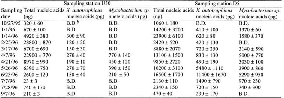
Table 1. QPCR prim ers . ... 138 Table 2. Cell counts in the East Drainage Ditch. ... 139 Table 3. Total nucleic acids, X autotrophicus nucleic acids, and ... ... 140
Mycobacterium sp. nucleic acids at stations U50 and D5.
APPENDIX A
Table 1. RFLP analysis of PCR-amplified 16S rRNA genes from ... 168 station D5.
From a quantitative point of view, microorganisms play an important role in the degradation of organic compounds in the environment. Microorganisms are particularly suitable agents for such biodegradation because of their small size, ubiquitous
distribution, high specific surface area, potentially high rate of metabolic activity, genetic malleability, potentially rapidly growth rate, and unrivaled enzymatic and nutritional
versatility (Madsen, 1997). The types of microorganisms that may preside at any one place and time are dependent on the conditions of their environment. Microorganisms may perceive the introduction of organic chemicals into the environment as either new nutritional opportunities or toxic threats, and it is expected that microbial communities will adapt in response to unfamiliar compounds in their milieu. The physiological and genetic studies that comprise this thesis have been designed to obtain information on the types and distributions of degrading microorganisms living in the toluene-contaminated East Drainage Ditch, a freshwater stream located in the Aberjona Watershed.
Chapter One of this thesis is a literature review, and discusses relevant
background information regarding toluene in the environment, microbial communities in contaminated ecosystems, and the use of molecular tools to study microbial ecology.
An earlier study had demonstrated that biodegradation is the most significant sink for toluene in the stream (Kim, 1995). We successfully isolated several strains of
toluene-degrading bacteria from a contaminated reach of the stream, and these strains have been identified from 16S rDNA analyses as belonging to Xanthobacter
characterization of Mycobacterium sp. strains T103 and T104, while Chapter Three describes X autotrophicus strains T101 and T102.
Chapter Four describes the use of quantitative PCR and slot-blot hybridization assays to assess the relative distributions of the two indigenous species of toluene-degrading bacteria in the stream. The findings were consistent with earlier studies, in which plate counts and most probable numbers of the toluene-degrading population were more significant at contaminated reaches of the stream than at pristine reaches (DeJesus, 1994), and in which biodegradation rates were higher in summer than in winter months (Kim et al., 1995). The results lead us to a better understanding of the factors that influence the natural distributions of these bacteria, and give us a deeper appreciation of the intrinsic capacity of the indigenous microbial community to adapt to the introduction of contaminants in their environment.
Appendix A details the construction and analysis of a clone library of 16S rRNA gene sequences obtained from a contaminated reach of the stream. One of the clones had a restriction pattern identical to those of T103 and T104. Appendix B describes an
unsuccessful attempt to quantify distributions of the X autotrophicus and Mycobacterium
sp. using quantitative hybridization; the hybridization signals detected were at or below
REFERENCES
1. DeJesus, I. 1994. M.Sc. thesis. Massachusetts Institute of Technology, Cambridge.
2. Kim, H.-Y. 1995. Ph.D. thesis. Massachusetts Institute of Technology, Cambridge.
3. Kim, H., H. F. Hemond, L. R. Krumholz, and B. A. Cohen. 1995. In-situ biodegradation of toluene in a contaminated stream. 1. Field studies. Environ. Sci.
Technol. 29:108-116.
4. Madsen, E. L. 1997. Methods for determining biodegradability, p. 709-720. In C. J. Hurst, G. R. Knudsen, M. J. McInerney, L. D. Stetzenbach, and M. V. Walter (ed.), Manual of environmental microbiology. American Society for Microbiology,
Chapter One
TOLUENE IN THE ENVIRONMENT
Toluene is among the 50 largest-volume industrial chemicals produced, with production figures of the order of millions of tons per year (Smith, 1990). It is a clear and colorless liquid, and has a sweet smell. It is produced from petroleum refining, and as a by-product in styrene production and coke-oven operations. Toluene has many industrial uses, and it is widely used in refining gasoline, in chemical manufacturing, in printing and leather tanning, and in the manufacture of paints, lacquers, rubber, and adhesives. Many consumer products also contain toluene, and these include gasoline, nail polish, cosmetics, rubber, cement, paint brush cleaners, stain removers, fabric dyes, and inks. In addition, cigarette smoke and automobile exhaust are sources of toluene emission to the atmosphere.
Because toluene has many industrial applications, it has also been found to be a common water contaminant in the vicinity of chemical waste sites. Toluene is usually disposed of at hazardous waste sites as a used solvent, and it occurs at measurable levels in about 54% of groundwater samples and 28% of surface water samples (U.S. Public Health Service, 1989). The mean concentrations of toluene in groundwater and surface water samples were 0.2 and 0.1 tM, respectively. Drinking water can pose a potential health hazard if it is contaminated with toluene. The most important health concern for humans from exposure to toluene is its harmful effects on the nervous system. Short-term exposures to moderate amounts of toluene can result in fatigue, confusion, general weakness, drunken-type actions, memory loss, nausea, and loss of appetite. Long-term
exposures to high amounts of toluene have been shown to lead to permanent brain
damage. Other effects such as loss of memory, loss of muscle control, and problems with speech, vision, and hearing have also been reported. Although toluene has not been shown to be carcinogenic by itself, it is an enhancing agent in skin carcinogenesis induced by 7,12-dimethylbenz[a]anthracene (Dean, 1978).
Degradation of toluene in water occurs primarily by microbial action. The microbial degradation of toluene has the potential to be rapid, provided that a suitable terminal electron acceptor is available for its oxidation. Half-lives of less than one day under favorable conditions have been reported (Wakeham et al., 1983). Toluene is aerobically biodegraded by both ring attack and methyl-group hydroxylation (Smith,
1990). Figure 1 shows the five different degradation pathways exist for toluene (Zylstra, 1994). For example, toluene is degraded via catechol and subsequently the meta pathway by several strains of Pseudomonas by enzymes encoded on TOL plasmids (Nakazawa et
al., 1980; Franklin et al., 1981). These plasmids often contain two catabolic operons.
The 'upper' pathway operon encodes enzymes for the successive oxidation of toluene to the corresponding alcohol, aldehyde, and carboxylic acid derivatives. The 'lower' or meta-cleavage pathway operon encodes enzymes for the conversion of the carboxylic acids to catechols, whose aromatic rings are then cleaved (meta-fission) to produce the corresponding semialdehydes, which are in turn catabolised through the TCA cycle (Ramos et al., 1987). Burkholderia sp. strain JS 150 is unique in its ability to use multiple pathways for toluene degradation. It has been reported to synthesize four ring fission (lower) pathways and three distinct dioxygenases for the initial oxidation of substituted benzenes (Johnson and Olsen, 1997). This multiplicity of pathways enables strain JS 150
to grow, not just with toluene, but also with benzene, ethylbenzene, halogen-substituted benzenes, and naphthalene as sole carbon sources.
Although many pure cultures that can degrade toluene have been isolated and extensively studied, it is not often apparent whether the microorganisms that have been isolated in the laboratory are actually carrying out the degradation reactions in the
environment. It is also not clear how these microorganisms are distributed in their natural environments, and what factors influence their occurrence. A particular bacterium may dominate an enrichment culture or grow more easily in pure culture than the
microorganisms responsible for the degradation process. Identifying the microorganisms involved in the degradation process in-situ will lead us to the most active cultures for potential bioremediation studies.
MICROBIAL COMMUNITIES IN CONTAMINATED ECOSYSTEMS
Degradation of organic chemicals at hazardous waste sites by the indigenous microflora is an important process in the removal of contaminants. In some cases, biodegradation may be the only important process that can completely remove the chemical pollutants. Observations in the field suggest that natural assemblages of
indigenous microorganisms possess an innate capacity for biorestoration. In some cases, adaptation of microorganisms to pollutants may be required before degradation can occur. For example, Spain et al. (1984) found that increases in the p-nitrophenol-degrading
communities in a freshwater pond correlated with increases in biodegradation rates even though the total bacterial community did not change. They suggested that adaptation was
the result of selection of organisms able to grow at the expense of p-nitrophenol. During a long exposure period, it seems possible that a change in the community may occur that selects for a certain portion of the community and allows the number of degraders to increase.
Pignatello et al. (1983) studied the fate of pentachlorophenol (PCP) in man-made channels at a field site on the Mississippi River near Monticello, Minnesota. The PCP was degraded by both abiotic (primarily photolysis) and biotic (microbial degradation) processes. The biotic removal required a moderately long adaptive response by the aquatic microflora, but eventually became the predominant mechanism of PCP removal from the system. This adaptation in the streams was attributed to the time necessary for selective enrichment of an initially low population of PCP degraders on surface
compartments (Pignatello et al., 1985a). The extent of biodegradation in the streams increased with increasing PCP input, and this correlated with increasing numbers of PCP degrading microorganisms. Most of the PCP-mineralizing microorganisms that
developed in the channels were either attached to surfaces (e.g. rocks and macrophytes), or associated with sediments. The contributions of different stream compartments (or microbial habitats) to microbial degradation of PCP were also assessed (Pignatello et al., 1985b). Contributions to PCP loss were determined for rock (epilithic) surfaces,
macrophyte (epiphytic) surfaces, sedimentary, and water column communities by
measuring rates of PCP disappearance in stream water containing ambient concentrations of PCP in contact with the respective compartmental samples. Results indicated that the rock surface compartment was considerably more efficient at removing PCP than the other compartments.
MOLECULAR ANALYSES OF NATURAL MICROBIAL COMMUNITIES
Our knowledge of the community structure of natural microbial ecosystems is limited because the majority of the organisms present cannot be recovered in culture. The isolation and study of pure cultures of microorganisms may provide a glimpse at the diversity of the microbial community in the environment, but many viable
microorganisms resist cultivation. Discrepancies between direct counts and plate counts are typically several orders of magnitude and raise doubts as to whether cultivated bacteria are actually representative of the microbial communities from which they are isolated. The widespread view is that microbial ecosystems contain numerous novel and uncultivated species, and it has been estimated that only 20% of the bacterial species are actually known (Ward et al., 1990). Culture methods alone may therefore be inadequate for studying microbial communities.
As an alternative to reliance on cultivation, molecular approaches based on phylogenetic analyses of rRNA sequences have been used to determine the species
composition of microbial communities (Pace et al., 1986). Molecular approaches can provide genetic markers for the dominant bacterial species in natural microbial
populations (Giovannoni et al., 1990). Although any gene may be used as a genetic marker, rRNA genes offer distinct advantages (Britschgi and Giovannoni, 1991). 16S rRNA genes are highly conserved and can therefore be used to examine distant
microbial systematics and evolution has resulted in large computer data bases of 16S rRNA sequence information.
The analysis of extracted rRNA has been performed to identify bacteria in marine bacterioplankton (Giovannoni et al., 1990; Schmidt et al., 1991), in terrestrial hot springs (Stahl et al., 1985; Ward et al., 1990; Weller et al., 1991), and in endosymbiotic
associations (Amann et al., 1991). Results from these studies revealed large numbers of community members that have not been cultured, and supported the widespread view that microbial ecosystems contain novel, uncultivated species. For example, the 550C
cyanobacterial mat of Octopus Spring, Yellowstone National Park, has been well characterized with respect to component microorganisms by microscopic and culture methods. Nevertheless, 16S rcDNA sequences retrieved from the Octopus Spring mat revealed eight distinct community members which have not been cultivated from this community (Weller et al., 1991). Indeed, the use of such culture-independent methods can complement, and in some cases circumvent, the bias of culture-dependent techniques and provide a more objective approach by which to understand the composition and character of microbial communities.
CH3 CH:
I
.CH3
CH3 &OH 1t OH OH OH OH Ring Cle D E c aCH 3 OH HOH OH O&H OH OH OH OH avageCOO-KY
REFERENCES
1. Amann, R., N. Springer, W. Ludwig, H.-D. Gortz, and K.-H. Schleifer. 1991. Identification in situ and phylogeny of uncultured bacterial endosymbionts. Nature. 351:161-164.
2. Britschgi, T. B., and S. J. Giovannoni. 1991. Phylogenetic analysis of a natural marine bacterioplankton population by rRNA gene cloning and sequencing. Appl.
Environ. Microbiol. 57:1707-1713.
3. Dean, B. J. 1978. Genetic toxicology of benzene, toluene, xylenes and phenols. Mutat. Res. 47:75-97.
4. DeJesus, I. 1994. M.Sc. thesis. Massachusetts Institute of Technology, Cambridge.
5. Franklin, F. C. H., M Bagdasarian, M. M. Bagdasarian, and K. N. Timmis. 1981. Molecular and functional analysis of TOL plasmid pWWO from Pseudomonas
putida and cloning of the entire regulated aromatic ring meta cleavage pathway. Proc.
Nat. Acad. Sci. USA. 78:7458-7462.
6. Giovannoni, S. J., T. B. Britschgi, C. L. Moyer, and K. G. Field. 1990. Genetic diversity in Sargasso Sea bacterioplankton. Nature. 345:60-63.
7. Johnson, G. R., and R. H. Olsen. 1997. Multiple pathways for toluene
degradation in Burkholderia sp. strain JS 150. Appl. Environ. Microbiol. 63:4047-4052. 8. Kim, H.-Y. 1995. Ph.D. thesis. Massachusetts Institute of Technology,
Cambridge.
9. Madsen, E. L. 1997. Methods for determining biodegradability, p. 709-720. In C. J. Hurst, G. R. Knudsen, M. J. McInerney, L. D. Stetzenbach, and M. V. Walter (ed.),
Manual of environmental microbiology. American Society for Microbiology, Washington, D.C.
10. Nakazawa, T., S. Inouye, and A. Nakazawa. 1980. Physical and functional analysis of RP4-TOL plasmid recombinants: Analysis of insertion and deletion mutants. J. Bacteriol. 144:222-231.
11. Pace, N. R., D. A. Stahl, D. J. Lane, and G. J. Olsen. 1986. The analysis of natural microbial populations by ribosomal RNA sequences. Adv. Microb. Ecol. 9:1-55. 12. Pignatello, J. J., M. M. Martinson, J. G. Steiert, R. E. Carlson, and R. L. Crawford. 1983. Biodegradation and photolysis of pentachlorophenol in artificial freshwater streams. Appl. Environ. Microbiol. 46:1024-1031.
13. Pignatello, J. J., L. K. Johnson, M. M. Martinson, R. E. Carlson, and R. L. Crawford. 1985a. Response of the microflora in outdoor experimental streams to pentachlorophenol: environmental factors. Can. J. Microbiol. 32:38-46.
14. Pignatello, J. J., L. K. Johnson, M. M. Martinson, R. E. Carlson, and R. L. Crawford. 1985b. Response of the microflora in outdoor experimental streams to pentachlorophenol: compartmental contributions. Appl. Environ. Microbiol. 50:127-132.
15. Ramos, J. L., N. Mermod, and K. N. Timmis. 1987. Regulatory circuits controlling transcription of TOL plasmid operon encoding meta-cleavage pathway for degradation of alkylbenzoates by Pseudomonas. Mol. Microbiol. 1:293-300.
16. Schmidt, T. M., E. F. DeLong, and N. R. Pace. 1991. Analysis of a marine picoplankton community by 16S rRNA gene cloning and sequencing. J. Bacteriol. 173:4371-4378.
17. Smith, M. R. 1990. The biodegradation of aromatic hydrocarbons by bacteria. Biodegradation. 1:191-206.
18. Spain, J. C., P. A. Van Veld, C. A. Monti, P. H. Pritchard, and C. R. Cripe. 1984. Comparison of p-nitrophenol biodegradation in field and laboratory test systems. Appl. Environ. Microbiol. 48:944-950.
19. Stahl, D. A., D. J. Lane, G. J. Olsen, and N. R. Pace. 1985. Characterization of a Yellowstone hot spring microbial community by 5S rRNA sequences. Appl. Environ. Microbiol. 49:1379-1384.
20. U.S. Public Health Service. 1989. Toxicological profile for toluene.
Publication ATSDR/TP-89/23. Agency for Toxic Substances and Disease Registry. U.S. Public Health Service, Atlanta, GA.
21. Wakeham, S. G., A. C. Davis, and J. L. Karas. 1983. Mesocosm experiments to determine the fate and persistence of volatile organic compounds in coastal seawater.
Environ. Sci. Technol. 17:611-617.
22. Ward, D. M., R. Weller, and M. M. Bateson. 1990. 16S rRNA sequences reveal numerous uncultured microorganisms in a natural community. Nature. 345:63-65. 23. Weller, R., J. W. Weller, and D. M. Ward. 1991. 16S rRNA sequences of uncultivated hot spring cyanobacterial mat inhabitants retrieved as randomly primed cDNA. Appl. Environ. Microbiol. 57:1146-1151.
24. Zylstra, G. J. 1994. Molecular analysis of aromatic hydrocarbon degradation, p. 83-115. In S. J. Garte (ed.), Molecular environmental biology. Lewis Publishers, Boca Raton, Florida.
Chapter Two
TWO NEW MYCOBACTERIUM STRAINS AND THEIR ROLE IN TOLUENE DEGRADATION
IN A CONTAMINATED STREAM
STEPHEN T.-L. TAY', HAROLD F. HEMOND'*, MARTIN F. POLZ2, COLLEEN M. CAVANAUGH 2, INDHIRA DEJESUS', and LEE R. KRUMHOLZ3
Ralph M. Parsons Laboratory, Department of Civil and Environmental Engineering, Massachusetts Institute of Technology, Cambridge, Massachusetts 021391; Department
of Organismic and Evolutionary Biology, Harvard University, The Biological Laboratories, 16 Divinity Avenue, Cambridge, Massachusetts 021382; and Department of
Botany and Microbiology, University of Oklahoma, Norman, Oklahoma 730193
* Corresponding author. Mailing address: Ralph M. Parsons Laboratory, Department of Civil and Environmental Engineering, Massachusetts Institute of Technology, Room 48-311, Cambridge, MA 02139. Phone: (617) 253-1637. Fax: (617) 258-8850.
Submitted for publication in Applied and Environmental Microbiology, September 29 1997.
ABSTRACT
Two toluene degrading strains, T103 and T104, were independently isolated from rock surface biomass in a freshwater stream contaminated with toluene. The two strains exhibit different capacities for degradation of toluene and a range of aromatic compounds, and have major characteristics of the genus Mycobacterium. Both strains are aerobic, rod-shaped, Gram-positive, non-motile, acid-alcohol-fast, and produce yellow pigments. Their fatty acids include mainly straight-chain saturated and monounsaturated fatty acids with 10 to 20 carbon atoms, and large amounts of 10-methyloctadecanoic acid. Fatty acid analyses indicate that T103 and T104 are not identical strains but are related at the subspecies level. They have identical 16S rDNA sequences that are most similar to Mycobacterium aurum and
Mycobacterium komossense, and they constitute a new species of fast-growing
mycobacteria. Ecological studies reveal that toluene contamination within the stream has selectively enriched for toluene degrading bacteria in the epilithic microbial community, and indicate that strains T103 and T104 and other
microorganisms are involved in the biodegradation. Within a library of 16S rDNA clones constructed from DNA from rock biofilms, a clone was found with a 16S
INTRODUCTION
In the United States, toluene is ranked 27th among the top 50 chemical products by production volume, with 931 million gallons (3.5 x 10' liters) of toluene manufactured in 1994 (22). Industry uses toluene in refining gasoline, in chemical manufacturing, in the manufacture of paints, lacquers, and adhesives, and in some printing and leather tanning processes. Toluene is listed as a priority pollutant (50) as toluene contamination of
drinking water is a potential health hazard. In a survey conducted by the U.S. Environmental Protection Agency in 1988, toluene contamination was detected in groundwater, surface water, or soil at 29% of the hazardous waste sites surveyed. The average amounts of toluene detected were 0.2 tM in groundwater, 0.1 pM in surface water, and 77 ptg/kg in soil (50).
Since toluene is ubiquitous in the environment, it is not surprising that microorganisms capable of degrading toluene have been isolated from a variety of environments (41, 52, 55). For example, an assortment of Pseudomonas species that can grow on toluene as the sole source of carbon and energy have been isolated from polluted topsoil from different sites in the Netherlands (11). Pseudomonas sp. strains T2 and T3 were isolated from oil tanker ballast water that was contaminated with toluene (4).
Mycobacterium vaccae strain JOB5, originally isolated from soil inoculum on plates
incubated with 2-methylbutane (30), is able to grow on toluene (5).
From an ecological perspective, we are concerned with understanding the microbial communities that degrade toluene in nature. One expects to find toluene-polluted
environments enriched in toluene-degrading microorganisms. However, little is known about the abundance and the relative importance of these microorganisms in the
conversion of toluene in the environment. Therefore, studies that address the response of stream bacteria to effects of anthropogenic chemical impact are important in enhancing our understanding of microbial diversity.
In this paper, we describe the isolation and characterization of two closely related toluene degrading strains of a novel Mycobacterium species from rock surface biofilms in a toluene-contaminated reach of a small freshwater stream. We also present ecological data to assess the role of this novel Mycobacterium species in degrading toluene in the rock surface biofilms, and to determine the impact of toluene contamination on the stream's microbial community. Earlier field studies had demonstrated that
biodegradation is the most significant sink for toluene in this stream throughout the year, accounting for 40% to 70% of the toluene sink in spring and summer (20).
Microorganisms attached to stream sediments and rock surfaces are responsible for most of the biodegradation (7).
MATERIALS AND METHODS
Study site. The East Drainage Ditch is a small freshwater stream in an industrial area of Wilmington and Woburn, Massachusetts (7, 20), and forms part of the Aberjona Watershed, a 90 km2 area 20 km north of Boston. Toluene in the stream arises from a
source of subsurface contamination below a 80 m long culvert (10) located 1,600 m upstream from the confluence of the East Drainage Ditch with the Halls Brook Storage
Area. Toluene levels typically range from 0.6 to 4.2 1iM in the streamwater (20).
Sampling stations are 50 m (U50) and 100 m (U100) upstream of the upstream end of the culvert, and 5m (D5) and 50 m (D50) downstream of the downstream end of the culvert.
Collection and isolation of bacterial strains. Rocks were collected in July 1992 from the streambed at station D5 of the East Drainage Ditch. Rock biomass was scraped with sterile spatulas, serially diluted, and plated onto a mineral salts (MS) agar medium (38) supplemented with trace elements (54). The plates were incubated in a dessicator which contained a beaker of saturated toluene solution (15 ml), which was replaced every two to three days. The toluene partitioned via the air into the agar. Calculations based on Henry's Law (39) indicate a theoretical toluene concentration of approximately 110 gtM in the agar, although the actual concentration may be affected by compounding factors such as hydrophobic interactions between toluene and organic constituents of the agar. Plates were monitored for colonies for periods up to seven weeks. Colonies were picked and restreaked on fresh plates. Several colonies were obtained which grew only in the presence of toluene, and four were selected for further characterization. Colonies were numbered sequentially starting from T101. Strains T103 and T104 possessed traits typical of mycobacteria, and results of their characterization are presented in this paper.
Phenotypic characterization of isolates. Tests for Gram stain, oxidase activity, catalase activity, carbon source utilization, nitrate reduction, and acid-fastness (Ziehl-Neelsen method) were performed on strains T103 and T104 as previously described (27,
40). To test for growth on aromatic compounds, colonies were transferred onto plates of MS media and incubated in the same manner as with toluene; substrates were supplied by
diffusion from a reservoir in the dessicator that resulted in theoretical concentrations in the agar of 10 mg/1 of benzene, o-xylene, m-xylene, p-xylene, phenol, or chlorobenzene. The reservoirs were replenished with the relevant substrates every two to three days and plates were monitored for up to four weeks. Visible colonies were picked and restreaked on fresh plates that were further incubated either in the presence or absence of the relevant substrate.
SEM. Cells on agar plates incubated with toluene were observed by scanning electron microscopy (SEM), as described elsewhere (28). SEM was also employed to view rock surfaces with intact biofilms and rock surfaces after extraction of nucleic acids. Rocks were broken up with a hammer, and small fragments of rock surfaces were
prepared as for the cells before viewing with SEM.
Toluene biodegradation kinetics. The rates of toluene degradation were
determined with a headspace gas chromatography method described previously (7). Cells were initially grown in 1100-ml teflon-stoppered glass bottles with 600 ml of MS
medium initially containing 110 gM toluene. Toluene levels were monitored daily and replenished when depleted. After every five toluene feedings, the headspace was flushed with air to replenish the oxygen. Cells were harvested by centrifugation during
exponential phase, and resuspended in 100 ml of fresh MS medium. The kinetic
experiments were performed in 60 ml serum bottles with 19 ml of medium and 0.5 ml of the concentrated cell suspension (230 g±g protein/ml for strain T103 and 185 pg
protein/ml for strain T104). The bottles were capped with teflon coated stoppers (The West Co., Phoenixville, Pennsylvania). Toluene was injected at approximately 1.1 pM,
2.2 kiM, 5.4 4M, and 10.9 ptM (aqueous concentration). Control experiments were performed with autoclaved cell suspensions. All bottles were shaken in the dark at a temperature of 200C on a rotary shaker at 150 rpm, and assayed hourly for toluene until little or no toluene remained. Initial rates of toluene disappearance with time were determined and used to establish the Michaelis-Menten kinetic parameters. Protein was measured with the BCA protein assay, according to manufacturer's specifications (Pierce, Rockford, Illinois).
Fatty acid analyses. The bacteria were grown on Middlebrook 7H 10 agar (9) for 7 days at 280C. Cells were then harvested and saponified, and fatty acid methyl esters were prepared as described previously (36). The analysis was performed at Microbial ID Inc.
(MIDI, Newark, Delaware) using an HP5890 series II gas chromatograph (Hewlett-Packard Co.) and the MIDI Microbial Identification System software for identification of fatty acids. Strains T103 and T104, together with M. komossense type strain (ATCC 33013), were analyzed in duplicate; the profiles obtained were compared with profiles from the Microbial Identification System (MIS) library (37).
16S rDNA sequencing. For total genomic DNA preparation, strains T103 and T104 were cultured in nutrient broth (NB) (Difco Laboratories, Detroit, Michigan) in teflon-stoppered serum bottles. The bottles were shaken at 200C on a rotary shaker at 150
rpm. Cells were harvested by centrifugation after two to three days. Genomic DNA was extracted from pure cultures of the two strains using a miniprep for extraction of genomic DNA from bacteria, as described previously (1). The nearly full-length 16S rRNA gene was amplified from genomic DNA by PCR using a forward primer Eubac27F and a
reverse primer Universal 1492R (23). PCR reaction mixtures contained 2 mM MgCl2, 10 mM TrisHCl (pH 8.3), 50 mM KC1, 200 ptM deoxynucleotide triphosphates, 2.5 U/100pl of Thermus aquaticus DNA polymerase, 0.2 gM each of oligonucleotide primer, and DNA template at 1 ng/gl. Acetamide (5%, wt/vol) was added to the reactions to enhance denaturation during the amplification reaction (32). All reactions were overlain with mineral oil and run in triplicate. Thermal cycling was carried out in a PTC-100 Peltier-effect thermal cycler (MJ Research, Inc.) as follows: denaturation at 950C for 1.5 min,
annealing at 550C for 1.5 min, and extension at 720C for 1.5 min for a total of 30 cycles
(8). Following amplification, the PCR product was purified using the Wizard PCR Prep purification kit (Promega Corp., Madison, Wisconsin) and resuspended in sterile H,O. Both strands of the purified PCR products were sequenced by automated dye dideoxy terminator sequencing at Michigan State University Sequencing Facility with a 373A DNA sequencing system (Applied Biosystems, Foster City, California). Sequences for oligonucleotides complementary to the conserved regions of the eubacterial 16S rRNA were kindly provided by Debra J. Lonergan (United States Geological Survey, Reston, Virginia); these oligonucleotides were chosen to prime the sequencing reactions.
Sequencing reaction mixes consisted of 12 pmoles of sequencing primer, and 50 to 250 ng of PCR template in a total volume of 20 gl of sterile H20.
Phylogenetic analyses. The 16S rDNA secondary structures of strains T103 and
T104 were constructed manually using templates published in the Ribosomal Database
Project (RDP) (25) to aid in the identification of homologous sequence positions.
(42). All reference sequences and the basic alignment to which the sequences of strains T103 and T104 were added were obtained from the RDP. Only homologous sites at which the 16S rDNA sequences of strains T103 and T104 could be aligned
unambiguously with the reference sequences were included for further analyses.
Phylogenetic distance analyses including bootstrap were performed using the programs DNADIST with the Jukes-Cantor correction, SEQBOOT with 100 replicates, and FITCH with input randomization and global rearrangement, all contained in the PHYLIP 3.5 package (13) implemented through the GDE. Parsimony analysis, including bootstrap analysis, was performed using PAUP 3.1 (45). Bootstrap values are shown if they were greater than 50%. Maximum likelihood analysis was performed with the fastDNAml program available from the RDP (12, 29). Trees were constructed by allowing jumbled addition of taxa and global rearrangement of the branches.
Nucleotide sequence accession numbers and strain designations. The sequences for strains T103 and T104 have been deposited in the Genbank database under accession numbers U62889 and U62890. The Genbank accession numbers of the other sequences used in the analyses are as follows: Mycobacterium aurum, X55595; Mycobacterium
chelonae subsp. abscessus str. L948, M29559; Mycobacterium chitae, X55603;
Mycobacterium chlorophenolicus str. PCP-I, X79094; Mycobacterium diernhoferi str. SN
1418, X55593; Mycobacteriumfortuitum subsp. fortuitum, X52933; Mycobacterium
gilvum, X55599; Mycobacterium komossense str. Ko2, X55591; Mycobacterium neoaurum, M29564; Mycobacterium sphagni str. Sph29, X55590; Mycobacterium thermoresistible, X55602; Mycobacterium vaccae, X55601; Mycobacterium asiaticum
haemophilum, L24800; Mycobacterium tuberculosis str. H37/Rv, X52917;
Mycobacterium xenopi, X52929; Corynebacterium xerosis, M59058; Gordona terrae,
X79286; Nocardia otitidiscaviarum, M59056; and Rhodococcus equi, M29574. The primary literature references for these sequences can be found in the RDP (25).
Extraction and purification of genomic DNA from rock biofilms. Rocks were collected in autoclaved 500 ml polypropylene bottles from station D5 in July 1993. The extraction protocol was based on a rapid freeze-thaw method developed by Tsai and Olson for direct DNA extraction from soil and sediments (49). The crude nucleic acids extract was further purified by gel electrophoresis. Polyvinylpyrrolidone (2% wt/vol) (PVP) was added to low-gel-temperature agarose (1.25% wt/vol) (Mallinckrodt Inc., Paris, Kentucky) to eliminate comigration of humic acids with nucleic acids by retarding the electrophoretic mobility of the phenolic groups of humic acids (53). Electrophoresis was performed overnight in PVP-low melt agarose at low voltage (5V/cm of gel length) to prevent overheating. TAE buffer (40mM Tris-acetate, 1mM EDTA) was used. The genomic DNA separated in the PVP-low melt agarose gel was visualized by ethidium bromide staining (35), and excised and purified with GeneClean II (Bio 101, Inc., La Jolla, California).
Construction of environmental 16S rDNA clone library. The 16S rRNA genes were amplified by PCR from the extracted environmental genomic DNA sample. These genes were cloned into the PCR II vector (Invitrogen, San Diego, California) and transformed into competent cells according to manufacturer's instructions. 46 colonies were picked for plasmid isolation and restriction analysis. Plasmid isolation was performed with an alkali lysis plasmid mini-prep, based on a method described
previously (1). The extracted plasmid DNA of individual clones were separated on a agarose gel and purified with GeneClean II (Bio 101, Inc., La Jolla, California). The 16S rRNA gene inserts were amplified by PCR from the purified plasmid DNA of individual clones. All reactions were performed in quadruplicate. Following amplification, the PCR products of each clone were pooled and purified using a Wizard PCR prep purification kit, and resuspended in sterile H20.
Restriction fragment length polymorphism (RFLP) analysis. Aliquots of purified PCR products from the individual clones as well as from strains T103 and T104 were simultaneously digested with the 6-base specific PstI and the 4-base specific Rsal restriction endonucleases (5 U per reaction), according to manufacturer's specifications (Promega Corp., Madison, Wisconsin). Digested DNA was analyzed by gel
electrophoresis in 2% agarose, carried out at 9 V/cm for 1 hr. Gels were stained in an aqueous solution of ethidium bromide (1 pg/ml) and photographed under UV
illumination. One of the environmental clones possessed an identical RFLP pattern to those of strains T103 and T104. The 16S rDNA sequence of this clone was determined by automated dye dideoxy terminator sequencing, as described earlier.
Bacterial counts. To determine viable counts of heterotrophic bacteria, rock samples were collected from stations U100, U50, D5, and D50 along the East Drainage Ditch on September 7, 1993. Rock biomass was scraped with sterile spatulas, suspended in 10 ml of MS medium, and the resulting cell suspension successively diluted to obtain
10- 2 to 10-7 g biomass/ml dilutions. Petri dishes containing 1% PTYG agar (3) were
incubated at 200C for up to three weeks. Heterotrophic colonies were counted on the plates every three days, until no new colonies were observed. Since counts were based on the highest dilution plates, which contained few colonies, colony overgrowth on the plates was not a problem.
Toluene degrading bacteria were enumerated with MS agar medium inoculated in duplicate with 100 til of each dilution. After inoculation, the plates were placed in dessicators and toluene was supplied to the plates as described earlier. After several weeks of incubation, colonies growing on the highest dilution plates were picked and transferred to new plates for a second incubation. These colonies were incubated both in the presence or absence of toluene to confirm the abilities of these isolates to grow with toluene as an energy source. Colonies that grew only in the presence of toluene were counted as toluene-degrading colonies.
Toluene levels in the stream. Duplicate water samples were collected at stations U100, U50, D5, and D50 on September 19, 1993, using 40 ml EPA vials provided with hollow screw caps and teflon-coated silicone septa. Mercuric chloride was added to the samples to a final concentration of 15 mg/1l. Toluene was quantitated using headspace gas chromatography as described previously (7).
RESULTS
Enrichment and isolation. Two mycobacterial toluene degrading strains, T103 and T104, were independently isolated from the East Drainage Ditch. Strains T103 and
T104 originated from two yellow colonies 1 to 2 mm in diameter that appeared between three and eight days on toluene-incubated MS plates inoculated with 10-6 g biomass, indicating that these pure cultures were present in the rock surface biomass at a density of 106 cells/g biomass; biomass scrapings averaged 0.018 g fresh weight/cm2 of rock
surface. Both strains grew slowly (relative to other non-mycobacterial toluene-degrading strains similarly isolated) on solid media when incubated with toluene. Mycobacteria-like colonies were not detected on plates with smaller amounts of biomass.
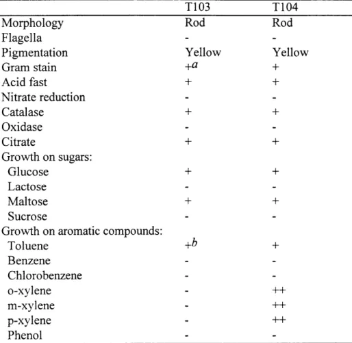
Morphological and phenotypic characteristics. Strains T103 and T104 had morphologically similar rod-shaped cells when grown on solid media (Figure 1). Cells of both strains were nonmotile, and there was no evidence of flagella. The strains were aerobic, and tested Gram-positive (Table 1). The strains also tested positive for acid-alcohol-fastness, a defining characteristic of the mycobacteria (51).
Strain T104 grew better when incubated with o-, m-, or p-xylene than with toluene (Table 1); visible yellow colonies appeared on plates within three days. Strain T103 did not show any capacity to utilize the xylenes. Neither strain was able to grow on benzene, phenol, or chlorobenzene.
Toluene biodegradation kinetics. Strains T103 and T104 had maximal velocities
(Vmax) of 1.0 ± 0.1 and 6.0 ± 1.3 pmoles toluene/mgrotein-hr respectively; their
half-saturation constants (Ks) were 0.6 ± 0.4 and 3.8 ± 1.9 iM respectively (Figure 4). Fatty acid analyses. Strains T103 and T104 contained mainly straight-chain saturated and monounsaturated fatty acids, as well as substantial amounts of tuberculostearic (10-methyloctadecanoic) acid (Table 2). Such high levels of
tuberculostearic acid as are present in these strains are typical of mycobacteria (16). Eleven fatty acids, including tuberculostearic acid, accounted for more than 80% of the total fatty acid composition. The coefficient of variation ([sample standard
deviation/mean] x 100) for each fatty acid that accounted for more than 5.1% of the total fatty acid content in each case was less than 11%.
Strains T103 and T104 did not show any species match with existing mycobacterial entries in the MIS library. Mycobacterium aurum was the most closely related species in the MIS library to the two strains, giving weak similarity indices of 0.31 ± 0.01 and 0.24 ± 0.05 with strains T103 and T104, respectively. Multiple analyses of the same strain result in linkages among samples of that strain at a level of 2 Euclidean distances or lower; the Euclidean distance scale also permits determination of the relatedness of samples at the genus, species, and subspecies levels (approximately 25, 10, and 6
Euclidean distances respectively) (37). The slight phenotypical differences between T103 and T104 were confirmed by fatty acid analysis. T103 and T104 link at a Euclidean distance of 6.9 (Figure 2), indicating that they are not identical strains but are related at the subspecies level. T103 and T104 link with Mycobacterium komossense and
Mycobacterium aurum at Euclidean distances of 10.5 and 14.3 respectively, indicating
that they belong to the Mycobacterium genus, but are different species from
Mycobacterium komossense and Mycobacterium aurum.
16S rDNA sequence analyses. The sequences of approximately 1425 nucleotide bases, corresponding to the E. coli 16S rDNA sequence from nucleotide 55 to 1501, were obtained in both directions for the two strains. The sequences of the two strains were
identical. The secondary structure of the T103/T104 sequence was identical to the secondary structure of other fast-growing mycobacteria. The T103/T104 sequence contains the shortened stem structure bounded by positions 455 to 477 (E. coli
numbering) that typically distinguishes the fast-growing mycobacteria from the slow-growing ones (44).
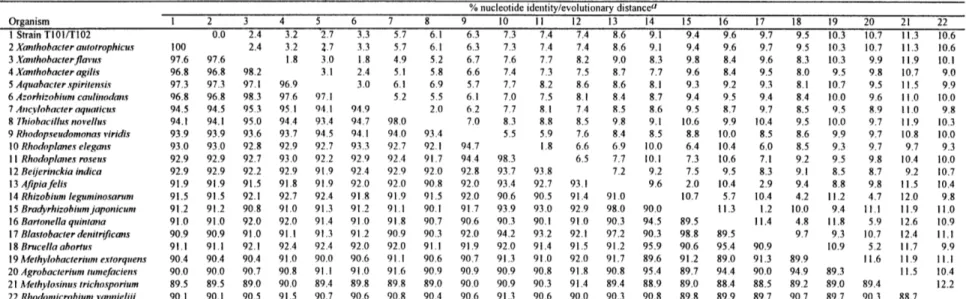
Phylogenetic analyses performed on the final data set of 1,165 nucleotides revealed that the T103/T104 sequence was very similar to the 16S rDNA sequences of the other mycobacteria. An identity matrix constructed with the aligned sequences obtained from the RDP (Table 3) showed that the level of identity between the T 103/T 104 sequence and the sequences of the representative mycobacteria was greater than 96%. The T103/T104
sequence was most identical to the sequences of Mycobacterium aurum (identity 99.0%) and Mycobacterium komossense (identity 98.9%). The levels of identity between the T103/T104 sequence and the sequences of the fast-growing mycobacteria, the slow-growing mycobacteria, and other non-mycobacterial nocardioform bacteria ranged from 96.9 to 99.0%, 96.1 to 97.3%, and 93.0 to 95.7%, respectively.
Distance and bootstrap analyses also showed that strains T103 and T104 belong to the fast-growing members of the Mycobacterium genus (Figure 3). In the distance analysis, all the mycobacteria fell into a closely related, coherent group, distinct from the
other high G+C Gram-positive bacteria examined; the bootstrap value was 81%. Within the genus, the fast-growing mycobacteria are set apart from the slow-growing species; the
slow-growing mycobacteria define a distinct line of evolutionary descent, with a
bootstrap value of 87%. Although strains T103 and T104 are positioned within the group of fast-growing mycobacteria, the precise relationships among the fast-growing species
remained unresolved because of low bootstrap values. Similar tree topologies were also observed in the parsimony analyses (data not shown). An examination of nine most parsimonious trees revealed that the mycobacteria clustered together in these trees, with the slow-growing mycobacteria forming a distinct clade. However, the mycobacteria cluster was not supported in the bootstrap results. Maximum likelihood analysis also showed that strains T103 and T104 fell within the fast-growing mycobacteria (data not shown), although three of the four other nocardioform bacteria (with the exception of
Corynebacterium xerosi) also formed a distinct sub-cluster within the fast-growing group
of mycobacteria. In these analyses, the slow-growing mycobacteria also constituted a distinct clade.
Extraction of genomic DNA. Scanning electron micrographs demonstrate the presence of bacteria on rock biofilms. Rod-shaped bacteria appear to predominate, although it was difficult to distinguish coccoid-shaped bacteria from mineral particles on the rock surfaces. Many of the rod-shaped bacteria were interconnected by an
extracellular matrix (Figure 5A). There was little evidence of remaining biological material on rock surfaces after DNA extraction (Figure 5B).
RFLP analysis. The amplified 16S rDNAs of 46 environmental clones from station D5 produced single bands of about 1,500 bp (data not shown), corresponding to the expected size of the 16S rRNA genes. From the 46 clones, 33 unique RFLP types were detected. The maximum number of clones associated with the same RFLP type was 6. Of the remaining clones, 27 possessed unique restriction patterns. The restriction pattern of one of these clones was identical to those of strains T103 and T104. The 16S
rRNA gene in this clone was sequenced and found to be identical to the 16S rRNA gene sequences of strains T103 and T104.
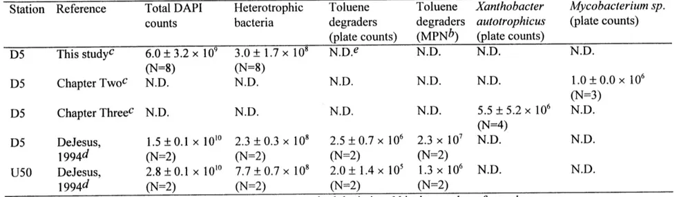
Bacterial counts and toluene levels. No toluene has been detected at station U100 (Figure 6), although very low levels of toluene (0.04 tM) have been measured at station U50. Downstream of the culvert, stations D5 and D50 had high toluene concentrations of 2.6 and 1.7 iM, respectively, in September 1993. The total heterotrophic bacterial plate counts at these stations ranged from 2 x 10⁸ to 7.7 x 10⁸ cells/g biomass. Toluene degrading bacteria were not detected for station U 100, where no toluene was present. The counts of toluene degrading bacteria for the other three stations increased with increasing toluene concentration, and made up 0.03, 1.09, and 0.35 % of the total heterotrophic plate counts at stations U50, D5, and D50, respectively.
DISCUSSION
Taxonomic and phylogenetic considerations. Strains T103 and T104 are both capable of degrading toluene and have major characteristics of the genus Mycobacterium. Strains T103 and T104 exhibit some differences in physiology. Unlike strain T103, strain T104 is able to grow on the xylenes. Also, their fatty acid profiles are sufficiently
different for them to be considered as different strains. Fatty acid analysis appears to be useful to distinguish closely-related strains, especially when 16S rDNA sequences cannot tell such strains apart.
T103 and T104 are thus different strains of a novel species of fast-growing
mycobacteria capable of growth with toluene as the sole carbon source. Compared to the slow-growing mycobacteria, many of which are human and animal pathogens, the fast-growing mycobacteria are common saprophytes in natural habitats (18). They have been isolated from a diverse array of habitats and are able to survive and multiply under a wide range of pH, temperature, and other environmental conditions. The fast-growing
mycobacteria are able to biotransform a variety of xenobiotic compounds and pollutants, including polycyclic aromatic hydrocarbons (15) and groundwater pollutant mixtures (5). There is a long history of the isolation from oil-contaminated soils of mycobacteria that have the capacity to degrade the aromatic fraction of the oil (47, 48).
Phylogenetic analysis of strains T103 and T104 indicates that they are most similar to Mycobacterium aurum and Mycobacterium komossense. Mycobacterium aurum is a fast-growing species commonly isolated from soil (51). Mycobacterium aurum strain MO 1, isolated from a mixed culture in a laboratory reactor, utilized morpholine as the main source of carbon, nitrogen, and energy (6, 26), while Mycobacterium aurum L1 metabolized vinyl chloride under aerobic conditions (17). Mycobacterium komossense is a fast-growing non-pathogenic species that has been repeatedly isolated from
Sphagnum vegetation of moors in south Sweden and the Atlantic coastal area of Norway
(19). It is unable to utilize benzoate and benzamide; it is not known if it possesses the ability to utilize other aromatic hydrocarbons or xenobiotic compounds.
Other closely related species have been found to degrade a range of aromatic hydrocarbons. Other than strains T103 and T104 however, Mycobacterium vaccae is the only Mycobacterium species known to grow on toluene. Mycobacterium vaccae strain
JOB-5 can also degrade acetone, cyclohexane, styrene, benzene, ethylbenzene,
propylbenzene, dioxane, and 1,2-dichloroethylene (5). A Mycobacterium sp., isolated from soil of a former coal gasification site, is able to degrade the polycyclic aromatic hydrocarbons phenanthrene, pyrene, and fluoranthene (2); this isolate is closely related to
Mycobacterium gilvum (16S rDNA identity of > 99.8%) (14).
Biodegradation kinetics. Toluene contamination of the East Drainage Ditch appears to selectively enrich for toluene degrading bacteria within the epilithic microbial community. That Ks values for strains T103 and T104 lie within the range of toluene concentrations observed in the stream suggests that these strains are adapted to the ambient level of toluene contamination in the stream. Non-carbon nutrients are unlikely limiting in this case, as these are present in the stream at high levels (46).
Maximal velocities and half-saturation constants have also been reported for other aerobic toluene-degrading bacteria. Robertson and Button (34) reported toluene
degradation by a marine Pseudomonas sp. strain T2 and by a terrestrial Pseudomonas
putida strain PpF 1 with Michaelian kinetics. Uptake for strain T2 was characterized by a
maximal velocity of 14 mg toluene/g cells-hr (or 0.30 pmoles toluene/mgprotein-hr,
assuming that 50% of a typical cell's dry weight is protein), and a half-saturation constant of 0.48 pM. Corresponding values for strain PpF 1 are 20 mg toluene/g cells-hr (or 0.43 ptmoles toluene/mgprotein-hr) and 0.68 rtM, respectively. Values reported for strain PpF1 are component values for the conversion of toluene to CO2 only.
In order to assess the role that strains T 103 and T 104 may play in toluene
the laboratory for rock biofilms and for pure cultures of our mycobacteria. Toluene biodegradation rates for East Drainage Ditch rocks with their natural biofilms under summer conditions were determined by Cohen et al. (7). The natural biofilm rate was observed to be first order for toluene concentrations up to 2.2 pM, and approached zero order for toluene concentrations greater than 4.3 pM, with a Vm. of 2.0 nmol/cm2 of rock surface-hr. These rates were determined in batch studies similar to those described in the Materials and Methods section, except that whole rocks were used instead of cell
suspensions.
Assuming mass transport to not be limiting, the portion of the toluene
biodegradation on the rock biofilm that may be attributed solely to strains T103 and T104 can be estimated from the cell densities of strains T103 and T104 on the rock surfaces and their individual kinetic parameters (Figure 4). The cell density (CD), estimated from plate counts of the mycobacterial isolates (106 cells/g biomass) and biomass density (0.018 g biomass/cm2 of rock surface), was determined to be 1.8 x 10⁴ cells/cm2 of rock surface. Since the biofilm data are most reliable for toluene concentrations greater than 4.3 pM (7), comparisons of biofilm rates and pure culture rates are performed for toluene concentrations greater than or equal to 4.3 p.M. Assuming a typical cell protein weight of 0.2 pg (24), and assuming that the mycobacteria are uniformly distributed throughout the biofilm, the relative contributions of strains T103 and T104 to the toluene biodegradation by the biofilm can be estimated as follows: